Exposure to cigarette smoke condensate and its
impact on human gingival fibroblast: mechanisms
of molecular pathways involved in
survival/apoptosis
Mémoire
Abdullah Alamri
Maîtrise en biologie cellulaire et moléculaire
Maître ès sciences (M. Sc.)
Québec, Canada
iii
Résumé
Notre objectif est d'étudier les effets de la fumée de cigarette (FC) sur les fibroblastes gingivaux humains. Nos travaux démontrent que FC réduit la viabilité cellulaire cellules vivantes par le biais de la voie apoptotique/nécrotique. Une analyse génomique a montré une surexpression significative de plusieurs gènes dont les gènes de Bax, récepteurs du TNF et caspases; mais une régulation des gènes Lymphotoxin alpha, BCLA1 et BIRC3. Une analyse protéique montant une augmentation des protéines Bax et p53 et de la caspase -3 confirmant l’effet nocif du FC biais un mécanisme apoptotique/nécrotique. Une exposition répétée au FC pendant de courtes périodes, favorise la prolifération cellulaire en augmentant l'activité de la télomérase. En conclusion, ces résultats démontrent qu’à hautes doses la FC est toxique mais à faibles doses la FC provoque une surcroissance cellulaire qui pourrait contribuer au développement des maladies parodontales et des caries.
v Abstract
The study was to investigate the effects of cigarette (CS) on normal human gingival fibroblasts. Our results showed that continuous exposure to CS led to reduced cell viability through an apoptotic/necrotic pathway. PCR arrays showed significant genes’ overexpression such as BCL2-associated X protein (Bax), Tumor necrosis factor receptor (TNF receptors) and Caspase genes. When fibroblasts were repeatedly exposed to small doses of CS, they showed higher proliferation rates. It is important to note that CS-treated fibroblasts showed a significant increase in telomerase activity when exposed periodically to low level of CS. In conclusion, these results demonstrated that at high dose and longer period, CS was toxic to the gingival cells through an apoptotic pathway. However, at small doses with multiple short time exposures CS promoted cell growth and modulated telomerase activities. This may be contributing to oral disease initiation and development of oral pathologies such as periodontitis or cancer.
vii
TABLE OF CONTENTS
Section Page
Résumé (French) ………...………... iii
Abstract (English) ………. v
Table of contents ……….…….………....………. vii
List of tables……….…………... xi
List of figures……….………… xiii
Abbreviations ……….….………...…….…… xv
Avant-propos (Foreword)……….…….………. xvii
Chapter-1 (Introduction)
1 1.1 Introduction……… 21.1.1 The object of investigation……..……….……… 2
1.1.2 Cigarette smoking……….…..……… 3
1.1.3 Periodontal disease and cigarette smoking ………...………...……..… 4
1.1.4 Effect of cigarette smoke on oral cavity ……….…………..……... 5
1.1.5 Cigarette smoking and oral cancer ………...………..……… 7
1.1.6 Effect of cigarette smoke and human gingival tissue ………….……….…... 9
1.1.6.1 Effect of cigarette smoke on gingival epithelial cells ……….…..………..…… 9
1.1.6.2 Effect of cigarette smoke on gingival fibroblasts ……… 10
1.2 Hypotheses……… 10
1.3 Objectives……….. 11
Chapter-2 (Article-1)
13 Article 1: Long-term exposure of human gingival fibroblasts to cigarette smoke condensate reduces cell growth by modulating Bax, caspase-3 and p53 expression ……….……… 14Foreword……….………. 15
2.1 Abstract ………..………... 16
2.2 Introduction ……….… 18
2.3 Materials and methods ……….………..…… 20
2.3.1 Preparation of cigarette smoke condensate ………..…..………. 20
2.3.2 Gingival fibroblast cell culture ……….………….……….… 20
2.3.3 Cell viability/growth assay ……….……….……….…. 21
2.3.4 Apoptosis assay ……….………..………..…. 21
2.3.5 Propidium iodide staining and flow cytometry analysis of cell cycle distribution following contact with cigarette smoke condensate ……….……….……….……. 22
2.3.6 Caspase-3 activity assay……….………....…... 22
2.3.7 Visualization by immunofluorescence of casapase-3 and Bax in the cytosol and mitochondria of cells exposed to cigarette smoke condensate ……….. 22
2.3.8 Real-time PCR array………..….…….……….…… 23
2.3.9 Western blots ……….…………..……… 24
2.3.10 IL-6 and IL-8 measurement following fibroblast exposure to cigarette smoke condensate ……… 24
2.3.11 Statistical analyses. ……….………….…….………….. 25
2.4 Results……… 26
2.4.1 Cigarette smoke condensate reduced gingival fibroblast growth.……….…….. 26
2.4.2 Cigarette smoke condensate promoted human gingival fibroblast apoptosis ..……….………… 26
2.4.3 Cigarette smoke condensate activated apoptotic genes of human gingival fibroblast ……….… 31
2.4.4 Cigarette smoke condensate induced Bax and p53 protein expression by human fibroblasts ……….. 31
2.4.5 Cigarette smoke condensate increased caspase-3 activity in gingival fibroblasts.……….. 33
2.4.6 Bax and caspase-3 displayed cytosolic and mitochondrial colocalization ………..……… 35
2.4.7 Cigarette smoke condensate decreased IL-6 and IL-8 secretion by the gingival fibroblasts ………… 38
2.5 Discussion ………..……….…….. 41
2.6 Conflict of interests. ……….……….……… 44
2.7 Acknowledgments. ……….……….……….. 44
2.8 References. ……….……….………….. 45
Chapter-3 (Article-2)
51 Article 2: Repeated exposure to cigarette smoke condensate induced gingival fibroblast proliferation through the activation of cell cycle genes and telomerase activity………. 52Foreword……….………. 53
3.1 Abstract ………..………... 54
3.2 Introduction ………...……… 56
3.3 Materials and methods ……….………. 58
3.3.1 Preparation of cigarette smoke condensate ………..……….………..………… 58
3.3.2 Gingival fibroblast cell culture …….……….………. 58
3.3.3 Effect of repeated exposure to cigarette smoke condensate on gingival fibroblast proliferation.……. 58
3.3.4 Human oncogenes and tumor suppressor gene profiling through quantitative real-time PCR array.… 59 3.3.5 Telomerase activity assay ……….………...….……… 60
3.3.6 Telomere length ………...……….…..…... 60
3.4 Results………..………. 62
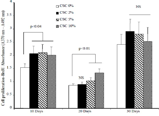
3.4.1 Repeated exposure to cigarette smoke condensate promoted fibroblast proliferation ……….... 62
3.4.2 Repeated exposure to cigarette smoke condensate modulates genes that promote oncogenesis..…….. 62
3.4.3 Cigarette smoke condensate increased telomerase activity in primary human gingival fibroblasts … 64 3.5 Discussion ………..………..……….. 65
3.6 Conclusion. ……….……….……… 68
3.7 Acknowledgments. ………..…….. 68
3.8 Contributions of the authors………..………. 68
3.9 Conflict of interests. ……….……….…… 68
ix
Figure 3-1: Cigarette smoke condensate increased gingival fibroblast proliferation……….. 76 Figure 3-2: Cigarette smoke condensate exposure increased telomerase activity in gingival human
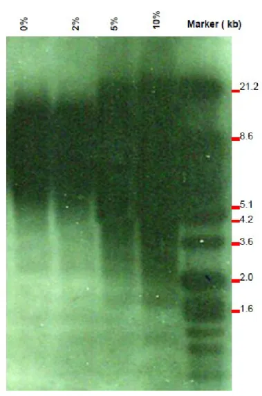
fibroblasts……….….. 77 Figure 3-3: Telomere length analyses……….. 78 Table 3-1: Oncogenes & tumor suppressor genes PCR array (PAHS-502Z): Functional gene grouping….. 79 Table 3-2: Oncogenes and tumor suppressor genes modulated by cigarette smoke condensate ……… 80 Table 3-3: Both onco/tumor suppressor and transcription factor genes modulated by cigarette smoke
condensate ………..……….. 81 Table 3-4: Apoptotic, cell adhesion molecules and cell cycle genes modulated by cigarette smoke condensate
………..…... 82
Chapter-4 (Discussion)
834.1 Discussion……… 84 4.2 References……… 89
xi
LIST OF TABLES
Table number Title of the Table Page
Table 1-1 Dental and gingival conditions associated with tobacco use… 7
Table 2-1
PCR arrays showing apoptotic activated/repressed genes following exposure of human gingival fibroblasts to cigarette
smoke condensate………. 32
Table 3-1 Oncogenes & tumor suppressor genes PCR array (PAHS-502Z): Functional gene grouping………. 79
Table 3-2 Oncogenes and tumor suppressor genes modulated by cigarette smoke condensate……….. 80
Table 3-3 Both onco/tumor suppressor and transcription factor genes modulated by cigarette smoke condensate………... 81
Table 3-4 Apoptotic, cell adhesion molecules and cell cycle genes
xiii
LIST OF FIGURES
Figure
number Title of the figure Page
Figure 1-1 Person suffered of oral cancer………... 5
Figure 2-1 Effect of cigarette smoke condensate on gingival fibroblast viability………... 28
Figure 2-2 Gingival fibroblast survival after cigarette smoke condensate exposure………...…... 29
Figure 2-3 Effect of cigarette smoke condensate on cell cycle
progression in gingival fibroblasts……… 30
Figure 2-4 Modulation of Bax and p53 protein expression by
cigarette smoke condensate ……….. 34
Figure 2-5 Caspase-3 activity following cigarette smoke condensate stimulation……….. 35
Figure 2-6
Bax localized in the cytosol and mitochondria of cigarette smoke condensate -exposed gingival
fibroblasts……….. 37
Figure 2-7
Caspase-3 localized in the cytosol and mitochondria of cigarette smoke condensate -exposed gingival
fibroblasts……….…. 38
Figure 2-8 Cigarette smoke condensate inhibited IL-6 and IL-8 secretions by gingival fibroblasts…………...………... 40
Figure 3-1 Cigarette smoke condensate increased gingival
fibroblast proliferation………... 76
Figure 3-2
Cigarette smoke condensate exposure increased telomerase activity in gingival human
fibroblasts………. 77
xv
ABBREVIATIONS
AFLP Amplified Fragment Length Polymorphism
bp Base pair
cDNA Complementary deoxyribonucleic acid dNTP deoxyribonucleotide-triphosphate dscDNA double strand cDNA
dTTP 2'-deoxythymidine 5'-triphosphate EDTA Ethylenediaminetetraacetic acid
g Gram H2O Water Kb Kilo bases μg Microgram min Minute (s) ng Nanogram nm Nanometer OD Optical density
PCR Polymerase chain reaction Q-RT-PCR Quantitative real-time PCR RNA Ribonucleic acid
RT Reverse Transcriptase
s Second (s)
SDS Sodium Do-decyl Sulfate TAE Tris Acetic acid EDTA
TBE Tris
TE Tris EDTA
U Units
UV Ultraviolet
V Volt
Bax BCL2-associated X protein
TNF Tumor necrosis factor
xvii
Avant-propos (Foreword)
The work with this thesis was performed at the «La Faculté de Médecine Dentaire de l’Université Laval », as a part of the Master degree in Cellular and Molecular Biology program at the Laval University. The work was started in September 2012.
I was fortunate performing my research about a very interesting subject related to the link between cigarette smoke and oral disease that involve gingival fibroblasts. With the help of different people from Dr Rouabhia’s research team, I was able to generate two major publications.
1.
Long-term exposure of human gingival fibroblasts to cigarette smoke condensate reduces cell growth by modulating Bax, caspase-3 and p53 expression. This study was already published.2.
Repeated exposure to cigarette smoke condensate induced gingival fibroblast proliferation throughthe activation of cell cycle genes and telomerase activity; this paper is under preparation for submission.
Different coauthors are listed in each publication, thanks to their active implications in my research activities. Coauthors roles were highlighted in the particular foreword of each publication.
First of all, I would like to thank my supervisor, Professor Mahmoud Rouabhia, for giving me the opportunity to complete my master thesis. I have learned many things since I became Dr. Rouabhia’s student. He spends very much time instructing me how to perform all needed protocols to move further adequately with my research project. He also significantly contributed explaining to me how to search appropriate literature, how to collect and analyses data, how to write a manuscript. Etc. Thanks Dr. Rouabhia for your availability, your kindness, for the useful comments, remarks and engagement through the learning process of my master thesis. It's hard to find the right words to say to him, so I will simply say thanks.
My thanks also go to Mr. Eric Jacques, Researcher assistance in the laboratory of Professor Mahmoud Rouabhia, for his help and support, with technical protocols including western blot, BrdU, cytometry, etc. I would also like to thank Dr. Abdelhabib Semlali, assistance professor in genome research chair, King Saud University. Dr Semlali gave me a lot of advice about my career development and for their advice, encouragement, patience, and support during my MSc training. I would also like to thank Dr. Mohammed Alanazi, Vice Dean of Faculty of Science. He has been a mentor guiding me to build my career, and he has been a role model for me. I would like to thank my parents, have given me their unconditioned love and supported me during my M. Sc. Training. I would like to thank my family, my wife and my kids for their love and support, thank you for always making me smile.
1
Chapter 1:
2
1.1 Introduction
1.1.1 The object of investigation
Oral cancers constitute a significant health concern for a large number of populations worldwide A profound understanding of the early effects of Cigarette smoke (CS) on gingival cells/tissues will likely provide critical insight into the prevention and therapy of CS-related damage and cancer. Oral cancer consistently ranks as one of the top ten cancers worldwide (approximately 5% of cancers in men and 2% in women), with broad differences in geographic distribution (1, 2). Overall, 10.5 adults per 100,000 will develop oral cancer, with greater prevalence in males compared to females (2). The oral cancer induced by cigarette smoke (CS) is the sixth most common malignancy. It is characterized by a reduced survival rate and a high morbidity rate, which have not changed significantly in the past half-century. Most studies examining tobacco-induced carcinogenesis have focused primarily on the mutagenesis of the epithelial cells, the target cells in the most environmentally induced cancers (3). Our Laboratory recently demonstrated that acute exposure to CS led to a significant effect on the barrier and innate immune function of the gingival epithelial cells and tissues (4, 5). However, the CS effect on gingival fibroblasts has yet to be elucidated. The mechanisms of these effects remain largely unknown and, therefore, must be explored. To attain our goals, we were using normal human gingival fibroblasts cells (HGFs) being subjected to either acute (one) or chronic (multiple) exposures to whole CS. Then the non-treated and smoke-treated cells were used for investigating the effect of CS condensate exposure on gingival fibroblast viability/ growth, apoptosis and telomere activity.
3
1.1.2 Cigarette smoking
The health effects of smoking cigarettes are destructive and in many cases, deadly. Cigarette smoke is a complex mixture of chemicals of thousands of chemicals, (e.g., nicotine, cadmium, phenol, anthracyclic hydrocarbons, nitrosamines, heavy metals, and chemical carcinogens, etc.) (6). Approximately 7000 chemicals, have been identified in cigarettes and cigarette smoke to date, 250 of which are poisonous and 70, carcinogenic and placed them in group 1 (carcinogenic to humans). Science is far from finished in its exploration of the composition of manufactured tobacco products, and the chemical count is still increasing, and that can affect the health of individuals who inhale it (7). Worldwide, tobacco use causes more than 5 million deaths per year, and current trends show that tobacco use will cause more than 8 million deaths annually by 2030 (2). Cigarette smoking is responsible for more than 480,000 deaths per year in the United States, including an estimated 42,000 deaths resulting from secondhand smoke exposure (2). This is about one in five deaths annually or 1,300 deaths every day (2). The diseases associated with smoking include: chronic obstructive pulmonary disease (COPD), asthma, laryngeal, lung, throat, cervical, kidney, coronary heart disease, stroke, abdominal aortic aneurysm, acute myeloid leukemia, esophageal, stomach, cataracts, pneumonia, periodontitis, and bladder, and pancreatic cancers.
In the oral cavity, cigarette smoking is a major risk factor for cigarette smoking is a risk factor for oral cancer, oral mucosal lesions, and periodontal disease (8). Human gingival fibroblasts (HGFs) are the main cellular component of periodontal connective tissues.
4
1.1.3 Periodontal disease and cigarette smoking
Smoking is one of the most significant risk factors associated with the development of periodontitis. Periodontal disease is defined as being conditions that range from simple gum inflammation to serious disease that results in significant damage to soft tissue and bone that support the teeth. In the worst cases, there was a loss of teeth. It is the leading cause of tooth loss among adults. Periodontal disease is very common affecting about 90% of the worldwide population (8). Experimental evidence accumulated over the last two decades has indicated that cigarette smoking is probably a real risk factor for periodontitis (9) and even promotes its development (1, 10). Smokers have both increased prevalence and more severe extent of periodontal disease, as well as higher incidence of tooth loss, compared to non-smokers. Several studies have associated cigarette smoking and subgingival infection with periodontal pathogens (11). Other studies have investigated the association between smoking and loss of periodontal bone height (12). It is evident that smokers suffer greater bone loss, greater attachment loss, and deeper periodontal pockets than non-smoking patients (13). Additionally, smoking can lower the chances of success of some treatments (13, 14). Three common bacteria are involved in periodontal disease. Porphyromonas gingivalis, Aggregatibacter actinomycetemcomitans, and Prevotellaintermedia, are all present in higher amounts in smokers than non-smokers, there is a lot of research that indicates the type of bacteria in smokers is more likely to cause periodontal disease. This is due to a higher quantity of bacteria that is present in smokers (15). The contact between cigarette smoke and oral bacteria promotes such oral diseases as periodontitis and oral candidiasis (16).
5
1.1.4 Effect of cigarette smoke on oral cavity
There are several ways that smoking disturbs our oral health that includes lesions in the mouth; the most common being gum disease. Smoking and tobacco products can lead to periodontal disease by affecting the attachment of soft tissue and bone to teeth. It seems that smoking interferes with the normal function of gum tissue cells. Smoking may affect wound healing, making smokers more susceptible to these diseases where weakens the blood flow to the gums. The following oral diseases and conditions are caused by smoking (17, 18).
Figure 1-1: showing person suffered of oral cancer that occurs with lump or thick portion of the lip
with white patches on the gums (19)
Smoker’s melanosis is associated with cigarette and pipe smoking. It refers to brown/dark spots inside the mouth, in the cervical margins of teeth. These spots are caused by tar and other products following cigarette of combustion (20).
Coated tongue is an extrinsic staining, such as dietary substances (e.g., coffee or tea), may contribute to the discoloration. This can be due to the formation of a colored layer composed of mainly bacteria, food particles in the mouth. Tobacco habit promotes plaque accumulation. Treatment consists of mechanical scaling and polishing with mild abrasives. However, the stains will quickly reappear with continued tobacco use (21)
6
Smoker’s palate also known as nicotinic stomatitis is an abnormal manifestation occurring in the hard palate of smokers using pipe, cigar or cigarette smoker. The palate of 45 years old men’s exhibiting a diffuse keratotic shape, with irritation to the minor salivary glands. Patient palate can present numerous papules with punctate red centers. This represents the irritated minor salivary glands with inflamed duct orifices (22, 23). Histologic analyses showed hyperkeratosis and acanthosis of the palatal epithelium. There is also inflammation in the connective tissue and minor salivary glands, and squamous metaplasia of the salivary gland ducts (22, 23). Nicotinic stomatitis increases the risk for squamous cell carcinoma different regions including the tonsillar, retromolar, and respiratory tract. Interestingly, Smoker’s Palate manifestation can be reversed following tobacco cessation.
There are other diseases that affect the oral cavity following smoke. These include oral thrush, which is a type of fungal infection that occurs in the mouth. Also gingivitis, an inflammatory manifestation being caused by bacterial infection can be promoted by cigarette smoke leading to gum disease (Table 1) (24). There is a link between smoking and the accumulation of dental plaque (biofilms), gingival recession, and deepening of periodontal pockets (25-27). Several biological mechanisms suggested host immunity decreasing with smoking (28). This can be through, harmful effects on the function of neutrophil (29) and reducing blood flow to the gums (30). Cigarette smoking has been associated with impaired healing and less improvement in pocket reduction following simple pocket-reduction surgery (31).
Mucosal conditions. Burns and kiratotik patches are common on the lips at the contact site with the cigarette when used (21). The lesions appear on the surface of the mucosa of the lower and upper lips showing white areas flat or slightly elevated with red striations (21).
7 Other injuries caused by tobacco smoke may have surface of wrinkles ranging from white, opaque to translucent, and located in an area where snuff. The solution of these pests is to stop smoking. A biopsy may be needed to rule out histological analyses especially if the lesion is associated with compaction, ulceration, erythema and non-resolution within two weeks of stopping tobacco cessation (32).
Table 1-1: Dental and gingival conditions associated with tobacco use (21). Dental
Conditions Mechanism Significance Managementa
Discolouration Combustion by-products Esthetics Plaque trap
Mechanical polishing Whiteners
Abrasion (mild) Smokeless tobacco Pipe smoking Dentinal/tooth sensitivity Direct restorations Desensitization Abrasion (severe) Pipe smoking Smokeless tobacco Pulp exposure Occlusal disharmony Endodontic/prosthodontics options Gingival Conditions ANUG Tar/nicotine-induced plaque accumulation Ischemia Severe gingival destruction Ultrasonic debridement Antibiotic therapy (rare)
Smoker’s melanosis
Stimulation of melanocytes
Must rule out melanoma or systemic conditions
Biopsy may be required to rule out melanoma
Association with gingivitis and
periodontitis
Increased calculus and plaque deposits Ischemia Periodontal destruction Periodontal therapy (debridement, excisional procedures) Poor wound healing Ischemia Postsurgical care Osteitis Regenerative periodontal techniques contraindicated a All treatment should coincide with the cessation of the tobacco habit. This Table groups multiple observations
made in smokers and there consequences on the oral health. ANUG, acute necrotizing ulcerative gingivitis.
1.1.5 Cigarette smoking and oral cancer
Cancer is a disease in which the normal body cells transform to malignant cells, as a result of changes or mutations in the normal cell genome resulting in uncontrolled cell proliferation. These changes (mutations) produce proteins that disrupt the balance between normal regulatory processes
8
(e.g. cell cycle) and programmed cell death (apoptosis) leading to cell overgrowth. The mutations are heritable and allow the cancer cells to spread and invade other tissues and organs (metastasis), which can be fatal. Almost 90% of cancer-related deaths are due to metastasis. Although cancer comprises at least 100 different types, and each is classified by the type of cell that is initially affected, all share one important characteristic: the abnormal, uncontrolled cell growth (33).
Oral cancer is considered one of the top ten cancers worldwide, with considerable differences in geographic distribution. It is a primary cause of global morbidity and mortality. Squamous cell carcinoma (SCC) accounts for more than 90% of oral malignancies and occurs most frequently in middle-aged to elderly patients who are heavy smokers and drinkers (34). This type of cancer represents approximately 5% of cancers in men and 2% in women (35). In USA, the National Cancer Institute Survey has shown that oral cancer rates have increased approximately 15% from the mid-1970s until recently (2004). This survey reported significant disparities in some population groups, with higher rates of increase in minority men. Overall, 10.5 adults per 100,000 will develop oral cancer, with greater prevalence in males compared to females. Oral cancer in USA is also dependent on the ethnicity since the rates are higher for Hispanic and Black males than for White males. As for worldwide population, oral cancer rates increase with age. In 2010, the statistics worldwide estimated that the oral cavity, pharynx (other than naso-pharynx), and larynx cancers account for 683,000 new cancer cases (5.2% of global cases) and 356,000 deaths (4.4% of global cancer deaths). The tongue, lip and floor of the mouth constitute the three most common sites for SCC development. SCC can develop from precancerous lesions (leukoplakia and erythroplakia) or even from the normal epithelium. Oral cancer such as squamous cell carcinoma (OSCC) often develops after the age of 50, with the highest peak in the sixth decade of life (36, 37). The exposure to exogenous carcinogens such as tobacco smoke and alcohol represents the primary risk factor for
9 oral cancer. Studies have shown that oral cancers are highly linked with alcohol consumption and cigarette smoking with an 80% of cases of oral cancers (38). A large number (3 to 6%) of oral cancers is associated with leukoplakia lesions, with an increasing frequency with longer follow-up periods (39, 40). Genetic susceptibility (predisposition) to OSCC is also a major risk factor especially in young patients. This is linked to the efficiencies of carcinogens metabolism, DNA repair, and cell cycle control, alone or in combination (41).
1.1.6 Effect of cigarette smoke and human gingival tissue
Upon entrance into the oral cavity, cigarette smoke reaches the oral mucosa where epithelial cells and fibroblasts interact. Cigarette smoke can have harmful effects on epithelial cells and fibroblasts.
1.1.6.1 Effect of cigarette smoke on gingival epithelial cells.
In the oral cavity, the epithelium forms the first line of defense against toxic agents and bacteria such as periodontopathogens (42). In addition to its function as a protective physicochemical barrier to the outside environment, oral epithelium has a number of metabolic and immunological roles, including fluid and ion transport regulation, mucus production/elimination and participation in innate and adaptive immunity, as well as the modulation of inflammation, cell migration and repair processes (43). These functions are essential to maintaining mucosal homeostasis. Despite the fact that human oral epithelial cells are the first cell type to be exposed to cigarette smoke, few studies have addressed the effect of cigarette smoke on these cells and the possible initiation and development of oral diseases that include cancer and periodontitis. In a recent study, our team reported that exposure to whole cigarette smoke markedly inhibits epithelial cell growth through an apoptosis/necrosis pathway that involves Bax and Bcl-xL proteins and caspase-3 activity. Cigarette smoke also disrupts epithelial cell migration, which may negatively affect periodontal
10
wound healing (4). It has also been reported that nicotine, one of the cigarette smoke constituents increased the secretion of IL-8 from human gingival epithelial cells (44). Also, cigarette smoke was shown to be able increasing the expression of matrix metalloproteinase (MMP)-2, MMP-9 and MMP-28 by human keratinocytes (45).
1.1.6.2 Effect of cigarette smoke on gingival fibroblasts
Fibroblasts are the predominant cell type inhabiting the gingival connective tissue, play a critical role in gingival tissue structures, extracellular matrix synthesis (26, 46) and tissue repair (27) (28). Available studies showed that exposure of the gingival mucosa to cigarette smoke could have a significant effect on fibroblast function and consequently that of gingival tissue. Indeed, nicotine and cigarette smoke condensate were reported to inhibit fibroblast proliferation and altered their capacity producing proteolytic enzymes (29, 30). Moreover, cigarette smoke extract was found to impair the wound healing process by inhibiting fibroblast recruitment and proliferation. It has been reported that cigarette smoke modulates pulmonary fibroblast-mediated contraction through possible enlarged air spaces that develop in the injuries associated with pulmonary diseases (31). Altogether, these data suggested a significant harmful effect of cigarette smoke on gingival fibroblast. However, critical investigation still to be performed to shed light on the direct effect of cigarette smoke on gingival fibroblasts.
1.2 Hypotheses:
Oral cancer induced by cigarette smoke (CS) is the sixth most common malignancy. It is characterized by a reduced survival rate and a high morbidity rate, which have not changed significantly in the past half-century. Gaining a better understanding of the link between CS and oral cancer is, therefore, critical. Most studies have used the epithelial cells as a model for examining tobacco-induced carcinogenesis. Previous work from our group clearly demonstrated
11 that acute exposure to CS significantly affected the barrier and innate immune function of gingival epithelial cells and tissues. Epithelial cells are in contact with gingival fibroblasts. It’s known that the fibroblast-epithelial cells interaction is very important for tissue homeostasis, and the impairment of this interaction may lead to oral cancer development. Fibroblasts can secrete many growth factors such as TGFb and other cytokines that can influence the function of epithelial cells. Deregulation of the fibroblast functions would have consequences on the function of epithelial cells. However, the mechanisms related to CS effects on gingival fibroblasts remain largely unknown.
1.3 Objectives
The general objective of this study was to investigate the harmful effect of Cigarette smoke
condensate (CSC) on human gingival fibroblast (HGF).
Specific objectives:
1. Investigate the effect of a long-term chronic exposure to CSC on gingival fibroblast viability and apoptosis
2. Investigate the effect of a short but repeated exposure to CSC on gingival fibroblast behavior
13
Chapter 2
14
Article – 1
Long-term exposure of human gingival fibroblasts to cigarette smoke condensate
reduces cell growth by modulating Bax, caspase-3 and p53 expression.
Alamri A
1,2, Semlali A
2, Jacques E
1, Alanazi M
2, Zakrzewski A
1, Chmielewski
W
1, Rouabhia M
1.
1Oral Ecology Research Group, Faculty of Dentistry, Laval University, Quebec, QC, Canada and, 2Genome Research Chair, Department of Biochemistry, College of Science King Saud
University, Riyadh, Kingdom of Saudi Arabia
J Periodontal Res. 2014 Aug 19. doi: 10.1111/jre.12223.
PMID: 25139560
15 Foreword
This article includes an introduction, an experimental protocol, results, discussion and conclusion. This publication was obtained thanks to the contribution of the following authors: Alamri A, Semlali A, Jacques E, Alanazi M, Zakrzewski A, Chmielewski W, Rouabhia M.
Alamri A conducted all the experiments with the help of Semlali A, Jacques E and Rouabhia M.
Alamri A, Semlali A, Jacques E, Chmielewski W and Rouabhia M, analyzed and interpreted the data.
Alamri A, Semlali A, Jacques E drafted the first version of the manuscript.
Mahmoud Rouabhia completed the manuscript with the help of Alanazi M, Zakrzewski A and Chmielewski W.
16
2.1 Abstract
BACKGROUND AND OBJECTIVE:
Smoking cigarettes increases the risk of oral tissue damage leading to periodontal disease. Gingival fibroblasts, the predominant cell type inhabiting gingival connective tissue, play a critical role in remodeling and maintaining gingival structure. The objective of this study was to investigate the effect of long-term exposure to cigarette smoke on human gingival fibroblast survival/apoptosis and the molecular pathways involved in these cell responses.
MATERIAL AND METHODS:
Human gingival fibroblasts were extracted from healthy non-smokers and cultured in the presence of cigarette smoke condensate (CSC). At the end of each time point, cell growth was evaluated by means of MTT assay. Apoptotic and necrotic gene's expression was investigated by polymerase chain reaction array and by annexin V/propidium iodide staining and cell cycle assays. Western blot was used to investigate Bax and p53 proteins. These tests were supported by caspase 3 activity analyses.
RESULTS:
High levels of CSC decreased cell growth and deregulated cell cycle progression by increasing the G0 /G1 and reducing the S and G2 /M phases of the gingival fibroblasts. Polymerase chain reaction
arrays revealed the activation of several apoptotic genes by CSC, including TNF receptors, caspases, Bax and p53. This was supported by increases in the Bax and p53 protein levels as well as by an elevated activity of caspase-3 in the CSC-exposed cells. Immunofluorescence staining demonstrated that both Bax and caspase-3 displayed a cytosolic and mitochondrial distribution in the CSC-exposed gingival fibroblasts, compared to controls. The damaging effect of CSC on
17 gingival fibroblast growth was also supported by the decrease in interleukin 6 and 8 secretion by the gingival fibroblasts.
CONCLUSION:
These results suggest that CSC may contribute to deregulating fibroblast functions. This can compromise fibroblast-epithelial cell interactions, which ultimately increases the risk of gingival tissue damage and the onset of periodontitis.
18
2.2 Introduction
The periodontium consists of a group of tissues that surround and support each tooth as well as retain it in the mandibular and maxillary bones. For these reasons, it is important to maintain healthy periodontium (1). Periodontal tissue health depends on the normal functions of periodontal cells. The attachment, migration, growth, and differentiation of periodontal cells are key steps in maintaining the functionality of periodontal tissues. Unfortunately, periodontal tissues are subjected to multiple insults which can lead to tissue dysfunction in the form of periodontal diseases (2).
Periodontitis is a complex chronic disease that leads to the destruction of tooth-supporting tissues, including alveolar bone, eventually resulting in tooth loss. The disease is believed to develop as a result of the host-mediated inflammatory response to pathogenic microflora residing in periodontal pockets (3). Among the different cell types in the periodontium that may be involved in the host immune response to periodontitis are gingival fibroblasts. As the main cellular component of periodontal connective tissue, these cells play a major role in periodontal health (4). Gingival fibroblasts are active in the inflammatory response by secreting inflammatory cytokines, such as IL-6 and IL-8, as well as inflammatory chemical mediators, such as PGE2,in response to stimuli
that include periodontopathic bacteria (5,6). Aside from the presence of periodontopathic bacteria in the oral cavity, several other factors actively contribute to the onset and progression of periodontitis, including such environmental factors as smoking (7).
It is well recognized that cigarette smoke can alter cell function and promote periodontal disease development and severity (7). Cigarette smoking is a risk factor in both the incidence and the progression of periodontal disease (8). Smokers exhibit significant damage to the alveolar bone which may augment tooth loss (9). Periodontitis severity has been shown to increase with smoking
19 intensity and duration (10). Furthermore, cigarette smoke also reduces the host response to periodontopathic bacteria, resulting in a more aggressive periodontal breakdown (11). Early studies have reported impaired phagocytosis by in situ smoke-exposed oral polymorphonuclear neutrophils (12). Smoking also appears to inhibit host defenses against microbial infection while promoting inflammatory reactions (13). Indeed, smokers are susceptible to colonization by P. gingivalis, a causative agent of periodontitis (14). Upon entering the oral cavity, cigarette smoke reaches the oral mucosa where epithelial cells and fibroblasts interact and maintain tissue integrity and function (15). Gingival fibroblasts, the predominant cell type inhabiting gingival connective tissue, play a critical role in remodeling and maintaining gingival structure and extracellular matrix (16,17) and are key players in tissue repair and wound healing through their adhesion, migration, growth, and differentiation, as well as through the production of extracellular matrix (16,17). Exposure of the gingival mucosa to cigarette smoke may have a significant impact on fibroblast function and consequently, that of gingival tissue. Cigarette smoke products, such as nicotine, act on periodontal cells by promoting the production of inflammatory cytokines (IL-1β and TNF-α) and inducing changes in cell cycle and differentiation marker values (18,19). Furthermore, nicotine has been shown to inhibit the growth of human periodontal ligament fibroblasts through apoptotic mechanisms (20). Tissue damage, as a result of smoking, cannot only impact the integrity of gingival tissue, but can also potentiate inflammatory responses as well as setting up an optimal environment for bacterial growth (21). Based on these data, we investigated the effects of cigarette smoke condensate (CSC) on normal human gingival fibroblast viability/growth, apoptotic process, and expression of specific genes followed by the production of proteins by these fibroblasts.
20
2.3 Materials and Methods
2.3.1 Preparation of cigarette smoke condensate. 1R3F cigarettes were purchased from the Kentucky Tobacco Research & Development Center (Orlando, FL, USA) and were used to prepare the cigarette smoke condensate solution as follows: Each cigarette was placed into one end of a silicone tube linked to an Erlenmeyer containing 200 ml of 0.09% sodium chloride. On the other end, a second silicone tube linked to a standard vacuum. The cigarette was lit and attached to a cigarette holder. The smoke was extracted by applying a vacuum to draw the smoke directly into the 0.09% sodium chloride solution. The process was repeated for a total of ten cigarettes. The cigarette smoke condensate (CSC) solution was then sterilized by filtration through a 0.22-m filter and subsequently stored at 4C until use.
2.3.2 Gingival fibroblast cell culture. Gingival fibroblasts were isolated from gingival mucosa biopsies taken from healthy non-smoker subjects. Written informed consent was obtained from each subject. The fibroblasts were cultured in Dulbecco’s modified Eagle’s medium (DMEM) (Invitrogen, Burlington, ON, Canada) supplemented with 10% fetal bovine serum (FBS) in a humidified incubator at 37C with 5% CO2. The medium was changed three times a week. When
the cells reached 90% confluence, they were detached with 0.05% trypsin-EDTA, seeded in 6-well plates at 4 x 10⁵ cells/well, and incubated for 24 h to allow for proper cell adhesion. Various
concentrations (0, 10, 30, or 50%) of CSC were added to the culture medium, after which time the cells were cultured for 5, 7, 10, and 14 days. Each medium with and without CSC were refreshed every two days.
21 2.3.3 Cell viability/growth assay. Following fibroblast culture in the presence or absence of CSC at various concentrations, fibroblast shape and density was determined by means of inverted microscopy observations and photos. Cell viability/growth was assessed by an MTT assay. Briefly, a stock solution (5 mg/ml) of MTT was prepared in PBS and added to each culture well at a final concentration of 1% (v/v). The fibroblast cultures were then incubated for 4 h at 37°C with the MTT. The supernatant was then removed, 2 ml 0.04 N HCl in isopropanol were added to each culture plate, and incubation was extended for another 15 min. Finally, 200 μl (in triplicate) of the reaction mixture was transferred to the wells of a 96-well flat-bottom plate, with absorbance measured at 550 nm by means of an enzyme-linked immunosorbent assay (ELISA) reader (X-Mark microplate spectrophotometer, BioRad Laboratories, Mississauga, ON, Canada). Baseline optical density accounting for the test plate was subtracted from each experiment. Tests of this assay repeatedly showed a linear relationship between cell number and optical density readings.
2.3.4 Apoptosis assay. An apoptosis/survival assay was performed on fibroblasts in contact with CSC for various periods. For this purpose, we used the Annexin V–fluorescein isothiocyanate detection kit (BD Bioscience, Mississauga, ON, Canada). Fibroblasts were detached from the culture plates with trypsin-EDTA and washed twice with culture medium. The resulting fibroblast pellets were suspended in binding buffer and incubated with Annexin V–fluorescein isothiocyanate and propidium iodide (PI), according to the manufacturer’s instructions. Stained cell suspensions were analyzed using the Beckman Coulter Epics Elite ESP cytometer. The cells were categorized as follows: viable cells refer to Annexin V and PI non-stained cells and apoptotic cells refer to the Annexin V-stained but PI non-stained cells.
22
2.3.5 Propidium iodide staining and flow cytometry analysis of cell cycle distribution following contact with cigarette smoke condensate. Gingival fibroblasts were seeded in a 6-well plate and left for 24 h to allow for cell adhesion, after which time the fibroblasts were subjected or not to CSC treatment (10, 30, or 50%) during 5 days, with medium changing every 48 h. The CSC-treated cells were harvested and 1 x 10⁶ cells/ml were fixed with cold 70% ethanol for 1 h on ice. Following
multiple washes in cold PBS, the cells were treated with RNase (10 µg/ml) at 37ºC for 1 h, after which time ropidium iodide (PI; 50 µg/ml) was added prior to analysis. Analyses were performed with an Epics® Elite ESP flow cytometer (Beckman Coulter, Miami, FL, USA). The single cell population was gated using pulse width vs. pulse area to exclude clumps and doublets and the scatter plot was used to exclude any obvious debris. The PI was detected using a FL4 channel vs. a cell count histogram plot.
2.3.6 Caspase-3 activity assay. Caspase-3 activity was measured on fibroblasts in contact or not with CSC using the caspase-3/CPP32 colorimetric assay kit (BioVision, Inc. Milpitas, CA, USA) according to the manufacturer’s instructions. Briefly, 2 x 10⁶ cells were suspended in lysis buffer.
Total protein extracts (100 g) were combined with reaction buffer and 200 M of DEVD-pNA substrate at 37C for 1 h. Thereafter, samples were read at 405 nm on a microplate reader and the percentage of activity was determined by comparing each sample with the optical density obtained with a cell extract prepared from cells cultured in the absence of CSC.
2.3.7 Visualization by immunofluorescence of casapase-3 and Bax in the cytosol and mitochondria of cells exposed to CSC. Cells were seeded on coverslips and allowed to reach half confluence. After exposing the cells to CSC for 5 days, the mitochondria were labeled with MitoTracker™ CMXRos-H2 (Molecular Probes, Inc., Eugene, OR, USA) according to the manufacturer’s
23 instructions. Briefly, on the final incubation day with CSC, MitoTracker was diluted to a final concentration of 1 µM in serum-free medium and was added to the cells for 45 min at 37ºC in a 5% CO2 humidified incubator. The cells were then washed in PBS, fixed, and permeabilized with
methanol for 15 min at -20ºC. Coverslips were blocked with rat serum and stained with either a mouse anti-human Bax or a mouse anti-human caspase-3 (10 µg/ml; Santa Cruz Biotechnologies, Santa Cruz, CA, USA) in 0.1% saponin for 1 h and with Alexa Fluor 488-conjugated secondary antibody (Molecular Probes (1:500 final) for 1 h in the dark. Cell nuclei were then stained with 1 mg/mL of Hoechst (Invitrogen, Carlsbad, CA, USA) for 10 min. The slides were subsequently mounted with the PBS/glycerol/gelatin and were visualized under a Zeiss Apotome® microscope with a 63 x /1.4 NA lens and AxioVision 4.8.2 scanning software (Carl Zeiss, Gottingen, Germany).
2.3.8 Real-time PCR array. Total RNA was extracted from cells cultured in the presence of either 0 or 30% CSC for 5 days by means of the RNAspin mini kit (GE, Mississauga, ON, Canada) according to the manufacturer’s protocol. RNA concentrations and quality were determined by the Experion automated electrophoresis station from Biorad (Mississauga, ON, Canada). RNA quality was optimal in all samples with 28S/18S rRNA ratios above 1.8. Real-time PCR arrays were performed by means of the RT2 Profiler PCR Array System from SuperArray Bioscience
(Frederick, MD, USA) according to the manufacturer’s instructions. Briefly, total RNA (1000 ng) was used to prepare cDNA with the RT2 first strand kit (QIAGEN, Germantown, MD, USA). PCR
arrays containing 88 gene-specific primers related to apoptosis were amplified. The results were analyzed using the ΔΔCt method and the fold changes between the non-stimulated and 30%
CSC-stimulated samples were calculated. Genes were selected if a fold change above 1.3 was observed and if baseline cycle thresholds for this gene were below 30 to eliminate feebly expressed genes.
24
2.3.9 Western blots. Following exposure to CSC, fibroblasts were resuspended in lysis buffer containing 25 mM of Tris-HCl, pH 8.0, 150 mM of NaCl, 1 mM of EDTA, 10% glycerol, 0.1% SDS and 1% Triton X-100. Proteinase inhibitor PMSF 300 mM was then added to the homogenized samples and protein concentration was determined using the Bradford assay. Equal amounts of total protein (20–30 g) in reducing sample buffer (containing 2.5% 2-mercaptoethanol) were boiled for 5 min and subsequently migrated using 4% stacking gel followed by 10 or 15% acrylamide SDS-PAGE. The gels were then transferred to PVDF membranes with a Tris-glycine refrigerated transfer buffer containing 15% methanol. The blots were incubated overnight with anti-Bax at 1:200 (Santa Cruz Biotechnology), anti-p53 at 1:500 (R&D Systems, Minneapolis, MN, USA) or anti--actin at 1:1000 (Sigma). The resulting membranes were washed and finally incubated with an anti-mouse peroxydase-conjugated antibody (BD Bioscience). Detection was performed thereafter by means of the ECL detection system (EMD Millipore Billerica, MA, USA) according to the manufacturer’s instructions. Luminescence was obtained by autoradiography. The modulation of protein production was determined by band scanning using Genetools software from Syngene (Frederick, MD, USA).
2.3.10 IL-6 and IL-8 measurement following fibroblast exposure to cigarette smoke condensate. Following exposure to CSC for various culture periods, supernatants were collected from each condition and analyzed by sandwich enzyme-linked immunosorbent assay (ELISA, R&D System). Immediately after, the supernatants were filtered through 0.22-μm filters and used to measure the IL-6 and IL-8 levels. ELISA plates were read at 450 nm and analyzed using a Microplate Reader Model 680 (Bio-Rad, USA). The minimum detectable concentrations were under 0.7 pg/ml for the
25 IL-6, and 3.5 pg/ml for the IL-8, as reported by the manufacturer. Data were reported as pg/ml per ug total protein in each condition to have useful comparisons.
2.3.11 Statistical analyses. Each experiment was performed at least three times, with experimental values expressed as means ± SD. The statistical significance of the differences between the control (absence of CSC) and the test (presence of CSC) values was determined by one-way ANOVA.
Posteriori comparisons were conducted using Tukey’s method. Normality and variance assumptions were verified by means of the Shapiro-Wilk test and the Brown and Forsythe test, respectively. All of the assumptions were fulfilled. P values were declared significant at ≤ 0.05. Data were analyzed using the SAS version 8.2 statistical package (SAS Institute Inc., Cary, NC, USA).
26
2.4 Results
2.4.1 Cigarette smoke condensate reduced gingival fibroblast growth. Following continuous exposure to CSC for various culture periods, we observed a reduction in cell density in those cultures put in contact with CSC. This treatment led to altered cell morphology, which went from a small elongated cell shape (control) to a large-sized cell with a faint cytoplasm beginning at 7 days and continuing up to 14 days of culture with CSC (Fig. 1A). To confirm these observations, we performed a quantitative measurement of cell viability/growth using an MTT assay. Exposure to CSC did indeed inhibit human gingival fibroblast growth (Fig. 1B). Cells grown without CSC showed increased optical density between cultures of 5 days (0.89 ± 0.03), 7 days (1.68 ± 0.05, p = 10-17), 10 days (2.38 ± 0.07, p = 10-20), and 14 days (2.77 ± 0.77, p = 10-17) (Fig. 1B). However,
cells grown in the presence of CSC showed a decrease in growth over the time course. We also demonstrated that cell growth in the CSC-exposed cultures (10, 30 or 50% CSC) was significantly (p < 10-10) lower than that in the non-stimulated controls at the different culture periods (5, 7, 10,
and 14 days). The growth rate of the fibroblasts differed depending on the CSC concentration. In the presence of CSC at 10,30, and even 50%, a significant proliferation of the gingival fibroblasts was observed after 5, 7, 10, and 14 days (Fig 1B) ,
2.4.2 Cigarette smoke condensate promoted human gingival fibroblast apoptosis. To determine whether the decrease in cell growth was due to an increase in apoptosis, cells exposed to CSC were detached and the percentage of viable or apoptotic cells was determined by means of an Annexin V-PI kit by flow cytometry after 5 days of incubation. As shown in Figs. 2A and 2B, the viable cell percentage was significantly higher in the control cells (80.7 ± 4.9%) than in the CSC-exposed cells in a dose-dependent manner (41.7 ± 6.9% with 10% CSC, p = 0.01; 34.9 ± 7.2%
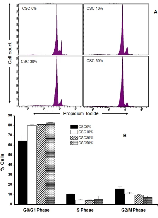
27 with 30% CSC, p = 0.01; and 25.4 ± 14.0% with 50% CSC, p = 0.05) (Fig. 2B). The number of apoptotic cells significantly increased with 10, 30, and 50% CSC compared to that observed in control cells. Similar results were obtained at 7 and 14 days (data not shown). CSC-induced cell growth decreases possibly took place through a deregulation of the cell cycle progression. To monitor the effect of CSC on the cell cycle, we treated gingival fibroblasts with various concentrations of CSC for 5 days, after which time we quantified the cell percentages in the different cell cycle phases. In the control group, less than 70% of the cells were in the G0/G1 phase, while over 10% were in the S phase and over 15% were in the G2/M phase (Fig. 3). However, following exposure to CSC, the values were significantly (p < 0.05) reduced. In contrast, in the fibroblast culture exposed to 10% CSC, over 80% of the cells were in the G0/G1 phase, with approximately 5% in S phase and close to 12% in the G2/M phase. Similar results were obtained with fibroblasts exposed to 30% and 50% CSC.
28
Figure 2-1: Effect of cigarette smoke condensate on gingival fibroblast viability. Gingival fibroblasts were seeded at 4 x 10⁵ cells per well and incubated with or without CSC at various
concentrations (0, 10, 30, and 50%) for 5, 7, 10, or 14 days. Photos were taken to determine cell shape (Panel A, Scale bars: 50 µm). MTT assay was performed to determine cell growth (Panel B). # = p < 10-15 obtained by comparing cell growth at 5 days to 7, 10 and 14 days without exposure to
29 Figure 2-2: Gingival fibroblast survival after cigarette smoke condensate exposure. Gingival fibroblasts were seeded at 4 x 10⁵ and stimulated with 0, 10, 30, or 50% CSC in DMEM/10% FBS
for 5 days. Apoptosis was measured with an Annexin V-PI kit by flow cytometry. (A) Representative result of three different experiments. Quadrant I3: Viable cells, Annexin V- and PI-negative. Quadrant I4: Apoptotic cells, Annexin V-positive and PI-PI-negative. Panel (B) refers to the percentage of viable and apoptotic cells compiled from three different experiments.
30
Figure 2-3: Effect of cigarette smoke condensate on cell cycle progression in gingival fibroblasts. The cell cycle phases were analysed following exposure of gingival fibroblasts to various concentrations (0, 10, 30, or 50%) for 5 days. The cells were then subjected to PI staining and cytometry analysis. Results are means ± SD (n = 3 for Fig. 3B).
31 2.4.3 Cigarette smoke condensate activated the apoptotic genes of human gingival fibroblasts. Because CSC increased the apoptotic activity of human fibroblasts, this possibly occurred due to the modulation of different apoptotic genes. To test this hypothesis, we performed real-time PCRs with arrays of 88 primers specific to genes involved in the apoptotic process following stimulation with 30% CSC during 5 days. Table 1 shows the genes displaying a significant fold change of gene expression after CSC exposure. TNF-induced apoptosis receptors 10A and 10B were strongly and significantly induced by the CSC. In addition, caspases 7 and 10, Bax and p53 were all overexpressed following cell exposure to the condensate. Among the repressed genes, Bcl-2-related protein A1 was greatly downregulated compared to what was observed in the fibroblasts not exposed to CSC (Table 2).
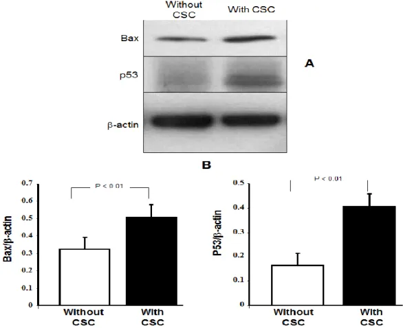
2.4.4 Cigarette smoke condensate induced Bax and p53 protein expression by human fibroblasts. As CSC caused the overexpression of several apoptotic genes in the human gingival fibroblasts, we examined whether this overexpression was mirrored with an increased protein production. We thus selected two key apoptotic proteins, Bax and p53, for this study. Using Western blot analysis on gingival fibroblasts after a 5-day exposure to 30% CSC, we revealed a significant induction of Bax and p53 proteins (Fig. 4). Indeed, both Bax and p53 proteins were significantly overproduced by almost 2-fold in the CSC-stimulated cells (Fig 4B). -actin was used as a loading control and did not vary between the controls and the CSC-stimulated cells. Bax and p53 protein expression also increased when the cells were exposed to 10 or 50% CSC for 5 days (data not shown).
32
Table 2-1: PCR arrays showing apoptotic activated/repressed genes following exposure of human gingival fibroblasts to cigarette smoke condensate.
Gene Description Fold Change
TNFRSF10A TNF receptor superfamily, member 10a 2.66 TNFRSF10B TNF receptor superfamily, member 10b 2.01 PYCARD PYD and CARD domain containing 1.95 BNIP3L Bcl2 interacting protein 3-like 1.84
CASP8 Caspase 8 1.73
CD40 CD40 molecule 1.73
BCL10 B-cell lymphoma 10 1.64
CASP7 Caspase 7 1.59
CASP10 Caspase 10 1.58
DIABLO DIABLO, IAP-binding mitochondrial protein 1.58
FAS Fas (TNF receptor superfamily) 1.56
TP53 Tumor protein p53 1.55
TNFRSF1B TNF receptor superfamily, member 1B 1.53
CASP6 Caspase 6 1.52
BAX Bcl2-associated athanogene 1.52
BFAR Bifunctional apoptosis regulator 1.48
BNIP3 Bcl2 interacting protein 3 1.48
LTBR Lymphotoxin beta receptor 1.44
LTA Lymphotoxin alpha -1.45
BCLA1 Bcl-2-related protein A1 -1.63
33 2.4.5 Cigarette smoke condensate increased caspase-3 activity in the gingival fibroblasts. Caspases are essential to the apoptotic response. Because some caspase genes were overexpressed when the fibroblasts were exposed to CSC, we investigated caspase activity by measuring caspase-3 activity in gingival fibroblasts following stimulation with CSC for 5 days. We then compared the activity of the CSC-stimulated cells with that of the control cells to calculate a percentage of 3 activity (Fig. 5). Cells grown in control medium for 5 days recorded 100 ± 28% caspase-3 activity, while cells grown with medium supplemented with 10, caspase-30, or 50% CSC showed higher caspase-3 activity ranging between 180 ± 42% and 190 ± 50%, which was a significant increase (p ≤ 0.01) compared to that recorded by the control. Caspase-3 activity was also measured after short (6 to 24 h) and long (7, 10, and 14 day) incubation times with CSC, with no significant increase of caspase-3 activity reported (data not shown).
34
Figure 2-4: Modulation of Bax and p53 protein expression by cigarette smoke condensate. Gingival fibroblasts were exposed to 0 or 30% CSC for 5 days and the total protein extracts were prepared. Bax and p53 expression was determined by Western blotting, and then scanned using Genetools software (Panel B). A -actin antibody was used as the loading control. The figure is representative of three different experiments.
35 Figure 2-5: Caspase-3 activity following cigarette smoke condensate stimulation. Gingival fibroblasts were exposed to CSC for 5 days and then suspended in lysis buffer. Protein extracts (100 µg) were assayed for caspase-3 activity by means of a CPP32 activity kit. The optical densities of the control cells and CSC-exposed cells were compared to determine a percentage of CPP32 activity. Data shown are the means SD of the casapase-3 activity in the samples calculated as a percentage of the control (non-CSC-exposed cells in which the caspase-3 activity was considered 100%). (n = 3).
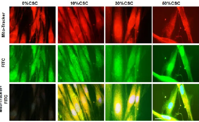
2.4.6 Bax and caspase-3 displayed cytosolic and mitochondrial co-localization. The subcellular location of Bax in the human gingival fibroblasts was determined by means of Zeiss Apotome fluorescence microscopy (Fig. 6) showing a diffuse cytosolic staining pattern with a superimposed punctate perinuclear component; low levels of diffuse nuclear staining were also observed (green
36
staining). The punctate component appeared similar to that observed with the mitochondria (red staining) following staining with mitochondrion-selective vital dye (MitoTracker, Life Technologies). Double staining with anti-bax and Mito-Tracker revealed that the punctate fraction of Bax co-localized with the mitochondria (overlapping red and green pixels seen as yellow). Similar analyses were conducted to localize caspase-3 in the CSC-exposed gingival fibroblasts, showing caspase-3 as co-localized with the mitochondria (Fig. 7). Overall data thus confirm that Bax and caspase-3 displayed cytosolic and mitochondrial distribution in the CSC-exposed gingival fibroblasts.
37 Figure 26: Bax localized in the cytosol and mitochondria of cigarette smoke condensate -exposed gingival fibroblasts. Following exposure to CSC for 5 days, cells were labeled with MitoTracker, fixed, permeabilized, and stained with mouse anti-human Bax monoclonal Alexa Fluor 488-conjugated secondary antibody. The stained cells were then examined under a Zeiss Apotome microscope. Bax-positive cells visualized with fluorescein were assigned the color green, while mitochondria labeled with MitoTracker were assigned the color red. When red and green images merge, the overlapping red and green pixels appear orange/yellow. Bar: 10 μm.
38
Figure 2-7: Caspase-3 localized in the cytosol and mitochondria of cigarette smoke condensate -exposed gingival fibroblasts. Following exposure to CSC for 5 days, gingival fibroblasts were labeled with MitoTracker, fixed, permeabilized, and stained with mouse anti-human casapse-3 monoclonal antibody, followed by Alexa Fluor 488-conjugated secondary antibody. The stained cells were then examined under a Zeiss Apotome microscope. Caspase-3-positive cells were visualized with fluorescein and assigned the color green, while mitochondria labeled with MitoTracker were assigned the color red. When red and green images merge, the overlapping red and green pixels appear orange/yellow. Bar: 10 μm.
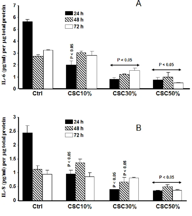
2.4.7 Cigarette smoke condensate decreased IL-6 and IL-8 secretion by the gingival fibroblasts. As shown in Fig. 8A, fibroblasts not exposed to CSC produced a high basal level of IL-6 after 24 h of culture under the appropriate conditions, while the cells exposed to CSC produced low levels of IL-6. The effect of CSC on IL-6 production was observable with each tested concentration. Of interest is that the low levels of IL-6 secreted by the fibroblasts were obtained
39 with an elevated CSC concentration. The decrease of IL-6 following cell exposure to CSC remained observable at 48 and 72 h (Fig. 8A). IL-8 was also modulated by the CSC. As shown in Fig. 8B, the unexposed fibroblasts produced a basal level of IL-8, which was significantly higher than that produced by the CSC-exposed cells. The reduction was observed with each tested CSC concentration. The effect of CSC on IL-8 secretion thus appeared to be linked to concentration level. Similar results were obtained at 48 and 72 h (Fig. 8B). We noted that the greater the CSC concentration, the lower the level of secreted IL-8.
40
Figure 2-8: Cigarette smoke condensate inhibited IL-6 and IL-8 secretions by gingival fibroblasts. Fibroblast cultures were exposed to various concentrations of CSC for 24, 48, and 72 h. Supernatant was then collected from each condition and used to measure IL-6 (panel A) and IL-8 (panel B) levels by ELISA. Data are expressed as means ± SD.
41 2.5 Discussion
In the present study, we demonstrated that the continuous exposure of normal human gingival fibroblasts to CSC resulted in shape change and growth inhibition as confirmed by optical microscope observations and MTT. Cigarette smoke induced both apoptosis and necrosis as of human gingival fibroblasts. Cells exposed to CSC showed significant increase in apoptotic/necrotic cell density, as ascertained by annexin-V/propidium iodide staining. These results are consistent with those reported for the effects of CSC on human bronchial (22) and nasal epithelial cells (23). The decreased growth of gingival fibroblasts following exposure to CSC may have occurred due to cell-cycle deregulation (24), as demonstrated in this study, where CSC increased the G0/G1 phases and decreased the S and G2/M phases, compared to that observed with the non-exposed cells. Our results also support a previous study showing that cigarette smoke compounds such as nicotine inhibited cell cycle progression by inducing the G1 arrest of various cell lines including
HaCaT, IHOK, HN4, and HN12 (25). Taken together, the cell damaging effects of CSC on gingival fibroblasts might potentiate inflammation, leading to periodontal disease.
Our PCR array data demonstrate that CSC modulated gene expression in gingival fibroblasts involved in the apoptotic/necrotic pathways, including Bax, previously shown to relate to apoptosis (26). Bax upregulation may be involved in the modulation of fibroblast apoptosis through the disruption of the pro-apoptotic–anti-apoptotic balance in the mitochondrial apoptosis pathway, as previously reported (27). Furthermore, by the opening of permeability transition pores, the activation and translocation of Bax to the mitochondria may induce the release of apoptotis-inducing proteins into the cytosol (27).