http://jhc.sagepub.com/
Journal of Histochemistry & Cytochemistry
http://jhc.sagepub.com/content/34/7/883
The online version of this article can be found at:
DOI: 10.1177/34.7.3519751
1986 34: 883
J Histochem Cytochem
E Van Vliet, M Melis, J M Foidart and W Van Ewijk
Reticular fibroblasts in peripheral lymphoid organs identified by a monoclonal antibody.
Published by:
http://www.sagepublications.com
On behalf of:
Official Journal of The Histochemical Society
can be found at:
Journal of Histochemistry & Cytochemistry
Additional services and information for
http://jhc.sagepub.com/cgi/alerts
Email Alerts:
http://jhc.sagepub.com/subscriptions
Subscriptions:
http://www.sagepub.com/journalsReprints.nav
Reprints:
http://www.sagepub.com/journalsPermissions.nav
Permissions:
This investigation was supported by project grant 13-27-66 from FUNGO, The Netherlands.
0022-1554/86/83.30
The Journal of Histochemistry and Cytochemistry
Copyright © 1986 by The Histochemical Society, Inc.
Vol. 34, No. 7, pp. 883-890, 1986
Printed in U.S.A.
Original
Article
Reticular
Fibroblasts
in Peripheral
Lymphoid
Organs
Identified
by a Monoclonal
Antibody1
ELS VAN
VLIET, MARLEEN MELIS, JEAN M. FOIDART, and WILLEM VAN EWIJKDepartment of Cell Biology and Genetics (E.VV; MM.; WVE.), Erasmus University, Rotterdam, The Netherlands; Laboratory ofExperimental Dermatology (J.M.F), University ofLilge, Lilge, Belgium
Received for publication June 11, 1985 and in revised form November 15, 1985; accepted November 22, 1985 (5A0434).
We have produced a panel of monodonal antibodies di-rected against nonlymphoid cells in central and peripheral lymphoid organs. In this paper we present the reactivity of one of these antibodies, ER-TR7. This antibody detects re-ticular fibroblasts, which constitute the cellular framework oflymphoid and nonlymphoid organs and their products. In frozen sections of the spleen incubated with this anti-body, the red pulp and white pulp are clearly delineated. Furthermore, the major white pulp compartments - the
follides and peniarteriolar lymphoid sheath as well as the
marginal zone - are recognized by their characteristic
label-ing patterns. In lymph nodes, the capsule, sinuses, follides,
Introduction
Peripheral lymphoid organs, such as the spleen and lymph nodes,
are highly compartmentalized. Within these organs T and B
lym-phocytes each have their own domains. In the spleen, B cells local-ize in follicles in the peripheral part of the white pulp (de Sousa,
1971; Gutman and Weissman, 1973; Nieuwenhuis and Ford, 1976)
and in the marginal zone (MZ) (Kumanaratne et al., 1981), which
separates the white pulp from the red pulp. T cells, on the other
hand, occupy the central area of the peni-arteniolar lymphoid sheath (PALS) (Mitchell, 1972; van Ewijk et al., 1974). In lymph nodes, B cells localize in the follicles in the outer cortex, whereas T cells occupy the paracortical area (Pammott et al. , 1966; van Ewijk and van den Kwast, 1980). Medullamy cords predominantly contain
plasma cells which migrate during differentiation from the outer
cortex into this region. T and B cells both enter the splenic white
pulp via the MZ (Fond, 1969; Nieuwenhuis and Fond, 1976;
Bnelinska and Pilgrim, 1982). In the lymph nodes they enter
through high endothelial post-capillary venules (HEV) located in
the pamacortex (Butcher et al., 1980). Upon entry, T and B cells segregate and migrate into their respective domains.
The factors that direct the migration and specific homing of B and T cells into their respective domains are still unknown. From
paracortex, and medullary cords are dearly delineated. In the thymus and bone marrow no such specialized compart-ments were demonstrated. ER-TR7 reacts with an intracellu-lar component of fibroblasts. Since ER-TR7 does not react with purified laminin, collagen types I-V. fibronectin, hep-aran sulfate proteoglycan, entactin, or nidogen, it detects a hitherto uncharacterized antigen. The possible role of the
ER-TR7 positive reticular fibroblasts in the cellular organi-zation of peripheral lymphoid organs will be discussed.
KEY WORDS: Monoclonal antibodies; Reticular fibroblasts; Spleen;
Lymph node; Immunohistology; Mouse.
light and electron microscopic studies it is known that both spleen and lymph nodes contain several nonlymphoid cell types, such as interdigitating cells (IDC), follicular dendnitic cells (FDC), macro-phages, and reticular fibroblasts (Veerman and van Ewijk, 1975; Humphrey and Gmennan, 1982). Morphologic observations
mdi-cate that FDC and IDC possibly play a role in the homing of B and T cells, respectively (van Ewijk et al., 1974, Dijkstra and Dopp,
1983). Little is known about the function of reticular fibmoblasts
in the lymphoid microenvironments. These cells constitute a
sup-portive cellular framework and they may also help direct in the
migration and localization of lymphocytes (de Sousa, 1969;
Bar-clay, 1981).
We recently produced a panel of monoclonal antibodies di-mected against nonlymphoid cells of the mouse thymus (van Vliet et al., 1984a). These antibodies provide a new approach for a detailed structural analysis ofthe nonlymphoid constituents of the
thymus. In this paper we present an extensive study of the
reactiv-ity of one of these antibodies, ER-TR7 (ER-TR = Erasmus
Univer-sity Rotterdam -Thymic Reticulum), which also reacts with the
stroma of peripheral lymphoid organs.
The purpose ofthis study is threefold: first, to analyze in detail the anatomy of peripheral versus central lymphoid organs using the monoclonal antibody ER-TR7, second, to describe the
meac-tivity of ER-TR7 in a variety of other tissues, and third, to analyze
the nature ofthe antigen detected by ER-TR7. The tissue distmibu-tion of the antigen expressed on stromal cells and detected by
884 VAN VLIET, MELIS, FOIDART, VAN EWIJK
Enzyme-linkedimmunosorbent assay (ELISA). ELISA was performed monoclonal antibody ER-TR7 was studied using the immunoper-oxidase technique on frozen sections.
Materials
and
Methods
Mice. Male and female C3H/He) and (CBA x C57BL/6)F1 mice,
aged 6-12 weeks, were used for this study. They were kept in our animal
colony under routine laboratory conditions.
Monoclonal antibody. Details of the production of rat monoclonal
antibodies directed against stromal cells of the mouse thymus have been published elsewhere (van Vliet et al., 1984a). We obtained seven hybrid
cell lines that produce antibodies against various stromal cell types in the thymus. In this study we describe the reactivity of one of these antibodies:
ER-TR7, an immunoglobulin G2a (IgG2a) antibody, which also reacts
with antigens of the reticular framework oflymphoid organs of the mouse.
Conjugate. Rabbit anti-rat immunoglobulin coupled to horseradish peroxidase (RaRa-Ig-HRP) (Dako, Copenhagen, Denmark) was used. To
prevent nonspecific binding of the conjugate, it was deaggregated by cen-tnifugation in a Beckman Aimfuge at 10’ g. The conjugate was optimally
diluted in PBS containing 0.5% bovine serum albumin (BSA) and 1%
normal mouse serum (NMS).
Preparation and incubation of frozen sections. Frozen sections were prepared and incubated with monoclonal antisera and photography was
performed as described elsewhere (van Ewijk et al., 1981).
Mouse fibroblast cell lines. Mouse fibroblast cell line 129 was initiated in our laboratory as a primary culture of strain 129 skin fibroblasts. A9 is a mouse L-cell derivative (Littlefield, 1964).
Preparation and incubation offibroblast cell lines. Cells were isolated
with a rubber policeman, fixed, and embedded in agar, and frozen
see-nons were cut as reported before (vanVliet et al., 1984b). Sections were
then incubated with ER-TR7 as described above.
Reticulin stain. Frozen tissue sections were stained for reticulin with
routine silver impregnation according to Gom#{246}rri(1952).
Further characterization of the antigen. The possible antigenic rela-tionship of the matrix component detected by ER-TR7 with laminin, types
I-V collagens, nidogen, entactin, fibronectin, and heparan-sulfate-rich
basement membrane proteoglycan was tested in the following ways.
Ouchterlony immunodiffusion. Double radial immunodiffusion in
1% agarose was performed in Immuno-Tek II OT agarose plates
(Behning-werke, Marburg, West Germany). ER-TR7 antibody was put into the cen-tral well and the connective tissue antigens were put in the peripheral wells. The proteins were allowed to diffuse overnight at room temperature
in a moist chamber.
Immunoelectropboresis. Immunoelectrophoresis in 1% agarose was
performed in electrophoresis base and agar gel plates (Hyland Laborato-ries, Costa Mesa, California). Laminin, fibronectin, or entactin was placed
in the well and electrophoresed for 45 mm at 30 mA in a barbital buffer system, pH 8.6. Either antibodies to these proteins on ER-TR7 were then
placed in the trough. The proteins were allowed to diffuse overnight at room temperature in a moist chamber.
Radioimmunoassay. Interstitial and basement membrane connective tissue antigens were iodinated with 125I by the chloramine-T method (McConahey and Dixon, 1966). Radioimmunoassay (RIA) was performed as described (Rohde et al., 1976), using purified antibodies to laminin,
entactin, types I-IV collagens, fibronectin, heparan sulfate proteoglycan, or ER-TR7 antibody.
as described previously (Voller et al., 1976). Serial dilutions of purified rabbit antibody to the connective tissue macromolecules were applied to microtiter wells (Cooke Laboratory Products Division, Alexandria, Vim-ginia) coated with fibronectin, laminin, or the other biochemically charac-tenized tested antigens.
Immunofluorescence. In order to determine whether the ER-TR7 anti-body bound to the same antigenic moieties as did the antibodies to the
matrix macromolecules, blocking studies were performed using mouse
skin sections as substrate according to a previously described protocol
(Yaoita et al., 1978). For example, purified antibody to laminin was
con-jugated with fluorescein isothiocyanate (Goldman, 1968). Unlabeled ER-TR7 antibody was reacted for 3 mm. Unbound reagents were washed way, and fluoresceinated antibody to laminin was applied for another 30 mm.
The sections were then extensively washed and mounted. Conversely, in
other studies, unlabeled rabbit antibody to matrix proteins was first ap-plied to tissue sections for 30 mm, unbound antibody was washed away,
and ER-TR7 antibody was applied for another 30 mm. After extensive
washings, bound rabbit and rat antibodies were detected using
fluores-cein-conjugated antibody to rabbit or rat immunoglobulin. Ifthe first an-tibody “blocked” binding of the second antibody, this would indicate
ci-then that they were binding to the same antigenic moieties or that there was stenic hindrance by the first antibody. The specificity of the ER-TR7
antibody was also assessed by measuring its binding to agarose beads
coated with connective tissue proteins, as described by Yaoita et al. (1978).
Finally, in other studies ER-TR7 antibody was preincubated overnight at
CC with 100 ig of each tested antigen in a test tube before
im-munofluorescence in order to block binding of antibody to the tissue section.
Immunoblotting. The reactivity ofER-TR7 with laminin, fibronectin, entactin, nidogen, and types I-V collagens was tested by
immunoperoxi-dase staining of their polypeptides, after electrophoretic transfer from
polyacrylamide gels to nitrocellulose sheets as described by Towbin et al. (1979).
Results
In the first part of this section we will describe the localization of cells expressing the antigen detected by ER-TR7 in peripheral lym-phoid organs. In the second part we will describe the distribution patterns of ER-TR7 observed in central lymphoid organs such as thymus and bone marrow. To obtain further information on the nature of the antigen detected by ER-TR7, we studied the reactiv-ity of ER-TR7 with various nonlymphoid tissues and with fibmo-blast cell lines, and we compared the reactivity pattern of ER-TR7 with a conventional reticulin stain. We also analyzed the possible
antigenic relationship of the antigen detected by ER-TR7 with
var-ious purified connective tissue components.
Anatomical
Distribution
of ER-TR7
Positive
Cells
in
Spleen
andLymph
Node
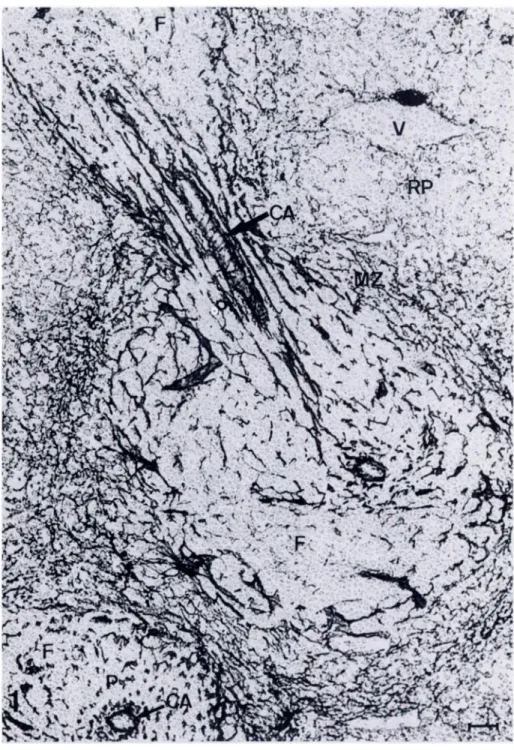
Frozen sections incubated with ER-TR7 followed by RaRa-Ig-HRP and diaminobenzidine (DAB) clearly show the two major com-partments in the spleen - the white pulp and the med pulp. Figure
1 shows that the white pulp is located around central arterioles which branch from the splenic artery. This area can be easily dis-tinguished from the med pulp, which is characterized by a ran-domly distributed meshwork. Within the white pulp, three dis-tinct areas can be delineated by their characteristic labeling
RETICULAR FIBROBLASTS IN PERIPHERAL LYMPHOID ORGANS 885
Figure 1. Immunoperoxidase staining pat-tern of frozen section of spleen. Section in-cubated with monoclonal antibody ER-TR7, followed by RaRa-lg-HRP and DAB. Note both longitudinal and transverse sections ofthe PALS. CA, centralarteriole; F, follicle; P, periarteriolar lymphoid sheath; MZ, mar-ginal zone; RP, red pulp; V, venule. Original magnification x 280. Bar = 25 pm.
patterns. In a longitudinal section ofthe spleen, the PALS contains
a network of fibers, concentrically arranged in sheaths, parallel to
the central arteriole. The central arteriole is outlined by a brightly
stained wall. Follicles at the periphery ofthe PALS are virtually un-stained by the antibody, except for the outer boundary of the
folli-dc with the MZ. The staining pattern in the MZ is a meticular meshwork, which is far more dense than in the red and white pulp. The marginal zone gradually merges into the reticulum of the
cords in the red pulp. Red pulp sinuses clearly stand out as
nega-tive areas. Tmabeculae and the splenic capsule are strongly positive.
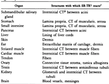
In frozen sections of the lymph node, the cortex and the medulla can be delineated with ER-TR7 (Figure 2a). As in splenic sections, follicles located in the outer cortex are unstained, except for the outer boundary. The interfollicular areas of the outer cor-tex and the paracortex show a characteristic fine reticular staining pattern. The walls of HEV located in the paracortical area also me-act strongly with this antiserum. The capsule stains intensely with ER-TR7, whereas the subcapsular sinus is negative. In the medulla, strongly stained cords can be distinguished from negative sinuses.
f
p
m
b
--a . , _fr-- , I,,:i
:=-T
-::
l’i; a’ #{149}. / l.#{149} , , - . . . -. .:
-. 5’.. . .;; ..Y , ;:S*/;.; .d
886 * I .‘ . pVAN YLIET, MELIS, FOIDART, VAN EWIJK
: Figure 2. Immunoperoxidase staining
pat-tern of (A,B) mesenteric lymph node, (C) thymus, and (D) bone marrow. A,C,D, incu-bation with ER-TR7; B, negative control. ca, capsule; c, cortex; f, follicle; hey, high en-dothelial venule; m, medulla; mc, medul-lary cord; ms, medullary sinus; p, paracor-tex; ss, subcapsular sinus; t, trabeculae; v, blood vessel. Original magnifications: A,C
x 60 (bar = 115 pm); B,D x 140 (bar =
50 pm).
in negative control sections - sections incubated with
RaRa-Ig-HRP and DAB only (Figure 2b).
Taken together, results of labeling of frozen sections of periph-eral lymphoid organs with ER-TR7 demonstrate the various
do-mains, to which T and B lymphocytes localize.
Anatomical
Distribution
ofER-TR7
Positive
Cells
in
Thymus
and
Bone
Marrow
To study a possible compamtmentalization of thymus and bone marrow, we incubated frozen sections ofthese organs with ER-TR7.
Frozen thymus sections incubated with ER-TR7 show staining
mainly of the capsule, blood vessels, and trabeculae (Figure 2c).
The border between cortex and medulla is not clearly outlined, although these regions can be distinguished.
In frozen sections of bone marrow plugs labeled with ER-TR7 (Figure 2d), a slight meticular pattern can be noted, together with strong staining of the walls of blood vessels. However, in contrast with the spleen and lymph node, this labeling pattern does not demonstrate any compamtmentalization within the bone marrow.
Anatomical
Distribution
ofER-TR7
Positive
Cells
in Nonlymphoid
Organs
We incubated frozen sections of a variety of nonlymphoid organs and tissues with ER-TR7 in order to study reactivity patterns in
RETICULAR FIBROBLASTS IN PERIPHERAL LYMPHOID ORGANS 887
these organs. The results are summarized in Thble 1. A few
exam-ples are shown below to illustrate the specificity of ER-TR7.
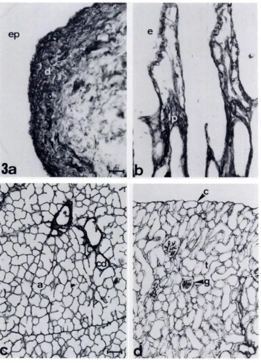
In frozen sections of skin incubated with ER-TR7 (Figure 3a) the dermis was confluently labeled, whereas the epidermis was negative. Sections of the small intestine incubated with ER-TR7 show a similar confluent staining of the lamina propria (Figure
3b). No staining of the intestinal epithelium was observed.
Simi-lamly, in sections of the stomach a positive lamina propnia and a
negative epithelium were observed (not shown). The ovarian
stro-ma was positive, whereas the follicles were negative (not shown).
ER-TR7 stains the connective tissue that forms a supporting
net-work between parenchymal cells in various organs, such as salivary gland, kidney, testis, liver, and pancreas. Cardiac and striated muscle also contain such an ER-TR7-positive connective tissue net-work. Examples of this staining pattern are in sections of salivary gland and kidney are shown in Figure 3c and d. In the salivary gland the epithelial cells ofthe acini and collecting ducts are
nega-tive, whereas the reticulam connective tissue around the acini and
ducts can be seen as thin lines (Figure 3c). A similar staining of
connective tissue elements, but no staining of muscle fibers, was
observed in striated muscle sections. In the testis the interstitial tissue between seminiferous tubuli is positive. Both glomeruli and interstitial connective tissue between kidney tubules are strongly labeled with ER-TR7 (Figure 3d). In the liver the lining of
sinu-soids are positive, whereas parenchymal cells are not labeled.
ER-TR7 reacts with the extracellulam matrix of cartilage and with the
dermis in sections of ear (not shown). Blood vessels always showed
a strongly positive wall.
In conclusion, in all the organs mentioned above, connective tissue compartments can be identified with the present mono-clonal antibody.
Comparison of the Reactivity Pattern of
ER-TR7
with a Conventional Reticulin Stain
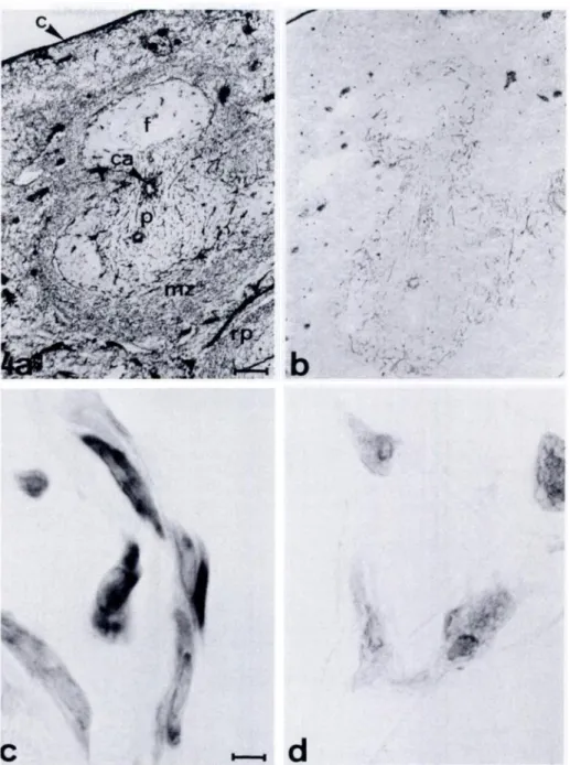
Further study of the nature of the antigen detected by ER-TR7 in-volved conventional silver impregnation of spleen sections. This method is known to detect reticulin, which is defined as the con-nective tissue that stains with silver (Hay et al., 1978). Figures 4a
and b show serial frozen spleen sections stained respectively with ER-TR7 or by conventional silver impregnation. Silver impregna-tion resulted in a reticular labeling pattern in the splenic white
pulp that was in general similar to the staining pattern observed
with ER-TR7, although less dense. However, no labeling ofthe
ER-TR7-positive meshwork of the splenic red pulp and MZ was seen
after silver impregnation. From this observation we tentatively conclude that ER-TR7 reacts with meticulin but also with other con-nective tissue components yet to be determined.
Reactivity ofER-TR7 with Fibroblast Cell Lines
To study the reactivity of ER-TR7 with fibmoblasts, we incubated frozen sections ofcell pellets ofmouse fibmoblast cell lines 129 and A9 with ER-TR7. ER-TR7 was shown to react with the cytoplasm offibmoblasts ofeach ofthese cell lines (Figure 4c). 80-90% of the cells are ER-TR7 positive. A negative control section is shown in Figure 4d.
Table 1. Reactivity ofER-TR7 with various nonlymphoid organs ofthe mouse
Organ Structures with which ER-TR7 react?
Submandibular salivary Interstitial CTb between acini
gland
Stomach Lamina propnia, CT of musculanis, serosa
Small intestine Lamina propria, CT of musculanis, serosa
Pancreas Interstitial CT between acini
Liver Lining of liver cords
Skin Dermis
Ear Extracellular matrix of cartilage, dermis
Striated muscle Interstitial CT between muscle fibers
Cardiac muscle Interstitial CT between muscle fibers
Tendon Fibers
Ovary Connective tissue stroma, tunica albuginea
Testis Interstitial CT between seminiferous tubuli
Kidney Glomeruli and interstitial CT between
tubuli
Brain Blood vessels, meninges
a In tissues tested ER-TR7 reacts with blood vessel walls and capsule.
bCT = connective tissue.
Characterization
of the
Antigen
Detected
by
ER-TR7
The reactivity ofER-TR7 with purified laminin, fibronectin, types I-V collagens, heparan sulfate proteoglycan, entactin, and nido-gen was investigated by the Ouchterlony technique, immunoelec-trophomesis, RIA, ELISA, immunoelectroblotting and indirect immunofluorescence blocking and inhibition studies. ER-TR7 reacted with none of the tested matrix components in any of the test systems, whereas the control antisera specific for these matrix components detected them in mouse tissue sections. These results show that ER-TR7 does not detect a strict basement membrane component or any major collagen type or fibmonectin. In addition, ER-TR7 reacts in immunofluorescence studies with interstitial
stroma and matrix cartilage but not with the basement membrane
matrix deposited by EHS sarcoma or L2 tumors, two transplant-able munine and rat tumors that synthesize a matrix of basement membrane (Timpl et al., 1979; Wever et al., 1981). In culture, ER-TR7 reacts with munine fibroblasts but not with L2 cells, vascular endothelial cells, or glomerular epithelial cells, which suggests that the antigen detected by ER-TR7 is synthesized by mesen-chymal cells rather than by epithelial or endothelial cells.
Discussion
In this study we analyze the anatomical distribution of the cellular
framework of lymphoid and nonlymphoid organs, detected by
monoclonal antibody ER-TR7 and the nature of the antigen de-tected by ER-TR7. Our results clearly demonstrate that ER-TR7 can be used to study the micro-anatomy of various organs. In sum-mary, we have demonstrated that 1) ER-TR7 outlines the various compartments of peripheral lymphoid organs by characteristic labeling patterns; 2) no such compartments are found in central
lymphoid organs; 3) ER-TR7 delineates various types of connective
888 VAN VLIET, MELIS, FOIDART, VAN EWIJK
Figure
a
Immunoperoxidase staining pat-tern of(A) skin, (B) small intestine, (C) sali-vary gland, (D) kidney. a, acinus; c, cap-sule; cd, collecting duct; d, dermis; e, epithelium; ep, epidermis; g, glomerulus; Ip, lamina propria; t, tubules. Original mag-nifications: A,B x 350 (bar = 20 pm); C,Dx 140 (bar = 50 pm).
detected is not a basement membrane component, nor any major collagen type or fibmonectin.
Our results show, furthermore, that ER-TR7 reacts with the
ba-sic cellular framework in peripheral lymphoid organs. This
cellu-lam framework consists of the reticular fibmoblasts, described by M#{252}ller-Hermelink et al. (1974), Veerman and van Ewijk (1975), Villena et al. (1983), and their products. The intracellular reactiv-ity of ER-TR7 with fibmoblast cell lines and the confluent staining in the dermis of the skin and the lamina propria of the intestine shows that ER-TR7 not only detects intracellular components of fi-broblasts, but also reacts with extracellular products. The spleen sections incubated with ER-TR7 and stained by conventional silver
impregnation clearly demonstrate that the antigen detected by ER-TR7 has a tissue distribution wider than that of reticulin. The major difference between these two staining procedures is that me-ticular components in the marginal zone and in med plup are de-tected by ER-TR7 antibodies but not by silver impregnation. Al-though the tissue distribution suggests that this antibody detects a major component of the extmacellulam matrix such as collagen, the fact that ER-TR7 does not react with a variety of collagens, gly-coproteins, or basement membrane proteoglycan with compara-ble tissue distributions demonstrates that ER-TR7 does not detect a strict basement membrane component or any major collagen type, or the biochemically characterized glycoprotein fibmonectin.
Figure 4. (A) Immunoperoxidase staining pattern ofspleen with ER-TR7. (B)Silver im-pregnation pattern of spleen. (C,D) lm-munoperoxidase staining pattern of frozen sections of a pellet of A9 cells, incubated with ER-TR7(C)and a negative control sac-tion (D). c, capsule; ca, central arteriole; f, follicle; p, periarteriolar lymphoid sheath;
mz, marginal zone; rp, red pulp; I, trabecu-lae. Original magnifications: A,B x 60 (bar
= 115 pm); C,D x 875 (bar = 8 pm). . ‘. V . ,. .. ,. .. ..,.. .,. . -.- . I ‘ ‘ . . -, .. . , --1 .
:
...- .-
p / “ -. . % .,. , . #{149}. :_‘,. , ..-‘:;
- ‘ ,i,..
*__,_ . \#{149} :‘ ‘ ! . ‘-S
. S4’
)1
I
C
RETICULAR FIBROBLASTS IN PERIPHERAL LYMPHO1D ORGANS
The tissue distribution of the antigen recognized by ER-TR7 is clearly distinct from those reported for all other biochemically characterized connective tissue macromolecules - the ER-TR7
an-tigen is a ubiquitous component of stromal (intemstitital) matrix cartilage and ofat least some basement membrane zones. ER-TR7
is particularly useful as a tool for distinguishing various lymphoid
compartments in central and peripheral lymphoid organs. Thus, based on the distribution pattern of ER-TR7-positive fibmoblasts, the various compartments in the spleen - med pulp, MZ, PALS, and follicles - and in lymph nodes - pamacortex, follicles, and medulla- can be clearly distinguished. This antibody also shows that the thymus, a lympho-epithelial organ, shows only
mesen-L:#{149}1..
-b
889
chymal components in the medulla and in the capsule. As can be judged from the staining pattern, these fibroblasts are not only present as components in a general framework structure, but also create various microenvimonments in the different lymphoid com-partments. In the spleen this is most obvious in the marginal zone and in the central part of the white pulp (T zone). We speculate that this arrangement of fibroblasts participates in two major functions of the spleen - phagocytosis of blood-borne substances (e.g. , erythrocytes, antigens) and initiation of the immune me-sponse. The dense meshwork in the marginal zone might then
function as a filter, slowing the flow of blood into this particular
890 VAN YLIET, MELIS, FOIDART, VAN EWIJK
McConahey PJ, Dixon F) (1966): A method oftmace iodination of proteins
for immunological studies. Int Arch Allergy Appl Immunol 29:185
Yaoita H, FoidantjM, Katz 51(1978): Localization ofthe collageneous
corn-ponent in skin basement membrane.
J
Invest Dermatol 70:191mononuclear phagocytes associated with this meticulam meshwork can optimally clear the in-flowing blood (van Vliet et al., 1985). The typical arrangement of fibroblasts in lymphoid organs might also guide migration of lymphocytes after their entry into the
splenic white pulp or into the lymph node paracortex (de Sousa,
1969; Barclay, 1981), and promote the intercellular contact be-tween lymphocytes and the cell types that regulate the ultimate homing of lymphocytes into their respective domains.
Acknowledgments
thank Mr T van Osforprinting thephotographs andMrs. C. Me:jer-ink for excellent typing assistance.
Literature
Cited
Barclay AN (1981): Different reticulan elements in rat lymphoid tissue identified by localisation of Ia, Thy-i and MRC OX2 antigens. Immunol-ogy 44:727
Brelinska R, Pilgrim C (1982): Significance of subcompantments of mar-ginal zone for direction oflymphocyte traffic within spleen pulp. Cell Tis-sue Res 226:155
Butcher EC, Scollay RG, Weissman IL (1980): Organ specificity of lympho-cyte migration: Mediation by highly selective lymphocyte interaction with organ-specific determinants on high endothelial venules.
J
Immunol10:556
de Sousa MAB (1969): Reticulum arrangement related to the behaviour of
cell populations in the mouse lymph node. Adv Exp Med Biol 5:49
de Sousa MAB (1971): Kinetics ofthe distribution ofthymus and marrow cells in the peripheral lymphoid organs of the mouse: Ecotaxis. Clin Exp Immunol 9:371
Dijkstra CD, Dopp EA (1983): Ontogenetic development ofT and B
lym-phocytes and non-lymphoid cells in the white pulp of the rat spleen. Cell
Tissue Res 229:351
Ford WL (1969): The kinetics of lymphocyte recirculation within the rat spleen. Cell Tissue Kinet 2:171
Goldman M (1968): Labeling agents and procedures for conjugation. In
Goldman M, ed. Fluorescent Antibody Methods. New York, Academic
Press, 97
Gom#{246}mmiG (1952): Microscopic histochemistry. Principles and practice. Chicago, University of Chicago Press.
Gutman GA, Weissman IL (1973): Homing properties of
thymus-independent folliculan lymphocytes. Transplantation 16:621
Hay ED, Hasty DL, Kiehnau H (1978): Fine structure ofcollagens and their relation to glucosaminoglycans (GAG). Thromb Haemost 63(suppl):129 HumphreyJH, Grennan D (1982): Isolation and properties ofspleen fol-licular dendnitic cells. Adv Exp Med Biol 149:823
Kumararatne DS, Bazin H, MacLennan 1CM (1981): Marginal zones: The
major B cell compartment of rat spleens. Eur) Immunol 11:858
Littlefield )W (1964): Three degrees of guanylic acid, inosinic, and pyrophosphorylase deficiency in mouse fibroblasts. Nature 203:1142
Mitchell) (1972): Antigens in immunity. XVII. The migration of antigen-binding bone marrow derived and thymus derived spleen cells in mice. Im-munology 22:231
MUllen-Henmelink HK, Heusermann U, Stutte H) (1974): Enzyme
histochemical observation on the localisation and structure of the T cell
and B cell regions in human spleen. Cell Tissue Res 154:167
Nieuwenhuis P, Ford WL (1976): Comparative migration of B and T lym-phocytes in the rat spleen and lymph nodes. Cell Immunol 23:254
Pamrott DMV, de Sousa MAB, East ) (1966): Thymus-dependent areas in the lymphoid organs of neonatally thymectomized mice.
J
Exp Med123:191
Rohde H, Nowak H, Becker Y, Timpl R (1976): Radioimmunoassay for the
amino-terminal peptide ofprocollagen pal (1)-chain.) Immunol Methods 11:135
Timpl R, Rohde H, Robey PG, Rennand SI, FoidantJM, Martin GR (1979):
Laminin: A glycoprotein from basement membranes. ) Biol Chem
254:9933
Towbin H, Staehelin T, Gordon
J
(1979): Electrophometic transfer of pro-teins from polyacrylamide gels to nitrocellulose sheet: Procedure and some applications. Proc NatI Acad Sci USA 76:4350van Ewijk W, van den Kwast ThH (1980): Migration of B lymphocytes in
lymphoid organs oflethally irradiated, thymocyte-reconstituted mice. Cell Tissue Res 212:497
van Ewijk W, van Soest PL, van den Engh GJ (1981): Fluorescence analysis
and anatomic distribution of mouse T lymphocyte subsets defined by
monoclonal antibodies to the antigens Thy-i, Lyt-1, Lyt-2 and T-200.
J
Im-munol 127:2594van Ewijk W, Verzijden )WM, van der Kwast ThH, Luijcx-Meijer SWM
(1974): Reconstitution of the thymus dependent area in the spleen of
lethally irradiated mice. A light and electron microscopical study of the
T cell microenvironment. Cell Tissue Res i49:43
van Vliet E, Melis M, van Ewijk W (1984a): Monoclonal antibodies to
stromal cells of the mouse thymus. Eur) Immunol 14:524
van Vliet E, Melis M, van Ewijk W (1984b): Immunohistology of thymic nurse cells. Cell Immunol 87:iOi
van Vliet E, Melis M, van Ewijk W (1985): Marginal zone macrophages in
the mouse spleen identified by a monoclonal antibody ) Histochem
Cytochem 33:40
Veenman A)P, van Ewijk W (1975): White pulp compartments in the
spleen of rats and mice. A light and electron microscopic study of
lym-phoid and nonlymphoid cell types in T and B areas. Cell Tissue Res
156:417
Villena A, Zapata A, Rivera-Pomar )M, Bamnitia MG, Fonfnia ) (1983): Structure of the nonlymphoid cells during the postnatal development of the rat lymph nodes. Cell Tissue Res 229:219
Voller A, Birdwell DE, Barlett A (1976): Microplate enzyme immunoassays
for the immunodiagnosis ofvirus infections. In Rose N, Fishman M, eds.
Manual of Clinical Immunology. Washington, DC, American Society for
Microbiology, 506
Wever U, Albrechtsen R, Ruoslahti E (1981): Laminin, a noncollagenous component of epithelial basement membranes synthesized by a rat yolk sac tumor. Cancer Res 41:1518