QUANTITATIVE ESTIMATION OF ESTROGEN AND
ANDROGEN RECEPTOR-IMMUNOREACTIVE CELLS IN
THE FOREBRAIN OF NEONATALLY
ESTROGEN-DEPRIVED MALE RATS
J. BAKKER,*‡ C. W. POOL,† M. SONNEMANS,† F. W. VAN LEEUWEN† and
A. K. SLOB*
*Department of Endocrinology and Reproduction, Faculty of Medicine and Health Sciences, Erasmus University Rotterdam, P.O. Box 1738, 3000 DR Rotterdam, The Netherlands †Graduate School Neurosciences Amsterdam, Netherlands Institute for Brain Research,
Meibergdreef 33, 1105 AZ, Amsterdam, The Netherlands
Abstract––Using quantitative immunocytochemical procedures, the total number of estrogen and andro-gen receptors was estimated in a large number of hypothalamic and limbic nuclei of male rats, in which brain estrogen formation was inhibited neonatally by treatment with the aromatase inhibitor 1,4,6-androstatriene-3,17-dione. The highest densities of estrogen receptor immunoreactivity were observed in the periventricular preoptic area and the medial preoptic area. Neonatally estrogen-deprived males showed a higher estrogen receptor immunoreactivity than control males in the periventricular preoptic area and the ventrolateral portion of the ventromedial nucleus of the hypothalamus, i.e. those brain areas in which sex differences have been reported, with female rats showing a greater estrogen binding capacity than male rats. The highest densities of androgen receptor immunoreactivity were found in the septohypothalamic nucleus, the medial preoptic area, the posterior division of the bed nucleus of the stria terminalis and the posterodorsal division of the medial amygdaloid nucleus. No significant differences in distribution or total numbers of androgen receptors were found between neonatally estrogen-deprived males and control males.
These findings suggest that neonatal estrogens, derived from the neural aromatization of testosterone, are involved in the sexual differentiation of the estrogen receptor system in the periventricular preoptic area and the ventromedial hypothalamus. The role of neonatal estrogens in the development of the forebrain androgen receptor system is less clear.? 1997 IBRO. Published by Elsevier Science Ltd.
Key words: neonatal ATD treatment, androgen receptor, estrogen receptor, estrogens, hypothalamus,
preoptic area.
In mammals, testosterone and estradiol derived from
neural aromatization of testosterone act
syner-gistically in the developing male brain to organize its
structures and functions, i.e. to masculinize and
defeminize the neural substrates that later control
sexual behavior and neuroendocrine function. In
the rat brain, masculinization results in the display of
male-typical sexual behavior (mounts, intromissions
and ejaculations) in adulthood. Defeminization
re-sults in a loss of cyclic release of gonadotropins
necessary for ovulation and a loss of female-typical
sexual behavior (lordosis, presenting, ear wiggling,
hop and dart) in adulthood (e.g., Ref. 37). Thus,
gonadally intact male rats display the complete
pat-tern of male coital behavior when pair tested with an
estrous female and no female-typical sexual behavior
when pair tested with a sexually active male.
How-ever, lordosis behavior can be induced in castrated
male rats by administering estradiol and
progester-one, although the behavior displayed is usually less
intense than that of females, and considerably more
estradiol is required for its stimulation.
20,42,54The development of the female rat brain is
gener-ally believed to proceed in the absence of testosterone
and estradiol. Gonadally intact female rats display
periods of behavioral proceptivity, i.e. seeking out
the male partner, and receptivity, i.e. the display of
lordosis in response to mounts, around the time of
ovulation.
12However, male-typical sexual behavior,
such as intromission-like and ejaculation-like
behav-ior, can be induced in ovariectomized female rats
by administering testosterone or estradiol, although
‡To whom correspondence should be addressed at:Depart-ment of Biology, Boston University, 5 Cummington Street, Boston, MA 02215, U.S.A. (present address).
Abbreviations: AR, androgen receptor; Arc, arcuate
nucleus; ATD, 1,4,6-androstatriene-3,17-dione; BNST, bed nucleus of the stria terminalis; DAB, 3,3 *-diaminobenzidine tetrahydrochloride; ER, estrogen receptor; IOD, integrated optical density; IR, immuno-reactive, immunoreactivity; MePD, posterodorsal medial amygdaloid nucleus; mPOA, medial preoptic area; PVP, periventricular preoptic area; SDN-POA, sexually dimor-phic nucleus of the preoptic area; TBS, Tris-buffered saline; VMH, ventromedial nucleus of the hypothalamus.
Pergamon
PII: S0306-4522(96)00423-X
Printed in Great Britain. All rights reserved.0306–4522/97 $17.00+0.00higher doses are required for its activation than in
castrated male rats.
8,11,27,41These sex di
fferences in responsiveness to
testoster-one and estradiol might be influenced by sex di
ffer-ences in the concentration and distribution of
androgen and estrogen receptors in the brain. To
date, there is evidence indicating that estrogen and
androgen receptor concentrations are sexually
di-morphic in several areas of the rat brain. Adult
female rats have a higher estrogen binding capacity in
the periventricular preoptic area (PVP), the medial
preoptic area (mPOA), the arcuate nucleus (Arc)
and ventromedial nucleus of the hypothalamus
(VMH) than adult male rats.
17,18Sex differences have
also been found in basal estrogen receptor (ER)
mRNA levels in the ventrolateral portion of the
VMH and in the Arc, with female rats showing
higher concentrations than male rats.
35The sex
dif-ferences in ER biosynthesis are already present early
in development.
24,33Using immunocytochemical
procedures, sex differences have been found in both
newborn and adult rats, with females showing a
higher density of ER immunoreactivity (ER-IR) than
males.
57Sex di
fferences with respect to the androgen
receptor (AR) have also been reported: nuclear AR
concentrations were found to be higher neonatally in
male rats than in female rats; this was most clearly
seen in the amygdala.
38Adult male rats, castrated
and treated with physiological doses of testosterone,
exhibit significantly higher levels of AR binding
compared to similarly treated females in the bed
nucleus of the stria terminalis (BNST), PVP and
VMH.
47Presumably, perinatal androgens are
involved in the sexual differentiation of brain ER and
AR.
We have shown previously that male rats in
which brain estrogen formation was inhibited
neo-natally by administration of the steroidal
aromatiz-ation blocker 1,4,6-androstatriene-3,17-dione (ATD)
can readily show male-typical as well as
female-typical sexual behavior (e.g., Refs 3 and 16). In
addition, partner preference of ATD males is also
incompletely di
fferentiated: when given free access
to an estrous female and a sexually active male,
ATD males approached both the estrous female to
whom they showed male-typical sexual behavior
(mounts, intromissions, but rarely ejaculations) and
the sexually active male to whom they showed
feminine sexual behavior (lordosis, presenting,
ear wiggling, hop and dart).
3,6Furthermore,
ATD males showed a response to estrogens
which di
ffered from that of control males. After
castration and estradiol treatment in adulthood,
ATD males displayed high levels of feminine sexual
behavior and showed a clear-cut preference for a
sexually active male over an estrous female.
Simi-larly treated control males displayed high levels of
masculine sexual behavior and showed a clear
pref-erence for an estrous female over a sexually active
male.
4,5In the present study, we evaluated the possible
contribution of neonatal estradiol in the sexual
dif-ferentiation of brain ER and AR using quantitative
immunocytochemical techniques.
EXPERIMENTAL PROCEDURES Animals and treatments
All animal experiments were carried out in accordance with the guidelines of the Animal Facility Department of the Faculty of Medicine and Health Sciences, Erasmus University Rotterdam, which in general follow the NIH guidelines. Male and female Wistar rats (Wu strain, outbred), obtained commercially (Harlan, Zeist, The Netherlands), were housed in single-sex groups of two or three. Food and water were available ad libitum. Female rats were time-mated and parturition occurred 22 days later. Within 2–4 h after birth, newborn males received sub-cutaneously a silastic capsule (SR3: i.d. 1.5 mm, o.d. 2.1 mm, length 5 mm) containing crystalline ATD under ice anesthesia. Control males received empty silastic capsules. The implants were removed at 21 days of age and the animals were housed two to three to a cage of the same treatment. The males were left undisturbed until the age of approximately six months. One week before ER immunocytochemistry, nine ATD and nine control males were castrated under ether anesthesia through a midline abdominal incision.
Immunocytochemistry
Males were anesthetized with sodium pentobarbital (100 mg/rat, i.p.), given an intracardiac injection of heparin (1000 U/rat) and perfused via the aorta with 0.1 M phosphate-buffered saline (pH 7.3), followed by 4% paraformaldehyde in 0.1 M phosphate buffer (300 ml per animal). Brains were removed and postfixed in 4% para-formaldehyde for 2 h and placed in 0.1 M phosphate-buffered saline. Sections of 50 µm were cut on a Vibratome using ice-cold Tris-buffered saline (TBS; 0.05 M, 0.9% NaCl, pH 7.6). TBS was used to rinse the sections for at least 1 h (three times) between all steps of the immunocyto-chemical procedure. All antibodies were diluted in 0.05 M TBS containing 0.5 M NaCl and 0.5% Triton X-100 (pH 7.6). For ER-IR, the antibody ER-21 (gift of G. Greene, University of Chicago), which is a rabbit polyclonal anti-body directed against the N-terminus (amino acids 1–21), i.e. the estrogen binding domain of the ER, was used (1 µg/ml). ER-21 has been validated previously.14,15,39For
AR-IR, the antibody PG-21 was used (gift of G. Greene, University of Chicago), which is a rabbit polyclonal anti-body directed against a synthetic peptide corresponding to the first 21 amino acids of the rat AR (2 µg/ml; e.g., Ref. 44). PG-21 has been validated extensively (e.g., Refs 19, 55 and 56).
Free-floating sections were incubated with primary antiserum (ER-21 or PG-21) and left for 1 h at room temperature and subsequently at 4)C overnight. Goat anti-rabbit immunoglobulin (Betsy, 1:100; Netherlands Institute for Brain Research) was applied for 1 h at room tempera-ture followed (after rinsing with TBS) by peroxidase– antiperoxidase (1:1000; Netherlands Institute for Brain Research) for 1 h. All incubations were done on a rocking table. As chromogen, 3,3*-diaminobenzidine tetrahydro-chloride (DAB; 0.5 mg/ml) with 0.01% H2O2 and 0.2%
nickel ammonium sulfate in 0.05 M TBS (pH 7.6) was used. Sections were then mounted on chrome–alum-coated slides, dehydrated and coverslipped using entellan.
Quantitative analysis
The densitometrical analyses were performed on an IBAS-KAT image analysis system (Kontron). The image
analyser was connected to a Bosch TYK9B TV camera equipped with a chalnycon tube mounted on a Zeiss micro-scope. The microscope was equipped with a planapo objec-tive, a light source connected to a stabilized power supply and a scanning stage under control of both a joystick and the image analyser. All measurements were done using a 560-nm small band filter (Schott), which coincides with the absorption maximum of the DAB–nickel precipitate in the sections.
For each section, the analysis consisted of the following steps: (i) loading of an image containing no immunostaining at a #10 magnification for measurement of the back-ground; (ii) loading of a#2.5 magnification image covering that part of the section which contained the area(s) of interest; (iii) loading of 12 (4#3) 768#512 images with #10 magnification; (iv) manual outlining of the measuring areas (anatomical structures) in the reconstituted #2.5 magnification image. With exception of the sexually dimor-phic nucleus of the preoptic area (SDN-POA), the outlines of the measuring areas were based on the immunocyto-chemical staining. For the sexually dimorphic nucleus, some sections were counterstained with Cresyl Violet in order to delineate more faithfully the SDN-POA boundaries; (v) feeding in the computer the names and the left/right side dimensions of the outlined structures; (vi) calculation of a mask for the immunoreactive cells in each outlined structure and measurement of the total area of that mask in each outlined structure (area mask); (vii) measurement of the area of the outlined structure (area field); (viii) calculation of the optical density of the area mask (ODmask), which
measures the concentration of DAB in the outlined area (mg/µm3); (ix) calculation of the integrated optical density
(IOD; IODmask=ODmask#area mask). In this way, the total
amount of DAB, which is an estimate for the total number of ER- or AR-IR nuclei, was measured in each outlined area. The data obtained were first analysed for normal distribution using the Kolmogorov–Smirnov test and the group means for each area of interest were subsequently subjected to Student’s t-test. The P=0.05 level was used as the upper limit for statistical significance.
RESULTS
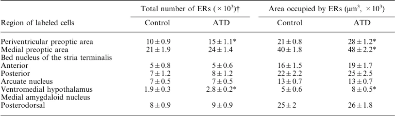
Estrogen receptor immunoreactivity
In both ATD and control males, dense
popu-lations of ER-IR cell nuclei were observed in the
PVP, mPOA, BNST, specifically in the anterior and
posterior medial divisions, Arc, ventrolateral
por-tion of the VMH and in the posterodorsal medial
amygdaloid nucleus (MePD). Furthermore, the PVP
and mPOA contained the greatest numbers of ERs.
Examples of ER-IR cell nuclei in the PVP, mPOA
and VMH are shown in Fig. 1. Analysis with
Student’s t-test showed that the total number of
ER-IR nuclei, i.e. the IOD, of ATD males was
significantly higher compared to control males in
the PVP (t=3.39, d.f.=16, P=0.01) and in the VMH
(t=2.42, d.f.=15, P=0.05) (Table 1). ATD males had
similar densities of ERs as control males in the
mPOA, BNST, Arc and MePD (Table 1). Analyses
with Student’s t-test revealed that the total area
occupied by ER-IR cells, i.e. the area mask, was
significantly higher in the PVP (t=4.8, d.f.=16,
P<0.001), the mPOA (t=2.6, d.f.=16, P=0.02) and
the VMH (t=3.6, d.f.=15, P=0.01) of ATD males
compared to control males (Table 1).
Androgen receptor immunoreactivity
In both ATD and control males, AR-IR cell nuclei
were found in the septohypothalamic nucleus, the
mPOA, the SDN-POA, the anterior and posterior
divisions of the BNST, the Arc, the VMH and
through the rostrocaudal extent of the medial
amygdaloid nucleus, i.e. anterior, anteroventral,
anterodorsal, posteroventral and MePD (see Table
2). Furthermore, the greatest numbers of ARs were
observed in the septohypothalamic nucleus, the
mPOA, the posterior division of the BNST, and the
MePD. Examples of AR-IR cell nuclei in the mPOA
containing the SDN-POA, the posterior division of
the BNST, and posterior medial amygdaloid nucleus
are presented in Fig. 2. Analysis with Student’s t-test
revealed that the total amount of AR-IR nuclei, i.e.
the IOD, was similar in ATD and control males for
each area measured (Table 2). Also, no differences
between ATD and control males were found in the
total area occupied by AR-IR cells, i.e. the area mask
(Table 2).
DISCUSSION
Estrogen receptor immunoreactivity
Neonatal inhibition of brain estrogen biosynthesis
in male rats increased ER levels in the PVP, an area
known to be involved in gonadotropin release, and
the ventrolateral portion of the VMH. As described
in the Introduction, sex di
fferences have been
reported in the PVP and VMH, with female rats
showing a higher estrogen binding capacity
18,33or a
higher ER-IR
57than male rats. Unfortunately, no
female controls were used in the present study.
There-fore, it is not known how closely the ER levels found
in the PVP and VMH of neonatally ATD-treated
males approximate to those found in normal females.
It could be possible that neonatally ATD-treated
males are intermediate between normal males and
females with respect to their ER levels, as was found
earlier with respect to their partner preference.
4This
would suggest that either androgens or prenatal
estrogens are also involved in the sexual di
fferen-tiation of ER levels in the PVP and VMH. Some
supportive evidence for a possible contribution of
prenatal androgens or estrogens to the sexual di
ffer-entiation of ER levels includes the finding that
neo-natal castration of male rats did not entirely feminize
their ER levels in the VMH.
34On the other hand,
neonatal castration of male rats increased ER levels
in the PVP, Arc and mPOA to levels
indistinguish-able from those found in normal females,
34suggest-ing that the sexual di
fferentiation of brain ER takes
place primarily neonatally under the influence of
androgens or estrogens.
Neonatal inhibition of brain estrogen biosynthesis
did not a
ffect ER levels in the Arc and mPOA,
although the total area occupied by ER-IR cells in
the mPOA was higher in neonatally ATD-treated
males than in control males (see Fig. 1). Ku¨hnemann
et al.
33,34reported that castration immediately after
birth increased ER levels in the Arc and mPOA,
whereas castration one day after birth failed to do so.
Presumably, the sex di
fferences in brain estrogen
levels in the mPOA and Arc might represent an
extremely rapid response to the hypothalamic surge
in estradiol occurring in males between 0 h in utero
and 1 h after delivery.
46Since ATD treatment was
not initiated until 2–4 h after birth, ATD males were
most likely exposed to this hypothalamic surge in
estradiol. Therefore, ATD males’ brains could
al-ready have been partly organized in a male direction,
i.e. ER levels were already lowered partly in the
mPOA and almost completely in the Arc. In support
of the role of neonatal estrogen in the sexual
differ-entiation of ER in the preoptic area are the findings
of DonCarlos et al.
25Female rats neonatally treated
with the synthetic estrogen, diethylstilbestrol, showed
at the age of 28 days reduced ER mRNA levels in the
Fig. 1. Photomicrographs showing ER-IR in neonatally ATD-treated male and control male rats. (A, B) The PVP of a control male (A) and of an ATD male (B). (C, D) The mPOA of a control male (C) and of an ATD male (D). (E, F) The VMH of a control male (E) and of an ATD male (F). Scales: 1 cm=195 µmpreoptic area, comparable to the levels seen in intact
males. The non-aromatizable androgen,
dihydrotes-tosterone, had no e
ffect on ER mRNA in the
pre-optic area of females.
The sex di
fferences in ER levels are presumably
established within 24 h after birth for the PVP and
mPOA, whereas the sex difference in the VMH
emerges later, between five and 10 days after birth.
33Thus, sex differences in ER levels in different brain
areas seem to be expressed asynchronously,
pro-viding a possible mechanism for variation in the
duration of critical periods for androgen- or
estrogen-mediated sexual differentiation of specific
neural functions. For instance, masculinization and
defeminization of sexual behavior are probably two
separable processes, which occur at slightly di
fferent
times during development (e.g., Refs 7, 13 and 52).
Our behavioral data suggest that defeminization of
the male rat brain takes place primarily neonatally
under the action of estradiol and that
masculiniz-ation of the brain occurs mainly both prenatally and
immediately after birth under the synergistic action
of testosterone and estradiol.
The sex di
fferences in ER levels in the PVP, mPOA,
Arc and VMH may contribute to the regulation
of sex-specific estrogen-dependent functions. For
instance, female rats are capable of showing a
pre-ovulatory luteinizing hormone surge after adult
gona-dectomy and treatment with a large dose of estradiol,
whereas male rats are not.
40Neonatal castration
makes it possible for males to show a preovulatory
luteinizing hormone surge in response to estrogen,
whereas neonatal treatment of female rats with
tes-tosterone eliminates this capacity to exhibit
estrogen-induced surges (for overview see Ref. 26). Whether
functional responses, such as an estrogen-induced
Table 1. Estrogen receptor immunoreactivity of neonatally 1,4,6-androstatriene-3,17-dione-treated male and control malerats in various hypothalamic and limbic nuclei
Total number of ERs (#103)† Area occupied by ERs (µm3,#103)
Region of labeled cells Control ATD Control ATD
Periventricular preoptic area 10&0.9 15&1.1* 21&0.8 28&1.2*
Medial preoptic area 21&1.9 24&1.4 40&1.8 48&2.2*
Bed nucleus of the stria terminalis
Anterior 5&0.8 5&0.6 16&1.5 19&1.7
Posterior 7&1.2 8&1.2 22&2.2 25&2.5
Arcuate nucleus 7&0.5 7&0.5 13&0.7 13&0.7
Ventromedial hypothalamus 1.9&0.3 2.8&0.2* 5&0.6 8&0.5* Medial amygdaloid nucleus
Posterodorsal 8&0.9 9&0.9 25&2 26&1.8
†The total number of ER-IR nuclei is estimated by taking the product of the optical density, which measures the concentration of the chromogen DAB in each outlined area, and the area occupied by ERs.
*Significantly (P<0.05) higher compared to control males. Data are expressed as mean&S.E.
Table 2. Androgen receptor immunoreactivity of neonatally 1,4,6-androstatriene-3,17-dione-treated male and control male rats in various limbic and hypothalamic nuclei
Total number of ARs (#103)† Area occupied by ARs (µm3,#103)
Region of labeled cells Control ATD Control ATD
Septohypothalamic nucleus 17&2.4 17&2.9 51&5.3 50&7.0
Medial preoptic area 16&1.2 17&1.6 50&3.0 50&3.0
Sexually dimorphic nucleus 6&0.5 5&0.6 13&1.0 11&0.8 Bed nucleus of the stria terminalis
Anterior 10&0.7 10&1.0 40&2.2 38&3.0
Posterior 22&2.3 25&4.2 71&5.3 77&9.0
Arcuate nucleus 2&0.2 3&0.4 6&0.3 7&0.8
Ventromedial hypothalamus 11&0.7 11&1.4 41&2.3 41&3.9 Medial amygdaloid nucleus
Anterior 4&0.5 5&0.8 20&2.3 25&3.3
Anterodorsal 6&0.6 5&0.7 27&2.2 23&2.5
Anteroventral 4&0.4 3&0.3 13&1.0 12&1.0
Posterodorsal 19&1.8 19&3.3 64&4.7 57&6.0
Posteroventral 4&0.4 4&0.7 15&1.4 16&2.1
†The total number of AR-IR nuclei is estimated by taking the product of the optical density, which measures the concentration of the chromogen DAB in each outlined area, and the area occupied by ARs.
luteinizing hormone surge, are directly proportional
to the number of ER-containing neurons in a
particular brain area remains to be investigated.
Interestingly, a co-existence of ER and neurotensin
has been found in the PVP.
2Neurotensin appears
to be involved as an interneuron in the regulation of
the release of luteinizing-releasing hormone (e.g.,
Ref. 1).
For activation of feminine sexual behavior in the
male rat, a much higher dose of estradiol is required
than in the female rat.
20,28,42,54This is confirmed by
previous behavioral findings in our laboratory.
4,5Neonatally ATD-treated male rats, castrated in
adulthood and subsequently treated with estradiol,
show high levels of feminine sexual behavior and a
partner preference for a sexually active male, whereas
similarly treated control males show high levels of
masculine sexual behavior and a partner preference
for an estrous female. Only when treated with
ex-tremely high doses of estradiol (blood serum levels
Fig. 2. Photomicrographs (1 cm=310 µm) showing AR-IR of neonatally ATD-treated male and control male rats. (A, B) The mPOA including the sexually dimorphic nucleus of a control male (A) and of an ATD male (B). (C, D) The MePD of a control male (C) and of an ATD male (D). (E, F) The VMH of1000 pmol/l; normal blood serum levels
~7 pmol/l),
normal males show feminine sexual behaviors
(unpublished observations in our laboratory). These
findings suggest that the reduced capacity to respond
to estrogen in adulthood is associated with lower
levels of ER in the male rat brain.
Meredith et al.
39reported that the ER-21
anti-serum is able to detect both occupied and unoccupied
ERs in the guinea-pig, since ER-21 immunostaining
was not affected by a high dose of 17â-estradiol
(50 µg). However, in a pilot study, we found a more
intense staining with the ER-21 antiserum after the
male rats were castrated. It has been reported that
castration leads to an up-regulation of ER mRNA,
49whereas acute estradiol treatment down-regulates ER
mRNA.
35In the present study, castration has
pre-sumably led to an up-regulation of ER in both ATD
and control males, although the possibility cannot be
ruled out that the ER-21 antiserum could be more
e
fficient at detecting unoccupied ERs in the rat brain
as opposed to the guinea-pig brain. If ER levels were
up-regulated by castration, then the ER levels in the
present study represent the maximal capacity rather
than physiological levels.
Androgen receptor immunoreactivity
Neonatal inhibition of brain estrogen biosynthesis
in male rats did not a
ffect forebrain AR levels. This
suggests that neonatal estrogens do not play a
signifi-cant role in the sexual di
fferentiation of the forebrain
AR system. However, it is not clear to what extent
ARs are sexually di
fferentiated, i.e. sexually
dimor-phic, in adulthood. Reports on sex differences in
androgen binding capacity have been conflicting.
Biochemical assays of androgen binding in whole
preoptic area and/or hypothalamus dissections have
not revealed sex differences.
29,48However, measuring
AR binding in more discrete nuclei within the
pre-optic area and hypothalamus has demonstrated sex
di
fferences.
47In particular, the caudal part of the
mPOA showed sex differences, with males having
more testosterone target neurons that project to the
midbrain than females.
36This might not be proof for
sex di
fferences in ARs, since testosterone can be
aromatized to estradiol within the CNS. Therefore, a
labeled cell following an injection of radioactively
labeled testosterone may represent estrogen binding
to ERs. Sex differences in AR levels have been found
during the first 10 days of life, especially in the
amygdala.
38In this latter study, however, males and
females were left gonadally intact, which makes it
very likely that the sex differences in AR levels reflect
sex di
fferences in endogenous testosterone or
di-hydrotestosterone levels. Presumably, there are no
large sex di
fferences in the distributions and densities
of AR-containing cells, although small, subtle
quantitative di
fferences in certain regions of the male
and female rat brain cannot be ruled out. It seems
unlikely that possible sex di
fferences in AR levels
are caused by the actions of estrogen during early
development, since neonatally ATD-treated males
showed similar distributions and densities of AR-IR
as control males. However, no female controls were
used in the present study, so it is not known whether
or not any sex differences in AR levels would have
been found using the assay conditions of the present
study. Therefore, we cannot determine from the
present data whether or not sexual di
fferentiation of
forebrain ARs occurs and, if it happens, whether it
takes place pre- or neonatally under the influence of
androgens or estrogens.
9,10,43,45,51,53Future studies
should address these questions.
Interestingly, a dense cluster of AR-IR was found
in the SDN-POA (see Fig. 2), as was reported earlier
for AR mRNA by Simerly et al.
50This was not seen
with ER-IR. In the adult rat, the volume of the
SDN-POA is determined by the perinatal action of
androgens.
21,22,23,30,32Jacobson et al.
31have found
that gonadectomized and adrenalectomized male
rats showed a higher percentage of radioactively
labeled cells in the SDN-POA following exposure to
[
3H]testosterone than gonadectomized and
adrenal-ectomized females.
31This sex di
fference might be due
to a difference in AR levels in the mPOA.
29In the
present study, no e
ffect of neonatal ATD treatment
was found on the number of ARs in the SDN-POA
or on the SDN-POA occupied by ARs. We expect
that the volume of the SDN-POA of neonatally
ATD-treated males is smaller than that of control
males, since perinatal ATD treatment to male rats
reduced SDN-POA volume in adulthood.
30Presum-ably, the absence of changes in the number of ARs in
the SDN-POA or in the SDN-POA occupied by ARs
suggests that there are either no changes in the size of
the SDN-POA or that AR immunostaining in the
SDN-POA has increased with ATD treatment to
compensate for the smaller size. We are currently
studying the SDN-POA volume in neonatally
ATD-treated male rats.
It has been suggested that the binding of the PG-21
antiserum to androgen receptors requires occupation
of the receptor. Zhou et al.
58reported that
testoster-one withdrawal by castration eliminated all nuclear
staining, whereas dihydrotestosterone or testosterone
treatment 15 min before perfusion restored nuclear
staining. In the present study, the ATD and control
males were left gonadally intact for the AR
immuno-cytochemistry. We do not believe that the number of
occupied ARs di
ffers between neonatally
ATD-treated males and control males, since neonatally
ATD-treated males and control males have similar
levels of endogenous testosterone.
6CONCLUSIONS
The available evidence points to sex di
fferences in
ER levels in the PVP, mPOA, Arc and VMH. The
development of these sex di
fferences in forebrain ER
levels seems to be partly dependent on the neonatal
action of estrogens, derived from the neural
aroma-tization of testosterone. To date, it is not clear
whether or not sexual di
fferentiation of forebrain
ARs occurs and, if so, whether there is any
contri-bution of neonatal estrogens to the development of
the AR system.
Acknowledgements—We thank Dr G. Greene for generously
providing the ER-21 and PG-21 antisera used in this study. REFERENCES
1. Alexander M. J., Mahoney P. D., Ferris C. F., Carraway R. E. and Leeman S. E. (1989) Evidence that neurotensin participates in the central regulation of the preovulatory surge of luteinizing hormone in the rat. Endocrinology 124, 783–788.
2. Axelson J. F., Shannon W. and Van Leeuwen F. W. (1992) Immunocytochemical localization of estrogen receptors within neurotensin cells in the rostral preoptic area of the rat hypothalamus. Neurosci. Lett. 136, 5–9.
3. Bakker J., van Ophemert J. and Slob A. K. (1993) Organization of partner preference and sexual behavior and its nocturnal rhythmicity in male rats. Behav. Neurosci. 107, 1049–1058.
4. Bakker J., van Ophemert J. and Slob A. K. (1996) Sexual differentiation of odor and partner preference in the rat.
Physiol. Behav. 60, 489–494.
5. Bakker J., Brand T., van Ophemert J. and Slob A. K. (1993) Hormonal regulation of adult partner preference behavior in neonatally ATD-treated male rats. Behav. Neurosci. 107, 480–487.
6. Bakker J., van Ophemert J., Timmerman M. A., de Jong F. H. and Slob A. K. (1995) Endogenous reproductive hormones and nocturnal rhythms in partner preference and sexual behavior of ATD-treated male rats.
Neuroendo-crinology 62, 396–405.
7. Baum M. J. (1979) Differentiation of coital behavior in mammals: a comparative analysis. Neurosci. Biobehav. Rev. 3, 265–284.
8. Baum M. J., So¨dersten P. and Vreeburg J. T. M. (1974) Mounting and receptive behavior in the ovariectomized female rat: influence of estradiol, dihydrotestosterone, and genital anesthetization. Horm. Behav. 5, 175–190.
9. Baum M. J., Woutersen P. J. A. and Slob A. K. (1991) Sex difference in whole-body androgen content in rats on fetal days 18 and 19 without evidence that androgen passes from males to females. Biol. Reprod. 44, 747–751.
10. Baum M. J., Brand T., Ooms M. P., Vreeburg J. T. M. and Slob A. K. (1988) Immediate postnatal rise in whole body androgen content in male rats: correlation with increased testicular content and reduced body clearance of testosterone. Biol. Reprod. 38, 980–986.
11. Beach F. A. (1942) Male and female mating behavior in prepuberally castrated female rats treated with androgen.
Endocrinology 31, 373–378.
12. Beach F. A. (1976) Sexual attractivity, proceptivity, and receptivity in female mammals. Horm. Behav. 7, 105–138. 13. Beatty W. W. (1992) Gonadal hormones and sex differences in nonreproductive behaviors. In Handbook of Behavioral
Neurobiology, Sexual Differentiation (eds Gerall A. A., Moltz H. and Ward I. L.), Vol. 11, pp. 85–128. Plenum,
New York.
14. Blaustein J. D. (1992) Cytoplasmic estrogen receptors in rat brain: immunocytochemical evidence using three antibodies with distinct epitopes. Endocrinology 131, 1336–1342.
15. Blaustein J. D. (1993) Estrogen receptor immunoreactivity in rat brain: rapid effects of estradiol injection.
Endocrinology 132, 1218–1224.
16. Brand T., Kroonen J., Mos J. and Slob A. K. (1991) Adult partner preference and sexual behavior of male rats affected by perinatal endocrine manipulations. Horm. Behav. 25, 323–341.
17. Brown T. J., Hochberg R. B., Zielinski J. E. and MacLusky N. J. (1988) Regional sex differences in cell nuclear estrogen-binding capacity in the rat hypothalamus and preoptic area. Endocrinology 123, 1761–1770.
18. Brown T. J., MacLusky N. J., Shanabrough M. and Naftolin F. (1990) Comparison of age- and sex-related changes in cell nuclear estrogen-binding capacity and progestin receptor induction in the rat brain. Endocrinology 126, 2965–2972.
19. Clancy A. N., Whitman C., Michael R. P. and Albers H. E. (1994) Distribution of androgen receptor-like immunoreactivity in the brains of intact and castrated male hamsters. Brain Res. Bull. 33, 325–332.
20. Davidson J. M. (1969) Effects of estrogen on the sexual behavior of male rats. Endocrinology 84, 1365–1372. 21. Do¨hler K. D., Coquelin A., Davis F., Hines M., Shryne J. E. and Gorski R. A. (1982) Differentiation of the sexually
dimorphic nucleus in the preoptic area of the rat brain is determined by the perinatal hormone environment. Neurosci.
Lett. 33, 295–298.
22. Do¨hler K. D., Coquelin A., Davis F., Hines M., Shryne J. E. and Gorski R. A. (1984) Pre- and postnatal influence of testosterone propionate and diethylstilbestrol on differentiation of the sexually dimorphic nucleus of the preoptic area in male and female rats. Brain Res. 302, 291–295.
23. Do¨hler K. D., Srivastava S. S., Shryne J. E., Jarzab B., Sipos A. and Gorski R. A. (1984) Differentiation of the sexually dimorphic nucleus in the preoptic area of the rat brain is inhibited by postnatal treatment with an estrogen antagonist. Neuroendocrinology 38, 297–301.
24. DonCarlos L. L. and Handa R. J. (1994) Developmental profile of estrogen receptor mRNA in the preoptic area of male and female neonatal rats. Devl Brain Res. 79, 283–289.
25. DonCarlos L. L., McAbee M., Ramer-Quinn D. S. and Stancik D. M. (1995) Estrogen receptor mRNA levels in the preoptic area of neonatal rats are responsive to hormone manipulation. Devl Brain Res. 84, 253–260.
26. Dyer R. G. (1984) Sexual differentiation of the forebrain—relationship to gonadotrophin secretion. In Progress in
Brain Research (eds De Vries G. J. et al.), Vol. 61, pp. 233–236. Elsevier Science, Amsterdam.
27. Emery D. E. and Sachs B. D. (1975) Ejaculatory pattern in female rats without androgen treatment. Science 190, 484–486.
28. Goy R. W. and McEwen B. S. (1980) Sexual Differentiation of the Brain. MIT Press, Cambridge, MA.
29. Handa R. J., Reid D. L. and Resko J. A. (1986) Androgen receptors in brain and pituitary of female rats: cyclic changes and comparisons with the male. Biol. Reprod. 34, 293–303.
30. Houtsmuller E. J., Brand T., de Jonge F. H., Joosten R. N. J. M. A., van de Poll N. E. and Slob A. K. (1994) SDN-POA volume, sexual behavior, and partner preference of male rats affected by perinatal treatment with ATD.
Physiol. Behav. 56, 535–541.
31. Jacobson C. D., Arnold A. P. and Gorski R. A. (1987) Steroid autoradiography of the sexually dimorphic nucleus of the preoptic area. Brain Res. 41, 349–356.
32. Jacobson C. D., Csernus V. J., Shryne J. E. and Gorski R. A. (1981) The influence of gonadectomy, androgen exposure, or a gonadal graft in the neonatal rat on the volume of the sexually dimorphic nucleus of the preoptic area.
J. Neurosci. 1, 1142–1147.
33. Ku¨hnemann S., Brown T. J., Hochberg R. B. and MacLusky N. J. (1994) Sex differences in the development of estrogen receptors in the rat brain. Horm. Behav. 28, 483–491.
34. Ku¨hnemann S., Brown T. J., Hochberg R. B. and MacLusky N. J. (1995) Sexual differentiation of estrogen receptor concentrations in the rat brain: effects of neonatal testosterone exposure. Brain Res. 691, 229–234.
35. Lauber A. H., Mobbs C. V., Muramatsu M. and Pfaff D. W. (1991) Estrogen receptor messenger RNA expression in rat hypothalamus as a function of genetic sex and estrogen dose. Endocrinology 129, 3180–3186.
36. Lisciotto C. A. and Morrell J. I. (1994) Sex differences in the distribution and projections of testosterone target neurons in the medial preoptic area and the bed nucleus of the stria terminalis. Horm. Behav. 28, 492–502. 37. MacLusky N. J. and Naftolin F. (1981) Sexual differentiation of the central nervous system. Science 211,
1294–1302.
38. Meaney M. J., Aitken D. H., Jensen L. K., McGinnis M. Y. and McEwen B. S. (1985) Nuclear and cytosolic androgen receptor levels in the limbic brain of neonatal male and female rats. Devl Brain Res. 23, 179–185.
39. Meredith J. M., Auger C. J. and Blaustein J. D. (1994) Down-regulation of estrogen receptor immunoreactivity by 17â-estradiol in the guinea pig forebrain. J. Neuroendocr. 6, 639–648.
40. Neill J. D. (1972) Sexual difference in the hypothalamic regulation of prolactin secretion. Endocrinology 90, 1154–1159.
41. Oboh A. M., Paredes P. G. and Baum M. J. (1995) A sex comparison in increments in Fos immunoreactivity in forebrain neurons of gonadectomized, testosterone-treated rats after mounting an estrous female. Neurobiol. Learning
Memory 63, 66–73.
42. Olster D. H. and Blaustein J. D. (1988) Progesterone facilitation of lordosis in male and female Sprague–Dawley rats following priming with estradiol pulses. Horm. Behav. 22, 294–304.
43. Pang S. F., Caggiula A. R., Gay V. L., Goodman R. L. and Pang C. S. F. (1979) Serum concentrations of testosterone, oestrogens, luteinizing hormone and follicle-stimulating hormone in male and female rats during the period of neural sexual differentiation. J. Endocr. 80, 103–110.
44. Prins G. S., Birch L. and Greene G. L. (1991) Androgen receptor localization in different cell types of the adult rat prostate. Endocrinology 129, 3187–3199.
45. Resko J. A., Feder H. H. and Goy R. W. (1968) Androgen concentrations in plasma and testis of developing rats.
J. Endocr. 40, 485–491.
46. Rhoda J., Corbier P. and Roffi J. (1984) Gonadal steroid concentrations in serum and hypothalamus of the rat at birth: aromatization of testosterone to 17â-estradiol. Endocrinology 114, 1754–1760.
47. Roselli C. E. (1991) Sex differences in androgen receptors and aromatase activity in microdissected regions of the rat brain. Endocrinology 128, 1310–1316.
48. Roselli C. E., Handa R. J. and Resko J. A. (1989) Quantitative distribution of nuclear androgen receptors in microdissected areas of the rat brain. Neuroendocrinology 49, 449–453.
49. Shughrue P. J., Bussnell C. D. and Dorsa D. M. (1992) Estrogen receptor messenger ribonucleic acid in female rat brain during the estrous cycle: a comparison with ovariectomized females and intact males. Endocrinology 131, 381–388.
50. Simerly R. B., Chang C., Muramatsu M. and Swanson L. W. (1990) Distribution of androgen and estrogen receptor mRNA-containing cells in the rat brain: an in situ hybridization study. J. comp. Neurol. 294, 76–95.
51. Slob A. K., Ooms M. P. and Vreeburg J. T. M. (1980) Prenatal and early postnatal sex differences in plasma and gonadal testosterone and plasma luteinizing hormone in female and male rats. J. Endocr. 87, 81–87.
52. Ward I. L. and Ward O. B. (1985) Sexual behavior differentiation: effects of prenatal manipulations in rats. In Handbook of Behavioral Neurobiology (eds Adler N., Pfaff D. and Goy R. W.), Vol. 7, pp. 77–98. Plenum, New York.
53. Weisz J. and Ward I. L. (1980) Plasma testosterone and progesterone titers of pregnant rats, their male and female fetuses and neonatal offspring. Endocrinology 106, 81–87.
54. Whalen R. E., Luttge W. G. and Gorzalka B. B. (1971) Neonatal androgenization and the development of estrogen responsivity in male and female rats. Horm. Behav. 2, 83–90.
55. Wood R. I. and Newman S. W. (1993) Intracellular partitioning of androgen receptor immunoreactivity in the brain of the male Syrian hamster: effects of castration and steroid replacement. J. Neurobiol. 24, 925–938.
56. Wood R. I. and Newman S. W. (1993) Mating activates androgen receptor-containing neurons in chemosensory pathways of the male Syrian hamster brain. Brain Res. 614, 65–77.
57. Yokosuka M. and Hayashi M. (1995) Sex difference in the distribution of estrogen receptor and aromatase in the rat brain. Soc. Neurosci. Abstr. 21, 423.16.
58. Zhou L., Blaustein J. D. and De Vries G. J. (1994) Distribution of androgen receptor immunoreactivity in vasopressin-and oxytocin-immunoreactive neurons in the male rat brain. Endocrinology 134, 2622–2627.