Brief communication
Detection of apple chlorotic leaf spot virus using a 5
%
nuclease assay with a fluorescent 3
% minor groove
binder-DNA probe
Michel A. Salmon *, Marina Vendrame, Jean Kummert, Philippe Lepoivre
Unite´ de Phytopathologie, Faculte´ Uni6ersitaire des Sciences Agronomiques de Gembloux(FUSAG), Passage des De´porte´s2, 5030Gembloux, Belgium
Received 29 October 2001; received in revised form 11 February 2002; accepted 12 February 2002
Abstract
The development of a real-time 5% nuclease RT-PCR assay for the detection of apple chlorotic leaf spot virus (ACLSV) from infected plant material is described. A short fluorogenic 3% minor groove binder-DNA hydrolysis probe was used to circumvent genome variability between isolates and target a short conserved sequence. The covalent attachment of the minor groove binder moiety at the 3% end of the probe increased the probe/target duplex stability and raised the melting temperature to a range suitable for real-time analysis. The method is rapid, sensitive and takes place within a single tube without post-PCR handling of the amplification products. © 2002 Elsevier Science B.V. All rights reserved.
Keywords:Apple chlorotic leaf spot6irus; 5% nuclease assay; Minor groove binder; RT-PCR
www.elsevier.com/locate/jviromet
Apple chlorotic leaf spot 6irus (ACLSV) is
known to infect a wide range of fruit tree species as apple, pear, peach, quince, plum, cherry, apri-cot and many ornamental rosaceous species. Al-though most strains are latent in fruit trees, others can be responsible for russet ring on apple fruits and for serious diseases in stone fruits, including plum bark split, plum pseudopox and peach dark green sunken mottle (Dunez and Delbos, 1988). In Japan, the virus causes top-working disease and induces lethal decline in apple trees
propa-gated on Maruba kaido (Malus prunifolia var.
ringo) rootstocks (Yanase, 1974). ACLSV is a
member of the Tricho6irus genus from the
Clos-tero6irus group (Martelli et al., 1994). Complete
nucleotide sequences of the ACLSV genome were determined from isolates of plum, apple and cherry trees (German et al., 1990; Sato et al., 1993; German-Retana et al., 1997; Jelkmann, 1996). ACLSV genome consists of a single strand poly-adenylated RNA molecule of 7545 – 7555 nu-cleotides excluding the poly-A tail, with three open reading frames (ORFs 1, 2 and 3) encoding proteins with molecular masses of 216.5, 50.4 and 21.4 kDa, respectively. The 216.5 kDa protein * Corresponding author. Tel.: + 81-622-434; fax: +
32-81-610-126.
E-mail address:lepoivre.p@fsagx.ac.be (P. Lepoivre).
0166-0934/02/$ - see front matter © 2002 Elsevier Science B.V. All rights reserved. PII: S 0 1 6 6 - 0 9 3 4 ( 0 2 ) 0 0 0 3 5 - 6
M.A. Salmon et al./Journal of Virological Methods104 (2002) 99 – 106 100
contains two consensus sequences associated with the RNA polymerase and NTP-binding helicase. The 50.4 kDa protein is suggested to be the movement protein, while the 21.4 kDa product is the viral coat protein (German et al., 1992).
Determination of the ACLSV sequence has led to the development of molecular tests based on reverse transcription and subsequent amplification of target sequences from the virus genome (Kum-mert et al., 2000; Kinard and Scott, 1996). To overcome the problem of inhibitory substances like phenolic compounds or polysaccharides that could interfere with template preparation, an im-munocapture RT-PCR (IC-RT-PCR) method has been developed, leading to increased sensitivity of 10 fg of purified virus (Candresse et al., 1995). More recently, a polyvalent nested RT-PCR using degenerated and inosine containing primers (PDO RT-PCR) was developed to detect Tricho-,
Capillo- and Fo6ea6iruses, including ACLSV
(Foissac et al., 2001). Although these techniques are highly specific and more sensitive than sero-logical methods, they require a post-PCR detec-tion step based on agarose gel electrophoresis or microplate/membrane hybridization with a la-belled probe.
In this paper, a detection protocol is described based on a real-time homogenous 5% nuclease RT-PCR assay using a fluorescent minor groove binder-DNA probe. The assay combines PCR amplification and DNA hybridization within a single tube and is based on the cleavage by the 5%3% exonuclease activity of Taq DNA poly-merase of a probe that hybridizes specifically to the target PCR product during amplification (Holland et al., 1991; Lie and Petropoulos, 1998; Livak et al., 1995; Bustin, 2000). The probe car-ries both a fluorescent reporter dye at the 5% end, a quencher and a minor groove binder (MGB) at the 3% end. The MGB binds the minor groove of double strand DNA and stabilizes the probe/ target duplex through Van der Waals contacts, hydrophobic and electrostatic interactions. There-fore the melting temperature (Tm) of the probe increases dramatically so that a Tm in a range of 65 – 70 °C can be reached with a probe of 12 – 18 nucleotides (Kutyavin et al., 1997, 2000; Wal-burger et al., 2001). The hybridized probe is then
cleaved by the enzyme during strand elongation, resulting in the separation of the fluorescent dye and the quencher, and a subsequent increase in fluorescence. Repeated cycles result in the expo-nential amplification of the target sequence with a parallel increase of the fluorescence intensity. Measurement of the fluorescence throughout the reaction eliminates the need for further detection procedures. Interpretation of the data can be achieved as a simple qualitative conclusion from the presence or the absence of amplified DNA within minutes at the end of the run.
Specific primers for ACLSV were chosen by alignment of the published sequences correspond-ing to isolates P863 from Prunus domestica (Ger-man et al., 1990), PBM1 from cv. Hauszwetsche (Jelkmann, 1996), Balaton-1 from cherry (Ger-man-Retana et al., 1997) and P205 from apple tree (Sato et al., 1993). Primers 5F (5%
GCCTA-CAAATTAGGTGAGAGGCTC) and 8R (5%
TTCCAATGGATCATGAGGTC) were selected by computer analysis with PILEUP, FASTA and PRIME programs (Wisconsin Package Version 10.1, Genetic Computer Group, USA). They am-plify a region of 288 nucleotides that overlaps the 3% end of ORF1 and the 5% end of ORF2 (Kum-mert et al., 2000). Synthesis and purification of the primers were carried out by Eurogentec (Seraing, Belgium).
In order to design a probe which is dedicated to detect most or all ACLSV isolates independently of their host plant or their geographical origin, the products amplified with primers 5F and 8R were sequenced from five apple trees (91328, A1, B3, B8 and C4) and two plum trees (H2 and H4) from the Gembloux collection (Belgium). Total RNA from bark tissues was extracted using the RNEasy Plant RNA kit according to manufactur-er’s instructions (Qiagen, Hilden, Germany). RT-PCR amplifications were undertaken using the Qiagen OneStep RT-PCR Kit (Hilden, Germany). The kit uses a hotstart polymerase and allows reverse transcription and PCR to be performed in a single tube. A 25-ml reaction mixture containing total RNA (200 – 400 ng), 0.4mM of each dNTP, 0.6 mM of both 5F and 8R primers and the reagents from the kit (5 × buffer, enzyme mix) was submitted to cDNA synthesis (30 min at
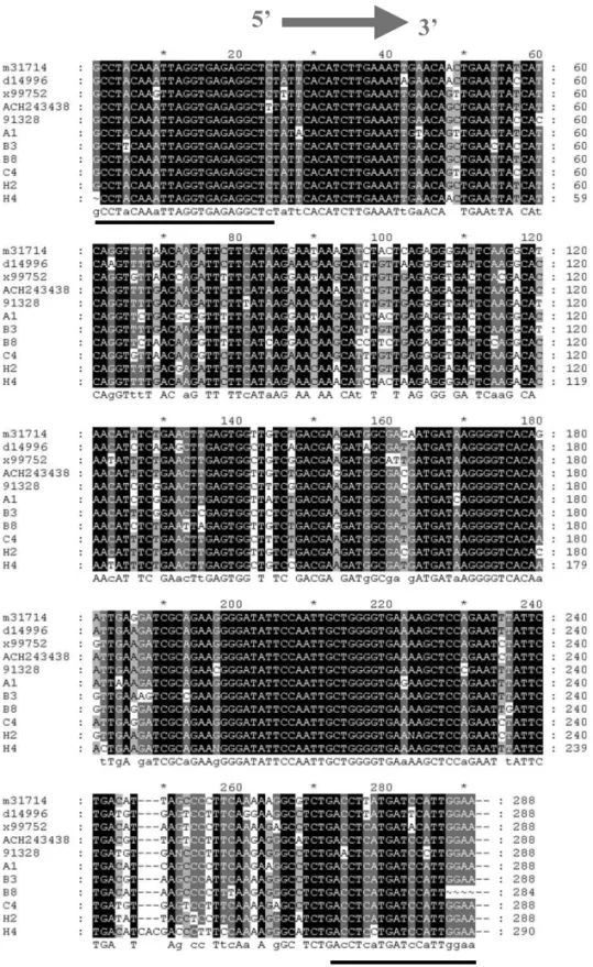
Fig. 1. Sequence alignment of seven RT-PCR products and the corresponding region from full length clones m31714 (P863), ACH243438 (PBM1), x99752 (Balaton-1) and D14996 (P205). The nucleotide sequence of MGB 26 is indicated by the arrow, and primers are underlined.
M.A. Salmon et al./Journal of Virological Methods104 (2002) 99 – 106 102
50 °C), Taq polymerase activation (15 min at 95 °C) and amplification with 40 cycles (30 s at 95 °C, 1 min at 55 °C and 1 min at 60 °C). Targets from all isolates tested were amplified successfully and the products were cloned into the pCR2.1 vector following the instructions of the supplier (TA Cloning, Invitrogen, Groningen, The Netherlands). All inserts were sequenced by Euro-gentec (Seraing, Belgium).
The alignment of these new sequences together
with all four sequences available from GenBank is illustrated in Fig. 1. It was used to define a fluorogenic probe for real-time detection. A con-ventional TaqMan probe of 30 – 35 nucleotides with a suitable Tm could not be found without multiple mismatches between the target and the probe, because of sequence heterogeneity between isolates. Therefore, a shorter 3% MGB DNA probe of 17 nucleotides was designed. The general rules outlined by Livak et al. (1996) were employed and
Fig. 2. Comparison of fluorogenic assay and gel electrophoresis to detect ACLSV from infected reference trees of the Gembloux collection. Panel A: gel electrophoresis and ethidium bromide straining. Panel B: amplification plot showing cycle number versus normalized fluorescence.
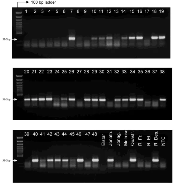
Fig. 3. Detection of ACLSV from field isolates by agarose gel electrophoresis with ethidium bromide staining. Samples 1 – 48: apple trees from the CTIFL collection. Elstar to Reinette Des.: apple cultivars from a nursery (La Hulpe, Belgium). NTC: no template control.
the software Primer Express 5.1 (Applied Biosys-tems, Forster City, USA) was used for Tm
calcula-tion. The probe (MGB26: 5% ATTCACATCTT-GAAATT) was supplied by Applied Biosystems, with VIC as the reporter dye at the 5% end, a non fluorescent quencher and the MGB moiety at the 3% end. The MGB, which raised the Tmto 64.7 °C,
prevented the 3% extension of the probe.
The first tests for real-time detection of ACLSV field isolates using MGB26 probe were performed on apple (A1, A4, B3, B4, C4, C7, D4, LP680, 91325, 91327, 91328, 10291) and pear trees (F1, H1, I1) from Gembloux (Belgium). The probe was
included in the RT-PCR mixture at a final concen-tration of 200 nM and fluorescence was recorded at the extension step of each amplification cycle using the GeneAmp 5700 Sequence Detection Sys-tem (Applied BiosysSys-tems, Forster City, USA). The threshold cycle (Ct) was calculated by plotting fluorescence versus cycle number. As demonstrated in Fig. 2, all positive trees as detected by conven-tional agarose gel electrophoresis were also positive in real-time analysis (A1, B3, C4, C7, D4, H1, 91327, 91328). Ct values were between 23 and 28,
except for sample 91327, for which a later Ct(33)
M.A. Salmon et al./Journal of Virological Methods104 (2002) 99 – 106 104
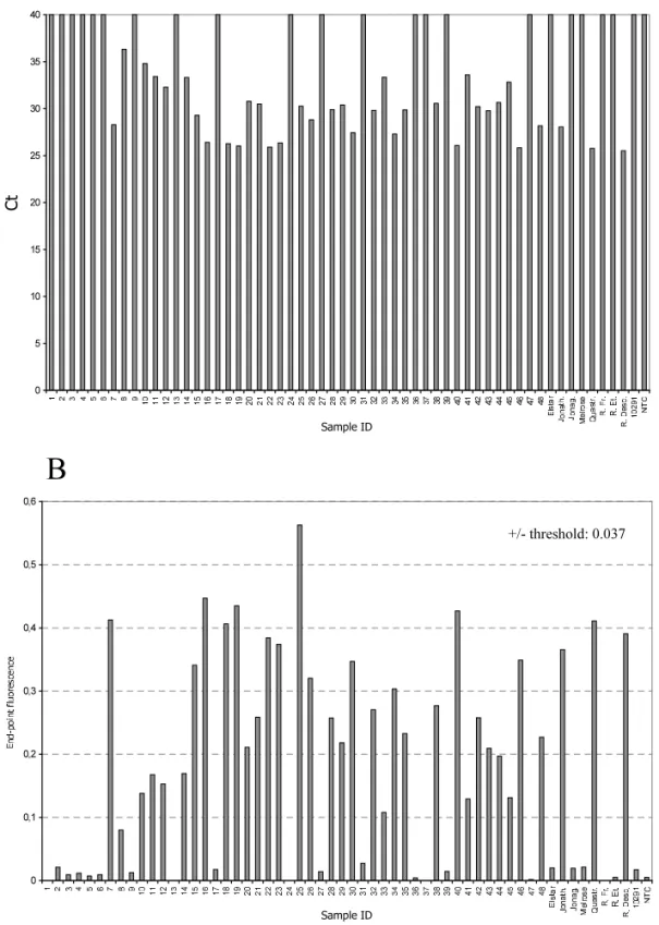
Fig. 4. Real-time detection of ACLSV from field isolates (CTIFL collection and nursery). The upper bar graph panel (A) represents the Ctvalues for each sample tested. The lower panel (B) shows the end-point fluorescence values. Samples that were negative on
gel were used for the calculation of a positive-negative threshold, which was fixed at 0.037 as the mean end-point fluorescence — 3 × S.D. (Chebychev’s equivalent).
faint signal in agarose gel. A1 isolate produced an early Ct(25) and high fluorescence although there was a mismatch between the probe and the target, which was thought to destabilize the duplex, espe-cially since the probe is short. It seems therefore that a perfect match is not absolutely required for the assay, but we could imagine that a mismatch at the 3% end of the duplex where the MGB binds, would have more serious effects by lowering the
Tmof the probe below the range suitable for the
assay.
Using serial dilutions of total RNA from the ACLSV-infected tree H1, the sensitivity of the fluorogenic 5% nuclease assay and agarose gel elec-trophoresis was compared. The threshold of sensi-tivity of gel electrophoresis was shown to be within the 200 – 20 pg range of total plant RNA. The fluorogenic assay proved to be equally sensi-tive (data not shown).
To expand the number of samples for assay validation, total RNA preparations of 48 samples obtained from twigs of dormant wood taken from trees containing apple latent viruses were used (samples numbered from 1 to 48 in Figs. 3 and 4). These samples representing isolates of different geographical origins (USA, Japan, Europe, New
Zealand) were received from the Centre Interpro-fessionnel et Technique des Fruits et Le´gumes (CTIFL, Lanxade, France). Apple cultivars taken in a nursery (Elstar, Jonathan, Jonagold, Melrose, Quastresse, Reinette de France, Reinette Etoilee and Reinette Descadre) were also used. In order to establish the correlation between gel elec-trophoresis and real-time detection, all samples were tested using both techniques. Comparison of the results demonstrated a perfect correlation for all 56 samples tested. In addition, all Ct from
real-time measurements reflected the intensity of the bands on gel (Figs. 3 and 4).
Fig. 5 represents a histogram of the frequency distribution where the Ct values obtained for all
tested samples including two negative controls were plotted. The distribution of the data is to a large extend bimodal, with all negative samples forming the large peak at 40 and all positive tests displaying values between 22 and 36, with most of them having a Ct in the 22 – 31 range.
Interpreta-tion of the data is therefore straightforward. In summary, the 5% nuclease assay allowed sen-sitive and unambiguous detection of all ACLSV field isolates tested and correlated perfectly with agarose gel electrophoresis, which remains until
M.A. Salmon et al./Journal of Virological Methods104 (2002) 99 – 106 106
now the reference technique for the detection of PCR products. Direct evidence is provided that short MGB-DNA hydrolysis probes are particu-larly suitable for the detection of microorganisms with high genome variability between strains.
Acknowledgements
This work was supported by the General Direc-torate for Technologies, Research and Energy of the Wallonia Region, Belgium (DGTRE), in the framework of the research agreement N° 001/ 4542.
References
Bustin, S.A., 2000. Absolute quantification of mRNA using real-time reverse transcription polymerase chain reaction assays. J. Mol. Endrocrinol. 25, 169 – 193.
Candresse, T., Lanneau, M., Revers, F., Grasseau, N., Mac-quaire, G., German, S., Malinovsky, T., Dunez, J., 1995. An immunocapture PCR assay adapted to the detection and the analysis of the molecular variability of the Apple Chlorotic Leafspot virus. Acta Hortic. 386, 136 – 147. Dunez, J., Delbos, R., 1988. Closteroviruses. In: Smith, I.M.,
Dunez, J., Lelliot, R.A., Phillips, D.H., Archer, S.A. (Eds.), European Handbook of Plant Diseases. Blackwell, Oxford, pp. 5 – 7.
Foissac, X., Svanella-Dumas, L., Dulucq, M.J., Candresse, T., Gentit, P., 2001. Polyvalent detection of fruit tree Tricho,
Capillo and Fo6ea6iruses by nested RT-PCR using
degener-ated and inosine containing primers (PDO RT-PCR). Acta Hortic. 550, 37 – 43.
German-Retana, S., Bergey, B., Delbos, R.P., Candresse, T., Dunez, J., 1997. Complete nucleotide sequence of the genome of a severe cherry isolate of Apple Chlorotic Leaf Spot Trichovirus. Arch. Virol. 142 (4), 833 – 841. German, S., Candresse, T., Lanneau, M., Dunez, J., 1992.
Genomic organization of Apple Chlorotic Leaf Spot Clos-terovirus (ACLSV). Acta Hortic. 309, 31 – 38.
German, S., Candresse, T., Lanneau, M., Huet, J.C., Pernol-let, J.C., Dunez, J., 1990. Nucleotide sequence and ge-nomic organization of Apple Chlorotic Leaf Spot Closterovirus. Virology 179, 104 – 112.
Holland, P.M., Abramson, R.D., Watson, R., Gelfand, D.H., 1991. Detection of specific polymerase chain reaction prod-ucts by utilizing 5%3% exonuclease activity of Thermus
aquaticus DNA polymerase. Proc. Natl. Acad. Sci. USA
88, 7276 – 7280.
Jelkmann, W., 1996. The nucleotide sequence of a strain of Apple Chlorotic Leaf Spot Virus (ACLSV) responsible for plum pseudopox and its relation to an apple and plum bark split strain. Phytopathology 86 (Suppl. 11), S101. Kinard, G.R., Scott, S.W., 1996. Detection of Apple Chlorotic
Leaf Spot and Apple Stem Grooving Viruses using RT-PCR. Plant Dis. 80 (6), 616 – 621.
Kummert, J., Vendrame, M., Steyer, S., Lepoivre, P., 2000. Development of routine RT-PCR tests for routine certifica-tion of fruit tree multiplicacertifica-tion material. OEPP/EPPO Bull. 30, 441 – 448.
Kutyavin, I.V., Lukhtanov, E.A., Gamper, H.B., Meyer, R.B., 1997. Oligonucleotides with conjugated dihydropyrroloin-dole tripeptides: base composition and backbone effects on hybridization. Nucleic Acid Res. 25 (18), 3718 – 3723. Kutyavin, I.V., Afonina, I.A., Mills, A., Gorn, V.V.,
Lukhtanov, E.A., Belousov, E.S., Singer, M.J., Walburger, D.K., Lokhov, S.G., Gall, A.A., Dempcy, R., Reed, M.W., Meyer, R.B., Hedgpeth, J., 2000. 3%-Minor groove binder-DNA probes increase sequence specificity at PCR exten-sion temperature. Nucleic Acid Res. 28 (2), 655 – 661. Lie, Y.S., Petropoulos, C.J., 1998. Advances in quantitative
PCR technology: 5% nuclease assay. Curr. Opin. Biotech-nol. 9, 43 – 48.
Livak, K., Flood, S.J.A., Marmaro, J., Giusti, W., Deetz, K., 1995. Oligonucleotides with fluorescent dyes at opposite ends provide a quenched probe system useful for detecting PCR product and nucleic acid hybridization. PCR Meth-ods Appl. 4, 357 – 362.
Livak, K., Marmaro, J., Flood, S., 1996. Guidelines for de-signing TaqMan fluorogenic probes for 5% nuclease assays. Perkin-Elmer Res. News 57, 1 – 5.
Martelli, G.P., Candresse, T., Namba, S., 1994. Trichovirus, a new genus of plant viruses. Arch. Virol. 134, 451 – 455. Sato, K., Yoshikawa, N., Takahashi, T., 1993. Complete
nucleotide sequence of the genome of an apple isolate of Apple Chlorotic Leaf Spot Virus. J. Gen. Virol. 74, 1927 – 1931.
Yanase, H., 1974. Studies on apple latent viruses in Japan. Bull. Fruit Tree Res. Stn. Jpn. C-1, 47 – 109.
Walburger, D.K., Afonina, I.A., Wydro, R., 2001. An im-proved real time PCR method for simultaneous detection of C282Y and H63D mutations in the HFE gene associ-ated with hereditary hemochromatosis. Mutat. Res. 432, 69 – 78.