UOJM | www.uojm.ca May 2018 | Volume 8 | Issue 1
68
Anatomical Variation of the Brachial Plexus: An
Ancillary Nerve of the Middle Trunk Communicating
with the Radix of the Median Nerve
T
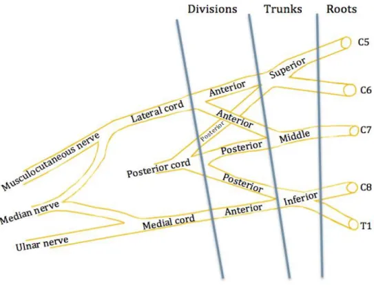
he brachial plexus is formed by contributions from the anterior rami of spinal nerves C5-T1. Spinal nerves C5-C6 continue to form the superior trunk; C7 continues to form the middle trunk, and C8-T1 forms the inferior trunk. Each trunk then divides into anterior and posterior divisions. The anterior divisions of the superior and middle trunk form the lateral cord, while the anterior divi-sion of the inferior trunk forms the medial cord. Finally, the lat-eral cord and the medial cord unite to form the median nerve proper (Figure 1) (1).Studies by Miller et al elucidate the origins of the brachial plexus. The brachial plexus is a pervasive structure apparent since the existence of amphibians. The structure began as no more than several nerve segments in amphibians that progressed and dif-ferentiated into its current intricate and complex state (2-5). The evolution of the brachial plexus can be attributed to the diver-sification of the corresponding blood supply and musculature. The orientation of the axillary artery is responsible for the intrin-sic variation, meanwhile the foundation of the brachial plexus is dependent on the musculature. For instance, the branching
Keywords: Brachial plexus; Variation; Anatomy; Median nerve; Middle trunk; Cadaver
Purpose: Variations in brachial plexus anatomy are common. As such, the knowledge of variations is essential for surgeons and
anesthesiologists to decrease the risk of iatrogenic injuries. Moreover, brachial plexus variations often co-exist with aberrant vascu-lature. The median nerve is formed from contributions by the lateral and medial cords. This case report details a unique variant in the formation of the median nerve.
Methods: The anatomical variant presented was identified during an upper-limb dissection of an adult cadaver.
Results: The anatomical variant presented demonstrates a bifurcation of the middle trunk of the brachial plexus that coalesces to
the radix of the median nerve. Although prior studies have demonstrated median nerve brachial plexus variations, the aforemen-tioned variant arises directly from the middle trunk and communicates directly with the median nerve, while previously menaforemen-tioned variants often connect to the medial or lateral cords.
Conclusion: The communicating branch between the anterior division of the middle trunk and radix of the median nerve
repre-sents a unique and uncommon anatomical variation.
Minh Huynh
1, Stewart Spence
1, Johnny W. Huang
1 1 Faculty of Medicine, University of OttawaABSTRACT
RÉSUMÉ
Objectif: Les variations de l’anatomie du plexus brachial sont fréquentes. En tant que tel, la connaissance des variations est
essen-tielle pour les chirurgiens et les anesthésiologistes afin de diminuer le risque de lésions iatrogènes. De plus, les variations du plexus brachial coexistent souvent avec un système vasculaire aberrant. Le nerf médian est formé de contributions par les cordes latérales et médiales. Ce rapport de cas détaille une variante unique dans la formation du nerf médian.
Méthodes: La variante anatomique présentée a été identifiée lors d’une dissection d’un cadavre chez un adulte.
Résultats: La variante anatomique présentée montre une bifurcation du tronc médian du plexus brachial qui coalesce avec la base
du nerf médian. Bien que des études antérieures aient démontré des variations médianes du plexus brachial, la variante susmen-tionnée provient directement du tronc central et communique directement avec le nerf médian, alors que les variantes mention-nées précédemment se connectent souvent aux cordons médiaux ou latéraux.
Conclusion: La branche communicante entre la division antérieure du tronc central et la base du nerf médian représente une
variation anatomique unique et peu commune.
UOJM | www.uojm.ca May 2018 | Volume 8 | Issue 1
69
pattern of the musculocutaneous nerve is dictated by the mus-cle it innervates. In primitive species, such as the lemuroids, the coracobrachialis muscle contains three heads. Consequently, their brachial plexus contains additional nerve branches supe-rior to the musculocutaneous nerve from the lateral cord. These additional nerve fibres are not found in higher-level species. In apes, the coracobrachialis and biceps are almost completely fused, thus, these nerve fibres have completely regressed (4,5). Normally, the development of the brachial plexus in humans starts at the 34th to 35th day of intrauterine life and proceeds to the definitive adult pattern by the 46th to 48th day (2-5). Any alteration in the signaling between mesenchymal cells and neuronal growth cones can lead to significant brachial plexus variations (1-3).
METHODS AND RESULTS
The anatomical variant presented was identified during routine upper-limb dissection of an adult cadaver through the Faculty of Medicine, Ottawa, Ontario. Upon dissection, anatomical vari-ations of the brachial plexus were carefully noted. The anatomi-cal variant presented demonstrates a bifurcation of the middle trunk of the brachial plexus that coalesces to the radix of the median nerve. Permission to take the photo in this article was
granted by the University of Ottawa Faculty of Medicine by the Department of Anatomy.
DISCUSSION
Previous Descriptions of Similar Variants
Common aberrations in the structure of the brachial plexus have a tendency to share vestigial features of more primitive species. For example, in humans, the correct anatomical ori-entation of the axillary artery is deep to the plexus. Conversely, the majority of primitive species have their artery superficial to the nerves and represent one of the most common human variations. Therefore, brachial plexus variations are due to er-rors during the embryological developmental process and are often remnants of vestigial structures (4,5). The purpose of this case report is to detail a unique variant in the formation of the median nerve.
Several anatomical median nerve variants have been previously reported. For example, in a study of 130 brachial plexus variants in 1999, Uzun and Bilgic found that 14 of 130 brachial plexuses (10.80%) demonstrated “a connection between the medial ra-dix of the medial nerve and ventral division of the middle trunk.”
Figure 1. Illustration demonstrating normal brachial plexus anatomy.
UOJM | www.uojm.ca December 2017 | UOJM ePub
CASE REPORT
2
heads. Consequently, their brachial plexus contains additional nerve branches superior to the musculocutaneous nerve from the lateral cord. These additional nerve fibres are not found in higher-level species. In apes, the coracobrachialis and biceps are almost completely fused, thus, these nerve fibres have completely regressed [4,5]. Normally, the development of the brachial plexus in humans starts at the 34th to 35th day of in-trauterine life and proceeds to the definitive adult pattern by the 46th to 48th day [2-5]. Any alteration in the signaling be-tween mesenchymal cells and neuronal growth cones can lead to significant brachial plexus variations [1-3].
METHODS AND RESULTS
The anatomical variant presented was identified during rou-tine upper-limb dissection of an adult cadaver through the Faculty of Medicine, Ottawa, Ontario. Upon dissection, ana-tomical variations of the brachial plexus were carefully noted. The anatomical variant presented demonstrates a bifurcation of the middle trunk of the brachial plexus that coalesces to the radix of the median nerve. Permission to take the photo in this article was granted by the University of Ottawa Faculty of Medicine by the Department of Anatomy.
DISCUSSION
Previous Descriptions of Similar Variants
Common aberrations in the structure of the brachial plexus have a tendency to share vestigial features of more primitive species. For example, in humans, the correct anatomical orien-tation of the axillary artery is deep to the plexus. Conversely, the majority of primitive species have their artery superficial to the nerves and represent one of the most common human variations. Therefore, brachial plexus variations are due to er-rors during the embryological developmental process and are often remnants of vestigial structures [4,5]. The purpose of this case report is to detail a unique variant in the formation of the median nerve.
Several anatomical median nerve variants have been previously reported. For example, in a study of 130 brachial plexus variants in 1999, Uzun and Bilgic found that 14 of 130 brachial plexuses (10.77%) demonstrated “a connection between the medial ra-dix of the medial nerve and ventral division of the middle trunk.” Additionally, 2 of 130 plexuses contained a contribution from the posterior division of the middle trunk connecting to the medial cord [6]. In 2006, Goyal and colleagues reported a bi-lateral cadaveric anomaly. On the right side, the median nerve
Figure 1. Illustration demonstrating normal brachial plexus anatomy.
10 148
149
Figure 1. Illustration demonstrating normal brachial plexus anatomy 150
UOJM | www.uojm.ca May 2018 | Volume 8 | Issue 1
70
Additionally, 2 of 130 plexuses contained a contribution from the posterior division of the middle trunk connecting to the medial cord (6). In 2006, Goyal and colleagues reported a bi-lateral cadaveric anomaly. On the right side, the median nerve was formed by the lateral and medial cords with a supplemen-tary branch from the lateral cord. The proximal aspect of the lateral cord gave rise to a small communicating branch, which eventually joined the median nerve. In the left axilla the me-dian nerve was formed by a similar, but more complex union of two lateral cord roots and one medial cord root. The anomalous proximal contribution of the lateral cord diverged from the
an-terior division of the middle trunk and lateral cord proper and provided an additional small communicating branch that trav-eled medially to reach the medial cord. Finally, the distal contri-bution of the lateral cord united with the medial cord normally to form the median nerve proper (7). More recently, Padur and colleagues, studying lateral cord variations in 82 cadaveric bra-chial plexuses, demonstrated 6 anomalies. Notably, in one limb the median nerve was formed by three roots; two of the roots arose directly from the medial side of the lateral cord, and the last root arose from medial cord. The three roots joined to form the trunk of the median nerve, and encircled the axillary artery
UOJM | www.uojm.ca December 2017 | UOJM ePub
CASE REPORT
4 Figure 2. Median nerve variant demonstrating additional ancillary branch (MNV) arising from the middle trunk (MT) and uniting to form the median nerve (Mn) at the union of the lateral (LC) and medial cords (MC) of the median nerve. Abbrevia-tions: Ulnar nerve (Un), median nerve (Mn), musculocutaneous nerve (Mc), medial cord (MC), lateral cord (LC), median nerve variant (MNV), normal anterior division (NAd), superior trunk (ST), middle trunk (MT)
11 151
Figure 2. Median nerve variant demonstrating additional ancillary branch (MNV) arising from the 152
middle trunk (MT) and uniting to form the median nerve (Mn) at the union of the lateral (LC) and medial 153
cords (MC) of the median nerve. Abbreviations: Ulnar nerve (Un), median nerve (Mn), musculocutaneous 154
nerve (Mc), medial cord (MC), lateral cord (LC), median nerve variant (MNV), normal anterior division 155
(NAd), superior trunk (ST), middle trunk (MT) 156
Figure 2. Median nerve variant demonstrating additional ancillary branch (MNV) arising from the middle trunk (MT) and uniting to form the median nerve (Mn) at the union of the lateral (LC) and medial cords (MC) of the median nerve. Abbreviations: Ulnar nerve (Un), median nerve (Mn), musculocutaneous nerve (Mc), medial cord (MC), lateral cord (LC),
me-dian variant (MNV), normal anterior division (NAd), superior trunk (ST), middle trunk (MT).
UOJM | www.uojm.ca May 2018 | Volume 8 | Issue 1
71
(8).
Discussion of Variant and Clinical Implications
Most commonly, reported median nerve variants demonstrate either a) additional roots arising from the lateral or medial cords uniting to form the median nerve; or b) roots diverging from the middle trunk to join the lateral or medial cords. The anatomical variant presented, however, demonstrates an uncommon ad-ditional anterior division arising directly from the middle trunk and uniting to form the median nerve at the union of the lateral and medial cords of the median nerve (Figure 2).
Although the anatomical variation presented may not have im-pacted limb function directly, variations in brachial plexus anat-omy may have clinical significance for surgical and anesthesio-logical operations performed around the brachial plexus and its vasculature (e.g., neck surgeries). Moreover, previously reported data suggests that brachial plexus and median nerve variations correlate with variations in the anatomy of the axillary artery (9). For example, Singhal et al, reporting several anomalous cord variations in a single cadaveric brachial plexus, identified an ab-normal relationship of the median nerve to the axillary artery, such that “the median nerve lay medial to the axillary artery” (9). Current anesthesiological methods for assessment of brachial plexus anatomy prior to surgical operation include: 1) blind approach; 2) peripheral nerve stimulation whereby a nerve is stimulated to produce paresthesia; and 3) ultrasound guidance. The first and second methods generally increase the risk of complications if aberrant anatomy exists, whereas ultrasound guidance renders a lower potential risk of operational com-plications, given the increased rate of detection of anatomical abnormalities. As demonstrated in this case, the potential for anatomical and vascular variations to exist is not uncommon. This emphasizes the need for thorough preoperative evalua-tion of brachial plexus anatomy, including ultrasound guidance, in order to detect anatomical variations, and decrease the rate of complications (10).
REFERENCES
1. Williams PL, Bannister LH, Berry MM, et al. Nervous system. In: Gray’s anato-my, 38th ed. New York: Churchill Livingstone; 1992. 1266– 1272 p. 2. Moore KL, Persaud TVN. The Developing Human: clinically oriented
embry-ology. 10th ed. Philadelphia, PA: Saunders/Elsevier; 2004. 10 p.
3. Sannes HD, Reh T, Harris W. Development of nervous system. In: Axon Growth Guidance. New York: Academic press; 2000. 189-197 p.
4. Miller RA. Comparative studies upon the morphology and distribution of brachial plexus. Am J Anat. 1934;54(1):143-7.
5. Miller RA. Observations upon the arrangement of the axillary artery and brachial plexus. Am J Anat. 1939;64(1):143-3.
6. Uzun A. Some Variations in the Formation of the Brachial Plexus in Infants.
Turk J Med Sci. 1999;29(5):573-8.
7. Goyal N Harjeet, Gupta M. Bilateral variant contributions in the formation of median nerve. Surg Radiol Anat. 2005;27(6):562-5.
8. Padur AA, Kumar N, Shanthakumar SR, Shetty SD, Prabhu GS, Patil J. Unusual and Unique Variant Branches of Lateral Cord of Brachial Plex-us and its Clinical Implications - A Cadaveric Study. J Clin Diagn Res. 2016;10(4):AC01-AC04.
9. Singhal S, Rao VV, Ravindranath R. Variations in brachial plexus and the relationship of median nerve with the axillary artery: a case report. J Bra-chial Plex Peripher Nerve Inj. 2007;2:21.
10. Macfarlane A, Brull R. Ultrasound Guided Supraclavicular Block. Journal of New York School of Regional Anesthesia. 2009;12:6-10.