Interactions between β-lactoglobulin and
nutraceutical ligands riboflavin,
vitamin D
3and Lysozyme
Formation, physico-chemical and biological characterization of
functional delivery scaffolds
Thèse
Fatoumata Diarrassouba
Doctorat en sciences et technologie des aliments
Philosophiae Doctor (Ph.D)
Québec, Canada
iii
Résumé
La protéine majeure du lactosérum, la β-lactoglobuline (βlg) est bien reconnue pour ses propriétés structurales intéressantes lui permettant d’établir des interactions avec des ligands de taille et de caractéristiques différentes. La riboflavine (RF) et la vitamine D3 (D3) ont été sélectionnées comme modèles de
petits nutraceutiques amphiphiles et hydrophobes, respectivement, et le lysozyme (Lyso), comme ligand protéique de plus grande taille.
La capacité de la βlg à lier la RF a été étudiée par des méthodes spectroscopiques. La βlg et la RF forment le complexe βlg-RF dont la photoactivation génère une activité antiproliférative contre les cellules cancéreuses de la peau, démontrée en utilisant le protocole du NCI/NIH Developmental Therapeutics Program. La cytotoxicité serait probablement due à la génération d’espèces oxydatives réactives résultant de l’interaction entre la RF et la βlg.
L’impact de la formation du βlg-D3 sur la solubilité et stabilité de la D3 a été étudié
en utilisant des méthodes spectroscopiques et de chromatographie. Les résultats ont démontré que le complexe βlg-D3 était stable aux pHs gastrique et intestinal et
augmentait la solubilité de la D3. De plus, une matrice protéique appelée coagulum
enrichie en D3 (94.5 ± 1.8 % de taux d’encapsulation) été formée à partir du
complexe βlg-D3 grâce à l’aptitude de la βlg à s’auto-associer.
Les images de microscopie électronique ont montré que les interactions électrostatiques entre la βlg et Lyso ont pour leur part, abouti à la formation de microsphères pouvant encapsuler la D3 à un taux élevé (90.8 ± 4.8 %).
La capacité des matrices à base de βlg à transporter, protéger et à améliorer la solubilité, la stabilité et la biodisponibilité de la D3 a été évaluée en effectuant des
expériences in vitro et in vivo chez des modèles animaux. Les matrices protéiques à base de βlg ont significativement augmenté la solubilité, stabilité et biodisponibilité de la D3 (p < 0.001). Ces études prouvent que la βlg, grâce à ses
caractéristiques suturales, pourrait former des matrices protéiques compatibles avec une administration orale et les aliments, tout en préservant l’activité biologique de la RF, de la D3 et donc possiblement d’autres molécules bioactives.
v
Abstract
The major whey protein, β-lactoglobulin (βlg) is well recognized for its interesting structural properties and ability to interact with ligands with varying size and characteristics. Riboflavin (RF) and vitamin D3 (D3) were selected as small amphiphilic and hydrophobic nutraceutical models, respectively, and Lysozyme, as a larger size ligand model.
Spectroscopic methods were used to demonstrate interaction between βlg and RF. βlg and RF form a complex, which was irradiated according to the NCI/NIH Developmental Therapeutics Program. The βlg-RF complex exhibited an important anti-proliferative activity against skin melanoma cancer cell lines, probably due to the generation of reactive oxygen species as the result of the interaction between RF and βlg.
The impact of the βlg-D3 complex on the solubility and stability of the D3 was studied using spectroscopic methods and chromatography. The findings indicate that the βlg-D3 complex is stable at the gastric and intestinal pHs and increases the solubility of the vitamin. A βlg-based scaffold, named coagulum, enriched with D3 (94.5 ± 1.8 % of encapsulation efficiency) was prepared by using the capacity of βlg to self-aggregate.
Electronic microscopy images showed that microspheres, with high D3
encapsulation efficiency (90.8 ± 4.8 %), were formed as the result of electrostatic interactions between βlg and Lyso.
The efficiency of βlg-based scaffolds to improve the solubility, stability and bioavailability of the D3 was evaluated by performing in vitro and in vivo experiments using animal model. The βlg-based scaffolds significantly increased the solubility, stability bioavailability of D3 (p < 0.001). Overall, the present study showed that βlg, due to its structural properties, can be used to form protein-based
matrices compatible with a food and an oral administration while preserving the biological activity of RF, D3 and possibly other bioactive molecules.
vii
Preface
The present thesis is submitted to the Faculty of Graduate Studies of Laval University (Faculté des études supérieures de l'Université Laval) to meet the requirements for achieving the Philosophiae Doctor es Sciences (Ph. D.) degree at the Faculty of Agriculture and Food Science (Faculté des Sciences de l’Agriculture et de l’Alimentation).
The research was mostly conducted at the Department of Food Science and Technology, Faculty of Agriculture and Food Science, Laval University. Additional experimental work was performed at the EA-CIDAM (Ingénierie Développement Aliment Médicament), Laboratoire de Biopharmacie, Faculté de Pharmacie, Clermont-Ferrand, France, with Professor Eric Beyssac, Associate Professor PhD Ghislain Garrait, PhD Li Liang and PhD Pedro Alvarez, as collaborators. PhD Garbiel E. Remondetto and Professor Muriel Subirade were the co-supervisor and supervisor, respectively.
In the introduction section, the work presented in this thesis describes the use of bovine milk whey β-lactoglobulin in the formulation of food grade oral delivery platforms. The first chapter of the thesis is a literature review on β-lactoglobulin – based delivery scaffolds, a version of which will be published in the book titled ‘Engineering Foods for Bioactives Stability and Delivery’ to be published by Springer.
The rationale for this work, hypothesis and objectives are presented in the second chapter. The main experimental work of this thesis has been published or submitted for publication in relevant scientific journals. Versions of seven manuscripts were presented in chapters 3 to 9 as follows:
- Chapter 3. Fatoumata Diarrassouba, Gabriel Remondetto, Li Liang and Muriel Subirade. 2013. Nanocomplex formation between riboflavin and β-lactoglobulin: spectroscopic investigation and biological characterization. Food Research International. 52(2):557–567.
- Chapter 4. Fatoumata Diarrassouba, Gabriel Remondetto, Li Liang, Ghislain Garrait, Eric Beyssac and Muriel Subirade. 2013. Effects of gastrointestinal pH conditions on the stability of the β-lactoglobulin/vitamin D3 complex and on the
solubility of vitamin D3. Food Research International. 52(2):515 – 521.
- Chapter 5. Fatoumata Diarrassouba, Ghislain Garrait, Gabriel Remondetto, Pedro Alvarez, Eric Beyssac and Muriel Subirade. 2014. Increased stability and protease
resistance of the β-lactoglobulin/vitamin D3 complex. Food Chemistry.
145:646-652.
- Chapter 6. Fatoumata Diarrassouba, Gabriel Remondetto, Ghislain Garrait, Pedro Alvarez, Eric Beyssac and Muriel Subirade. Increased water solubility, stability and bioavailability of vitamin D3 upon sequestration in β-lactoglobulin - Based Coagulum. Submitted to the Journal of Controlled Release.
- Chapter 7. Fatoumata Diarrassouba, Ghislain Garrait, Gabriel Remondetto, Pedro Alvarez, Eric Beyssac and Muriel Subirade. Self-assembly of β-lactoglobulin and egg white lysozyme as a potential carrier for nutraceuticals. Submitted to the Journal of Physical Chemistry B. Manuscript ID jp-2013-08298b.
- Chapter 8. Fatoumata Diarrassouba, Ghislain Garrait, Gabriel Remondetto, Pedro Alvarez, Eric Beyssac and Muriel Subirade. Food proteins-based microspheres for increased uptake of vitamin D3. Submitted to the Journal of Controlled Release. - Chapter 9. Fatoumata Diarrassouba, Ghislain Garrait, Gabriel Remondetto, Pedro Alvarez, Eric Beyssac and Muriel Subirade. Comparative study between different β-lactoglobulin-based scaffolds for the delivery of vitamin D3.This manuscript is still in preparation. A comparative study was performed between the βlg-based scaffolds prepared in the present thesis, the βlg/vitamin D3 complex, and the free
vitamin D3. The efficiency of the different systems to stabilize and enhance the
bioavailability of vitamin D3 were evaluated.
Finally, the conclusion of this work including the main findings and research outcome and perspectives are presented in chapter 10, which is followed by the references and appendices. Appendix I represents the list of contributions including the list of manuscripts and conferences. Appendices 2 to 6 represent the posters and oral presentations at different conferences and oral presentation competitions.
ix To my beloved dad Soumana Diarrassouba,
Humble and accomplished scientist, your exceptional human and scientific
qualities are recognized worldwide. You have deeply touched everyone who has been in contact with you, You are my inspiration, role-model, and mentor. You are my hero dad.
xi
Acknowledgements
The present PhD thesis is the outcome of converging efforts of many years of hard work and dedication, which would have not been possible without the constant support and love from my family, friends, supervisors and co-workers.
Family and friends.
First and foremost, I would like to express all my love and gratitude to my son Soumana for being such a great, loving and compassionate human being. He is always supportive, always ready to give me hug whenever he feels like I need comfort and warmth. Thank you son and know that I am so grateful to be your mom and so proud of you.
To moms Assitan Samaké and Heidi Rempel, dads Irv Rempel and Soumana Diarrassouba, without whom I would not have been at this stage of my personal and professional live. I love you dearly and I thank you so much for your unconditional love and support. This PhD thesis is the result of your joined blessings. God bless you all. A special ‘thank you’ to Mom Heidi for your patience and precious help in proofreading all my papers during these years, from my Masters degree to the present PhD thesis. You have been a great inspiration for me, a role model who will always inspire me in all areas in my life.
To my brothers Ibrahim, N’Faly, Jamy, Brad and sister Kim, thank you for being so supportive and thoughtful.
I am very much grateful to my dear friends Anne and David Ehret for all the help and invaluable support, encouragement and advice. David I have no word strong enough to express all my gratitude for all your help in editing my work. Your contribution to this thesis was deeply appreciated. Anne thank you for motivating me, your invaluable support and commitment to our twining project Teriya.
I am deeply thankful to my dear friend Earla Legault for all the love, good spirit, beautiful smile and for introducing me to the group of the ‘Healing Chicks’: Darlene, Irene, Debbie, Patsy, Roxanne, Carmen, Margot, Leigh-Ann, Jinder, Paula, Christine and Osa. Thank you for all the positive thoughts, great and unique moments we shared. Thank you Earla for your help and priceless contribution to our twining project Teriya.
I would like to specially dedicate this PhD thesis to Amine, Alya, Tamou Jelti and Adama Berthe. You are my family, my friends and I love you dearly. Thank you for being there at any moment of the day and night for me and Soumana.
My deep appreciation and love to my dear buddies Mariama Abdou Goubé and Fatou Thiam. Thank you for all the inspiring discussions, great moments we shared and unforgettable memories.
Thank you to my friends Ibrahima and Kadiatou Sow, OumouTouré Fall, Solange N’Gazo aand Rémy and Hapsatou Maiga.
I am very grateful to my friends and colleagues who made this PhD a very enjoyable ride that was rich in learning opportunities, pleasant encounters and new friendships: Massama, Raquel, Romain, Li Liang, Pedro Álvarez, Ahmed Gomez, Shyam Suwal, Luis Felipe Gutiérrez, Luca Lo Verso, Sébastien Goumon, Abdelbasset Atia (Abdel), Élodie Rozoy, Sandra Laneuville, Sabrine, Nassim, Juan, Sergei Mikhailyn, Hasna Hanchi, Rima Atoum, Hany Geagea, Hélène Gaudreau, Charles Edmonds, Sophie Banville, Marina Bergoli, Mónica Araya-Farías and Franck.
To all my Teriya friends, in Mali, the Commune of Sanakoro – Djitumu, and the cities of Agassiz and Harrison-Hot-Springs: thank you for your enthusiastic commitment and accepting to be part of this exciting ride. Special ‘thank you’ to Monica for translating most of our documents for the twinning project.
I would like to thank Ghislain Garrait, Rachel, Quentin and Alexis for all the help and warm welcome which made me feel like I was home any time I traveled to Clermont-Ferrand (France). I am so grateful to you Ghislain and so glad that I was coached by you. Thank you so very much for your great and invaluable scientific contribution to my work.
xiii Supervisors and co-workers.
I am deeply grateful to my supervisor, Professor Muriel Subirade for accepting me as a PhD student in her prestigious Canadian Research Chair and team. Muriel impressed me with her pragmatism and dynamism. She inspired me with her excellent management skills. I learned a lot from her and acquired useful tools for my future academic carrier.
I am very thankful to my co-supervisor, Gabriel E. Remondetto for accepting to work with me despite his busy schedule and professional commitment. I was mesmerized by Gabriel’s multidisciplinary competence. He has a vast knowledge in so many research areas and is very knowledgeable. With his contribution, my work gained in meaningful insights and considerable depth.
I would like to thank Professor Jean Amiot for agreeing to pre-read this thesis and for his valuable comments.
I am thankful to Dr Michel Britten, for evaluating this thesis. Dr Britten is world-class scientist well known for his work in the dairy industry, on milk components, milk by-products and processed foods.
I am grateful Professor Dérick Rousseau for agreeing to be the external examiner of this thesis. I feel very privileged to receive an evaluation and a feedback on my work from such a world class and well renowned scientist in food research.
I am deeply grateful to Profesor Odile Chambin for accepting to take the trip to Québec city to evaluate this thesis, despite her extremely busy agenda. I am very appreciative for having such a well-known scientist in Pharmaceutical and polymeric delivery technologies to evaluate my work.
I must also thank Professor Eric Beyssac for inviting to carry out important parts of my research in his lab EA – CIDAM (Équipe d’Accueil, Conception Ingénierie Développement Aliment Médicament) at Clermont-Ferrand, France. Prof. Beyssac was always available to provide any support, scientific advice or resources, which helped my greatly to reach important results during both of my stays in his lab. I am thankful to the team of the EA – CIDAM lab: Xie, Roselyne, Pascale, Sandrine, Professor Cardot and Valérie for the good moments in the lab and positive encouragement in my studies.
It my absolute pleasure to thank Mr Bernard Pouliot for all the laughter and very joyful moments in his office when we were working on my different PowerPoint presentations and scientific posters. I am profoundly thankful for all the advice and learning opportunities, both socially and professionally. It was such an honor for me to get to know such a unique human being.
Finally, I would like to express my sincere acknowledgements and deep appreciation to the technicians without whom this PhD thesis would have never been completed with such an ease and an efficiency: Diane Gagnon, Pascal Dubé, Alain Gaudreau and Richard Janvier.
This thesis was sponsored by the Canada Research Chair on Proteins, Biosystems and Functional Foods and the Natural Sciences and Engineering Research Council of Canada (NSERC) through the Vanier Canada Graduate Scholarships (Diarrassouba, F.). Experimental works in France (EA-CIDAM Faculté de Pharmacie, Clermont-Ferrand, France) were funded by the Canada Graduate Scholarships – Michael Smith Foreign Study Supplements (Diarrassouba, F.) and Fonds de Recherche Nature et Technologies Québec (FRQNT) – International Training Program (Diarrassouba, F.).
xv
Table of contents
Résumé ... iii Abstract ... v Preface ... vii Acknowledgements ... xi Table of contents ... xvList of abbreviations ... xxvi
Introduction ... 1
Chapter 1. Literature review ... 5
1.1. Abstract ... 6
1.2. Introduction ... 6
1.3. Manufacture of whey proteins ... 8
1.4. Fractionation of βlg from the whey proteins ... 9
1.5. Structure of βlg ... 11
1.5.1. Basic structure of βlg ... 11
1.5.2. Structural transitions of βlg ... 17
1.6. Functionality of βlg ... 18
1.7. Formation and characterization of the βlg-ligand complexes ... 20
1.7.1. Structural basis for the formation of the βlg-ligand complexes ... 20
1.7.2. Fluorescence quenching upon ligand binding ... 21
1.7.2.1. Theoretical explanation ... 21 1.7.2.2. Fluorescence emission of βlg ... 23 1.7.3. Circular dichroism ... 24 1.7.3. Βlg-ligand complexes ... 25 1.8. Βlg-biopolymer self-assembly ... 29 1.8.1. Βlg auto-association ... 29
1.8.2. Βlg-based delivery systems: from molecule to particles ... 30
1.8.3. Βlg-based microparticles for oral delivery ... 34
1.8.4. Characterization of βlg-based nano and microparticles ... 38
1.9. Conclusion ... 41
Chapter 2 ... 43
Problem statement ... 43
Hypothesis and objectives ... 43
2.1. Problem statement ... 44
2.2. Hypothesis ... 46
2.3. Objectives ... 46
2.3.1. General objective ... 46
2.3.2. Specific objectives ... 46
Chapter 3. Nanocomplex formation between riboflavin and β-lactoglobulin: spectroscopic investigation and biological characterization ... 51
3.1 Abstract ... 52
3.3. Experimental section ... 54
3.3.1. Materials ... 54
3.3.2. Sample preparation ... 54
3.3.3. Circular dichroism measurement... 55
3.3.4. Steady state fluorescence measurement ... 55
3.3.4.1. Protein fluorescence spectra ... 55
3.3.4.2. Synchronous fluorescence spectra ... 56
3.3.5. Biological characterization of the βlg/RF nanocomplex ... 56
3.3.5.1. Cell line culture ... 56
3.3.5.2. Anti-proliferative activity assay ... 56
3.3.5.4. Light stability of the βlg/RF nanocomplex ... 58
3.4. Results and discussion ... 58
3.4.1. Influence of βlg–RF interaction on the structure of the protein ... 58
3.4.2. Influence of RF on fluorescence spectra of βlg ... 61
3.4.3. Analysis of fluorescence quenching mechanism ... 63
3.4.4. Fluorescence resonance energy transfer from βlg to RF ... 66
3.4.5. Binding constants and binding points of RF to βlg ... 71
3.4.6. Influence of RF on the synchronous fluorescence of βlg... 78
3.4.7. Anti-proliferative activity assay the βlg/RF nanocomplex ... 80
3.4.8. Light stability of the βlg/RF nanocomplex ... 85
3.5. Conclusion... 87
3.6. Acknowledgments ... 88
Chapter 4. Effects of gastrointestinal pH conditions on the stability of the β-lactoglobulin/vitamin D3 complex and on the solubility of vitamin D3. ………. 90
4.1. Abstract ... 91
4.2. Introduction... 92
4.3. Materials ... 95
4.3.1. Sample preparation ... 95
4.3.2. Influence of the pH on the stability of the βlg/D3 complex ... 95
4.3.3. Influence of the βlg/D3 complex formation on the solubility of vitamin D3 ... 97
4.3.4. Statistical analysis ... 97
4.4. Results and discussion ... 98
4.4.1. Influence of the pH on the interaction between βlg and D3 ... 98
4.4.2. Influence of the pH on the stability of the βlg/D3 complex ... 106
4.4.3. Influence of the pH on the fractional residual fluorescence ... 109
4.4.4. Influence of the βlg/D3 complex formation on the solubility of D3 ... 109
4.5. Conclusion... 111
4.6. Acknowledgements ... 112
Chapter 5. Increased stability and protease resistance of the β-lactoglobulin/vitamin D3 complex ... 115
5.1. Abstract ... 116
5.2. Introduction... 116
5.3. Experimental section ... 118
xvii
5.3.2 sample preparation ... 118
5.3.3. Stability of the βlg/D3 complex in refrigerated conditions ... 119
5.3.4. Uv – stability of the βlg/D3 complex ... 119
5.3.5. Intestinal stability of the βlg/d3 complex ... 120
5.3.6. In vitro study of the permeability of the βlg/D3 complex ... 120
5.3.7. In vivo study of the intestinal absorption of the βlg/D3 complex ... 121
5.3.7.1. Animal models ... 121
5.3.7.2. In vivo experiments ... 122
5.3.8. Statistical analysis ... 122
5.4. Results and discussion ... 123
5.4.1. Stability of the βlg/D3 complex in refrigerated conditions ... 123
5.4.2. UV-light - stability of the βlg/D3 complex ... 126
5.4.3. Intestinal stability of the βlg/D3 complex ... 130
5.4.4. In vitro study of the permeability of the βlg/D3 complex ... 137
5.4.5. In vivo study of the intestinal absorption of the βlg/D3 complex ... 140
5.5. Conclusion ... 143
5.6. Acknowledgements ... 144
Chapter 6. Increased water solubility, stability and bioavailability of vitamin D3 upon sequestration in β-lactoglobulin - based coagulum ... 146
6.1. Abstract ... 147
6.2. Introduction ... 148
6.3. Experiemental section ... 150
6.3.1. Materials ... 150
6.3.2. Sample preparation ... 150
6.3.3. Encapsulation of D3 in the βlg-based coagulum ... 150
6.3.4. Stability of D3 during long term storage at 4 oc ... 151
6.3.5. Stability of D3 to uv-light irradiation ... 151
6.3.6. Kinetic release of D3 in simulated intestinal fluid ... 152
6.3.7. In vivo study of the intestinal absorption of the D3 containing βlg/lyso 152 6.3.8. Statistical analysis ... 153
6.4. Results and discussion ... 153
6.4.1. Encapsulation efficiency of D3 in the βlg-based coagulum ... 153
6.4.2. Impact of the βlg-based coagulum on the long term stability of D3 ... 154
6.4.4. Impact of the βlg-based coagulum on the uv-light stability of D3 ... 157
6.4.5. Impact of the βlg-based coagulum on the intestinal kinetic release of D3 ... 159
6.4.6. In vivo study of the bioavailability of D3 ... 162
6.5. Conclusion ... 165
6.6. Acknowledgements ... 166
Chapter 7. Self-assembly of β-lactoglobulin and egg white lysozyme as a potential carrier for nutraceuticals ... 168
7.1. Abstract ... 169
7.2. Introduction ... 170
7.3. Experimental section ... 172
7.3.2. Sample preparation ... 172
7.3.3. Effect of protein ratio on the formation of the βlg/Lyso co-precipitate . 173 7.3.4. Effect of the ph on the formation of the βlg/Lyso co-precipitate ... 173
7.3.5. Characterization of the βlg/Lyso co-precipitate ... 174
7.3.5.1 size and surface charge ... 174
7.3.5.2 scanning and transmission electron microscopy for the determination of surface and interior morphology ... 174
7.3.6. Model nutraceutical loading studies ... 175
7.3.7. Statistical analysis ... 176
7.4. Results and discussion ... 176
7.4.1. Effect of the protein concentration on the formation of the βlg-Lyso co-precipitates ... 176
7.4.2. Effect of the pH on the formation of the βlg-Lyso co-precipitates ... 182
7.4.4. Characterization of the βlg/Lyso co-precipitates ... 188
7.4.4.1 scanning and transmission electron microscopy of surface and interior morphology ... 188
7.4.4.2. Determination of the amounts of proteins in the βlg/Lyso microspheres ... 190
7.4.5. Vitamin D3 encapsulation in βlg/Lyso microspheres ... 192
7.5. Conclusion... 195
7.6. Acknowledgments ... 196
Chapter 8. Food proteins-based microspheres for increased uptake of vitamin D3... 198 8.1. Abstract ... 199 8.2. Introduction... 200 8.3. Experiemental section ... 202 8.3.1. Materials ... 202 8.3.2. Sample preparation ... 202
8.3.3. Encapsulation of D3 in the βlg/Lyso microspheres ... 202
8.3.4. Stability of D3 during long term storage at 4 oc ... 203
8.3.5. Stability of D3 to UV-light irradiation ... 203
8.3.6. Kinetic release of D3 in simulated intestinal fluid ... 203
8.3.7. In vitro study of the permeability of the D3 ... 204
8.3.8. In vivo study of the intestinal absorption of the d3 containing βlg/Lyso 204 8.3.9. Statistical analysis ... 205
8.4. Results and discussion ... 206
8.4.1. Stability of D3 during long-term storage at 4 oc ... 208
8.4.2. Stability of D3 in response to uv-light irradiation ... 209
8.4.3. Kinetic release of D3 in simulated intestinal fluid ... 214
8.4.4. In vitro study of the permeability of D3 ... 221
8.4.5. In vivo study of the bioavailability of D3 ... 224
8.5. Conclusion... 226
8.6. Acknowledgments ... 226
Chapter 9. Comparative study between different β-lactoglobulin-based scaffolds for the delivery of vitamin D3 ... 228
xix 9.2. Introduction ... 230 9.3. Experiemental section ... 232 9.3. Experimental section ... 232 9.3.1 materials ... 232 9.3.2 sample preparation ... 233
9.3.3. Stability in refrigerated conditions ... 233
9.3.4. Uv – stability of the βlg-based scaffolds ... 234
9.3.5. Intestinal stability of the βlg-based scaffolds ... 234
9.3.6. In vivo study of the bioavailability of D3 ... 235
9.3.6.1. Animal models ... 235
9.3.6.2. In vivo experiments ... 235
9.3.7. Statistical analysis ... 236
9.4. Results and discussion ... 236
9.6. Conclusion ... 246
9.7. Acknowledgments ... 247
Chapter 10. Concluding chapter ... 248
10.1. Research outcome ... 249
10.2. Perspectives and future trends ... 252
References ... 255 Appendices ... 277 Appendix 1 ... 278 Appendix 2 ... 281 Appendix 3 ... 283 Appendix 4 ... 285 Appendix 5 ... 287 Appendix 6 ... 288
Index of tables
Chapter 1Table 1.1. Major whey proteins content in bovine milk, unprocessed whey (UPW), whey protein concentrates (WPC) and whey protein isolates (WPI) ... 9
Chapter 3
Table 3.1. Stern–Volmer quenching constants KSV and dynamic quenching rate
constant at different temperatures ... 65
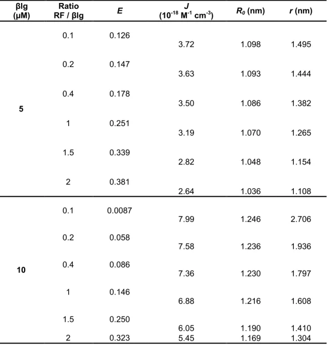
Table 3.2. Energy efficiency transfer and distance between the donor βlg and the acceptor RF ... 68
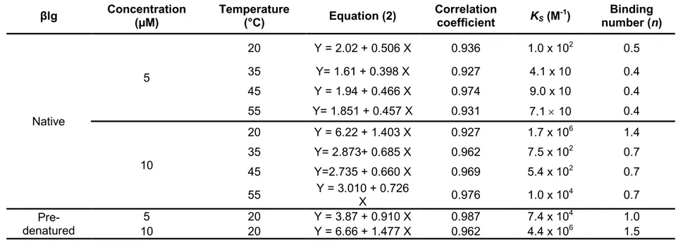
Table 3.3. Binding constant and binding number for static quenching of native and pre-denatured βlg by RF, at different temperatures and at neutral pH (7.4) ... 72
Chapter 4
Table 4.1. Influence of pH on the emission maximum wavelength of βlg and the βlg/D3 complex ... 103 Chapter 5
Table 5.1. TEER of the Caco-2 cells monolayers ... 139
Chapter 7
Table 7.1. Average Size (three measurements) of the βlg/Lyso self-assembly at ratio 2:1 and different pH values before and after overnight agitation ... 187
Chapter 8
Table 8.1. TEER of the Caco-2 cells monolayers ... 223
xxi
Index of figures
Chapter 1
Figure 1.1. Dimer of βlg with di-sulfides bonds in blue ... 13 Figure 1.2. Central cavity of βlg monomer with hydrogen bonds ... 15 Figure 1.3. Structures of βlg displaying the EF-loop in the closed (left) and open (right) conformation in the green circle. The conical β-barrel is formed by two sheets consisting of β-strands A-D and strands E-H.. ... 16 Figure 1.4. Different structures of microparticles obtained by microencapsulation: (a) microcapsule, (b) multilayer microcapsule, (c) microsphere and (d) multishell and multicore microsphere ... 35
Chapter 3
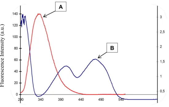
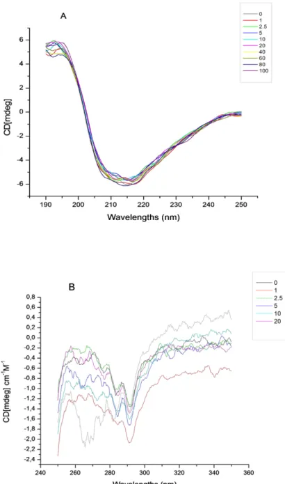
Figure 3.1. (A) Far-UV, (B) near-UV circular dichroic spectra of βlg in the absence and presence of increasing concentration of riboflavin in 10 mM phosphate buffer at pH 7.4. The concentrations of βlg are 10 μM for the far-UV region and 20 μM for the near-UV region. The concentrations of RF vary from0 to 40 μM. ... 60 Figure 3.2. Fluorescence emission spectra of βlg in the presence of different concentrations of RF; (A) [βlg] = 5 μM; (B) [βlg] = 10 μM; [RF] in μM, curves: 0, 1, 2.5, 5, 10, 15 and 20(λEX = 280 nm, T = room temperature, pH = 7.4). Insets: Stern–Volmer plots for the quenching of βlg by RF (A): [βlg] = 5 μM; (B): [βlg] = 10 μM. ... 62 Figure 3.3. Overlap of the fluorescence emission spectra of βlg (A) with the absorption spectra of RF (B). ... 70
Figure 3.4. The structure of βlg was updated from PBD (ID # 3BLG) 70
Figure 3.5. The evolution of fluorescence intensity of βlg alone as a function of temperature (20, 35, 45 and 55 °C). (A) [βlg] = 5 μM, (B) [βlg] = 10 μM; phosphate buffer 10 Mm at pH 7.4. Excitation at 280 nm. ... 75 Figure 3.6. The synchronous fluorescence spectra of βlg at 5 (A and B) and 10 μM(C and D). On panels A and C, Δλ = 15 nm; on panels B and D, Δλ = 60 nmin the presence of [RF] = 0, 1, 2.5, 5, 10, 15 and 20 μM. Solvent: phosphate buffer 10 mM at pH 7.4. Fluorescence scans were carried out from 200 to 400 nm. ... 79
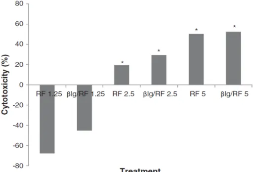
Figure 3.7. Cytotoxicity RF containing solutions on skin melanoma M21 cell lines irradiated in the absence and presence of βlg (25 min at 365 nm). The treatment consisted in the βlg/RF complex and RF solutions, each at a final concentration of 5, 2.5 and 1.25 μM. Growth inhibition and cytotoxicity were evaluated after an incubation period of 48 h for recovery. ... 82 Figure 3.8. Effect of irradiation of the βlg/RF complex, βlg and RF solutions during 25 min at 365 nm (4 W); βlg (20 μM): not irradiated (A) and irradiated (B); RF (20 μM): not irradiated (C) and irradiated (D); βlg/RF (20 μM): not irradiated (E) and irradiated (F). ... 84 Figure 3.9. Influence of light on the stability of the βlg/RF nanocomplex for a two week period. UV–vis spectra of RF (A) compared to that of the βlg/RF nanocomplex (B). C represents the impact of explosion to light on the concentration RF alone and the βlg/RF nanocomplex. UV–vis spectra and concentrations were determined at 445 nm. ... 86
Chapter 4
Figure 4.1. Fluorescence intensity of βlg (a and c) and the βlg/D3 complex (b and
d) at different pH (1.2, 2.0, 3.0, 5.0, 6.8, 7.0 and 8.0). Excitation at 280 nm (a and b) and 290 nm (c and d). ... 99 Figure 4.2. Synchronous fluorescence of Trp using ∆ʎ = 60 nm of βlg in absence (a) and presence of vitamin D3 (b). ... 101
Figure 4.3. Vitamin D3 binding capacity of βlg at different (1.2, 2.0, 3.0, 5.0, 6.8, 7.0
and 8.0). B. Fractional residual fluorescence of βlg. Fmax is the intensity at the
emission maximum (ʎmax) and F0 is the intensity for βlg at ʎmax. Excitation at 280
and 290 nm. ... 105 Figure 4.4. Relative solubility of vitamin D3 in the presence and absence of βlg. A
static concentration of vitamin D3 of 20 µM was incubated overnight at 4 oC with
increasing concentration of βlg (0, 1.25, 2.5, 5, 10, 20, 30, 60 and 80 µM). ... 110
Chapter 5
Figure 5.1. Stability of D3 and the βlg/D3 complex in refrigerated conditions (4oC)
during five weeks. A : Plot of the fitted curves of D3 and the D3/βlg. B. Equivalence
between D3 and the D3/βlg. The level of α was set at 0.05. The lower decision limit
(LDL) was 0.8 and the upper decision limit (UDL) was set at 1.25 (representing 25% difference). ... 125 Figure 5.2. UV-light Stability of D3 and the βlg/D3 complex during 24 hours (ʎ =
254 nm). A: Graph of the stability and B. the equivalence between D3 and the
xxiii
Figure 5.3. Stability of D3 and the D3/βlg complex in Simulated Intestinal Fluid
without pancreatin at pH 6.8, 37oC (USP-2 apparatus). A: Graph of the intestinal stability and B. The equivalence between D3 and the βlg/D3 complex. ... 133
Figure 5.4. Stability of D3 and the D3/βlg complex in Simulated Intestinal Fluid with
pancreatin at pH 6.8, 37oC (USP-2 paddle apparatus). A: Graph of the intestinal stability and B. The equivalence between D3 and the βlg/D3 complex. ... 136
Figure 5.5 A. Mean 25(OH)D3 in the plasma samples of the rats receiving water
(control), D3 and the βlg/D3 complex. Each error bar is constructed using 1
standard error from the mean (JMP, SAS Institute, Inc.). B. The equivalence between D3 and the βlg/D3 complex. ... 141 Chapter 6
Figure 6.1. Long term stability of D3 and the D3-loaded βlg-based coagulum in
refrigerated conditions (4oC). ... 155
Figure 6.3. Stability of D3 and the D3-loaded βlg-based coagulum in the simulated
intestinal fluid without and with pancreatin at 37oC. ... 161
Figure 6.4. Bioavailability of D3 in the rat experiment. Mean 25(OH)D3 (± SEM) in
rats forced fed the water (control), D3, the βlg/D3 complex and D3-loaded βlg-based
coagulum. D3 was measured by dosage of the serum level of the 25(OH)D3 was
significantly enhanced for the D3 entrapped in the βlg-based coagulum. ... 163 Chapter 7
Figure 7.1. Impact of the βlg:Lyso concentration ratio (v/v) on the protein self-assembly determined by the turbidity at 600 nm. ... 177 Figure 7.2. Impact of the βlg:Lyso concentration ratio (v/v) on the surface charge of the protein self-assembly at pH 6.8. ... 179 Figure 7.3. Impact of the pH on the formation of βlg/Lyso self-assembly as
determined by turbidity at 600 nm. The βlg:Lyso concentration ratio was 2:1 and the pH values were 4, 5, 6.8, 7.5, 8. 9, 10 and 11. ... 183 Figure 7.4. Impact of pH on the charge of the βlg/Lyso self-assembly at varying pH: 4, 5, 6.8, 7.5, 8, 9, 10 and 11. The βlg:Lyso concentration ratio was 2:1. ... 185 Figure 7.5A. TEM image of the βlg/Lyso self-assembly at pH 7.5, ratio 2:1. ... 189 Figure 7.5B. SEM image of the βlg/Lyso self-assembly at pH 7.5, ratio 2:1. ... 189
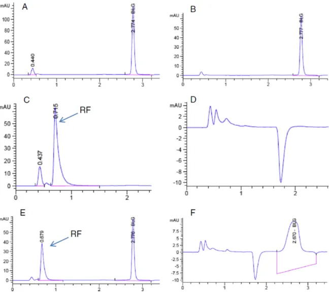
Figure 7.6. RP-HPLC profiles of amounts of βlg and Lyso in the supernatant (A) and the pellet (B) after centrifugation (5000 x g for 5 min) of the βlg/Lyso microspheres prepared using ratio 2:1 at pH 7.5. Absorbance for the proteins at 280 nm and D3 at 265 nm. ... 191
Figure 7.7. RP-HPLC profiles of D3 recovered from the supernatant (A) and the
pellet (B) after centrifugation (5000 x g for 5 min) of βlg/Lyso microspheres - containing D3.Absorbance for the proteins at280 nm and D3 at 265 nm. ... 194
Chapter 8
Figure 8.1. Stability of D3 and the βlg/D3/Lyso microspheres in refrigerated
conditions (4oC) during five weeks. A: Plot of the fitted curves of D3 and the
βlg/D3/Lyso microspheres. B. Equivalence between D3 and the βlg/D3/Lyso
microspheres. The level of α was set at 0.05. The lower decision limit (LDL) was 0.8 and the upper decision limit (UDL) was set at 1.25 (representing 25% difference). ... 208 Figure 8.2. UV-light stability of D3 and the βlg/D3/Lyso microspheres over 24 hours
(ʎ = 254 nm). A: Graph of the stability of D3. B. The equivalence between D3 and
the βlg/D3/Lyso microspheres. ... 212
Figure 8.3. Stability of D3 and the βlg/D3/Lyso microspheres in Simulated Intestinal
Fluid without pancreatin at pH 6.8, 37oC (USP-2 apparatus). A: Graph of the intestinal stability and B. The equivalence between D3 and the βlg/D3/Lyso
microspheres. ... 216 Figure 8.4. Stability of D3 and the βlg/D3/Lyso microspheres in Simulated Intestinal
Fluid with pancreatin at pH 6.8, 37oC (USP-2 apparatus). A: Graph of the intestinal
stability and B. The equivalence between D3 and the βlg/D3/Lyso microspheres.219
Figure 8.5. Bioavailability of D3 in the rat experiment. Mean 25(OH)D3 in rats forced
fed the water (control), D3 and D3 - entrapped βlg/Lyso microspheres. Each error
bar is constructed using 1 standard error from the mean. ... 225 Figure 8.6. Stability of the unprotected D3 and in the βlg/D3 complex, βlg/D3/Lyso
microspheres and βlg-based coagulum. and the βlg/D3/Lyso microspheres in
xxv
Figure 8.7. UV-light stability of D3 of the unprotected D3 and in the βlg/D3 complex,
βlg/D3/Lyso microspheres and βlg-based coagulum. ... 239
Figure 8.8. Stability of the unprotected D3 and in the βlg/D3 complex, βlg/D3/Lyso
microspheres and βlg-based coagulum in Simulated Intestinal Fluid without pancreatin at pH 6.8, 37 oC (USP-2 apparatus). ... 241 Figure 8.9. Stability of the unprotected D3 and in the βlg/D3 complex, βlg/D3/Lyso
microspheres and βlg-based coagulum in Simulated Intestinal Fluid with pancreatin at pH 6.8, 37 oC (USP-2 apparatus). ... 242 Figure 8.10. In vivo experiment of the bioavailability of D3. Mean 25(OH)D3 in rats
forced fed the water (control), unprotected D3, βlg/D3 complex, βlg/D3/Lyso
microspheres and βlg-based coagulum in the rat experiment. Each error bar is constructed using 1 standard error from the mean. ... 244
Chapter 9
Figure 9.1. Stability of the unprotected D3 and in the βlg/D3 complex, βlg/D3/Lyso
microspheres and βlg-based coagulum. and the βlg/D3/Lyso microspheres in
refrigerated conditions (4 oC) during five weeks. ... 237
Figure 9.2. UV-light stability of D3 of the unprotected D3 and in the βlg/D3 complex,
βlg/D3/Lyso microspheres and βlg-based coagulum. ... 239
Figure 9.3. Stability of the unprotected D3 and in the βlg/D3 complex, βlg/D3/Lyso
microspheres and βlg-based coagulum in Simulated Intestinal Fluid without pancreatin at pH 6.8, 37 oC (USP-2 apparatus). ... 241 Figure 9.3. Stability of the unprotected D3 and in the βlg/D3 complex, βlg/D3/Lyso
microspheres and βlg-based coagulum in Simulated Intestinal Fluid with pancreatin at pH 6.8, 37 oC (USP-2 apparatus). ... 242 Figure 9.5. In vivo experiment of the bioavailability of D3. Mean 25(OH)D3 in rats
forced fed the water (control), unprotected D3, βlg/D3 complex, βlg/D3/Lyso
microspheres and βlg-based coagulum in the rat experiment. Each error bar is constructed using 1 standard error from the mean. ... 244
List of abbreviations
ACN Acetonitrile
a.u. Arbitrary Units
βlg β-lactoglobulin
cm centimeter
Da Dalton
D3 Vitamin D3
DTP DevelopmentTherapeutics Program
HPLC High performance liquid chromatography
hr Hour(s) IU International units KDa Kilo-dalton L Liter m Meter mA Milliampere mg Milligram MeOH Methanol mm Millimeter mM Millimolar min Minutes mL Milliliters nm Nanometer PBS Phosphate-buffered saline pI Isoelectric point pH Potential of Hydrogen RF Riboflavin RP-HPLC Reversed – phase HPLC
rpm Revolutions per minute
s Seconds
SDS Sodium dodecyl sulfate
SEM Scanning electron microscopy
TEER Trans Epithelial Electric Resistance
TEM Transmission electron microscopy
TFA Trifluoroacetic Acid
µL Microliter μg Microgram µm Micrometer μmol Micromole µM Micromolar UV Ultraviolet V Volts v/v Volume/volume W Watt
xxvii
x g Relative centrifugal force
Common amino acid abbreviations
A Ala Alanine
C Cys Cysteine
D Asp Aspartic acid
E Glu Glutamic acid
F Phe Phenylalanine G Gly Glycine H His Histidine I Ile Isoleucine K Lys Lysine L Leu Leucine M Met Methionine N Asn Asparagine P Pro Proline Q Gln Glutamine R Arg Arginine S Ser Serine T Thr Threonine V Val Valine W Trp Tryptophan Y Tyr Tyrosine
1
Introduction
Active compounds such as probiotics, bioactive peptides and proteins, antioxidants, vitamins with physiological benefits resulting in enhanced general well-being and/or reduced risk of chronic disease beyond basic nutritional functions, are often referred to as nutraceuticals [1, 2]. In Canada, they fall under the regulation of the natural health products (NHPs), which came into effect on January 1, 2004 sites [3]. The market of NHPs is rapidly increasing worldwide. Since 2004, Health Canada has authorized over 50,000 NHPs for sale in Canada as well as 1,250 manufacturing sites [3]. According to the same source, within only five months (October 2011 to March 2012), the number of product applications completed per month has increased 109 %. The market value is estimated around 117 billion US dollars only from the three most important sources, including the Unites States, Japan and Western Europe [4]. This values increases between 300 and 400 billion US dollars, when other area such as East Europe, Latin America, Asia or Africa are considered.
Generally, bioactives are labile ingredients that are susceptible to elevated temperature, pressure, oxygen and light during food processing. Furthermore, they are prone to degradation during passage in the gastrointestinal (GI) tract when confronted to the pH, presence of enzymes and other nutrients [2]. Entrapment of bioactives in delivery platforms appears as an effective mean to preserve their integrity, to deliver and modulate their release in the body.
β-lactoglobulin (βlg) accounts for about 60 % and thus, is the major protein in bovine milk whey. This whey protein with high nutritional value, related to its high content in essential amino acids, is used in the food industry for its interesting techno-functional and structural properties [5]. Although its true physiological role is still elusive, βlg which is also member of the lipocalin family, has been attributed a transport function for small hydrophobic nutraceutical ligands as well as larger molecules. The interesting structural properties and physicochemistry of βlg
provide this protein with the ability to establish various types of interactions, including hydrophobic and electrostatic interactions, with ligands of different characteristics [5]. Reports indicate that the biological properties such as water solubility or photoreactivity of some of these ligands are improved upon binging to βlg [6-8].
Riboflavin (RF) and vitamin D3 (D3) are both small nutraceutical ligands with
important biological activities, but with dissimilar physico-chemical characteristics. RF is an endogenous photosensitizer, which implies that upon light irradiation at a certain wavelength, it can be photoactivated in aqueous solution and produce reactive oxidative species [9, 10]. Since βlg is composed of aromatic amino acids, including tryptophan and tyrosine, which are also capable of generating oxidative species, the association of the βlg and RF may produce cascade or cumulative oxidative reactions [11, 12]. While RF is an amphiphilic vitamin which binding to βlg has not yet been described in the literature, reports indicate that two molecules of D3 can bind to βlg to form a complex [13]. D3 is highly hydrophobic and sensitive to
light, which is problematic in food formulation. In Canada, foods that are currently fortified with D3 are dairy products or margarine, which have high fat content [14,
15]. The binding of D3 with βlg might improve these shortcomings and thus extend
some of its applications to foods with low fat contents.
βlg – based vehicles of varying can be formed by promoting hydrophobic interactions or through an electrostatically-driven process, which could lead to the self-aggregation of βlg or co-assembly with other food grade biopolymers of opposite charge [16-20]. The cationic protein Lysozyme (Lyso) offers such a possibility. Its isoelectric point is around 10.7, which provides a wide range for electrostatic interactions with βlg, which isoelectric point is 5.3 [5, 21]. Additionally, Lyso is one of the main components of airway fluid where it assumes the host defense [22]. As an enzyme, it damages or kills bacteria by lysing their cell wall peptidoglycan. Its antibacterial activity is therefore limited to gram positive bacteria. Lyso can also bind small ligands with pharmaceutical interest [23, 24]. Therefore, the complex formation between βlg and Lyso might lead to formation of a
3
surpamolecular matrix which can be used for the encapsulation of small bioactives that bind to both proteins. However, βlg is sensitive to environmental factors including the pH and temperature, which are inherent to the process of foodstuffs preparation. This instability can considerably impede the effectiveness of the delivery platform, especially those intended for food applications. The formation of complexes could contribute to enhance the stability of βlg.
Despites the challenges and limitations related to use of βlg in the formulation of delivery scaffolds, there is still a high interest in βlg-based vehicles for oral delivery of biologically active molecules. Ongoing research is seeking new alternatives to overcome such drawbacks and develop βlg-based carriers systems for optimal delivery of biologically active molecules of nutritional importance.
The following chapter presents an extensive work on the production of βlg, its structure and functionality as a platform of nano and microsize for the oral delivery of bioactives.
5
CHAPTER 1
Literature review
β-Lactoglobulin-based nano and microparticulate
systems for bioactives protection and delivery
A version of this literature review will be published as a chapter in the book on
Engineering Foods for Bioactives Stability and Delivery to be published by
Springer.
By :
Fatoumata Diarrassoubaa, Ghislain Garraitb, Gabriel Remondettoc and Muriel Subiradea
aChaire de Recherche du Canada sur les Protéines, les Bio-systèmes et les
Aliments Fonctionnels, INAF/STELA, Université Laval, Québec, QC, Canada
b ERT-CIDAM, Lab Biopharmacie, Faculté de Pharmacie, 28 Place Henri Dunant,
63001 Clermont-Ferrand, France
c Centre de Recherche et Développement, Agropur Coopérative, 4700 Armand
1.1. Abstract
The new paradigms in human nutrition and ever increasing demand of consumers for safe and healthy food products encourage research for functional foods and nutraceuticals as pharmaceutical surrogates. Food proteins are abundant and from renewable sources, with functional groups conferring interesting structural and functional properties. Their ability to bind small ligands, form aggregates and electrostatic complexes with other food macromolecules provides numerous applications for oral delivery technology. The current review focuses on the major milk protein β-lactoglobulin, its fractionation and isolation from whey proteins, techno-functional properties and applications in the formulation of nano and micro-sized oral delivery platforms.
1.2. Introduction
The development of the ideal oral delivery system has become the focus of tremendous research worldwide, particularly in food and pharmaceutical sciences. Along with the field of functional foods and nutraceuticals, the market of which is skyrocketing, the popularity of oral delivery systems has soared and the sales were already expected to reach over $ 67 billion a few years ago [25, 26]. The designation of ‘oral delivery system’ rhymes usually with ‘controlled delivery’ of bioactive compounds, which involves the efficiency of delivery and the enhancement of solubility, absorption, bioavailability, safety and duration of action [26]. In addition to increasing patient comfort and compliance, oral delivery systems can achieve sustain, controlled and site-specific release of bioactives [25-28]. Consequently, on a pharmaceutical point of view, serum concentrations are less prone to fluctuations; toxicity and the frequency of dosing are reduced [26]. In the food industry, delivery systems are used as carriers for functional ingredients which are hence protected from hostile environmental factors including heat, light, oxygen, high pressure and shear forces during processing, storage and utilization
7
until the intact bioactive ingredient is delivered in the intestines; the list of advantages is not exhaustive. The release rate of the bioactive molecule can then be modulated via the control of conditions including temperature, ionic strength, pH and digestive enzymes [29].
The release kinetics from the oral delivery system relies on the chemical composition and/or the structure of the carrier system. In general, mechanisms of release such as diffusion results from the physical (erosion) or chemical decomposition (degradation); swelling and osmotic pressure lead to an expansion of the carrier system, thus expulsing the bioactive out of the carrier matrix [26, 28]. Therefore, the chemical composition and structure of the oral delivery system are of utmost importance to achieve effective release of bioactives. With the size of oral delivery systems decreasing dramatically, concerns have been raised regarding the toxicity of such carriers and the resulting degradation products. Indeed, reduced size may lead to an increase in toxicity given that small sized carriers might reach and accumulate in regions within cells or tissue inaccessible to macroscopic particles of the same composition [30]. In fact, the size of oral delivery systems decreased from capsules, tablets or powder forms to microparticles, nanoparticles and nanocomplexes. Microparticles are less than about 1000 µm while nanoparticles and nanocomplexes are less than 100 nm in size [2, 29]. Therefore, biopolymers which are biocompatible and biodegradable with negligible to null toxicity are increasingly used to fabricate oral delivery systems. Milk whey proteins fit well to this criteria and as such, whey proteins – based delivery systems have been the focus of countless peer-reviewed articles [2, 18, 31-36]. However, whey proteins are composed of diverse proteins with different structures, properties and functions, which makes undistinguishable the role and degree of participation of each protein in the formation of the carrier system [37]. Most functional properties of whey isolates, including aggregates and gel formation, are attributed to its major protein that is β-lactoglobulin (βlg) [17, 38]. The present review is therefore consecrated on the major whey protein β-lactoglobulin (βlg) and its functionality as micro and nano-based oral delivery system.
1.3. Manufacture of whey proteins
Whey is the by-product of cheese production consisting of the serum recovered after isoelectric precipitation of the caseins [39]. The cow milk batch is initially centrifuged to separate the cream to obtain the skim milk, which pH is then decreased to 4.6 in order to precipitate the caseins or the curd, used for cheese production. The resulting serum contains the so-called whey proteins. Sweet whey results from a renneting process in which, the rennet enzyme cleaves kappa-casein liberating high levels of the kappa-caseino-maropeptide (13 mg/mL or 22 %). This protein is present in much lower quantities (0.25 mg/mL or 5 %) in acid whey, a by-product of acid caseins [38]. The protein content in whey depends essentially on two methods which are used to concentrate the different proteins at a yield of about 80 % purity [38]. Whey protein concentrates (WPC) are mainly obtained via ultrafiltration using a membrane with a cut-off of about 10,000 Da. Whereas, whey protein isolates (WPI) with higher protein content result from subsequent diafiltration of the WPC [39]. Individual proteins, including the major whey protein βlg, can then be separated from the whey proteins (Table 1.1).
9 Table 1.1. Major whey proteins content in bovine milk, unprocessed whey (UPW), whey protein concentrates (WPC) and whey protein isolates (WPI)
Protein Milk (g/l) UPW (%) WPC (%) WPI (%) Molecular Weight (KDa) / (pI)
β – lactoglobulin (βlg) 2 – 4 (61) 3.1 (51) 31 (80) 71 (pI = 5.3) 18.3 α – lactalbumin
(α-lac) 1 – 1.5 0.76 (15) (18) 11 (12) 11 (pI = 4.4) 14.2 Bovine serum albumin
(BSA) 0.1 – 0.4 0.15 (3) (1.8) 1.1 2.6 (3) (pI = 4.7) 66 Immunoglobulin(IgG) 0.6 – 1.0 0.37 (7) (5.4) 3.3 3.3 (4) 146 - 1,030 Total milk whey protein 5 – 7
Note: Adapted from [38-41]. Values are expressed as % w/w in powder, mg/ml in whey
and % of measured protein (in brackets) for whey protein concentrates (nominally 80% protein) in cheese whey and whey protein isolates obtained by ion exchange of the cheese whey. Lactoferrine, proteose peptone and caseinomacropeptide which are minor proteins also constituting whey proteins, are not presented in the table. Traces of IgA and IgM also exist in bovine milk, usually at around 1/10 the level of IgG.
1.4. Fractionation of βlg from the whey proteins
A number of separation methods have been optimized to produce high purity levels of βlg. Some of these methods have been scaled up for commercial applications while most of them remain at the laboratory scale, certainly due to their relatively high cost, low productivity, poor selectivity, presence of unwanted denatured proteins and inappropriateness for industrial implementation [41-43]. Mehra and O’Kennedy summarized the current fractionation methods into five categories: the selective precipitation, addition of salts or precipitants, ion-exchange chromatography, enzymatic hydrolysis and membrane filtration [41].
Variants of each method were published; however, their principles are presented as follows. The selective precipitation consists in adjusting the pH (4.65) of the whey protein which is then demineralized (reduction of the ionic strength) by electrodialysis or diafiltration before recovering βlg by centrifugation. The yield of this method is mitigated with 51.6 % of dried βlg concomitantly with 30.9 % of lipids, which is disadvantageous for some food applications (foaming or whipping). A recovery of 84 % of pure βlg from whey proteins was obtained by salting out at acidic pH (2.0) and increasing concentration of salt (NaCl from 7.0 to 30 %). Various salts (ferric chloride - FeCl3 – at pH 4.2) and precipitants have also been used [41, 42]. Anion-exchange chromatography at higher pH values (6.0 and above) or cation-exchange chromatography at a lower pH value (3.0) allow the selective binding and improved recovery of βlg (96 % to 100%) with high purity (67.7 to 99 %). The acidic and peptic resistance of βlg was exploited to optimize a method which allowed the production of high purity protein in its native conformation. In fact, during peptic digestion, βlg remains stable and thus is not hydrolyzed, which is not the case for other whey proteins. Finally, whey protein components of similar molecular weight can also be separated by membrane filtration in response to ionic strength (electrostatic forces) and pH modification. Subsequently additional microfiltration or other types of membrane of controlled pores can be used to further separate proteins of similar size.
11
Research is still ongoing to develop methods that ally speed, sensitivity, resolution, user-convenience with high efficiency. The challenge now resides in separating genetic variants of βlg (mainly variants A and B), given that milk source has a great impact on the protein composition of both milk and whey, and in term, on the molecular structure of the protein [44, 45].
1.5. Structure of βlg
The structure and amino acid composition of βlg have been the focus of extensive research [5, 46-48]. Thus, only the structural characteristics and physico-chemical properties related to bioactive protection and delivery are detailed in the present review.
1.5.1. Basic structure of βlg
Well defined crystal structures indicate that βlg is a globular protein pertaining to the lipocalin family, with a molecular weight of 18,3 KDa and an isoelectric point (pI) of 5.3 [5, 38, 48]. The two main genetic variants of βlg, A and B, differ from a mutation occurring at amino-acid sequence position 64 (AspA→GlyB) and 118
(ValA→AlaB). The overall conformation of the molecule remains fairly the same,
although the substitutions lead to dissimilarities in properties such as heat stability and resistance to pressure denaturation [48, 49]. The information presented in the current review will refer to βlg variant B.
The protein is constituted of 162 amino acids, including all 20 amino acids in relative amounts that make it exceptional and valuable nutritionally. In fact, compared to theoretical common values computed for proteins, βlg comprises about 17 % more essential amino acids [41]. The tertiary structure of βlg is
composed of 8 % of α-helix, 45 % of β -sheet and 47 % of random coil, as represented by the dimeric protein in Figure 1.1 [5, 47, 48].
13
Figure 1.1. Dimer of βlg with di-sulfides bonds in blue. Adapted from PDB 1B8E [48].
βlg has an overall radius of 2 nm, with almost 90 % of its mass within 1 nm of the
surface and nearly 60 % within 0.5 nm [50]. The structure of the monomer of βlg is
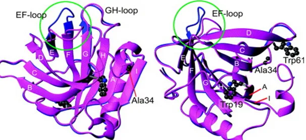
composed of nine strands of antiparallel β – sheets (strands A to I), eight (strands A to H) of which wrap around to form a flattened, conical barrel also called central calyx (Figure 1.2).
15
Figure 1.2. Central cavity of βlg monomer with hydrogen bonds (--). Adapted from PDB 1B8E [48].
The cylindrical shaped calyx has a length of 15 Å, with hydrophobic walls composed of two sheets made of stands A - D and stands E - H (Figures 1.2 and 1.3).
Figure 1.3. Structures of βlg displaying the EF-loop in the closed (left) and open (right) conformation in the green circle. The conical β-barrel is formed by two sheets consisting of β-strands A-D and strands E-H. Adapted from Sakurai and Goto 2006 [51].
17
Strand A (residues 16 – 27) participates in both sheets due to its 90º bend at its midpoint (Ser21), while the ninth strand (I) extends the EFGHA sheet. The neighboring strands within the sheets are connected via a loop [52]. The dimer interface is formed in part by strand I and the loop connecting strand A to B. Strand A is preceded by a 3-turn α-helix while another one lies in the A-B loop. The structure of βlg contains two disulfide bridges consisting of Cys66 – Cys160, connecting the C-D loop to the carboxyl-terminal region, and Cys106-Cys119 links strands G and H. An additional Cys at position 121 is buried and thus, remains free. At pH values from 5 to about 7, including that of the milk (~ pH 6.6), βlg exists in as a dimer in solution, below pH 3 and above 7, the monomeric conformation predominates [5, 52]. Upon modification of the pH, βlg undergoes different structural transitions categorized into distinct classes (M, Q, N and R) and their interrelation is as follows: M ↔ Q ↔ N ↔ R [53].
1.5.2. Structural transitions of βlg
Reports indicate that moderate to considerable pH-induced structural transitions of βlg occur between pH 2.5 and 8, while above pH 9, βlg undergoes significant and irreversible structural modification, also called base-induced unfolding of the protein [52-54]. Below pH 2, the volume and compressibility of the protein decrease, providing a more compact structure to βlg [54]. It is believed that further structural transition continues at pH values less than 1.0, even though no report exists at the moment. Between pH 2.5 to 8, the overall conformation of the protein is conserved extraordinarily well, despite significant structural changes.
The dimer-to-monomer transition occurring between pH 4.5 and 2.5 is the acid induced dissociation of the dimeric βlg into monomers. It includes the βlg transition from the monomeric (M) to the acidic form (Q) around pH 3, where the protein is believed to dimerize [53, 54]. This transition triggers a different orientation of the α-helix, thus affecting only the surface electrostatic properties of βlg. The native
dimeric (N) to the acidic (Q) form transition occurs between pH 6.0 and 4.5. This transition is accompanied with a change in the compactness of the protein translated into a slight expansion of the hydrodynamic volume of βlg [53, 54]. It also includes the pH 5 transition (dimer to octamer transition from pH 3.9 to 5), at which βlg undergoes an octamerization without significantly affecting its secondary structure. Above pH 5, the dimerization of βlg is due to the electrostatic interactions between Asp130 and Glu134 of one monomer with corresponding lysine residues of other monomers. The Tanford (N - R) transition occurs between pH 7 and 8 [53, 54]. This transition involves a conformational change of the EF loop (residues 85– 90), probably due to the cleavage of hydrogen bonds between the F and G strands [53]. Finally, the transition at pH 9.0 or above where βlg undergoes an irreversible base-induced unfolding [54]. These structural transitions are important for understanding the functional properties of the protein.
1.6. Functionality of βlg
The physiological role of βlg, albeit not fully understood, seems to be intimately related to its amino acid composition and tertiary structure. The health benefits of βlg and derived peptides go far beyond its undeniable nutritional value and well justify its use as functional food ingredients and nutraceuticals. Their effects on a number of disease conditions have been reviewed elsewhere [41, 55]. Examples of beneficial activities on human health include their role as hypotensive, anticancer, immunomodulatory, opioid agonist, mineral binding, antimicrobial, gut health enhancing, hypocholesterolemic, insulinotrophic and psychomodulatory agents. Functional advantages of βlg reside in its properties such as gel formation, foaming, emulsion stabilization, all of which find numerous applications in the food industry [2, 17, 56]. Gelation remains one of the most important techno-functional properties of βlg and is traditionally achieved though thermal treatment. The sequential gelation steps consists of the unfolding of polypeptide chains with
19 concomitant exposure of initially buried hydrophobic amino acid residues and subsequent self-aggregation of protein molecules into a three-dimensional network that entraps water by capillary forces [2].Contributing forces are hydrophobic effects, van der Waals, hydrogen bonding, and covalent interactions, which impact
can be determined via the use of destabilizing agents. As such, urea can be used
to block the formation of the hydrogen bonds, sodium dodecyl sulfate (SDS) to block the formation of the hydrophobic interactions, and 2-mercaptoethanol to block the formation of the disulfide bridge [57]. In absence of salt, βlg forms transparent ‘fine stranded’ gels at extreme pH values away from its pI and opaque ‘particulate’ gels near its pI.
Salt was used to induce cold-set gels made by prior heat denaturation of βlg [2, 57]. The solubility of βlg is greatly enhanced in presence of salt due its surface charge distribution at neutral pH, thus explaining the harvesting of the protein by dialysis or precipitation upon salting out and further growing of X-ray crystal structures [5, 58].
The structure of βlg justifies its classification as a member of the lipocalin family and calycin subclass, which is naturally involved in the transportation of small hydrophobic bioactives [59]. Reports indicate that βlg binds retinol, triglyceride and long chain fatty acids such as palmitic acid, resulting in enhanced intestinal uptake of these ligands. βlg might also have an important role in carrying implicated ligands in food systems and pharmaceutical preparations, as well as in digestion, absorption and metabolism of some of implicated ligands in neonates [41]. However, the sequence similarity of βlg with glycodelin, an important protein for fetal development expressed in the endometrium during the first trimester of human pregnancy, suggests further important biological functions [5, 51]. This is
confirmed by the absence of βlg from human and rodents milk and its presence in
the whey of ruminant’s milk including cow, thus making its true function still elusive [5, 43]. However, there is a general consensus about its role as ligands transporter.
Intensive literature exists on the large selection of ligands that bind to βlg [5, 46, 58]. This ligand binding property is mainly ascribed to the presence of the central calyx, which plays the role of a receptacle for small hydrophobic and amphiphilic molecules, consequently forming βlg-ligand complexes [6, 7].
1.7. Formation and characterization of the βlg-ligand complexes 1.7.1. Structural basis for the formation of the βlg-ligand complexes
The folding of the two β-sheets (A - D and E - H) into a central cavity paneled with hydrophobic amino acids (Figure 1.2 and 1.3) resembles a barrel with the EF loop (residues 85-90) acting as the gate [59]. The EF loop folds over the entrance of the calyx to form a closed conformation at pH lower than 6.5 and at pH above 7, it adopts an open conformation which exposes the interior of the calyx [60]. This conformational flexibility is attributed to the Tanford transition which is accompanied by the deprotonation of the carboxyl group of Glu 89, located on the EF loop. Glu 89 that has an anomalous pKa of 7.3 is normally buried and protonated in the “close conformation” at acidic pH. At pH values above 7, Glu 89 is exposed and deprotonated, thus triggering the opening of the EF loop. This consequently offers access to the central calyx [59]. The pH-controlled flipping of the EF loop seems to be crucial for the physiological significance of βlg since in the closed conformation, bound ligands might be protected in the acidic stomach in order to be further released within the intestines at higher pH value [5, 46, 53]. A number of methods have been used to study the binding of the ligands to βlg [7, 47, 61-64]. However, spectroscopic methods are among the most user-friendly, rapid, accurate and less cumbersome techniques used to investigate the protein-ligand interaction. Fluorescence spectroscopy is preferred to study the binding stoichiometric of the βlg-ligand complex while circular drichoism is interesting for
21
studying the influence of the ligand binding on the secondary and tertiary structure of the protein.
1.7.2. Fluorescence quenching upon ligand binding 1.7.2.1. Theoretical explanation
Fluorescence spectroscopy is principally used to study the microenvironment of species with intrinsic fluorescence emission aptitude, called chromophores or fluorophores, which typically contain aromatic molecules [65]. The fluorescence spectral data constitutes the emissions spectra, which is largely influenced by the chemical structure of the fluorophore and the solvent in which it is dissolved. By definition, the fluorescence emission spectrum is a plot of the fluorescence intensity versus wavelengths, usually in nanometers [65]. Fluorescence spectroscopy and mostly fluorescence quenching, is commonly used to characterize ligand binding to proteins. Fluorescence quenching refers to any process that decreases the fluorescence intensity of a sample caused by processes such as inner-filter effect, energy transfer, ground state complex formation and collisional processes [66, 67]. The inner-filter effect occurs when the fluorescence emission of the fluorophore is affected by the presence of an absorbing substance that absorbs the radiation going towards (excitation) or emanating from (emission) the fluorophore [68]. The resulting fluorescence
quenching will thus arise from the reduction of the radiation intensity that excites the fluorophore. The inner-effect can be corrected by carefully selecting the concentration of the ligand, so that the absorbance of the ligand at the excitation and emission wavelength is below 0.1 [68].
Collisional or dynamic quenching results from collisions involving both the fluorophore and quencher during the lifetime of the excited state, while static quenching refers to ground-state fluorophore–quencher complex formation [67]. In the case of collisional quenching, the quencher diffuses to the fluorophore during
![Figure 1.1. Dimer of βlg with di-sulfides bonds in blue. Adapted from PDB 1B8E [48].](https://thumb-eu.123doks.com/thumbv2/123doknet/7220778.202246/41.918.195.739.203.451/figure-dimer-βlg-sulfides-bonds-blue-adapted-pdb.webp)
![Figure 1.2. Central cavity of βlg monomer with hydrogen bonds (--). Adapted from PDB 1B8E [48]](https://thumb-eu.123doks.com/thumbv2/123doknet/7220778.202246/43.918.286.575.192.488/figure-central-cavity-βlg-monomer-hydrogen-bonds-adapted.webp)
![Figure 3.2. Fluorescence emission spectra of βlg in the presence of different concentrations of RF; (A) [βlg] = 5 μM; (B) [βlg] = 10 μM; [RF] in μM, curves: 0, 1, 2.5, 5, 10, 15 and 20(λEX = 280 nm, T = room temperature, pH = 7.](https://thumb-eu.123doks.com/thumbv2/123doknet/7220778.202246/90.918.151.736.148.820/figure-fluorescence-emission-spectra-presence-different-concentrations-temperature.webp)