UNIVERSITY OF QUEBEC – “Institut National de la
Recherche Scientifique, Centre Energie, Matériaux et
Télécomunications”
UNIVERSITY OF CALABRIA – Ph.D. School “Science and
Engineering of Environment, Construction and Energy”
Dual degree joint Philosophiae Doctor (Ph.D.) program
Nano Materials and Innovative Laser-Based Accelerators for
Cultural Heritage
Supervisor: Ph. D. student
Patrizio Antici Simona Veltri
Co-supervisor:
Marianna Barberio
Contents
Preface
1Chapter 1 - Introduction
51.1 Context and Main Scientific Objectives 5
1.2 Relevance of the Research Activity for European Union and Canada 9
1.3 Thesis Plan and main Results 11
Chapter 2 - State of the Art
192.1 Diagnostic methods: state of the art and experimental setups 20
2.2 Conservation Methods 43
Chapter 3 - Growth and Characterization of Nanomaterials and
Nanostructured Materials for Conservation of Cultural Heritage
71
3.1 Synthesis and Characterization of thin-transparent nanostructured films for surface protection
71
3.2 TiO2 and SiO2 nanoparticles film for cultural heritage: conservation
and consolidation of ceramic artifacts
84
3.3 AFM and Pulsed Laser Ablation Methods for Cultural Heritage: Application to Archeometric Analysis of Stone Artifacts
93
Chapter 4 - Innovative Laser-Plasma Diagnostics for Cultural
Heritage
105
4.1 One-shot PIXE diagnostic 105
4.2 Design of nanostructured targets: enhanced proton beams for one-shot
PIXE
121
4.3 Plasma Induced Luminescence in air: a new diagnostic method based on laser-plasma interaction
133
Chapter 5 - General Conclusions and Future Perspectives
141Appendix I: Publication List
Attendance at International Conference
147 148
1
Preface
Cultural Heritage, defined by the UNESCO as “the legacy of physical artefacts and
intangible attributes of a group or society that are inherited from past generations, maintained in the present and bestowed for the benefit of future generations” enriches our everyday lives in countless ways: Cultural Heritage ranges from cherished historic monuments and museums to traditional practices and contemporary art forms, includes new scientific discoveries and constitutes a source of identity and cohesion for communities disrupted by bewildering change and economic instability. This basic principle is again confirmed by the UNESCO when it says “Heritage constitutes a source of identity and cohesion for communities disrupted by bewildering
change and economic instability. Creativity contributes to building open, inclusive and pluralistic societies. Both heritage and creativity lay the foundations for vibrant, innovative and prosperous knowledge societies.[…] No development can be sustainable without a strong culture component […] International treaties endeavor to protect and safeguard the world’s cultural and natural heritage including ancient archaeological sites, intangible and underwater heritage, museum collections, oral traditions and other forms of heritage, and to support creativity, innovation and the emergence of dynamic cultural sectors.”
Following these principles for the sustainable development, in the last years the scientific communities put strong effort into research of innovative techniques in the field of Engineering, Physics, and Chemistry applied to Cultural Heritage (CH) for both, diagnostic and conservations, based on the applications of new materials and investigation methods.
The main challenge within this field is to obtain the greatest number of information without provoking damage to the artifacts, i.e. having an efficient method for diagnostics. Another challenge is the conservation of artifacts without modifying its aesthetical appearance. Chemical and morphological information on artworks are mainly obtained using surface spectroscopies or other methods based on particle accelerators and are most effective and sensible in laboratory. On the other hand, conservation and restoration of artifacts without changes in aesthetical aspect or chemical composition are currently obtained mostly using nanoscience and nanomaterials. The enhancement in the nanoscience and in the growth of nanomaterials allows the production of materials with new mechanical and optical properties and opens up new possibilities for the consolidation and restoration.
2
The ambitious aim of this Ph. D. thesis is to improve both fields of Diagnostic and Conservation in the Cultural Heritage. The main objectives of this work are 1) the development of much more sensitive and performing diagnostics and how this innovative diagnostics react on materials, 2) the improvement of new nanomaterials for the consolidation and conservation of artifacts.
As methodology, for the first topic I use laser-driven particle sources (in application for Cultural Heritage) and for the second topic the realization of thin nanostructured films and laser-generated nanoparticles working as protective materials for artifact surfaces. My thesis opens the field of new laser-driven particle accelerators for research in the field of Cultural Heritage. Moreover, the study of new nanostructured materials for consolidation and conservation allows pursuing new routes to sustainable and easily reversible restoration, without damages or irreversible changes in artwork aesthetical.
The Ph. D thesis has been performed in the framework of a Dual Ph.D. in collaboration between University of Calabria (UNICAL – Italy // GAP Research Group) and the Institute
National de la Recherche Scientifique – Centre Energie Materiaux and Communiction
3
EMT- Canada // iPatLab Research Group). Moreover, all the research activities were performed in strict collaboration between a network of National and International Collaboration: two partners of the “Istituto Nazionale di Fisica Nucleare” (INFN-Italy, INFN-CS and INFN-RM1), the “Lawrence Livermore National Laboratories” (LLNL - USA), the Laser Lab “LULI” (France), the “Sovraintendenza ai Beni Culturali Regione Calabria” (Italy), the ELI-ALPS research center (Hungary), and the ArcheoLab (Italy). Finally, the research activity related to this thesis was performed in different periods spent at UNICAL (18 months total), INRS-EMT (16 months total), and LLNL (2 months total) with the collaboration of researchers of all the involved institutions.
All the results obtained in this Ph. D work were published or accepted for publication in prestigious international Peer-Review Journals (i.e. Scientific Reports, Applied Surface Science, Applied Physics Letters, Applied Physics A, Surface and Coatings, Archeological Discovery), and presented in many prestigious international Conferences of Laser-Plasma, Nanoscience, and Cultural Heritage (see Appendix I for the detail of Publication).
All the project objectives were fully achieved including the challenging determination to open the first research line on Physics Applied to Cultural Heritage at the INRS-EMT, with the intention to setup dedicated experiment on CH investigation at the ALLS facility (the line is currently in preparation and the first experiment is scheduled in the second half of 2017).
The thesis is presented in 5 Chapters which include the Scientific Context, the State of the Art, the Objectives, the Results, the Conclusions of the Scientific Activities and the Future Perspectives. In detail, the Chapter 1 introduces the context of the Physics Applied to the Cultural Heritage, the Scientific Objectives with their relevance for the scientific communities (Canadian and European) and summarizes the thesis project plan and the main results. The Chapter 2 describes The State of the Art while Chapters 3 and 4 illustrate and discuss all the scientific results. The thesis closes with the General Conclusions and the Future Perspectives and, finally, the Appendix shows the Scientific Publications and the participation to the International Conferences.
4 Figure 2 - iPatLab Research Group during an experiment
5
1. Context and Main Scientific Objectives
The world of Cultural Heritage, in particular the field of preservation and restoration of Culture, is hindered by the lack of techniques and diagnostics suitable for the conservation and the preservation of artworks or monuments. There is therefore a strong and urgent need for the discovery and development of new techniques and methodologies for the conservation and restoration1,2.
One of the main challenges in the conservation of artifacts is that they can be damaged by a corrosive patina, which can change their aesthetical aspect. The patina can strongly modify the chemical composition, provoking in the worst case the complete destruction of artwork as in the case of biological films (biofilms) on ceramic and stones exposed to biological agents. Biofilm are mostly generated in humid atmospheric conditions or underwater archeological sites. Similar to the patina, this biofilm can deteriorate the artwork’s surface, causing in many cases the complete destruction of decorations, colorations, and pigments3,4. Several studies have demonstrated that the role of biological agents is one of the main causes of the deterioration of stones5,6.
Microorganism activity has a strong negative impact on the preservation on Cultural Heritage, because they attack the material producing nitric and sulfuric acid7 and as a consequence
destroying the surface or generating green patina that covers completely the surface8. Moreover,
in previous works9,10 it was found that the polymeric films, realized as protective film, do alter the artwork’s aesthetics, changing brightness and coloration. In addition, their effects on biological agents had not been tested. Great effort has been devoted to the employment of nanomaterials (nanofiber, nanoparticles, and quantum dots) embedded in thin transparent films: Silicon dioxide nanoparticles can be used for the consolidation of porous ceramics and stones while thin films of titanium dioxide (pure or doped) can be used to realize protective and transparent films to be placed on artworks. The research in these fields is in progress and several groups have proposed the growth of new materials and their applications for different artwork substrates11,12 (see Figure 1 as example of nanomaterial applications)
6
However, in the world of cultural Heritage, there is not only the need of preservation of the artworks, but also of diagnostic the status of an artwork. In fact, in the last years, strong exertion has been put into the research of innovative techniques in the field of Physics and Chemistry applied to Cultural Heritage for conservation and diagnostic too. Actually several groups explore the possibility to realize equipment to be used directly on side in the archeological sites or in museum. In fact, this need is due to that currently the best performing classical techniques of diagnostics and conservation (restoration and consolidation) require, generally, to move the artworks from museum, or archeological site, into a laboratory, or to make micro sampling of the artifacts (i.e. extract a small part of the artifact to be analyzed).
Chemical information on artworks (ceramic, bronzes, metals, pigments) is mainly obtained using surface spectroscopies (such as Photoluminescence, Raman, X-ray photoelectron spectroscopy (XPS), X-Ray-Fluorescence (XRF), Energy Dispersive X-ray spectroscopy (EDX) in SEM), while morphological information can be obtained with SEM. Complete chemistry of material bulks is known using sophisticated techniques of nuclear physics such as Proton Induced X-ray and Gamma Emission (PIXE and PIGE - see an example of PIXE analysis in Figure 2). All these methods are based on particle accelerators such as, e.g. the Accélerateur Grand Louvre d' Analyse Elémentaire -AGLAE in Paris and the Laboratory of Nuclear Techniques for Environment and Cultural Heritage-LABEC in Florence. Raman and Photoluminescence spectroscopy techniques, on the other side, require sophisticated spectrometers and lasers. SEM and XPS must be taken under vacuum conditions and PIXE and PIGE require the employment of large particle accelerators. Moreover, all these methods allow realizing only local analysis on artworks (beam
7
spot sizes are generally in the order of mm2). Several portable instrumentations, such as XRF and Raman, have been realized to make diagnostics directly in situ. However, all this portable equipment has efficiencies lower than when used in laboratory conditions. Moreover, they present the limiting factor of not being very tunable and adaptable, making their use limited only to a certain field of energy range and with that, to a certain field of interest.
The advent of lasers, which can produce compact bunches of coherent photons, has opened up possibilities of laser-based particle acceleration, as foreseen in reference13. The acceleration of electrons has been studied since the 70’-80’ through wake fields and has recently made a breakthrough by reaching the GeV limit14 in mono-energetic bunches15. First evidence of proton production by laser-target interaction was observed in experiments between 1978-1982, yielding very low maximum proton energies of ~0.56 MeV. In 2000, evidence of laser-accelerated proton beams with significantly improved beam characteristics was obtained, yielding maximum proton energies of ~58 MeV16. Collimated high-current multi-MeV beams of protons generated by
ultra-intense (>1018 W/cm2) short laser pulse (30 fs–10 ps) were discovered also by other research
groups17. The investigation of laser-driven proton acceleration and its use is currently challenging many research laboratories worldwide, with some significant results, in particular for the improved characteristics of laser-based particle sources such as compactness, its efficiency, its versatility and its tunability.
A Laser-based particle beam, moreover, presents the advantage of having a high current, strong laminarity at the source, short duration, and small source side. Focusing on laser-driven proton acceleration, today, existing multi-hundred-TW table-top laser systems generating on-target intensities of ~ 1019-1020 W/cm² can routinely reach proton energies of ~15-20 MeV with a typical laser-to-proton energy conversion efficiency of 1-6 %18,19,20 a current in the kA regime and a laminarity at the source 100 times better than conventional accelerators. These parameters make them a desired candidate for innovative applications requiring one or more of the above-mentioned properties, e.g. in the medical field21, for inertial confinement fusion22, warm dense matter studies23 or hybrid accelerators24. While there has been already many attempts to use and compare these laser based protons to conventional accelerated particle in some applications (medical field, etc), no effort has currently been put to test the efficiency and usage of laser-driven proton acceleration for purposes of Cultural Heritage. This opens up new opportunities to apply the field
8
of laser-driven particle accelerators on this research domain and to test and develop innovative and improved methods of diagnostics with new particle sources.
In my thesis I want to overcome some of the above mentioned existing bottlenecks proposing to improve new nanomaterials for the consolidation and conservation of the artifact and trying to develop new diagnostics methods based on Laser-Plasma Acceleration which are more sensitive and better performing.
In this scenario, there is the need to put emphasis in the research related to the following Activities that are the main scientific mission of the current Ph. D Thesis (See Figure 3 below):
01 Study and realization of Nanostructured Thin Films and Nanomaterials for consolidation and conservation of artworks
02 Study the use of Laser-Driven Particle sources for Cultural Heritage applications and compare them to conventional accelerators
a. Study the influence of particle (protons) irradiation on the stability of the artwork surface
b. Study of the effect of accumulated radiative dose during particle irradiation on artifacts materials
Figure 2 - Classical PIXE applied to the diagnostic of artworks of The Uffizi Museum of Florence
9
1.2 Relevance of the Research Activity for the European Union and for Canada
In the context of the Protection of Cultural Heritage, the European Commission and its member states (including Italy) are undertaking a variety of actions with the aim of promoting and stimulating the optimal use and the resources in science and technology. Not surprisingly, HORIZON2020 is premised on the fundamental idea that setting up synergies between different communities and research domains is a key element for guiding the development of science and technology worldwide and enhancing its efficiency. In order to reinforce their statement, the European Commission plans to name 2018 the “Year of the Cultural Heritage” (see Figure 4).In the upcoming HORIZON2020 Program, the European Parliament has recently included “Cultural Heritage” as a priority in the proposed funding program of €80 bn from 2013-2020. The inclusion of Cultural Heritage in HORIZON2020 takes the form of a series of amendments focusing on the contribution of culture to research excellence, social cohesion and growth. A key section reads:
“Accessibility and preservation of Cultural Heritage [..] is needed for the vitality of engagement within and across European cultures by also considering the importance of Cultural Heritage as strong economic driver in a post-industrial economy and its contribution to sustainable economic growth.”
The sections of European interest in Cultural Heritage are specified in the “Carvalho Report Specific Program”. In particular, it can be read in the CA 18 (Industrial Leadership) the objective:
To improve new nanomaterials for consolidation and conservation of artworks Nanomaterial synthesis
To develop more sensitive and performing diagnostics
studying also their effects on materials
Particles acceleration
10
“Applying design and the development of converging technologies to, create new business opportunities, including the preservation of Europe’s heritage and materials with historical or cultural value. Protecting the Cultural Heritage: assessment, monitoring and choice of conservation materials and techniques, with reference to the environment and energy management, use and maintenance, and integration into contemporary and historical urban surroundings and archaeological and cultural contexts”.
The following project endorses fully these objectives of the European Commission. The collaboration of Universities, Research Institutes, Regional and National Agencies and, indirectly, Industry, ensures the correct use of European resources in science and technology. The research in new Materials and Methods for Cultural Heritage will contribute to protect and preserve our common historical and archaeological patrimony. Moreover, the optimization of diagnostic and conservation technique can reduce the costs of restoration and conservation, favoring their usage also for small museums and small archeological sites and ensuring the accessibility and the preservation of Cultural Heritage for all European citizens.
Moreover, the project links different communities, such as the conventional accelerator community, the nanomaterials community and the laser community, which allows for cross-linked activities on different facilities and different fields of applications that build a unique compound, currently not existing elsewhere.
The research Field of Science Applied to the Cultural Heritage is relatively new for Canada and for the Quebec. New laws on the preservation of Cultural Heritage were introduced in 2012 and the Canadian scientific community is starting to work in this new research field and new funding programs have been opened in the last three years (e.g. Coopération Québec-Italie 2014-2017 and 2017-2019 Appel à projects en art et culture dans le cadre de la Sous-commission mixte).
This Ph.D. project is the first thesis in the field of the Physics Applied to the Cultural Heritage at the INRS-EMT center and should allow for new experiments devoted to the field on the proton beam line currently under setup, with the aim of producing a “laser-driven, few-shot
11
1.3 Thesis Plan and main Results
The entire research activity was organized in two main Activities, which are subdivided in different subtask. Each task corresponds to a well-defined research activity to realize the objectives mentioned before, and is structured as indicated below.
1.3.1 Activity I – Growth and Characterization of Nanostructured Thin Films and Nanoparticles for Conservation of Cultural Heritage
The duplex goal of these scientific research was: (i) the study and the synthesis of nanostructured protective films and nanoparticles for surface protection and the test of their morphological and biological properties against the more common bacteria. (ii) the use of equipment and methods that are proper of the domain of nanoscience and nanotechnology for the diagnostic and the removal of the surface corrosive patina.
The Activity I will be discussed in the Chapter 3 and the research activity performed within this topic can be summarized as followed:
01. Consolidation of ceramic artifacts using a colloidal solution of TiO2 and SiO2 NPs;
02. Selection of nanomaterials and hydrophobic agents for protective film realization;
12
03. Realization of protective film and deposition on different surfaces of interest for Cultural Heritage;
04. Testing of several deposition methods;
05. Testing of bactericidal and hydrophobic properties of protective films;
06. Studying the use of a Atomic Force Microscope as diagnostic for corrosive patina at nanometric scale;
07. Studying the use of Pulsed Laser Ablation as cleaning process on artworks and artifacts at both, nanometric and micrometric scale.
The results of Activity I produced 4 papers published in International Peer-Reviewed Journal and several participations in International Conferences (see Appendix I). The methodologies and the results, discuss in detail in Chapter 3 can be summarized as follows:
- TiO2 and SiO2 nanoparticle films for Cultural Heritage: conservation and consolidation of
ceramic and stone artifacts (Surface & Coatings Technology, 271 (2015) 174–180)
The work focuses on consolidating ceramic and stone artifacts with SiO2 and TiO2
nanoparticles. The nanoparticles were grown by laser ablation of a solution and their shape and dimensions were monitored by optical absorption and AFM morphology studies. The colloidal solutions are mainly made out of spherical nanoparticles with diameter of about 10 nm following a LogNorm distribution with a standard deviation of about 5 nm.
The colloidal solution was then deposited on pottery and stone surfaces to obtain a transparent film with thickness of about 100 nm. The work studies the influence of the nanoparticle deposition on artifacts evaluating 1) the penetration depth of particles into porous materials with XRF and PIXE spectroscopy, 2) the hydrophobicity of deposited film with contact angle measurements, and 3) the influence of the deposition for thermoluminescence dating processes. The results show the reversibility of the thin film deposition process with laser ablation in order to ensure that there are no damages to the artifacts.
13
- Synthesis and characterization of thin-transparent nanostructured films for surface protection (Superlattices and Microstructures, 101 (2017) 209-218 ; DOI:
10.1016/j.spmi.2016.11.023; ICACC’15 Conference 2015 “39th International Conference and Expo on Advanced Ceramics and Composites)
This work demonstrates that very thin and optically transparent nanocomposite films can be conveniently applied on the surface of materials relevant for Cultural Heritage, including bronze, granite, marble, and glass, and display potent antibacterial properties without affecting the aesthetics of the underlying material. The films contain very small loadings of TiO2, graphene, or
fullerene, and can easily be applied on large surfaces using conventional brushes or air-brushes. The antibacterial properties of the films are unaffected by two accelerated aging tests, suggesting their longevity in a real world setting. These nanocomposite films are very promising candidates for the preservation of statues, mosaics, floors, buildings, and other objects that are exposed to challenging environmental conditions.
- Implementation of methods used in nanoscience as diagnostics in the field of Cultural Heritage (AFM and Pulsed Laser Ablation Methods for Cultural Heritage: Application to
Archeometric Analysis of Stone Artifacts. Applied Physics A 120 (2015) 909-916; DOI 10.1007/s00339-015-9225-x)
This work introduce, for the first time, the use of the atomic force microscope (AFM) and of the pulsed laser ablation (PLA) as methods for morphological diagnostic with nanoscale precision of archeological artifacts and corrosive patina removal from stone artifacts extracted from the Church of Sotterra (located in Calabria, South Italy). The AFM microscopy was compared with different petrographic, chemical, optical and morphological analysis methods for identifying the textural characteristics, evaluating the state of preservation and formulating some hypotheses about the provenance and composition of the impurity patina located on the artifact surfaces. The results demonstrate that with the nanometric precision obtained with AFM microscopy, it is possible to distinguish the different states of preservation, much better than using conventional petrographic methods. The surface’s roughness is evaluated from very small artifact’s fragments, reducing the coring at micrometric scale with a minimal damage to the artworks. After the
14
diagnosis, we performed restoration tests using the PLA method and compared it with the more common micro-sandblasting under dry conditions. The work demonstrates that the PLA is highly effective for the removal of the superficial patina, with a control of a few hundreds of nanometers in the cleaning of surface, without introducing chemical or morphological damages to the artifacts. Moreover, PLA can be easily implemented in underwater conditions; this has the great advantage that stone and pottery artifacts for marine archeological sites do not need to be removed from the site.
- Laser Ablation Cleaning Effects on Thermoluminescence Dating Technique (Archaeological Discovery, 2 (2014) 58-64)
Here we present the study of laser ablation (LA) restoration techniques and of thermo luminescence dating process (TL). The main aim of the work is to demonstrate that LA doesn’t affect the possibility to date ceramic artifacts after restoration. We ablate Neolithic ceramics in air with a first harmonic of laser YAG (1064 nm) and dating the artifacts before and after the cleanness process. The obtained discrepancy before and after cleanness respectively is of about 200 years (3300 B.C. and 3100 B.C.), value that is below the limits of the experimental error. Moreover, the temperature of the artifacts during the LA was monitored in order to check that the temperature stayed below a certain threshold value. The results indicated that the maximum temperature reached was about 100˚C, not enough to empty the metastable traps that cause the luminescence signal. Therefore the artifact can still be used for dating techniques.
1.3.2 Activity II – Laser-driven proton sources for improved and innovative Applications in Cultural Heritage
The aim of this activity is to improve the field of Diagnostic in Cultural Heritage using Laser-accelerated beams and plasma. The work focuses in particular in the design and realization of experiments on laser-driven few-shots PIXE and Cathodo-Luminescence. The experiments were performed in the laser facilities of LLNL (one shot-PIXE) and ALLS (plasma induced
15
luminescence). The experiments design and realization are discussed in detail in the Chapter 4, where the main research activity can be summarized as follows:
01. Study the use of laser-driven particle sources for Cultural Heritage applications and compare them to conventional accelerators;
02. Perform numerical simulations for investigating the phenomena occurring during irradiation, using Monte Carlo codes;
03. Measure the improvement and the tunability/versatility of laser-driven sources (e.g. regarding penetration depth); Design the experimental setup, realizing preliminary tests on commercial materials of interest for Cultural Heritage (i.e. bronzes, marbles, glass, etc.,); 04. Perform experiments on original artworks and artifacts, demonstrating the effectiveness of new methodologies on some particular case studies; Realize monochromatic proton beams designing and realizing dedicated nanostructured target suitable for improving laser-driven proton acceleration experiments, in particular with regard to the Target-Normal-Sheath Acceleration (TNSA) acceleration mechanism, and the use of an energy selector;
05. Study a new method for performing analysis of the chemical composition and optical properties of materials using plasma induced luminescence in air.
The results of Activity II produced 3 papers published in International Peer-Reviewed Journals and several participations in International Conferences (see Appendix I with Publications and Conferences). The methodologies and the results, discuss in detail in Chapter 4 can be summarized as follows:
- Laser-Accelerated Proton Beams as Diagnostics for Cultural Heritage: One-Shot PIXE (Scientific Report, 7 40415 (2017); DOI: 10.1038/srep40415)
The aim of this study is to improve the field of Diagnostic in Cultural Heritage using Laser-accelerated beams developing of much sensitive and performing equipment for diagnostics on the different materials of interest for Cultural Heritage (i.e., stones, bronzes, marbles, pottery, etc…). This work introduces the first use of laser-generated proton beams as diagnostic for materials of interest in the domain of Cultural Heritage. Using laser-acceleration protons, as generated by interaction of a high-power short-pulse laser with a solid target, we are able to produce
proton-16
induced X-ray and Gamma-ray emission spectroscopies (PIXE/PIGE). Correctly tuning the proton flux on the sample, we are able to perform the PIXE in a single shot without provoking more damage to the sample than conventional methodologies. Modelling the interactions of high-fluxes (1013 particles shot) of ultra-short (ps-scale) tunable (keV - 60 MeV) laser-generated proton beams
with the most common Cultural Heritage materials, the results show that is possible to analyze artifacts up to a depth of a few hundreds of microns with a very high precision and a beam current at least three orders of magnitude higher than conventional accelerators. The results of this work demonstrate that laser-accelerated protons can be successfully used in one-shot proton induced X-ray spectroscopy thus strongly enhancing the diagnostic of many materials.
- Fabrication of nanostructured targets for improved laser-driven proton acceleration (Superlattices and Microstructures, 95 (2016) 159–163)
Here we present a novel realization of nanostructured targets suitable for improving laser-driven proton acceleration experiments, in particular with regard to the Target-Normal-Sheath Acceleration (TNSA) acceleration mechanism. The nanostructured targets, produced as films, are realized by a simpler and cheaper method which includes two step approach for the production of the gold nanoparticle layers: 1) Laser Ablation in Solution and 2) Spray dry technique using a colloidal solution on target surfaces of Aluminum and Mylar thin films and Carbon Nanotube buckypaper. The obtained nanostructured films appear, at morphological and chemical analysis, uniformly nanostructured without presence of oxides or external contaminants. The nanostructured layer were grown on these three materials because the wetting properties between them and the gold nanoparticles can ensure the realization of a uniform nanostructured film on the surface without local aggregation of the NPs25. Moreover, these are some of the most used target materials in laser-driven proton acceleration. The obtained targets show a broad optical absorption in the entire visible region and a surface roughness that is two times greater than non-nanostructured targets, enabling a greater laser energy absorption during the laser-matter interaction experiments producing the laser-driven proton acceleration.
17
- Laser Stimulated Plasma-Induced Luminescence: a new method for on-air material analysis (Applied Physics Letters, 110 021114 (2017); DOI: 10.1063/1.49734670)
In this research activity we develop a new method for performing analysis of the chemical composition and optical properties of materials using In-Air Plasma-Induced Luminescence. This is achieved by interaction of a focused high-energy laser with air, interaction that produces a sub-millimetric plasma. The energetic electrons generated and accelerated in the plasma at energies higher than 5 keV reach the target surface of the sample to be analyzed, causing luminescence emission and plasmonic resonance. Each material is characterized by different chemical and optical properties that can be determined with the above-described technique. As such, our method allows obtaining an exact analysis of the sample, covering surfaces in the range of tens of mm2, in
only a few minutes. The results show that the acquired information is identical to what obtained with more sophisticated methods, such as SEM-Cathodoluminescence and Photoluminescence.
19
2. State of the Art
Cultural Heritage takes up a very important worldwide role because it represents the memory, the history and the basis for the sustainable development of the human society. The world is rich and abundant in various cultural heritage buildings, monuments and objects of different sizes; each of these artworks is the fingerprint of a local culture. Our Heritage constitutes the root of present and future cultures, and it can and has to be protected and transferred to the future generations through the scientific skills.
Physical methods applied to the Cultural Heritage became, in this general view, increasingly important and more frequent. Today, it is necessary to perform any restoration process using scientific criteria. In order to do so, there is the crucial need of having as much information as possible about the artwork to get knowledge about the past and current state. Therefore, these methods allow answering questions about the state of deterioration of the work of any previous restorations, and the authenticity of the artifact etc. In recent times, the scientific applications in archaeology are heavily increasing and their particular role in connection with Cultural Heritage is also called Archaeometry. Literally, Archaeometry means measurement on ancient objects and Physics and Chemistry give a fundamental contribution to solving problems associated with the conservation and restoration. Archaeometry also includes the scientific study of an artifact based on the radiation-matter interaction and allowing getting any kind of information about the chemical compositions, the physical properties and the dating of the artifact. The modern approach of studying Cultural Heritage is highly interdisciplinary and in the last decades strong effort has been put into this research area.
Many groups currently explore the possibility to realize equipment for diagnostic and conservation that can be used directly in situ (i.e. in the archeological sites or in museums). As mentioned in the previous chapter, chemical information on artworks (ceramic, bronzes, metals, pigments) is, mainly, obtained using surface spectroscopies (such as Photoluminescence, Raman, X-ray photoelectron spectroscopy (XPS), X-Ray-Fluorescence (XRF), Energy Dispersive X-ray Fluorescence (EDX)) in SEM), while morphological information can be obtained with SEM.
Complete chemistry of material bulks is acquired using sophisticated techniques of nuclear physics such as Proton Induced X-ray and Gamma Emission (PIXE and PIGE). However, all these methods present the limiting factor of not being very tunable and adaptable (as already specified
20
in Chapter 1), making their use limited only to certain energy ranges and with that, to certain areas/fields of interest.
Regarding the conservation field, in the last years many research groups have studied the methods to improve materials for the consolidation and conservation of the artifacts. The current research is based on the fabrication of different protective layers. Some of the layer developments warrant the protection of the surface against condensed water1, or against organic contaminants and environmental toxins2, others employ the well-known photo catalysts activity of some nanomaterials to realize an hydrophobic and biocidal coating3.Other layer developments include Si-based resins4 to realize a protective coating for atmospheric pollutants, Oil-in-water (o/w) micro emulsions or micellar solutions5 for the solubilization of acrylic and vinyl polymers and the development of new protective films with very low environmental impact, New cellulosic titanium dioxide nanocomposite6 is employed for the protection of paper based materials against the damaging effect of ultraviolet radiation, pollutant gasses, mold and bacteria. All these methods allow to preserve the surfaces; some, specifically, are dedicated to the hydrophobicity while others to the bactericidal activity or to the polluted atmospheric environment.
This Chapter gives an overview of the main existing scientific methods applied to the diagnostic, the restoration, and the protection of Cultural Heritage.
2.1 Diagnostic methods: state of the art and experimental setups
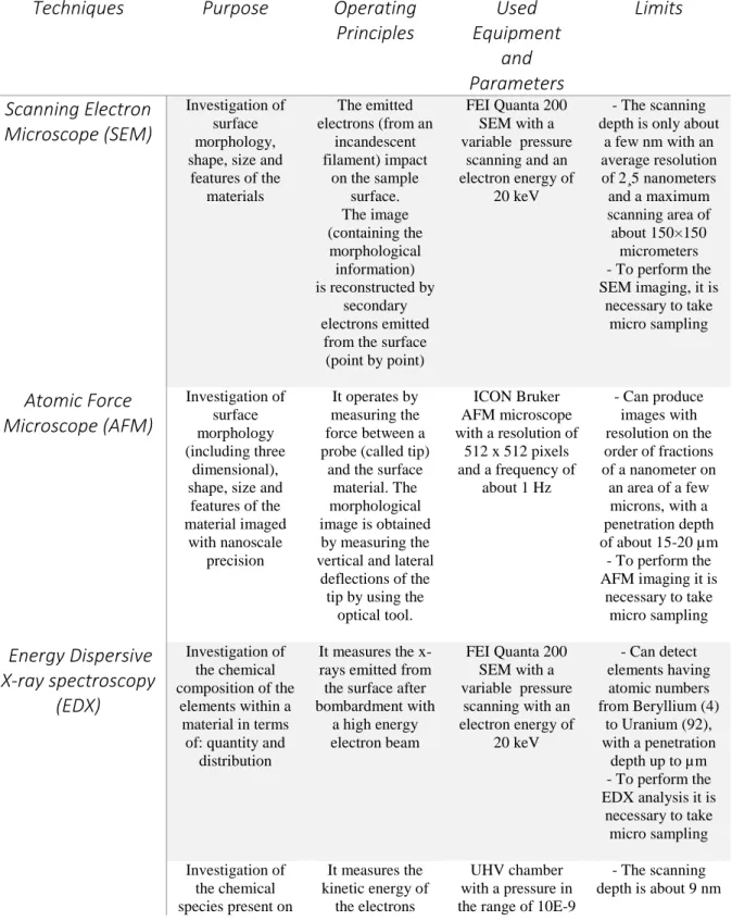
This first paragraph describes all the diagnostic techniques. The techniques will be described from a theoretical point of view, besides the description of the experimental setups finishing with the explanation of the PIXE and Photoluminescence spectroscopies, which are the two methods we have proposed to improve in the present study. The paragraph closes with a briefly summary shown in a table 1 of all the mentioned diagnostic techniques.
2.1.1 Scanning Electron Microscope (SEM) coupled with Energy Dispersive X-Ray (EDX) spectrometry
The scanning electron microscopy (SEM) is not only used to define the chemical composition of the material but also the surface morphology. This technique allows to acquire information about the topography of the sample underlining the texture (up to a few nm) and morphology: shape,
21
size and arrangement of the particles that make up the material. The ability of this instrument to provide very realistic images of the test sample with a high definition is due to employment of electronic waves instead of light waves (see Figure 1 below).
An electron gun (see Figure 2) contains a tungsten filament (cathode), which is displaced in the vacuum. When traversed by a current, this filament becomes incandescent and starts emitting electrons, which are accelerated and focused into a thin beam by means of electromagnetic lenses. Moreover, the beam's impact position on the sample surface, and then the scanning of the area, are controlled by electronically driven deflection coils. The image is acquired by secondary electrons emitted point by point from the sample surface hit by the primary electron beam7,8. Finally, the achieved image is a morphological image, constituted by secondary electrons emitted from the surface, point by point.
22
The high-energy electron beam (20 eV) interacting with the sample surface generates a series of signals:
- Secondary electrons (SE): originate from a depth of a few nm, have low energy and are mainly issued by the Auger effect 9. The collision of primary electron creates an electronic vacuum in a solid inner band. This band can be filled by decay of an electron from a higher band, with energy transfer liberated to an electron so that it can be expelled from the solid. The secondary electrons provide information on the surface topography and the electric or magnetic fields distribution.
- Electron back-diffused (BSE): derived from elastic interactions of the primary beam with the nuclei of the atoms in the sample. . These electrons allow obtaining information about the average atomic number, the crystalline structure and topography of the scanned area of the sample.
23
- X-rays: produced by fluorescence. They are characteristic of the elements making up the sample. The intensity of these radiation characteristics is proportional to the element concentration in the sample10.
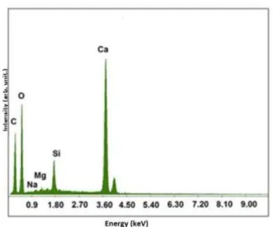
Microanalysis X-ray (Energy Dispersive X-ray- EDX) gives specific information on the chemical composition of the elements in the sample, in terms of quantity and distribution, by measuring the X-rays emission following the bombardment with a high-energy electron beam. The microanalysis is realized by means of a microprobe applied to the scanning microscope, comprising a window permeable to X-rays11. All elements having atomic numbers from 4 (Be) to 92 (U) can be detected, though not all instruments are equipped for 'light' elements (Z < 10). Qualitative analysis involves the identification of the lines in the spectrum, which can be quite easily performed due to the uniqueness of the X-ray spectra for each element. Quantitative analysis (determination of the concentrations of the elements present in the sample) entails measuring the line intensities (height of the line) for each element in the sample. Additional calibration allows measuring the percentage contribution of each element.
The EDX spectrum (shown in 2D images or 'maps', see Figure 3) can be obtained by scanning a single point or the whole area investigated by the microprobe and displaying the intensity of a selected X-ray line.
Three devices essentially constitute the experimental setup for EDX:
• Electron microprobe: it is a system for the beam formation. It generates electron beams characterized by high density, current stability and small diameters that may vary depending on the experimental setup used and that are adapted to obtain the highest possible resolution.
• X-ray detection systems: the detector is made of a single crystal of Si doped with Li. The detector allows the realization of a graph, where each peak corresponds to a specific chemical element. • Analysis of the detected X-rays: the amplified signal from the detector is conveyed into a device called multichannel analyzer (MCA), which acts as an analog-digital converter and sends the signal coming from the amplifier in discrete energy channels, whose amplitude is usually of 10 or 20 eV. The signals of the X-rays are then distributed according to their energy, thus obtaining the X-rays spectrum. The MCA is integrated in a computerized system that allows to display the obtained spectrum on a monitor and a post-processing of the data.
24
2.1.1.1 The SEM and EDX experimental setup used in this work
The equipment used for most of the analysis in this thesis is a FEI Quanta 200 SEM with a variable-pressure scanning electron microscope (ESEM™). The SEM images are taken under a STEREOSCAN SEM microscope working with an electron energy of 20 keV.
The Quanta SEM systems can be equipped with analytical systems, such as energy dispersive spectrometer, wavelength dispersive x-ray spectroscopy and electron backscatter diffraction. The microprobe equipped to perform EDX analysis works with a brightness of 100 A/cm2 sr, an energy spread of 2.5 -3 eV and a source size of 50 nm.
2.1.2 Raman
Raman microscopy is a spectroscopic technique used to observe vibrational, rotational, and other low-frequency modes in a system12. It provides information on the atom vibrations in a crystal lattice by studying the inelastic and Raman scattering of a monochromatic light (usually a laser in the visible range). The laser light interacts with molecular vibrations, phonons, or other excitations in the system, and the produced shift in energy gives information about the vibrational modes in the system13.
25
In this present thesis, we have not used this technique.
2.1.3 X-ray Photoelectron-XPS
The X-ray photoemission spectroscopy (XPS) allows the determination not only of the chemical species present, but also the nature of the bonds in which they are involved, through the position analysis (chemical shift) of photoelectric peaks compared with tabulated values referring to the pure atomic species14,15.
The XPS measures the kinetic energy and the number of electrons that are emitted from the surface of a sample after the irradiation with a beam of X-rays17 (Figure 4).
The XPS is based on the photoelectric effect: a photon, of well-defined energy hv and hitting a solid material, penetrates into the surface and is then absorbed by an electron. If the photon has a sufficiently high energy then the excited electron will emerge from the surface with a kinetic energy approximately equal to:
𝐸𝐾= ℎ𝜈 − 𝐸𝐵 − 𝜙𝑆 (1)
where ϕ represents the work function of the solid, 𝐸𝐵 the energy of the electron bond in the solid,
and hv the energy of the X-ray source.
26
From the above equation it is possible to conclude that, by measuring the kinetic energy (EK) of
the emitted electrons, one can retrieve the initial state energy (previous excitation), provided that the electron reaches the analyzer. This, since the electron will endure a variation of energy equal to
𝐸𝐾 = ℎ𝜈 − 𝐸𝐵 − 𝜙𝑎 (2)
due to the difference between the sample and the analyzer work functions.
From the description of the photoelectric effect, the distribution of electron energy photo-emitted directly represents the distribution of the electronic states of the surface of the solid (less than an amount equal to hv)16. The photoelectron spectrum can then be interpreted as a reflection of the
orbital diagram: each band of the photoelectron spectrum corresponds to the ionization of a single orbital and each orbital occupied with binding energy less than hv gives rise to a band in the spectrum.
The photoemission model described by equation (1) is known as an approximation to an electron. The spectrum (typical of the metals of the second column) is usually dominated by a few very narrow and intense peaks, which originate from the electrons emission from the core levels of the surface atoms, by the emission of electrons from the valence band, by some secondary structures that are attributable to the plasmons, to Auger electrons17 generated from vacation left after the emission of an electron from a core layer and at secondary electrons derived from inelastic processes.
The basic components of the XPS are:
- Vacuum system: this system involves the use of three pumps, which work according to three different principles (a mechanical rotary pump, a turbo molecular pump and a ionic pump)
- Excitation source: as X-ray source, in the XPS instruments are used the emission lines of a metal target called anode, which are subjected to bombardment with electrons having energies of the order of tens of keV. The energy of these lines, resulting from electronic transitions, depends only on the energy level of the anode component atoms. The bombarded anode causes the emission of characteristic radiation of the bombarded
27
element. This characteristic radiation is superimposed on a continuous spectrum called
Bremsstrahlung, which depends on the energy of the incident electrons. Anodes available on the instrument at our disposal are aluminum (Kα = 1486.6 eV)18 and magnesium (Kα =
1253.64 eV)18 since these two materials guarantee characteristic peaks with an energy that
allows to excite the deepest core level of all elements of the periodic table.
- Electron energy analyzer: the hemispherical electron analyzer (EA) is a tool that allows measuring the energy of a charged particle. The EA consists of two metal hemispheres on which a potential difference is applied so that only the charges with a determined energy (E) are able to have a curvature that allow them to reach the other terminal of the analyzer. The condition is:
Δ𝑉 = 𝑉 (𝑅1 𝑅2 − 𝑅2 𝑅1 ) (3) Where E = qV
This passing energy is called Energy pass. Over the output terminal of the charges, there is a channeltron that consists of a glass tube, whose walls are coated with a highly resistive materials. In this way, whenever the wall is hit by an electron, this will provoke an avalanche breakdown mechanism and for each electron entering in the channeltron, the number of generated electrons is about 104. The resulting current is then transformed into a series of pulses whose frequency is proportional to the intensity of the initial current.
- Acquisition system detection signal: the data acquisition is performed by employing a computer with Keithley interface. Through the interface, one can send the voltage to the analyzer lenses to fix the analytical power and receive its signals. Final data are the average of several acquisitions in order to reduce the effects of noise.
28
2.1.3.1 The experimental setup used in this work
XPS experiments are conducted in a UHV chamber equipped for standard surface analysis with a pressure in the range of 10−9 Torr by a non-monochromatic Mg-K𝛼 X-ray (h𝜈 = 1253.64 eV). The energies of XPS spectra are calibrated with the C 1s peak of a pure carbon sample (binding energy of 284.6 eV).
2.1.4 X-Ray Fluorescence (XRF)
The X-ray fluorescence (XRF) allows to attain the elemental (qualitative and quantitative) composition of a material. Since this method is fast and non-destructive to the sample, it is commonly used for applications in the field of Cultural Heritage.
The XRF technique is based on the “photoelectric effect”: when a primary X-ray excitation source from an x-ray tube or a radioactive source strikes a sample, if the primary x-ray has sufficient energy, it is absorbed by the atom by transferring all of its energy to an innermost electron. During this process, electrons are ejected from the inner shells, creating vacancies that are an unstable condition for the atom.
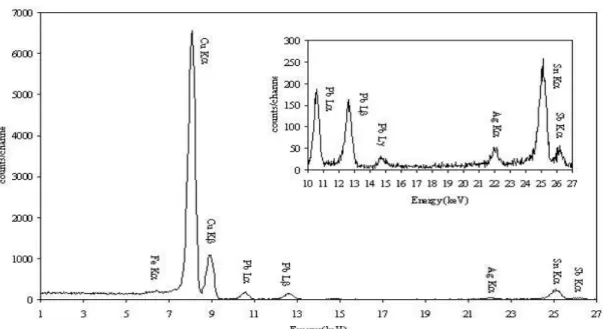
When the atom returns to its stable condition, electrons from the outer shells are transferred to the inner shells and in the process, giving off a characteristic x-ray whose energy is the difference between the two binding energies of the corresponding shells. The emitted x-rays produced from this process are called “X-ray Fluorescence,” or XRF. The process of detecting and analyzing the emitted x-rays is called “X-ray Fluorescence Analysis.” In most cases, the innermost K and L shells are involved in the XRF detection (see Figure 5). A typical x-ray spectrum from an irradiated sample will display multiple peaks of different intensities (see Figure 6).
29
The characteristic x-rays are labeled as K, L, M or N to denote the shells they originated from. Another designation alpha (α), beta (β) or gamma (γ) is made to mark the x-rays that originated from the transitions of electrons from higher shells. Hence, a Kα x-ray is produced from a transition of an electron from the L to the K shell, and a Kβ x-ray is produced from a transition of an electron from the M to a K shell, etc. Since within the shells there are multiple orbits of
Figure 5 - Sketch of X-ray Fluorescence spectroscopy
30
higher and lower binding energy electrons, a further designation is made as α1, α2 or β1, β2, etc. to denote transitions of electrons from these orbits into the same lower shell.
The components of a XRF spectrometer are:
- Primary X-rays source: is composed of a cathodic tube able to operate at high values of potential difference and of an anticathod made of Au.
- Sample holder chamber
- Collimator system: they are usually constituted of several parallel foils that are used to make parallel the obtained X-rays
- Analyzer crystal: it allows the identification of the elements by diffracting the secondary X-ray radiation coming from the sample
- Detector: it is composed by a counter able to distinguish the different wavelengths and intensities of the collected X-ray radiations, showing them as peaks19.
2.1.4.1 Experimental setup used in this work
XRF measurements were conducted by the X-123 SDD apparatus by Amptek (USA), equipped by a gold cathode and a beryllium revelator, operating at fixed angle.
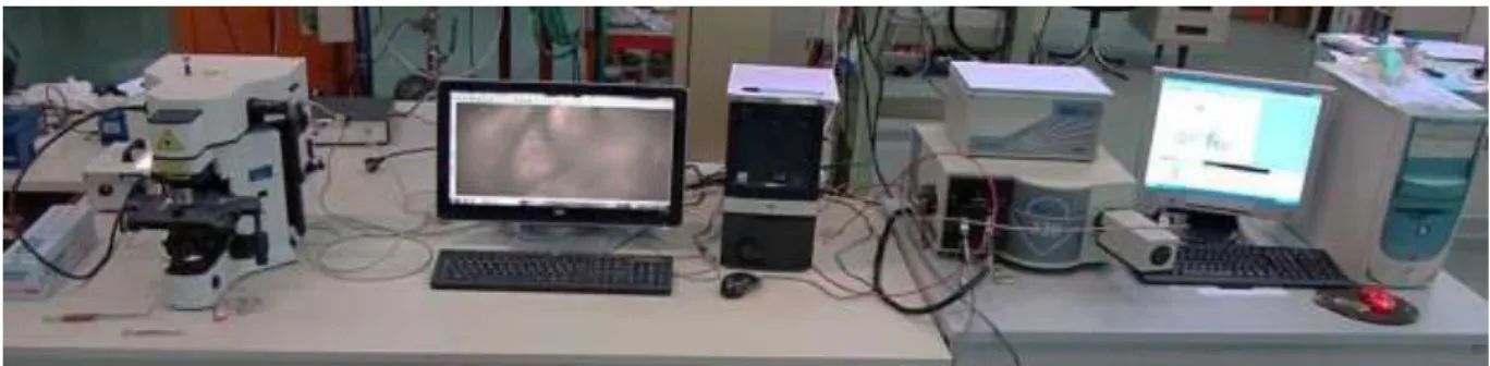
The device used for the XRF measurements is shown in Figure 7, and is composed by a “Mini-X” X-ray generator mounted on a MP1 tube equipped with X-123SDD detector. Mini-X is a self-contained, miniature X-ray tube system, which includes the X-ray tube, high voltage power supply
31
and USB controller.. It features a 50 kV/80 μA power supply, a gold (Au) or silver (Ag) transmission target, and a beryllium end window. The Mini-X is designed to generate voltages up to 50 kV. Beryllium window is on the front of the unit. The Mini-X is provided with two collimators.
The X-123SDD combines in single package Amptek’s high performance X-ray spectroscopy components: (1) the XR-100SDD silicon drift X-ray detector and preamplifier, (2) the DP5 digital pulse processor and MCA, and (3) the PC5 power supply. The X-123SDD uses a silicon drift detector (SDD) to improve the energy resolution and increase the count rates.The pulse processor is the DP5. The DP5 digitizes the preamplifier output, applies real-time digital processing to the signal, detects the peak amplitude, and bins this in its histogram memory. The spectrum is then transmitted to the user’s computer.
2.1.5 Photoluminescence
The Photoluminescence technique, widely used in the field of Cultural Heritage, is employed to identify mineralogical phases present on the surface of the materials, the presence of any coatings, “alteration patinas” or extrinsic too.
The luminescence is a phenomenon by which the radiative charge transitions happen with emission of electromagnetic radiation in the visible region and close to it (infrared and ultraviolet) as a result of the passage of radiative charged from the excited states to those with a lower energy.
If the emission process stops with the cause exciter, i.e. between the absorption and re-emission there is only the time interval necessary for the atomic transition (τc <10-8 s), then a
fluorescence process occurs. If the state of excitation is metastable and reissue is delayed (τc ≥ 10
-8s) and the emergence issue continues even after the removal of the cause exciter, then a
phosphorescence process occurs.
In relation to the phosphorescence the energy levels diagram phenomenon present a metastable m level in the band gap between e and g (see Figure 8). An electron excited to g can be trapped in m where it can remain until it receives an energy E sufficient to get it back in e; from here there can be a transition to g with emission of light.
32
The delay that is observed in the phosphorescence phenomenon corresponds to the time that the electron spends in the trap for electrons m. The average life spent in the trap at the temperature T is given by:
τ = S−1 ∙ ek.TE (4)
Where s is a constant called frequency factor, expressed in s-1; E is called the trap depth (energy
difference between m and e); k is the Boltzmann constant.
The phosphorescence process has an exponential temperature dependence. The complete model, based on the theory of energy bands is the result of the study by Randall and Wilkins20.
Randall and Wilkins assume that, when the electron is freed from the trap (m → e, Figure 8b), the probability that it returns to the ground state g(s → g) is much higher than the probability that it is trapped. The intensity of phosphorescence is proportional to the rate of recombination (e → g). In this case the transitions involved are m → e, for which I (t) is proportional to the escape velocity of electrons from the trap:
𝐼(𝑡) = −𝐶𝑑𝑛 𝑑𝑡 = 𝐶
𝑛
𝜏 (5)
Figure 8 - Energetic transition involved in the production of fluorescence (a) and phosphorescence (b)
33
Where C is a constant of proportionality and n is the number of electrons trapped in m. Integrating, we obtain:
𝐼(𝑡) = 𝐼0 . 𝑒−(
𝑡
𝜏) (6)
Where τ is the average life time, t is the time and 𝐼0 is the intensity at t = 0
The equation represents the decay of phosphorescence after the end of irradiation, at a constant temperature (Figure 8). The trend at constant temperature will be a simple exponential or a decay of the first order.
The implemented setup for the photoluminescence consist of:
- Horiba Jobyn Yvon Olympus microscope: it mounting objectives 10x, 50x, 100x. The microscope is equipped by a laser source by a Triax 320 (Horiba-Jobyn-Yvon) for the processing of the optical signal.
- Optical detenction system: it is composed of a monochromator in series to a CCD. The monochromator (is composed by some toroid mirrors that focused the input signal on a turret with three different diffraction greatings decomposing the signal into its components in various wavelengths) that focus the input signal, transfer it to the CCD (which is is a Si wafer detector with a resolution of 1024 x 128 pixels that has the task of transforming the incoming optical signal into an electrical signal by photoelectric effect, so that it is interpretable by the appropriate software). The sample positioned in the housing of the microscope is focused via remote with the 100x objective through a camera connected to a computer. After focusing the sample, due to the optics of the microscope, is sent on a UV laser beam (incidence angle zero), with respect to the normal of the sample. The photoluminescence signal is then collected and sent through an optical system (including a notch filter that excludes the output laser frequencies, to avoid overlap between the input signal and signal output) to an optical fiber that is connected to the entrance slit of the spectrometer.
34
2.1.5.1 Experimental setup used in this work
Photoluminescence (PL) measurements were taken by an Olympus microscope (Horiba-Jobyn Yvon) mounting objectives of 10×, 50× and 100× magnifications. The microscope is equipped by a 375 nm-laser source with a power of 15 mW for the generation of photo-luminescence, and a Triax 320 that works in the 200 - 1500 nm range.
2.1.6 Proton Induced X-ray Emission- PIXE
Particle-induced X-ray emission called PIXE is a technique that allows to investigate the elemental (qualitative and quantitative) composition of trace elements (ppm) of a wide range of material and then very important in the Cultural Heritage field. The advantages of this method are many, as high sensitivity (offers better peak to noise ratios and consequently much higher trace element sensitivities), multi-element analysis and measurements at atmospheric pressure.
PIXE is an elemental analysis technique which employs a charged particle beam from an accelerator to excite characteristic emission from the constituents of a material. When a material is exposed to an ion beam, atomic interactions occur, emitting x-rays that are specific to an element. In details, when the ion beam interact with a material, it creates electron “vacancies” in the inner atomic energy levels (the so called K, L and M shells) by ejecting one or more of its most tightlly bound electrons out of the atom21 (Figure 10).
35
When a beam of heavy charged particles of a few MeV per atomic mass unit penetrates into matter (considered as a compound), it gradually loses its energy with depth, until it is finally stopped. The energy loss occurs mainly through inelastic Coulombic encounters with bound electrons, and in contrast to the case of electron beams, the direction of travel of an ion beam is scarcely altered during the slowing-down process. The stopping power S(E) of an ion with energy E is defined as the energy loss per unit mass thickness traversed:
𝑆(𝐸) = −1 𝜌
𝑑𝐸
𝑑𝑥 (7)
where φ is the density of the stopping material and x is the distance. As defined here, S(E) is expressed in units of kiloelectron volt per gram per square centimeter. For the energy range of 1-4 MeV, which is the most important in PIXE, the uncertainty of the values calculated with the semiempirical equation is estimated to be less than 3%.
The stopping power for compounds or more complex matrices is obtained from those of the constituent elements throught the Bragg-Kleeman additivity rule:
Figure 10 - Sketch of atomic proton interactions and electronic transitions between K, L and M
36
𝑆𝑚𝑎𝑡𝑟(𝐸) = ∑ 𝑤𝑖 𝑛 𝑖=1
𝑆𝑖(𝐸) (8)
where 𝑤𝑖 and 𝑆𝑖(𝐸) are the mass fraction and stopping power of costituent element i, respectively.
The total path length R of an ion may easily be obtained by integration of the stopping powers:
𝑅 = ∫ 𝑎𝑏
0 𝐸0
− 𝑑𝐸
𝑆(𝐸) (9)
where 𝐸0 is the incident ion energy. Althought the total path length is larger than the projected
range, the difference between the two is smaller than 1% for the incident protons of a few megaelectron volts (MeV).
Many of the Coulombic interactions between protons or heavier ions and matter results in the ejection of inner-shell electrons. Those interactions and their cross sections are important in PIXE. Calculation of the x-ray production cross section from the ionization cross sections involves different atomic parameters; that is, fluorescence yields and fractional radiative widths in the case of the K shell, and fluorescence yield, Coster-Kronig yields, and fractional radiative widths for the
L shell. As to the K fractional radiative widths, either the experimental data of Salem et al22 or the
theoretical values of Scofield23 that are derived from Dirac-Hartree-Fock (DHF) calculations are considered accurate whitin 1%.
There is, however, a small failure of the theory in the atomic number region 22< Z <30, where experimenters24 agree on a deviation of a few percent. The K fluorescence yields are usually taken from Krause25. These data are quite accurate for atomic numbers aboce Z=20, but the
situation for the lighter elements is less clear26. For these lighter elements, one currently prefers
the data from Hubbell27. From the case of the L x-rays, Cohen and Clyton28 and Cohen29 suggest employing the fractional radiative widths of Salem et al12, and the Krause15 fluorescence and Coster-Kronig yields, and Cohen and Harrigan30 used this approach to convert their table of ECPSSR L ionization cross section31 into a very useful table of production cross sections for up to 16 individual L x-ray lines.
37
A very practical alternative to tabulated theoretical ionization and x-ray production cross sections, particularly with computer calculations in mind, are parameterized or analytical formulas which are obtained by fitting polynomial expressions to theoretical or empirical cross-sectional data. The equations derived by Johansson and Johansonn32 have been widely used for the PIXE
analysis in the past, but it is now known that they progressively underpredict cross sections with increasing Z of the target element.
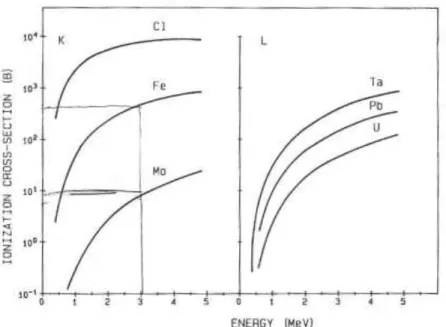
For the proton energy range of 1-4 MeV typically used in PIXE, the ionization (and x-ray production) cross sections increase with increasing proton energy and decrease with increasing atomic number of the target atom. This is illustred in Figure 11, which displays theoretical K and
L shell ionization cross sections21 for the selected target elements.
The very steep fall in the K ionization cross section with Z is particularly notable. Protons or other heavy charged particles that pass near atomic nuclei can be scattered elastically (i.e., without causing nuclear or atomic excitation). The cross section for the elastic scattering is strongly dependent on the scattering angle and on the atomic number (Z) of the scattering nuclide; it decreases with increasing angle and increases with increasing Z. The energy of the scattered particles also increases with Z. By measuring the elastically scattered particles, information can be
Figure 11 - K and L shell ionization cross sections in barns (1 barn=10-24 cm2)
as a function of proton energy and target atom. The values are theoretical ECPSSR predictions (Cohen and Harring, 1985)
38
obtained on the elemental composition of the sample and on the distribution of the elements within a certain depth.
In addition to elastic encounters between incident particles and target nuclei, various inelastic interactions or nuclear reactions are possible. These both include (𝑝, 𝛾)(𝑝, 𝑝′𝛾) and (𝑝, 𝛼𝛾) nuclear reactions when protons are used as incident particles. The cross sections for those reactions do not vary in a regular way with target nuclide and with incident particle energy. The cross sections generally increase with increasing incident particle energy but may exhibit intense resonance peaks at particular energies. In addition, because of the Coulomb barrier, the cross sections are smaller for the heavier target elements than for lighter ones.
By detecting the promptly emitted 𝛾-rays or charged particles for the above-mentioned nuclear reactions, an elemental analysis and the depth profiling of certain elements is possible. The analytical technique that employs these possibilities is referred to as nuclear reaction analysis.
2.1.6.1 Classical instrumentation
The ion beam used for the PIXE analysis is produced by a conventional accelerator. The energy range required, 1-4 MeV/u, means that relatively small (i.e. Accélérateur Grand Louvre d' Analyse Elémentaire called AGLAE - C2RMF in France allows to obtain several beam spot diameters from 20 µm to 500 µm, current intensities from 2 to 10 nA, with dimension of about 40 meters) accelerators are sufficient. Electrostatic accelerators are suitable sources33 and electrostatic accelerators are most commonly used for this purpose. They may be of a regular or modified Van de Graaff type, in which a high-voltage terminal is charged by a belt or a metallic chain. A second type of electrostatic accelerators uses a high-frequency voltage multiplication stage to charge a cavity, with the ions being accelerated in the electrostatic field created by the cavity.
Usually, in order to perform PIXE, a single-ended or tandem electrostatic machine of 2-3 MeV is chosen. Certain types of analysis, such as analyses by micro beam techniques, impose special requirements on ion source brightness, energy stability, beam emittance, and minimum scattering of the ion beam. Such requirements may play an important role in selecting an accelerator.
Commercially available vacuum chambers used as scattering chambers in nuclear physics experiments are generally not well suited for PIXE. Their shapes and dimensions hamper obtaining large solid angles for detection and introducing multi specimen holders and sample charges.