COMMUNAUTE FRANCAISE DE BELGIQUE ACADEMIE UNIVERSITAIRE WALLONIE-EUROPE
FACULTE UNIVERSITAIRE DES SCIENCES AGRONOMIQUES DE GEMBLOUX
Crucial role of reversible phosphorylation in the
mechanisms governing the biological functions
of class IIa Histone Deacetylases
Maud MARTIN
Dissertation originale présentée en vue de l'obtention du grade de docteur en sciences agronomiques et ingénierie biologique
Promoteurs: Dr. Franck Dequiedt Pr. Richard Kettmann
Martin Maud. (2009). Crucial role of reversible phosphorylation in the mechanisms governing the biological functions of class IIa Histone Deacetylases (thèse de doctorat). Gembloux Agricultural University, 178p., 2 tabl., 38 fig.
Summary:
Regulation of class IIa histone deacetylases (HDACs) phosphorylation is crucial because it provides the opportunity to control important developmental processes associated with these key enzymes. Indeed, the transcriptional repressor activity of class IIa HDAC is controlled via their phosphorylation-dependent nucleo-cytoplasmic shuttling. While a lot of efforts have been directed towards the identification of the inactivating kinases that phosphorylate class IIa HDACs, the identity of the antagonist phosphatase remained an open question. During this work, we found that protein phosphatase 2A (PP2A) is responsible for dephosphorylating the class IIa HDACs member HDAC7, thereby regulating its subcellular localization and repressor activity. In order to validate our model, functional consequences of these findings was illustrated during the two main biological processes involving HDAC7, i.e. T-cells apoptosis during negative selection and endothelial cells angiogenic activities during vascular network formation. Cellular PP2A represents a large population of trimeric holoenzymes containing a variable regulatory subunit, whose identity has a crucial role in determining the specificity of PP2A catalytic activity. In an effort to characterize the regulation of HDAC7 dephopshorylation, we identified the relevant PP2A holoenzyme regulating HDAC7 function during vasculogenesis and we found that, among diverse regulatory subunit isoforms, PP2A-Bα uniquely regulates endothelial cell angiogenic properties. PP2A-PP2A-Bα silencing using small interfering RNAs results in a significant inhibition of endothelial cell tube formation and migration. These results establish PP2A, and more precisely the Bα containg PP2A holoenzyme, as an essential element in the regulation of the class IIa HDACs HDAC7 and unravel a first developmental function for the PP2A regulatory subunit Bα in the genesis of blood vessels.
Martin Maud. (2009). Importance de la phosphorylation réversible dans les mécanismes contrôlant les foncions biologiques des Histone Désacétylases de class IIa (thèse de doctorat). Gembloux Faculté Universitaire des Sciences Agronomiques, 178p., 2 tabl., 38 fig.
Résumé:
La régulation de la phosphorylation des histone désacétylases (ou HDACs) de classe IIa est un processus crucial car il permet le contrôle des programmes développementaux majeurs associés à ces enzymes clé. En effet, l'activité répressive des HDACs de classe IIa est contrôlée via une translocation nucléo-cytoplasmique dépendant de leur phosphorylation. Tandis que beaucoup d'efforts ont été menés afin identifier les kinases phosphorylant les HDACs de classe IIa, la recherche de l'identité de la phosphatase antagoniste est restée marginale. Lors de ce travail, nous avons identifié la protéine phosphatase 2A (PP2A) en tant que phosphatase spécifique de HDAC7, un membre des HDACs de classe IIa. En déphosphorylant HDAC7, PP2A contrôle également sa localisation subcellulaire, ainsi que son activité répressive. Afin de valider notre modèle, les conséquences fonctionnelles de la régulation de HDAC7 par PP2A ont été illustrées dans les deux contextes biologiques majeurs impliquant HDAC7, c'est-à-dire le processus d'apoptose des thymocytes lors de la sélection négative et le processus de formation du réseau de vascularisation. La famille de phosphatases PP2A est constituée d'une vaste population d'enzymes hétérotrimériques, comprenant une sous-unité régulatrice variable dont l'identité détermine la spécificité de l'activité catalytique PP2A. Afin de mieux comprendre la régulation de la déphosphorylation de HDAC7, nous
avons identifié l'holoenzyme PP2A précise responsable de la régulation des fonctions de HDAC7 lors du processus de vasculogenèse. Nous avons en effet découvert que parmi de nombreuses isoformes de la sous-unité régulatrice de PP2A, la sous-unité Bα est la seule à contrôler les propriétés angiogéniques des cellules endothéliales. L'inhibition de PP2A Bα par utilisation d'ARN interférents mène à une diminution significative de la capacité des cellules endothéliales à former des vaisseaux et à migrer. Ces résultats établissent PP2A, et plus précisément l'holoenzyme PP2A contenant la sous-unité régulatrice Bα, en tant qu'éléments essentiels de la régulation de HDAC7 et associe, pour la première fois, un rôle développemental à la sous-unité Bα de PP2A durant la formation des vaisseaux sanguins.
Copyright. Aux termes de la loi belge du 30 juin 1994, sur le droit d'auteur et les droits voisins, seul l'auteur a le droit de reproduire partiellement ou complètement cet ouvrage de quelque façon et forme que ce soit ou d'en autoriser la reproduction partielle ou complète de quelque manière et sous quelque forme que ce soit. Toute photocopie ou reproduction sous
Aknowledgments - Remerciements
Almost four years of thesis led me to meet a lot of people. Through their help, motivation, experience or support, most of them have contributed, each in its manner, to the realization of this work. Let them find here all my gratitude.
Fistly, I would like to thank Pr André Théwis, Dr Jacqueline Destain, Dr Micheline Vandenbol and Dr Jean-Claude Twizere for their participation in this jury and especially, Pr Daniel Portetelle and Dr Veerle Janssens, for their critical review of this manuscript and their useful comments .
I would also express my gratitude to Dr Veerle Janssens and Pr Jozef Goris for their help in order to introducing us in the "PP2A world". In the same way, I would like to warmly thank Dr Michael Potente for our enriching collaboration and for teaching me the basis for the successful but delicate use of endothelial cells.
En revenant quelques années en arrière, je me souviens de mon arrivée à la faculté, au commencement de mes études. De nature assez ouverte, j'ignorais encore à quel domaine je me destinais. C'est en assistant au cours de biologie moléculaire, dispensé par le Pr Richard Kettmann, que cette curiosité naturelle a été guidée vers la recherche scientifique. Je tiens à remercier le Pr Richard Kettmann pour m'avoir ensuite accueillie au sein de son laboratoire et avoir accepté d'être co-promoteur de cette thèse. Je le remercie aussi très chaleureusment pour ses conseils éclairés et son éternelle bienveillance à mon égard.
Au commencement il y a donc une première accroche vers le métier de chercheur. Eh puis, il y a tout ce qui nous fait nous passionner et persévérer dans notre travail, malgré les vicissitudes de ce domaine…. Franck, ces quelques lignes auront beaucoup de mal à exprimer ma reconnaissance envers toi. Je te remercie très sincèrement pour le rôle primordial que tu as joué dans cette aventure. Si ces quelques années méritent bien le terme de "formation" doctorale, c'est en grande partie grâce à toi. Merci d'avoir accepté d'être mon promoteur, merci de m'avoir donné l'impulsion, la direction, l'envie et les moyens de mener à bien ce travail. Merci également d'avoir fait de ces années une expérience enrichissante, mais aussi un vrai plaisir!
Comment ne pas parler de l'équipe de biologie moléculaire et de l'ambiance de travail qui y règne? La vie d'un laboratoire implique que les personnes s'y succèdent à un rythme assez soutenu…mais cela ne veut pas dire pour autant qu'on oublie les rencontres que l'on y a faites. Ainsi, Emily, qui a accompagné et guidé mes premiers pas dans les manips, Marielle, dont les petites visites au labo sont toujours un plaisir, Caro, toujours présente pour partager les moments agréables comme les plus difficiles, et Véro, dont le passage fut bref mais suffisant pour nouer une agréable relation …les filles, merci pour tout, ce fut un plaisir de travailler, et papoter, avec vous!
La fin de cette thèse n'aurait pas été la même sans les joyeux drilles qui forment notre petite équipe actuelle. Pour cette convivialité à toute épreuve, je remercie donc Xavier, le nouvel expatrié, grâce à qui un grand moment de ski a été immortalisé, Pauline, dont la
présence féminine, dynamique et rafraichissante a beaucoup de valeur dans cette équipe d'homme, JC, qu'il est toujours agréable de croiser au labo à 23h lors de manips tardives, et enfin John pour son aide précieuse et notre agréable collaboration.
Je tiens également à associer à ces remerciements mes amis, dont certains ont déjà été cités précédemment, pour leur présence et leur soutien. Particulièrement, le groupe, élargi, des chimistes qui bougent encore et toujours, Annick, Ben, Nadège, Thomas, Ju et Valou, merci à vous pour tous ces moments et fous rires partagés.
Enfin, je tiens à remercier Didier, mon "coach personnel", pour sa présence et son inestimable appui, et mes parents pour leur soutien inébranlable au cours de toutes ces années. Je me souviens vous avoir entendu affirmer lors de ma remise de diplôme d'ingénieur que votre "boulot de parents" était maintenant terminé. Comme je le pressentais, vous m'avez prouvé à maintes reprises depuis lors que ce n'était pas tout à fait exact… et que je pouvais toujours compter sur votre aide et votre soutien en toutes circonstances!
Table of contents
Chapitre 1: Introduction ... 7
1.1. Class IIa Histone Deacetylases ... 9
I) Summary... 10
II) Introduction ... 10
III.a) Structure of class IIa HDACs: the C-terminal deacetylase domain ... 12
III.b) Structure of class IIa HDACs: the N-terminal adaptor domain ... 13
IV) Functions of class IIa HDACs ... 18
V) Regulation of class IIa HDACs... 23
VI) Biological functions of class IIa HDACs ... 31
VII) Therapeutic implications ... 41
VIII) Conclusions and perspective ... 43
1.2. Protein Phosphatase 2A... 44
I) Summary... 45
II) Introduction ... 45
III) Classification... 46
IV) Structure of PP2A ... 46
IV) Regulation... 50
V) Conclusions ... 56
Chapitre 2: Objectives ... 57
Chapitre 3: Results ... 60
3.1. Constitutive, selective and hierarchical phosphorylation of HDAC7 ... 61
I) Summary... 62
II) Introduction ... 62
III) Materials and Methods... 64
IV) Results... 69
V) Discussion ... 95
3.2. Regulation of HDAC7 by PP2A ... 100
I) Summary... 101
II) Introduction ... 101
III) Materials and methods ... 102
IV) Results... 105 V) Discussion ... 123 3.3. PP2A-Bα in angiogenesis... 125 I) Summary... 126 II) Introduction ... 126 III) Methods... 128 IV) Results... 131 V) Discussion ... 144
Chapitre 4: Conclusion and future prospects ... 148
Chapitre 5: Bibliography... 155
5.1. My bibliography... 156
I) Publications ... 157
II) Meeting abstracts... 157
Unravelling the mechanisms by which eukaryotic cells regulate gene expression is a fundamental issue in biology. Indeed, precise transcriptional regulation controls virtually all known biological processes governing cell growth, development, differentiation and survival. Some decade of stimulating studies has highlighted the importance of chromatin, the dynamic packaging structure of genomic DNA, in the events leading to either gene activation or gene silencing, or, in other words, leading to regulation of the "on-off" status of genes. The central structural elements of chromatin are specific proteins called histones. Significantly, their amino-terminal tail is subject to a diverse array of posttranslational modifications that largely impact on chromatin structure and accessibility, thereby playing an important regulatory role in the different cellular functions involving DNA, namely replication, transcription, recombination and repair. Among the various histone covalent modifications which combine together to constitute a "code" read by transcriptional regulators, lysine acetylation is certainly one of the most studied. This particular interest is mainly due to the correlation observed between the local acetylation status of chromatin in the vicinity of a promoter and its transcriptional activity: lysine hyperacetylation by histone acetyltransferases (or HATs) is favourable to transcription, whereas hypoacetylation by the opposing action of histone deacetylases (HDACs) is transcriptionaly unfavourable.
Hence, by leading to local closed conformation of chromatin, HDACs act as transcriptional repressors. Eighteen human HDACs have been identified to date. They are divided into different classes based on biochemical, structural and phyllogenetic criteria. The sub-class IIa HDACs which are HDAC4, 5, 7 and 9, was the focus of this work. This key family of transcriptional regulators share several similarities on sequence, domain organization and mechanism of regulation. In this introducing part, we first present a summary of our knowledge on class IIa HDACs, specifically on their inventive efficient signal-controlled regulation and on the functional relevance of these enzymes. Indeed, genetic inactivation studies in mice have established class IIa HDACs as key transcriptional
regulators of multiple crucial developmental and differentiation programs. .
1.1 Class IIa Histone Deacetylases: Regulating the
Regulators
I) Summary
In the last decade, the identification of enzymes that regulate acetylation of histones and non-histone proteins has revealed the key role of dynamic acetylation and deacetylation in various cellular processes. Mammalian histone deacetylases (HDACs), which catalyze the removal of acetyl-groups from lysine residues, are grouped into three classes, on the basis of similarity to yeast counterparts. An abundance of experimental evidence has established class IIa HDACs as crucial transcriptional regulators of various developmental and differentiation processes. In the past five years, a tremendous effort has been dedicated to characterizing the regulation of these enzymes. In this introduction, we summarize the latest discoveries in the field and discuss the molecular and structural determinants of class IIa HDACs regulation. Finally, we emphasize that comprehension of the mechanisms underlying class IIa HDAC functions is essential for potential therapeutic applications.
II) Introduction
Protein Nε-acetylation consists of the transfer of an acetyl moiety from an acetyl-coenzyme A (CoA) to the ε-amino group of a lysine residue. This modification was first identified on histones in the 1960s by Allfrey et al. (1964) who proposed that it could "influence RNA synthesis." Forty years later, it has become clear that lysine acetylation can target many cellular proteins in addition to histones. Similarly to phosphorylation, methylation or ubiquitination, lysine acetylation belongs to the panel of post-translational modifications used by the cell to alter the specific properties of a given protein (Kouzarides, 2000; Yang, 2005; Yang & Gregoire, 2005). Lysine acetylation is a dynamic, reversible and tightly regulated protein modification. In vivo, acetylation is controlled by the antagonistic activities of acetyltransferases and deacetylases. Because acetylation was originally characterized on histones, lysine acetyltransferases and deacetylases are known as histone acetyltranferases (HATs) and histone deacetylases (HDACs).
Eukaryotic HDACs are divided into two groups based on the specificities of their catalytic mechanism. Group I HDACs are zinc-dependent amidohydrolases. Eleven subtypes have been identified and named chronologically HDAC1 to HDAC11. While they share some degree of homology in their catalytic domain, group I HDACs are further subdivided into class I and class II enzymes on the basis of phylogenetic and structural criteria (Gregoretti et al., 2004). HDAC1, the founding member of class I, shows a high degree of sequence similarity with the yeast transcriptional regulator Rpd3 (Rundlett et al., 1996; Taunton et al., 1996). Together with HDAC1, four additional human Rpd3 orthologs, HDAC2, -3, -8 and -11 make up the mammalian class I HDAC family (Marks et al., 2003). Class II HDACs (HDAC4,-5,-6,-7,-9 and -10) are defined based on their sequence homology with Hda1in Saccharomyces cerevisiae (Fischer et al., 2002; Fischle et al., 2001; Fischle et al., 1999; Grozinger et al., 1999; Guardiola & Yao, 2002; Kao et al., 2002; Kao et al., 2000b; Miska et al., 1999; Petrie et al., 2003; Tong et al., 2002; Verdel & Khochbin, 1999; Wang et al., 1999a; Zhou et al., 2001). The Hda1-like sequences of class II HDACs correspond to their catalytic domain. These enzymes also contain additional domains that allow for further subdivision into class IIa (HDAC4, -5, -7 and -9) and class IIb (HDAC6 and -10). Recently, analysis of the silent information regulator (Sir2) protein in yeast and its homologues in higher eukaryotes (SirT1-7) has revealed a nicotinamide adenine dinucleotide NAD+-dependent HDAC activity. Because of the obvious differences in their catalytic mechanism and their unrelated sequences, SirT proteins constitute the second group of mammalian HDACs often referred as Class III enzymes (Haigis & Guarente, 2006).
This review is restricted to class IIa HDACs and will focus on the latest developments in the understanding of their regulation and biology. For more general information on HDACs, we respectfully refer the reader to previous excellent reviews from our colleagues (Bertos et al., 2001; Grozinger & Schreiber, 2002; de Ruijter et al., 2003; Legube & Trouche, 2003; Marks et al., 2003; Thiagalingam et al., 2003; Verdin et al., 2003; Yang & Seto, 2003; Blander & Guarente, 2004; Marmorstein, 2004; North & Verdin, 2004; Sengupta & Seto, 2004; Ekwall, 2005; Glozak et al., 2005; Nusinzon & Horvath, 2005; Yang & Gregoire, 2005).
III.a) Structure of class IIa HDACs: the C-terminal deacetylase domain
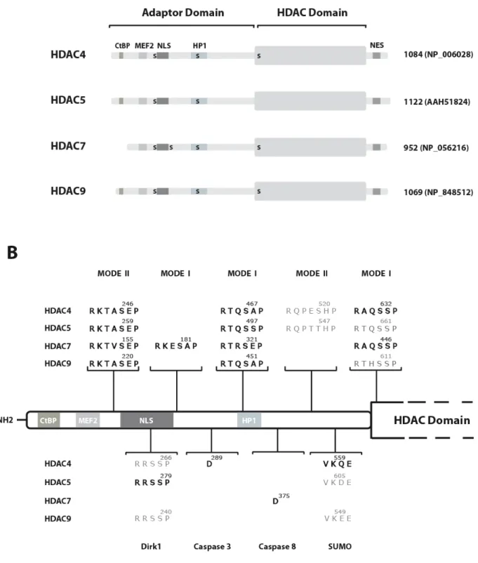
HDAC4, -5, -7 and -9 are defined by their particular bipartite structure (Figure 1A). The so-called HDAC domain is limited to a 400-450 amino acid region located at their C-terminus. This region, which shows the highest homology to yeast Hda1 (52, 53, 51 and 51% similarity for HDAC4, -5, -7 and -9 respectively) is highly conserved amongst class IIa members (around 80% sequence similarity). Structural analysis of FB188 HDAH, a bacterial HDAC-like protein with significant homology to the catalytic domain of class II HDACs has revealed that while the canonical fold of their catalytic pocket is very similar, there are several important differences between class I and class II HDACs that mainly concern the entrance region of the active site cavity and the outer charge transfer relay system (Finnin et al., 1999; Nielsen et al., 2005; Somoza et al., 2004; Vannini et al., 2004). This might explain why, in contrast to class I HDACs, researchers in the field have remained remarkably unsuccessful at obtaining enzymatically active class IIa HDACs in recombinant forms in vitro (Hassig et al., 1998; Hu et al., 2000). When successful, these attempts have mainly yielded protein preparations with low specific activity, especially when compared to the corresponding protein analyzed in vivo (Wang et al., 1999a). Indeed, when expressed as an isolated subdomain the C-terminal region of class IIa HDACs associates with full-HDAC activity in vivo (Fischle et al., 2001; Fischle et al., 2002; Wang et al., 1999a). Therefore, cumbersome transient transfection approaches followed by immunopurification have been required to measure significant deacetylase activity associated with class IIa HDACs (Verdin et al., 2004). This suggests that the enzymatic activity of class IIa members would rely on cellular co-factors or specific posttranslational modifications. Accordingly, there is evidence indicating that HDAC activity of class IIa members is dependent on an HDAC3/N-CoR/SMRT complex (Fischle et al., 2001; Fischle et al., 2002). Related to this, HDAC4 was shown to be sumoylated at lysine 559 (Kirsh et al., 2002). Because a K559R mutation abolished HDAC4-associated HDAC activity, the authors concluded that sumoylation was necessary for full enzymatic activity. However, binding of the K559R HDAC4 mutant to the N-CoR/SMRT/HDAC3 complex was not investigated in the study. Therefore, it cannot be formally excluded that the effect on the enzymatic activity was due to an overall alteration of the protein structure resulting in an inability to associate with the active HDAC3 complex. All these remaining questions illustrate the fact that further efforts should be dedicated to understanding how the activity of class IIa HDACs is controlled in vivo.
III.b) Structure of class IIa HDACs: the N-terminal adaptor domain
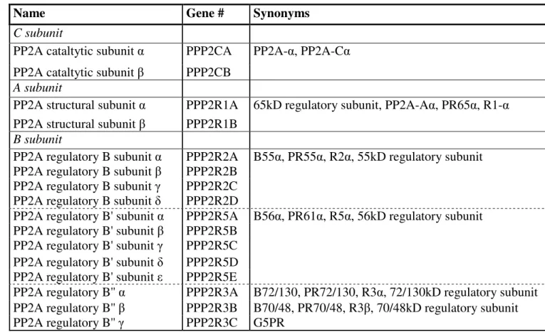
In addition to their Hda1-like domain, class IIa HDACs are comprised of a 450 to 600 amino acid extension at their N-terminus. Overall, the sequence homology between the members of the family in this region is much lower (30-45%) than that of the HDAC domain. Nevertheless, this region contains conserved amino acid motifs that are specialized for binding an array of proteins. Most of these are DNA-binding transcription factors, for which class IIa HDACs serve as transcriptional corepressors (Table 1). By far the best characterized of such interactions is the association with the MEF2 transcription factors which occurs via a 17 amino acid motif conserved in all class IIa HDACs (Dequiedt et al., 2003; Han et al., 2005; Kao et al., 2001; Lu et al., 2000b; Verdel & Khochbin, 1999; Wang et al., 1999a). Repression of MEF2-targeted promoters via recruitment of class IIa-associated HDAC activity has been extensively documented (Lemercier et al., 2000; Lu et al., 2000b; Miska et al., 1999; Wang et al., 1999a) and plays a key role in the biological functions of these enzymes (see below). Some interactions that take place in the N-terminal region of class IIa HDACs mediate the recruitment of their deacetylase activity into higher-order complexes, together with multiple other enzymes. Two examples of such interactions are interactions with COOH-terminal binding protein (CtBP) and Heterochromatin P1 (HP1). A 100 amino-acid motif has been implicated in the interaction of HDAC4 and HDAC5 with HP1α and associated histone methyl transferase (HMTase) SUV39H1 (Zhang et al., 2002b). The functional significance of the association between class IIa HDACs and HMTases remains unclear. However, considering that lysine deacetylation is a prerequisite to methylation, two attractive models emerge. First, this interaction would allow the recruitment of a deacetylase/methyltransferase complex that would be important for the establishment and maintenance of transcriptional silencing at promoters to which class IIa HDACs are recruited by DNA-binding transcription factors (Zhang et al., 2002b). Alternatively, when bound to methylated histones, the HP1/HMTase complex could propagate a repressive chromatin state through class IIa HDAC-mediated deacetylation and subsequent methylation of adjacent nucleosomal histone tails. On a similar vein, the N-terminus of HDAC4, -5 and -9 comprises an interaction motif for the transcriptional corepressor CtBP (Dressel et al., 2001; Zhang et al., 2001a). Since CtBP is part of a higher-order complex containing chromatin modifying enzymes such as class I HDACs and HMTs
(Shi et al., 2003), it is tempting to speculate that class IIa HDACs might use CtBP to recruit multiple enzymatic activities to their target promoters.
The adaptor domain of class IIa HDACs also contains motifs specifically involved in their regulation. Three clusters of arginine/lysine residues are found between aa 251-272 of HDAC4 and aa 264-285 of HDAC5. Mutational analyses confirmed the ability of these sequences to act as an autonomous nuclear import signal and enlightened their importance in the subcellular localization of HDAC4 and HDAC5 (McKinsey et al., 2000a; Wang & Yang, 2001). Despite a lack of experimental evidence, it is likely that this NLS also mediates the observed interaction between HDAC4 and importinα (Grozinger & Schreiber, 2000). These motifs are fairly well conserved in HDAC7 and HDAC9, althoug it has not been formally demonstrated, it is logical to assume that the corresponding regions constitute authentic nuclear import signals for these class IIa members.
A puzzling feature of the N-terminal adaptor domain of class IIa HDACs is its ability to inhibit transcription when tethered to a heterologous promoter, independently of the C-terminal catalytic domain (Sparrow et al., 1999; Wang et al., 1999a; Zhou et al., 2000a). This property has been assigned to a glutamine-rich domain (aa. 69-155 of HDAC4 that contains 26 glutamine residues) which organizes into a single α-helix and would be involved in homo- and/or hetero-oligomeric protein-protein interactions (Guo et al., 2007; Kirsh et al., 2002). Curiously, this region, which is conserved in HDAC4, -5 and -9 is not present in HDAC7. To date, very few experiments have been conducted to understand how the coiled-coil domain of HDAC4,-5 and -9 might repress transcription. Resolution of this question must await the identification of the cellular factors interacting specifically with this region.
The adapter domain of class IIa HDACs contains specific residues that are subjected to various post-translational modifications, such as proteolytic cleavage (Bakin & Jung, 2004; Liu et al., 2004; Paroni et al., 2004), ubiquitination (Li et al., 2004), sumoylation (Kirsh et al., 2002; Petrie et al., 2003) and most importantly phosphorylation. In response to various stimuli, a set of serine residues in the adapter domain of class IIa HDACs is phosphorylated, creating docking sites for the 14-3-3 proteins (Grozinger & Schreiber, 2000; Wang et al., 2000). Association with 14-3-3 induces nuclear export and cytoplasmic accumulation of class IIa HDACs with concomitant derepression of their target promoters (Grozinger & Schreiber, 2000; Kao et al., 2000b; Liu et al., 2005; Wang et al., 2000). This nuclear export mechanism allows for signal-dependent activation of class IIa HDAC target genes and has proven to be crucial for their regulation (see below). The 14-3-3 proteins are
a highly conserved family of proteins that have important roles in a wide range of cell signaling pathways (Dougherty & Morrison, 2004; Mackintosh, 2004). They bind to specific phosphoserine- or phosphothreonine-containing motifs within target proteins that generally match with either RxxpSxP (mode I) or RxxxpSxP (mode II) (Muslin et al., 1996). Their rigid structure and dimeric nature allow them to act as intra- and intermolecular adaptors and to alter specific properties of their target proteins (Yaffe, 2002). Mutational analyses identified two conserved 14-3-3 interacting motifs in HDAC5 and HDAC9 (centered on Ser259 and Ser497 in HDAC5 and Ser220 and Ser451 in HDAC9). Simultaneous mutations of these serine residues into alanines totally abolished interaction between HDAC5/9 and 14-3-3 (McKinsey et al., 2000b; Zhang et al., 2002a). Unexpectedly, the corresponding serine-to-alanine double mutants of HDAC4 and HDAC7 retained residual 14-3-3 binding (Wang et al., 2000; Kao et al., 2001; McKinsey et al., 2000). This observation led to the identification of Ser632 in HDAC4 and Ser449 in HDAC7 as additional 14-3-3 binding sites (Dequiedt et al., 2003; Grozinger & Schreiber, 2000; Kao et al., 2001; McKinsey et al., 2000a; Wang et al., 2000). Surprisingly, the corresponding motif is also conserved in HDAC5 and -9 (Figure 1B). In addition, we recently identified a cryptic 14-3-3 binding site (Ser181) in HDAC7 (Dequiedt et al., 2006; Dequiedt et al., 2005). This site was overlooked in previous mutational analyses because its phosphorylation seems dependent on prior phosphorylation of the most N-terminal serine residue, Ser155. Whereas Ser181 is uniquely present in HDAC7, sequence analyses disclosed other putative cryptic 14-3-3 motifs in the adaptor domain of HDAC4, -5 and -9 (Figure 1B). The functionality and possible biological relevance of these additional sites should be investigated comprehensively.
Figure 1: Domain organization of human class IIa HDACs. (A) Schematic representation of HDAC4, HDAC5, HDAC7 and HDAC9. For each protein, the longest isoform is shown, with the total number of residues and the corresponding database accession number. The CtBP, MEF2 and HP1-binding regions along with the NES and NLS are depicted as solid boxes.
14-3-3 binding sites are indicated with an S. NES, Nuclear export signal; NLS, nuclear localization signal. (B) Identification of post-translational modifications sites in the adaptor domain of class IIa HDACs. Sequence corresponding to the Nterminal regions of HDAC4, -5, -7 and -9 were examined for the presence of 14-3-3-binding motifs (mode I and mode II), Dirk1 phosphorylation target sequence, sumoylation consensus and caspase 3 and 8 cleavage sites. For each identified motif, the position of the critical residue is indicated. Motifs are written in bold when the corresponding post-translation modification was demonstrated experimentally.
Partner Function Reference(s)
MEF2 DNA anchoring transcriptional factor (Dressel et al., 2001; Kao et al., 2001; Lemercier et al., 2000; Lu et al., 2000a; Lu et al., 2000b; Miska et al., 1999; Sparrow et al., 1999; Verdel & Khochbin, 1999; Wang et al., 1999a) NF-AT3C DNA-anchoring transcriptional factor (Dai et al., 2005)
DnaJ (Mrj) bridging co-factor (Dai et al., 2005) Runx 2/3 DNA-anchoring transcriptional factor (Vega et al., 2004b)
GATA 1/2 DNA-anchoring transcriptional factor (Ozawa et al., 2001; Watamoto et al., 2003)
FOXP3 DNA-anchoring transcriptional factor (Li et al., 2007) Nkx2-5 DNA-anchoring transcriptional factor (Song et al., 2006) CAMTA2 bridging co-factor (Song et al., 2006) SRF DNA-anchoring transcriptional factor (Davis et al., 2003) Myocardin bridging co-factor (Xing et al., 2006a) NRSF DNA-anchoring transcriptional factor (Nakagawa et al., 2006) BCl6 DNA-anchoring transcriptional factor (Lemercier et al., 2002) BCor BCL6 co-repressor (Huynh et al., 2000) BCor-L1 BCoR homolog co-repressor (Pagan et al., 2007) PLZF DNA-anchoring transcriptional factor (Lemercier et al., 2002) TR2 nuclear hormone receptor (Franco et al., 2003) LXR nuclear hormone receptor (Ghisletti et al., 2007) NCoR transcriptional co-repressor of nuclear
hormone receptor
(Fischle et al., 2001; Fischle et al., 2002; Huang et al., 2000; Kao et al., 2000a)
SMRT transcriptional co-repressors of nuclear hormone receptor
(Fischle et al., 2001; Fischle et al., 2002; Huang et al., 2000; Kao et al., 2000a)
mSin3A transcriptional co-repressor adaptator (Kao et al., 2000a) RIP140 transcriptional co-repressors of nuclear
hormone receptor
(Castet et al., 2004) REA transcriptional co-repressors of nuclear
hormone receptor
(Kurtev et al., 2004) ARR19 transcriptional co-repressors of nuclear
hormone receptor
(Jeong et al., 2004) ET-A G protein-coupled endothelin receptor (Lee et al., 2001) ANCO1 ankyrin repeat-containing transcriptional
co-repressors of nuclear hormone receptor
(Zhang et al., 2004)
RFXANK ankyrin repeat-containing co-factor (McKinsey et al., 2006; Wang et al., 2005)
NF-Y DNA anchoring transcriptional factor (Basile et al., 2006; Imbriano et al., 2005)
TEL transcriptional co-repressor (Petrie et al., 2003) CtBP transcriptional co-repressor (Zhang et al., 2001a) HP1 adaptator protein associated with HMTase (Zhang et al., 2002b)
TIP60 histone acetyltransferase (Lee et al., 2001; Li et al., 2007; Xiao et al., 2003)
IκBα NF-κB inhibitor, transcriptional repressor (Aguilera et al., 2004) 53BP1 DNA damage response protein (Kao et al., 2003)
HIF1α transcription factor (Kato et al., 2004; Qian et al., 2006) ICP0 immediate-early protein of HSV-1 virus (Lomonte et al., 2004)
EBNA2/EBNALP Barr nuclear antigen and Epstein-Barr nuclear antigen leader protein, transcriptional co-activators
(Bryant & Farrell, 2002; Portal et al., 2006)
α-actinin 1/4 actin-binding protein (Chakraborty et al., 2006)
14-3-3 chaperone proteins (Grozinger & Schreiber, 2000; Kao et al., 2001; McKinsey et al., 2000a; Wang et al., 2000; Zhang et al., 2001b)
Importinα nuclear import receptor (Grozinger & Schreiber, 2000) Calmodulin calcium-binding protein (Berger et al., 2003; Youn et al.,
2000)
Ubc9 SUMO E2 conjugating enzyme (Zhao et al., 2005) CaMKI protein kinase (Backs et al., 2006) CaMKII protein kinase (Backs et al., 2006)
PKD1 protein kinase (Dequiedt et al., 2005; Parra et al., 2005; Vega et al., 2004a)
MARK2/3 protein kinase (Dequiedt et al., 2006) ERK1/2 protein kinase (Zhou et al., 2000b) PP1/MYPTI protein phosphatase (Parra et al., 2007) PP2A protein phosphatase (Martin et al., 2008)
Table 1. Interaction partners of class IIa HDACs
IV) Functions of class IIa HDACs
Co-repressors of transcription
Class IIa HDACs are thought to act as transcriptional corepressors by deacetylating nucleosomal histones. Since these enzymes do not bind directly to DNA, the current model posits that their deacetylase activity is recruited to specific promoters by sequence-specific DNA binding proteins. Interactions with distinct transcription factors would thus dictate targeting specificity of class IIa HDACs. The canonical example of this model is illustrated by the interaction between class IIa HDACs and MEF2 transcription factors.
Historically, MEF2 was the first cellular partner identified for class IIa HDACs (Lemercier et al., 2000; Lu et al., 2000b; Miska et al., 1999; Sparrow et al., 1999; Wang et al.,
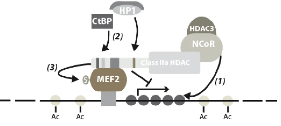
1999a). The MEF2 family consists of four MADS-box transcription factors (MEF2A, -B, -C and -D) involved in numerous signal-dependent pathways of cellular differentiation (Black & Olson, 1998). Members of the MEF2 family can act as repressors or activators of transcription depending on the mutually exclusive recruitment of specific chromatin modifying factors (McKinsey et al., 2001a; Youn et al., 2000). MEF2 associates with class IIa HDACs through a region located at the junction of the MADS/MEF2 domain which does not overlap with the DNA binding region of MEF2 (Lu et al., 2000b). This and other observations are compatible with a simple model in which, through interaction with the N-terminus of class IIa HDACs, DNA-bound MEF2 would recruits the HDAC activity associated with their C-termini to deacetylate local chromatin and repress transcription. This would establish class IIa HDACs as general transcriptional repressors of the multitude of promoters that are under the control of MEF2 transcription factors (McKinsey et al., 2002a). Accordingly, the ability of class IIa HDACs to act as potent inhibitors of MEF2-dependent transcription has been extensively documented (Dressel et al., 2001; Lemercier et al., 2000; Lu et al., 2000b; Miska et al., 1999; Sparrow et al., 1999; Wang et al., 1999a). Several experimental observations suggest that class IIa HDACs might control MEF2 transcriptional activity through additional mechanisms. MITR, a splice variant of HDAC9 that comprises only the N-terminal adaptor domain efficiently impairs MEF2-dependent transcription (Zhang et al., 2001b; Zhou et al., 2000a). Therefore, in addition to the HDAC activity associated with their C-terminus, class IIa HDACs might also repress MEF2 transcriptional activity through association of their N-terminal domain with co-repressors such as HP1 or CtBP (Zhang et al., 2002a; Zhang et al., 2001a; Zhang et al., 2001b). Recently, HDAC4 and -5 were shown to promote sumoylation of MEF2 which results in inhibition of its transcriptional activity (Gregoire et al., 2006; Gregoire & Yang, 2005; Zhao et al., 2005). As opposed to the original model, these results illustrate the multiple levels of regulation of MEF2 by class IIa HDACs and emphasize the need for further work to achieve comprehensive understanding of these mechanisms (Figure 2).
Figure 2: Cartoon depicting various mechanisms involved in the repression of MEF2-dependent transcription by class IIa HDACs. Via their N-terminal adaptor domain, class IIa HDACs interact with the MADS domain of DNA-bound MEF2. At MEF2-targeted promoters, class IIa HDACs establish a repressed chromatin state by recruiting the N-CoR/HDAC3 complex through their C-terminus (1) and co-repressors such as HP1 or CtBP via their N-terminus (2). In addition, class IIa HDACs may also inhibit MEF2 transcriptional activity by promoting its sumoylation (3).
Members of the NFAT family of transcription factors play prominent roles as regulators of calcium-inducible gene expression in diverse cell types (Hogan et al., 2003). A recent study indicated that class IIa HDACs interact with NF-AT3c to repress its transcriptional activity (Dai et al., 2005). In contrast to the interaction with MEF2, the recruitment of class IIa HDACs by NFAT is indirect and relies on a bridging co-factor, the chaperone mammalian relative of DnaJ (Mrj). However, the biological relevance underlying NFAT transcriptional repression through Mrj and class IIa HDACs remains obscure.
Members of the Runx family of transcription factors have important functions in several developmental programs (Otto et al., 2003). Runx1 is involved in hematopietic development and mice deficient in Runx2 display alterations in bone development (Komori et al., 1997; Otto et al., 1997). In the case of Runx3, recent studies have suggested pleitropic functions ranging from immune cell differentiation to tumour development (Puig-Kroger & Corbi, 2006). Several lines of evidence implicate class IIa HDACs as corepressors of Runx-mediated transcription. A recent study reported that HDAC4 associates with Runx2 via its N-terminal
adaptor domain and inhibits its ability to activate transcription (Vega et al., 2004b). Accordingly, reduced histone acetylation of a Runx2 target promoter was observed following overexpression of HDAC4. However, rather than the classical model in which Runx2 would tether HDAC4 to DNA to deacetylate nucleosomal histones, the authors of the study provided evidence that HDAC4 interferes with the ability of Runx2 to bind to its target promoter. In preosteoblasts, HDAC4 and 5 regulate Runx2 activity to control osteoblast differentiation. In this case, the repression of Runx2 activity may rely on the recruitment of class IIa HDACs to Runx2 target promoters through interaction with Smad3 (Kang et al., 2005). Finally, HDAC4 and -5 were reported to directly deacetylate Runx2 and Runx3 which would lead to their ubiquitin-mediated degradation and repression of their transactivation activity (Jeon et al., 2006; Jin et al., 2004). Interestingly, the model that emerges from these observations is dramatically different from what has been established for the regulation of MEF2. While in some instance, class IIa HDACs would act as corepressors of Runx-dependent transcription by promoting local deacetylation of histones, they may also inhibit Runx activity by hindering its DNA binding abilities and by promoting its deacetylation-mediated degradation. Deacetylation of Runx2 by HDAC4/5 reinforces the accumulating evidence suggesting that class IIa HDACs might also target non-histone proteins for deacetylation.
During the past few years, class IIa HDACs have been shown to repress an increasing number of transcriptional regulators through direct or indirect interaction. Interestingly, many of these are important regulators of key genetic programs during development, such as members of the Gata (Ozawa et al., 2001; Watamoto et al., 2003), Forkhead (Li et al., 2007), NK-2 homeodomain-containing (Song et al., 2006) and signal transducer and activator of transcription (STAT) (Xiao et al., 2003) families of transcription factors.
Unfortunately, for the majority, these interactions remain as yet mechanistically and functionally poorly characterized. Future studies will hopefully shed some light on these interesting issues.
Class IIa HDACs are also candidates for cooperation and regulation of the members of the nuclear hormone receptor family. As a general mechanism, the conserved zinc finger DNA-binding domain of nuclear receptors shows ability to interact with HDAC4 (Franco et al., 2003). At least in the case of TR2, this interaction is of functional consequence as the recruitment of HDAC4 leads to a reduction of acetylated histone associated with a TR2-related promoter. More recently, a study identified HDAC7 as a novel corepressor of the androgen receptor (AR) (Karvonen et al., 2006). Class IIa HDACs also interact with RIP140,
for the first two, and by the androgen receptor for the latter respectively (Castet et al., 2004; Jeong et al., 2004; Kurtev et al., 2004).
Interestingly, among the proteins interacting with class IIa HDACs, several contain ankyrin-repeat regions, indicating that similar structural determinants mediate association of class IIa HDACs with these transcription factors. Hence, class IIa HDACs interact with and repress the transcriptional activity of RFXANK, a positive regulator of MHC II genes expression (McKinsey et al., 2006; Wang et al., 2005), of ANCO 1, a nuclear receptor cofactor (Zhang et al., 2004), of B-CoR and B-CoR L1 (Huynh et al., 2000; Pagan et al., 2007), and CAMTA (Song et al., 2006). Class IIa HDACs also interact with ANKRA, a protein related to RFANX, but the functional implications of this interaction are unknown (McKinsey et al., 2006; Wang et al., 2005).
Atypical roles
As transcriptional coactivators of HIF-1α--Recent findings show that general HDAC inhibition may not only lead to gene activation but also to gene repression (Glaser et al., 2003; Nusinzon & Horvath, 2005). Supporting this in the case of class IIa HDACs, the C-terminus of HDAC4 and -7 but not HDAC5 have been shown to bind to hypoxia-inducible factor 1α (HIF-1α), a transcription factor that controls expression of genes responsive to low oxygen tension. Surprisingly, association with HDAC7 leads to increased HIF-1α transcriptional activity under hypoxic conditions (Kato et al., 2004). The exact mechanism underlying the transcriptional activation of HIF1-α by some members of class IIa HDACs remained unclear until recently, when it was reported that inhibition of HDAC4 by small interfering RNA had a positive effect on HIF-1α acetylation which correlated with reduced stability (Qian et al., 2006).
As SUMO E3 ligases--In addition to being sumoylated, HDAC4, and potentially other class IIa members can act as SUMO E3 ligases, a property dependent on their N-terminal domain (Gregoire et al., 2006; Gregoire & Yang, 2005; Zhao et al., 2005). Interestingly, MEF2 was identified as a relevant target for this novel class IIa-associated enzymatic activity. HDAC4 promoted sumoylation of MEF2 which inhibits its transcriptional activity. However, whether this effect is indirect or results from an intrinsic SUMO-ligase activity of HDAC4 is still matter of debate (Gregoire et al., 2006).
HDAC4 as a component of the DNA damage response pathway--Given the recent interaction unravelled between HDAC4 and the repair factor 53BP1, HDAC4 was though to be a
component of the DNA damage response (Kao et al., 2003). Indeed, in response to DNA damage, HDAC4 is recruited to the same dots, or repair foci, together with 53BP1. Moreover, silencing of HDAC4 abrogates DNA damage-induced G2 delay and increases radiosensitivity. Because drugs that inhibit transcription do not reverse these effects, the role of HDAC4 in DNA damage response is likely not restricted to transcriptional repression. Two recent studies shed new light on this mechanism by showing the DNA damage dependent-recruitment of HDAC4 by a repressor complex containing P53 and NF-Y, a complex that is involved in the repression of G2/M promoters (Basile et al., 2006; Imbriano et al., 2005). HDAC7 as a mitochondrial protein implicated in programmed cell death--Sequence analysis identified a mitochondrial targeting motif in the N-terminal domain of human HDAC7 (Bakin & Jung, 2004). This motif targets HDAC7 to the mitochondria where it is N-terminally processed and can be located in the mitochondrial inner membrane space. The fact that HDAC7, like other pro-apoptotic mitochondrial factors, relocalizes to the cytoplasm in response to apoptosis points to a possible role in the initiation of programmed cell death. However, no additional experimental data is currently available to strengthen the significance of mitochondrial HDAC7in the process of apoptosis.
V) Regulation of class IIa HDACs
Sub-cellular distribution
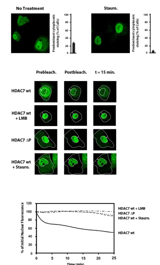
As expected from their ability to deacetylate histones, class IIa HDACs are found in the nucleus of most cell lines. However, depending on cell lines examined, a significant portion of the molecules can also be found in the cytoplasm, suggesting that these enzymes may shuttle between the nucleus and the cytoplasm (Fischle et al., 2001; Miska et al., 1999). Early confocal microscopy experiments with Leptomycin B and recently using the fluorescence loss in photobleaching (FLIP) technology confirmed that class IIa HDACs are subject to CRM1-dependent nuclear export (Dequiedt et al., 2006; Grozinger & Schreiber, 2000; Kao et al., 2001; Miska et al., 1999; Wang et al., 2000). Cytoplasmic accumulation of class IIa HDACs renders them unable to impact on transcription since it sequesters them away from their histone substrates and renders them enzymatically inactive as HDACs (Fischle et al., 2001; Fischle et al., 2002). The nucleo-cytoplasmic distribution of Class IIa HDACs is controlled by two separate domains: a nuclear localization signal (NLS) present in the N-terminal adaptor
(McKinsey et al., 2001b; Wang & Yang, 2001). This property of class IIa HDACs has attracted a lot of attention in the past few years and has emerged as a major mechanism in the regulation of these enzymes.
Re-localization of class IIa HDACs to more specific sub-cellular compartments or structures could also participate in the regulation of class IIa HDACs. It was proposed that mitochondrial targeting and processing of HDAC7 may act as an irreversible intracellular reservoir to sequester HDAC7, that would otherwise be available for nucleo-cytoplasmic trafficking (Bakin & Jung, 2004). Class IIa HDACs can localize into specific sub-nuclear structures and HDAC5 has been found to form dot-like nuclear structures, termed matrix associated deacetylase bodies (MAD bodies) (Downes et al., 2000; McKinsey et al., 2000b; Miska et al., 1999; Wang et al., 1999a). In response to DNA damage, HDAC4 is recruited to nuclear repair foci, together with the DNA damage response protein 53BP1 (Kao et al., 2003). Additionally, ICP0, a protein encoded by the HSV-1 virus, has been shown to co-localize with and to promote the nuclear redistribution, in ring-shaped structures, of class IIa HDACs (Lomonte et al., 2004).
Phosphorylation of 14-3-3 binding sites
Several lines of evidence suggest that phosphorylation of their 14-3-3 sites regulates subcellular localization of class IIa HDACs. Inhibition of class IIa HDAC phosphorylation, by protein kinase inhibitors or serine-to-alanine mutation of the 14-3-3 consensus sites, leads to their nuclear accumulation. In contrast, activation of phosphorylation by overexpression of protein kinases leads to cytoplasmic accumulation of class IIa HDACs.
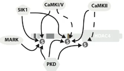
To date, four families of class IIa HDAC kinases have been identified (Figure 3). Historically, members of the Ca2+/calmodulin-dependent kinase family (CaMK), specifically CaMKI and IV, were the first kinases shown to promote nuclear export of class IIa HDACs. Subsequent studies have confirmed the role of these kinases in signal-dependent activation of class IIa HDAC target genes (Chawla et al., 2003; Davis et al., 2003; Kao et al., 2001; Linseman et al., 2003; McKinsey et al., 2000a; McKinsey et al., 2000b). Recently, CaMKII was shown to specifically phosphorylate HDAC4, promote its cytoplasmic accumulation and lead to hypertrophic growth in cardiomyocytes (Backs et al., 2006). In contrast, observations supporting a biologically relevant role for CaMKI/IV in class IIa HDAC phosphorylation remain equivocal and argue that CaMK might not be the sole kinases responsible for regulation of class IIa HDAC nuclear export (Chawla et al., 2003; Dequiedt et al., 2005; Vega
et al., 2004a; Zhang et al., 2002a). Recently, protein kinase D (PKD), a downstream effector in PKC signaling, was shown to directly phosphorylate class IIa HDACs on their 14-3-3 binding sites and induce their cytoplasmic accumulation. Experimental evidence supports a role for the PKD-class IIa HDAC axis in T-cell apoptosis (Dequiedt et al., 2005; Parra et al., 2005), cardiac hypertrophy (Vega et al., 2004a) and B-cell receptor signalling (Matthews et al., 2006). Whether PKD is also involved in other class IIa HDAC-regulated biological processes remains unknown. In addition to CaMK and PKD, two members of the microtubule affinity-regulating kinase (MARK)/Par-1 family, MARK2 and MARK3, were identified as potent class IIa HDAC kinases and were shown to induce their cytoplasmic accumulation (Chang et al., 2005; Dequiedt et al., 2006). Interestingly, CaMK, PKD and MARK fall within the CaMK super family and possess related catalytic domains (McKinsey, 2007). While constitutively active in most cell lines (Dequiedt et al., 2006), MARK, via its upstream kinase LKB1, is also considered a stress-responsive kinase. Indeed, the LKB1/MARK pathway is activated by high osmolarity (Wang et al., 2007) or focal cerebral ischemia (Schneider et al., 2004). The identification of MARK as a class IIa HDAC kinase thus suggests that class IIa HDACs may be implicated in these stress signalling processes. The recent identification of AMPK/SNF1-like kinases as a new family of class IIa HDAC kinases further supports this idea. In Caenorhabditis elegans, KIN-29 was shown to phosphorylate the class IIa HDAC, HDA-4 in vitro and antagonize its repression of chemoreceptor gene expression in sensory neurons (van der Linden et al., 2007). In addition, salt-inducible kinase (SIK1), a mammalian KIN-29 homolog, phosphorylates two 14-3-3 binding sites of HDAC5, thereby alleviating its repression of MEF2-targeted genes in skeletal muscle (Berdeaux et al., 2007).
The existence of multiple families of kinases targeting class IIa HDACs, each with multiple isoforms, reflects the numerous biological roles of these transcriptional repressors. This diversity allows class IIa HDACs to respond to different signaling pathways, with each signaling pathway providing a functional relevance for a given kinase. Some kinases have been shown to act preferentially on specific members of the class IIa family, which emphasizes the importance that distinct class IIa HDACs may have in specific genetic program (Backs et al., 2006; Chawla et al., 2003; Liu et al., 2005). CaMKII, for example, is able to drive HDAC4, but not HDAC5, out of the nucleus, probably because HDAC4 possesses a non-conserved CaMKII binding site (Backs et al., 2006).
Some class IIa HDAC kinases may display preferences toward specific 14-3-3 sites. In vitro, PKD is much less effective at phosphorylating Ser259 than Ser498 of HDAC5 (Huynh &
but not other 14-3-3 sites of HDAC7 (Dequiedt et al., 2006). CaMKI and -II target different sites in HDAC4 (Backs et al., 2006). These differences suggest that phosphorylation of each 14-3-3 site may have a specific impact on the regulation of class IIa HDACs and that multisite phosphorylation by distinct protein kinases may constitute a tightly regulated mechanism to induce expression of specific target genes in response to specific signals (Figure 3).
Figure 3: The 14-3-3 binding sites of Class IIa HDACs are phosphorylated by members of multiple families of protein kinases. Phosphorylation of HDAC4 is illustrated as an example as it is the only class IIa member phosphorylated by CaMKII. CaMKI and CaMKIV show a moderate preference for the most N-terminal site, whereas CaMKII exhibits some specificity for the internal site. PKD can phosphorylate indiscriminately the three 14-3-3 binding sites. In contrast, MARK selectively targets the most upstream serine residue. The two most N-terminal sites are optimal recognition motifs for SIK1 and are efficiently phosphorylated by this kinase.
Dephosphorylation at the 14-3-3 binding sites
It is logical to envision the phosphorylation-dependent regulation of class IIa HDACs as a reversible mechanism. In this context, relocalization of class IIa HDACs to the nucleus and reinstatement of their transcriptional inhibition would be expected to occur through dephosphorylation by a cellular phosphatase (Figure 4). Supporting this idea, Calyculin A, an inhibitor of protein phosphatases PP1 and PP2A, has been shown to promote cytoplasmic
retention of HDAC4 and to reduce its interaction with importin α (Grozinger & Schreiber, 2000).
Until recently, the identity of such an activating class IIa HDAC phosphatase was still an open question. A very recent study showed that, few hours after its signal-dependent nuclear export, HDAC7 becomes dephosphorylated and re-enters the nucleus. Myosin phosphatase, containing PP1β and myosin phosphatase targeting subunit 1 (MYPT1), was identified as an HDAC7-associated complex. This complex seems able to dephosphorylate the 14-3-3 binding sites of HDAC7 and promote its nuclear localization after signal-dependent export (Parra et al., 2007). In addition, results from our laboratory reveal that another cellular phosphatase, PP2A, stably associates with the N-terminal domain of class IIa HDACs. Our observations demonstrate that PP2A constitutively dephosphorylates class IIa HDACs in vivo and regulates their subcellular localization and repressor activity, thereby controlling their biological functions (Martin et al., 2008). The identification of multiple class IIa HDAC phosphatases may signify that class IIa HDACs dephosphorylation is regulated by different signaling pathways and would allow for flexible control of class IIa HDAC functions. Sequential or coordinated actions of the various protein kinases and phosphatases on the N-terminus of class IIa HDACs would constitute a tightly regulated mechanism to rapidly, appropriately and reversibly induce expression of specific target genes in response to specific signals.
Association with 14-3-3 and other cellular proteins
Association with 14-3-3 proteins soon appeared as a critical factor in the regulation of class IIa HDAC subcellular localization (Grozinger & Schreiber, 2000; Kao et al., 2001; McKinsey et al., 2000b; Wang et al., 2000). As observed for most 14-3-3 binding partners (Muslin et al., 1996), this association is dependent on the phosphorylation of interacting motifs within class IIa HDACs. 14-3-3 proteins are thought to regulate the subcellular localization of class IIa HDACs by modulating the function of their NES and NLS. First, binding of a 14-3-3 dimer would mask the N-terminal NLS and prevent class IIa HDAC recognition by importin α (Grozinger & Schreiber, 2000). Alternatively, binding of 14-3-3 proteins could unmask the C-terminal NES of class IIa HDACs or provide a NES in trans, facilitating nuclear export (McKinsey et al., 2000b). It is thus well-established that signal-dependent phosphorylation of the conserved serine residues and subsequent 14-3-3 binding are critical in the control of the
precise sequence of events involved in this mechanism. Whether binding of 14-3-3 precedes and/or promotes nuclear export or whether it takes place in the cytoplasm and prevents nuclear import of class IIa HDACs remains unclear. Clues about this issue came from experiments using HDAC5 constructs harboring inactivated NES. These mutants are unable to exit the nucleus despite intact phosphorylation 14-3-3 binding sites. Importantly, these mutants cannot efficiently inhibit muscle differentiation, indicating that release from their targeting DNA-binding factor is the crucial step in the relief of their transcriptional repression (Lu et al., 2000a; McKinsey et al., 2000a). In this context, 14-3-3-dependent nuclear export of class IIa HDACs may serve as a supporting mechanism to ensure maximal activation of their target genes (McKinsey et al., 2000a). Interestingly, the fact that 14-3-3 proteins are also regulated by phosphorylation may provide an additional level of regulation of class IIa HDAC nucleo-cytoplasmic shuttling (Ellis et al., 2003; Fu et al., 2000; Shen et al., 2003; Tzivion & Avruch, 2002).
In addition to 14-3-3 proteins, the subcellular localization and repressor activity of class IIa HDACs are controlled by additional cellular factors. The nuclear export of class IIa HDACs is inhibited by Leptomycin B, which suggests that the export receptor CRM1 may interact with the leucine-rich nuclear-export signal (NES) found in class IIa HDACs (Harrison et al., 2004; Wang & Yang, 2001). Whereas no CRM1-class IIa HDACs association has ever been formally documented, overexpression of CRM1 promotes cytoplasmic accumulation of HDAC7 (Gao et al., 2006).
Overexpression of MEF2 was reported to promote nuclear localization of class IIa HDACs. While direct interaction between both proteins was required for nuclear targeting of class IIa HDACs, no experiment was conducted to explain the underlying mechanism (Borghi et al., 2001; Chan et al., 2003; Miska et al., 1999; Wang & Yang, 2001).
Signaling
Multiple signaling pathways impact on class IIa HDAC localization, the effects of which can most of the time, be attributed to the activation of one or more class IIa HDACs protein kinases. In agreement with this, class IIa HDAC mutants in their phosphorylatable serine residues remain nuclear and are thus effective blockers of related developmental programs.
Agonists that stimulate PKD, through PKC-dependent or –independent mechanisms, induce class IIa HDAC phosphorylation and nuclear export. This is the case for the PKC activator PMA, for the B- and T-cell receptors and for several other cell surface receptors,
such as the serotonin receptor and the G protein coupled receptors (GPCR) ETR, α1-AR and
PGF2α. Signaling via Rho-GTPases, mediators of GPCR signaling, and via phospholipase C, a mediator that lies downstream of α1-AR, also activates PKD and triggers
phosphorylation-dependent nuclear export of HDAC5 (Chang et al., 2005; Dequiedt et al., 2005; Harrison et al., 2006). Some of these cell surface receptors also activate calcium fluxes and thus part of their effects could be mediated through activation of CaMKs (Backs et al., 2006). It has been shown that cardiac hypertrophic stimuli, such as pressure overload or expression of activated calcineurin, activate a cardiac kinase(s) that phosphorylates the regulatory serine residues within HDAC5 and HDAC9. However, the identity of this kinase remains unknown (Chang et al., 2004; Zhang et al., 2002a).
Neuronal receptors involved in synaptic activity also regulate class IIa HDACs localization in a way that depends on the phosphorylatable serine residues. Whereas neuronal activity-dependent nucleocytoplasmic shuttling of HDAC4 and HDAC5 was induced through both L-type calcium channels and synaptic NMDA receptors, differences in the activation thresholds for HDAC4 and HDAC5 nuclear export were observed (Chawla et al., 2003). Another neuronal signaling pathway converges on class IIa HDACs. In cultured cerebellar granules, HDAC4 accumulates in the nucleus in response to death stimuli. Interestingly, this translocation can be prevented by a survival factor, the neurotrophin BDNF. Treatment with a CaMK inhibitor promoted HDAC5 nuclear translocation, even with BDNF treatment, suggesting that CaMK may be a downstream effector of BDNF in this signaling pathway. Accordingly, the BDNF receptor can activate phospholipase C, which can then produce a rise in intracellular calcium and activate CaMK (Bolger & Yao, 2005). Of note, phospholipase C can also contribute to activate PKD. HDAC5 was reported to undergo similar nuclear translocation in response to low potassium, or to CaMK inhibition (Linseman et al., 2003).
Besides the above examples, very few signals have been reported to induce nuclear import of class IIa HDACs. Heat shock stimulation induced HDAC4 nuclear translocation (Dai et al., 2005), hypoxic conditions, in the presence of HIF1-α, drive HDAC7 to the nucleus (Kato et al., 2004) and hormone occupancy of the androgen receptor induces nuclear transfer of HDAC7 and -4 (Halkidou et al., 2004; Karvonen et al., 2006). The mechanism by which these signals lead to class IIa HDACs nuclear accumulation is not known and undoubtedly deserves further investigation.
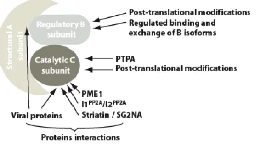
Figure 4: Model of class IIa HDAC regulation. The transcriptional repressor activity of class IIa HDACs is controlled through phosphorylation-dependent nucleo-cytoplasmic relocalization. In response to extra- or intra-cellular signals, PKD, CaMK, SIK1 or MARK phosphorylate specific regulatory serine residues in the adaptor domain of class IIa HDACs. Phosphorylation of class IIa HDACs is associated with displacement from their target promoters, association with 14-3-3 proteins and nuclear export through a CRM1-dependent pathway. This depletes the nuclear compartment of class IIa HDACs and allows for expression of a specific set of target genes. Import of class IIa HDACs to the nucleus and reinstatement of their transcriptional inhibition is achieved through dissociation from 14-3-3 proteins (by a still unknown mechanism) and dephosphorylation by cellular phosphatase such as PP2A or Myosin phosphatase. In the cytoplasm, dephosphorylated class IIa HDACs may be more sensitive to Ubiquitin-mediated proteolytic degradation.
Other posttranslational modifications
In addition to their 14-3-3 binding sites, other class IIa HDAC residues are subject to phosphorylation. Dyrk1B phosphorylates HDAC5 at a serine residue shared by HDAC4 and HDAC9, but not by HDAC7. Phosphorylation by Dyrk1 reduces their nuclear accumulation and leads to MEF2 activation (Deng et al., 2005). This residue does not correspond to a
14-3-3 binding site and its mutation does not prevent CaMK-induced nuclear export (McKinsey et al., 2000a), but it lies within the nuclear localization region of class IIa HDACs. In contrast, oncogenic Ras was found to promote the nuclear localization of HDAC4 by stimulating its phosphorylation by extracellular signal-regulated kinase-1 and -2 (ERK1/-2) (Zhou et al., 2000b). However, no experiment demonstrated direct phosphorylation of HDAC4 by ERK1/2 nor identified the target residue(s).
HDAC4 and -9 are subject to sumoylation, at a lysine residue also conserved in HDAC5 (Kirsh et al., 2002; Petrie et al., 2003; Tatham et al., 2001). SUMO is believed to alter the interaction properties of its target, often affecting its localization within the cell (Seeler & Dejean, 2001). The functional relevance of class IIa HDAC sumoylation is still unclear, but sumoylation abolished HDAC4-associated HDAC activity (Kirsh et al., 2002). Interestingly, indirect evidence suggests that class IIa HDAC sumoylation might be coordinated with phosphorylation. Indeed, by a still-unknown mechanism, two proteins involved in the process of sumoylation, the SENP “SUMO remover” and PIAS, an E3 ligase, promote phosphorylation of 14-3-3 binding sites of HDAC5 (Chang et al., 2005). In addition, HDAC4 and HDAC7 are cleaved by caspases. In both cases, cleavage separates the N-terminal NLS from the C-terminal NES. Whereas the caspase-generated N-terminal fragment of HDAC4 retains repressor ability (Liu et al., 2004; Paroni et al., 2004), cleavage of HDAC7 abolishes its transcription repressor activity (F. Scott and F. Dequiedt, unpublished observations).
VI) Biological functions of class IIa HDACs
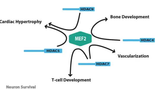
The existence of multiple regulatory pathways converging on class IIa HDACs emphasizes the importance of these enzymes in various biological processes. In the past 5 years, genetic inactivation studies in mice conducted by the Olson laboratory have been instrumental in elucidating the biological functions of these enzymes. These studies have revealed the key role of class IIa members in several important developmental and differentiation processes. Surprisingly, despite the large number of transcriptional regulators targeted by class IIa HDACs, most of their functions seem to involve the transcriptional repression of MEF2 transcription factors. In the following sections, we will review the latest insights into the biological functions of these fascinating enzymes.
Chondrocytes hypertrophy
HDAC4-null mice die during the perinatal period due to severe growth retardations and numerous skeletal abnormalities that result from excessive hypertrophic chondrocyte differentiation and inadequate endochondral ossification (Vega et al., 2004b). This remarkable phenotype was originally attributed to the ability of HDAC4 to repress Runt-related transcription factor-2 (Runx2), a well-recognized positive regulator of chondrocyte hypertrophy (Komori et al., 1997; Otto et al., 1997). Indeed, HDAC4 physically associates with Runx2 and inhibits its transcriptional activities. In addition, the phenotypic abnormalities of HDAC4 KO mice are strikingly reminiscent of those observed in mice with ectopic expression of Runx2 in prehypertrophic chondrocytes (Takeda et al., 2001; Ueta et al., 2001). The precise mechanism by which HDAC4 inhibits Runx2-mediated transcription remains obscure. Original observations suggested that repression of Runx2 by HDAC4 might occur independently of its HDAC catalytic activity. Instead, HDAC4 was thought to impede on Runx2 DNA binding by direct association with its Runt domain (Vega et al., 2004b). However this model failed to explain why HDAC4 mutants consisting of the C-terminal catalytic domain of HDAC4, and thus lacking the Runx2 binding region, retain significant repressive activity. The recent observation that HDAC4 and HDAC5 deacetylate Runx2 and Runx3 provides an explanation for these apparent discrepancies (Jeon et al., 2006; Jin et al., 2004). In addition to preventing Runx2 from binding to DNA, HDAC4 may deacetylate Runx2 and promote its ubiquitin-mediated degradation. This double-targeting of Runx2 by HDAC4 would thus solely explain its key role as a negative regulator of chondrocyte hypertrophy.
This apparently satisfying model was recently challenged by a study published by the same authors and which unravelled the unexpected role of MEF2C in chondrocyte hypertrophy (Arnold et al., 2007). This study reports that homo- or even heterozygous mutation of Mef2c is associated with severe skeleton defects resulting from reduced chondrocyte hypertrophy and ossification of endochondral bones. Given the preponderance of the class IIa HDACs-MEF2 axis in several developmental programs (figure 5, see below), this unexpected finding raises the possibility that bone defects associated with deletion of Hdac4 could, at least partly, result from hyperactivation of MEF2C. Supporting this hypothesis, a genetic antagonism exists between HDAC4 and MEF2: the excessive endochondral ossification observed in HDAC4 null mice is partially abolished by deletion of one Mef2c allele. Conversely, inactivation of Hdac4 in the presence of a heterozygous Mef2c allele