Successful controlled limb reperfusion after severe prolonged ischemia
J. O. Defraigne, J. Pincemail, C. Laroche, F. Blaffart, and R. Limet
From the Department of Cardiovascular Surgery, University Hospital of Liège, Domaine Universitaire du Sart-Tilman, 4000 Liège, Belgium.
Controlled limb reperfusion was performed in two patients who were admitted for lower limb-threatening ischemia as a result of embolism. After embolectomy, the inflow blood was drained with a cannula and mixed with a crystalloid solution to obtain an hyperosmolar, hypocalcemic, alkalotic, and substrate-enriched (aspartate, glutamate) reperfusate. This reperfusate was reinjected with a roller pump for 30 minutes through two cannulas inserted in the profunda and superficial femoral arteries. Temperature, intraarterial pressures, and flow were closely controlled. No complications occurred, and complete recovery of motor and sensory functions were observed, with restoration of pedal pulses.
Revascularization of a limb after a severe and prolonged period of ischemia may be associated with several local and systemic complications ("revascularization syndrome") that lead to high rates of mortality and amputation.1-3 Current therapies are generally directed against complications after they occur, once revascularization is completed. Nevertheless, a substantial percentage of the injury is generated on reperfusion, and some
experimental and clinical data suggest that careful control of both the composition and the physical conditions of the initial reperfusion may prevent the development of this injury.4-5
Here we report our initial clinical experience with controlled limb reperfusion in two patients with severe limb-threatening ischemia.
CASE REPORTS
Case 1. A 75-year-old woman was admitted for acute bilateral lower limb ischemia (pallor, cyanosis, profound sensory loss, and complete paralysis). Symptoms were present since 4 hours before admission. Eighteen months earlier the patient underwent an aortobifemoral bypass procedure with terminoterminal proximal anastomosis. Two months before, the patient was asymptomatic with bilaterally palpated pedal pulses. On examination, no arterial pulses were palpated bilaterally and no distal venous and arterial Doppler signals were audible.
Considering the bilaterality of the ischemia and the absence of claudication before die present episode, massive embolism was considered. No arteriogram was obtained, and the patient was immediately transferred into the operating room.
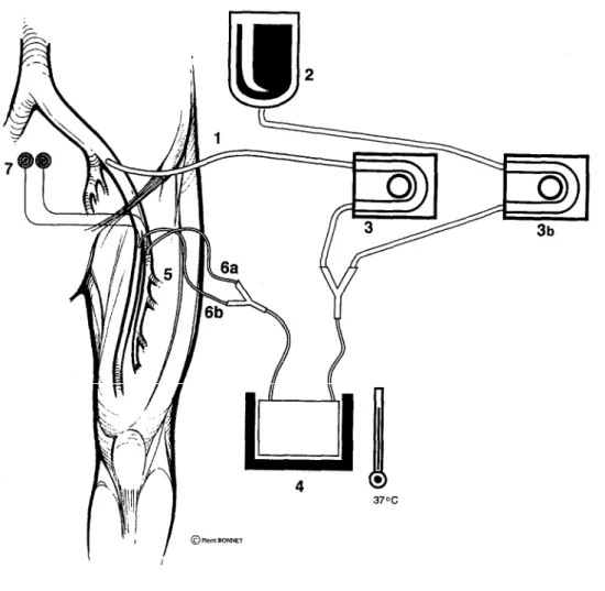
After induction of general anesthesia, heart rate, electrocardiogram, central venous pressure (CVP), blood pressure, urine output, and rectal temperature were monitored continuously. The common, superficial, and profunda femoral arteries and the graft limbs were exposed bilaterally via groin incisions. The right and left limbs were treated sequentially. After administration of heparin (3 mg/kg), a 3 cm longitudinal arteriotomy was performed on the graft limbs and prolonged on the origin of the superficial femoral arteries. Proximal and distal thrombectomies were performed with a Fogarty catheter. A wire-enforced 22F cannula (inflow cannula, Jostra Medizintechnick, Hechinger, Germany) was introduced into the graft limb from the arteriotomy to aspirate oxygenated blood and was connected to the inflow line of the reperfusion set (HP Medica, Augsburg, Germany; Fig. 1). The inflow line was placed in the head of a roller pump. A second line was connected to a crystalloid crystalloid solution (Dr. Franz Köhler Chemie, Asbach-Hahnlein, Germany) and placed in the head of a second roller pump. The crystalloid solution contained citric acid (0.1230 g/L), sodium citrate (2.4860 g/L), NaH2PO4 (0.0935 g/L), NaCl (1.2150 g/L), Trometanol (4.9080 g/L), glucose (7.8250 g/L), sodium glutamate (4.8090 g/L), and sodium aspartate (4.3990 g/L). The two outflow lines coming from the pumps were connected with an Y piece to form the delivery line of the controlled reperfusate, which was passed through a heat exchanger (Fig. 1). Two reperfusion cannulas (9F catheter with self-inflating balloon and additional pressure monitoring line [Research Medical, Midvale, Utah]) were connected at the end of the delivery line. After deairing, these cannulas were introduced into the superficial and profunda femoral arteries. Thereafter, controlled limb reperfusion was started and continued for 30 minutes in each limb, treated successively. The flow rates of both roller pumps were
adjusted to mix the crystalloid solution and the oxygenated blood (ratio 1:6). Samples were taken from the arterial blood (inflow line) and from the controlled reperfusate (delivery line) and were analyzed at sequential time intervals (Table I). Compared with the arterial blood, the controlled reperfusate was hypocalcemic, alkalotic, and hyperglycemic. At the end of the reperfusion, the arteriotomies were closed with a Dacron patch and the limb reperfused with normal blood.
Fig. 1. Controlled limb reperfusion. 1, Inflow cannula; 2, asanguineous crystalloid solution; 3 and 3b, roller
pumps; 4, delivery line of controlled reperfusate; 5, heat exchanger; 6a and 6b, outflow cannulas inserted into profunda and superficial femoris arteries; 7, pressure monitor.
The rectal temperature dropped from 35.7° C to 34° C during the reperfusion procedure. The intraarterial pressures in the profunda and superficial femoral arteries were maintained between 45 and 70 mm Hg. As the flow reached 120 ml/min for the left limb and 230 ml/min for the right limb, the patient received 1750 ml of crystalloid solution. Systemic arterial pressure remained stable, but an increase of die CVP to 15 mm Hg required administration of furosemide (Lasix, Hoechst, Germany, two intravenous boluses of 20 mg and 10 mg), with a resultant decrease of the CVP. During the controlled reperfusion, total urine output reached 500 ml. Hyperglycemia required administration of two 10 mg intravenous boluses of insulin.
After partial neutralization of heparin with protamine, bilateral fasciotomies of the legs were performed 1 hour after revascularization. At this moment, the muscles were not edematous and contracted under mechanical stimulation. After surgery, complete recovery of sensory and motor functions were observed in both legs and pedal pulses were palpated bilaterally. Only a small nonsignificant increase in the limb perimeters was observed. The maximal serum peak of creatinine kinase reached 7169 U/L (normal range, 0 to 100 U/L), and the increase of die plasma myoglobin level was moderate (396 µg/L; normal range, 5 to 90 ugr/L). The renal function remained stable, with a maximum plasma creatinine level to 1.1 mg/dl (normal range, 0.6 to 1.1 mg/dl) that was
not significantly different from the preoperative value (0.9 mg/dl). The patient was treated with low molecular weight heparin and thereafter with warfarin derivatives and was discharged on the tenth postoperative day. Case 2. A 81-year-old woman was admitted for severe ischemia of the right lower limb (pallor, cyanosis, complete anesthesia, and severe motor function impairment) that began suddenly 8 hours before admission. Past medical history included chronic atrial fibrillation. No femoral and distal pulses were palpated, and no distal Doppler signals were audible. An arteriogram showed occlusion of the external iliac artery with
revascularization of distal branches of the profunda femoral artery. Considering atrial fibrillation and sudden onset, the diagnosis of embolism was retained.
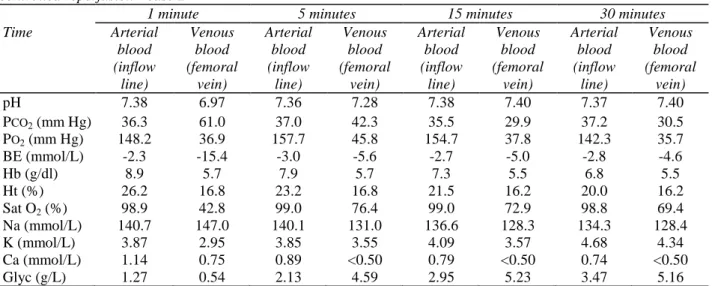
General anesthesia and monitoring of the vital signs were performed as in Case 1. After exposure of the femoral vessels and administration of heparin (3 mg/kg), a longitudinal arteriotomy was performed on the common femoral artery. Embolectomy was achieved with a Fogarty catheter. Thereafter, controlled limb reperfusion was performed according to the procedure described for Case 1, with the inflow cannula passed into the external iliac artery from the arteriotomy. After 30 minutes the controlled limb reperfusion was stopped, the arteriotomy was closed with a Dacron patch, and the limb was reperfused with normal blood. Samples were taken from the arterial blood (inflow line) and from the venous effluent (femoral vein) and were analyzed at sequential time intervals (Table II). No hyperkaliemia was observed in the venous blood, and oxygen uptake occurred in the limb as shown by the arteriovenous difference in PO2. During the controlled reperfusion, the rectal temperature dropped from 36.5° C to 35° C. The intraarterial pressures in the profunda and superficial femoral arteries were maintained between 45 mm Hg and 70 mm Hg by adjusting the flow rate, which varied from 180 to 250 ml/min, approximately 1500 ml of the crystalloid solution being perfused. An increase of the CVP to 22 mm Hg required administration of 20 mg of furosemide and withdrawal of 300 ml of blood from the inflow line. The CVP decreased progressively, and the total urine output reached 700 ml during the procedure. To manage
hyperglycemia, two boluses of 10 mg of insulin were administered intravenously. After partial neutralization of heparin with protamine, fasciotomies were performed one half hour after revascularization. At this moment, no marked muscle edema was observed. After surgery, complete functional recovery was observed and pedal pulses were palpated bilaterally. The measurements of the limb perimeters did not show limb edema. The maximal serum peak of creatinine kinase reached 2000 U/L (normal range, 0 to 100 U/L) and the increase of the plasma myoglobin level was moderate (284 µg/L; normal range, 5 to 90 ugr/L). The plasma creatinine level (1.5 mg/dl; normal range, 0.6 to 1.1 mg/dl) was transiently increased after 24 hours compared with the preoperative value (0.9 mg/dl) and returned to normal value within 48 hours, with a diuresis of 2500 ml on the first postoperative day. The patient, who was discharged on the tenth postoperative day, was treated with low molecular weight heparin because of a fear of hemorrhage with warfarin derivatives in an old patient.
Table I. Blood measurements in arterial blood and in controlled reperfusate after 15 and 30 minutes of
controlled reperfusion of right limb—case 1 Arterial blood before controlled reperfusion Arterial blood (15 min) Controlled reperfusate (15 min) Arterial blood (30 min) Controlled reperfusate (30 min) pH 7.31 7.57 7.50 7.37 7.50 PCO2 (mm Hg) 44.1 43.9 27.9 42.8 28.4 PO2 (mm Hg) 280.4 257.5 260.9 274.5 242.4 BE (mmol/L) -3.4 +0.8 +0.1 +0.7 +0.3 Hb (g/dl) 9.4 7.3 6.6 7.2 6.2 Ht (%) 27.6 21.5 19.4 21.2 18.2 Sat O2 (%) 98.4 98.4 99.2 99.0 98.5 Na (mmol/L) 139.0 133.0 130.9 131.6 129.2 K (mmol/L) 3.07 3.39 2.82 3.35 2.78 Ca (mmol/L) 1.05 0.75 0.22 0.71 0.20 Glyc (g/L) 2.63 3.70 6.4 4.31 6.7
BE, Base excess; Hb, hemoglobin; Ht, hematocrit; Sat O2, saturated in oxygen; Glyc, glycemia.
DISCUSSION
muscle cells. Rhabdomyolysis and resultant leakage of the myocytes content into the venous effluent onsets the development of a postrevascularization syndrome that is characterized by several injuries, both local (limb edema, no reflow phenomenon, and compartment syndrome) and systemic (hyperkaliemia, rhythm disturbances, myoglobinuria with renal failure, pulmonary microembolism).1-3 Depending on the extent of the collateral circulation, the duration of ischemia is only one determining cause among others.6 In our first patient, limb-threatening ischemia was present in spite of a limited period of ischemia (4 hours). No collaterally was possible because of the obstruction of the previous aortobifemoral bypass graft implanted in a termino-terminal way on the native aorta.
The contribution of the reperfusion to the pathogenesis of these damages has been underlined. By an analogy with the myocardium,7 various interventions designed to modify the biochemical and physical conditions of the initial reperfusion may protect the skeletal muscle and prevent the postrevascularization syndrome. In our patients, the arterial blood was modified by mixing with a crystalloid solution and reinfused in the outflow vessels to allow progressive restoration of the metabolism. Compared with the arterial blood, the reperfusate was hyperosmolar, hypocalcemic, and substrate-enriched. Oxygen was provided during the reperfusion because of its importance for the metabolism. Although some authors have shown improved microvascular permeability after reperfusion with hypoxemic blood,8 induction of hypoxia could be hazardous in aged patients under general anesthesia, as in the present report. Local hypoxemia during the reperfusion could be eventually achieved by isolated limb perfusion with extracorporeal circulation coupled to an oxygenator, which is, however, more technically consuming. Calcium concentration was markedly decreased to limit calcium influx by adding citrate to the blood; nevertheless, the reperfusate was not calcium-free to avoid the calcium paradox.4 To minimize the cellular and extracellular swelling, a marked increase of the osmolarity above 380 mosm/L was obtained by high concentrations of glucose. In addition to its osmotic effect, glucose may perhaps initiate anaerobic energy production at the start of reperfusion. In fact, glucose uptake by the muscle is increased in the postishemic period and is dependent on the arterial glucose concentration.4,5,9 Physiologic colloid osmotic pressure (26 mm Hg) was achieved by the albumin concentration present in the blood and was not markedly affected by the decrease of the hematocrit level (20%). To improve oxygen uptake and metabolic processes that are pH-dependent, the
reperfusate was rendered markedly alkalotic with a buffer to reverse tissue acidosis, despite the low flow of the reperfusate. As shown by the arteriovenous difference in PO2 (Table II), oxygen uptake was not affected by the increased pH of the reperfusate. Amino acids are released by the skeletal muscle after ischemia, and glutamate uptake is related to arterial plasma levels.4,5,10 Thus the reperfusate was enriched in glutamate and aspartate, which, as in the cardiac muscle, are used in the Krebs cycle to produce adenosine triphosphate for cell repair and subsequent function. Finally, the high levels of systemic heparinization used in controlled reperfusion might avoid propagation of thrombus in both the arterial and venous circulation.4,5 The prevention of oxygen free radical production with a xanthine oxidase inhibitor (i.e. allopurinol) may be useful. However, reperfusion injury has a complex pathophysiologic basis and cannot be used as a synonym for free radical injury because a
modified reperfusate without free radical scavengers is still superior to an unmodified reperfusate. Furthermore, red blood cells are potent free radical scavengers.4,5
Table II. Blood measurements in arterial blood and in venous effluent at various time intervals during
controlled reperfusion—case 2
1 minute 5 minutes 15 minutes 30 minutes
Time Arterial blood (inflow line) Venous blood (femoral vein) Arterial blood (inflow line) Venous blood (femoral vein) Arterial blood (inflow line) Venous blood (femoral vein) Arterial blood (inflow line) Venous blood (femoral vein) pH 7.38 6.97 7.36 7.28 7.38 7.40 7.37 7.40 PCO2 (mm Hg) 36.3 61.0 37.0 42.3 35.5 29.9 37.2 30.5 PO2 (mm Hg) 148.2 36.9 157.7 45.8 154.7 37.8 142.3 35.7 BE (mmol/L) -2.3 -15.4 -3.0 -5.6 -2.7 -5.0 -2.8 -4.6 Hb (g/dl) 8.9 5.7 7.9 5.7 7.3 5.5 6.8 5.5 Ht (%) 26.2 16.8 23.2 16.8 21.5 16.2 20.0 16.2 Sat O2 (%) 98.9 42.8 99.0 76.4 99.0 72.9 98.8 69.4 Na (mmol/L) 140.7 147.0 140.1 131.0 136.6 128.3 134.3 128.4 K (mmol/L) 3.87 2.95 3.85 3.55 4.09 3.57 4.68 4.34 Ca (mmol/L) 1.14 0.75 0.89 <0.50 0.79 <0.50 0.74 <0.50 Glyc (g/L) 1.27 0.54 2.13 4.59 2.95 5.23 3.47 5.16
The conditions of reperfusate delivery were also controlled. By adjusting the flow rate, the intraarterial pressure was maintained above 45 mm Hg to overcome the residual tone in small muscular arteries, and below 70 mm Hg to prevent edema observed with higher pressures. As the peripheral vascular resistance may be different in the profunda and superficial femoral arteries, clamps on the cannulas allowed selective control of the pressure in each vessel. In our first case, the peripheral resistance were higher (lower flow achieved) in the left limb when compared with the right limb, but the clinical outcomes were similar in both legs. We have no explanation for this. Normothermia during controlled reperfusion helps to optimize the cellular metabolism and the ionic transport. Finally, the duration of the reperfusion was limited to 30 minutes because of adverse effects beyond this time period (cell dehydration by the hyperosmolar reperfusate).4,5
Although no side effects were reported in one series,4,5 controlled limb reperfusion may potentially cause systemic alterations. Decrease of the total calcium concentration was observed both in the venous and arterial blood. The volume overload and the resultant increase of the CVP, which occurred in our two patients, was treated with diuretics, and no fluid infusion nor blood transfusion were given by the anesthesiologist. Nevertheless, a continuous increase of the CVP during controlled reperfusion may be a sufficient reason to terminate the procedure. Finally, hyperglycemia related to the high levels of glucose in the crystalloid solution required administration of insulin, which rapidly normalized the glycemia. Principle fasciotomies were
performed because of potential medicolegal implications. Nevertheless, based on the macroscopic aspects of the muscles 1 hour after reperfusion, our feeling was that these fasciotomies could have been avoided. In the future, with gaining experience, fasciotomies will perhaps no longer be performed.
CONCLUSION
Initial results obtained with controlled limb reperfusion are promising. The procedure is safe and easily performed in the operating room. Additional randomized investigations are necessary, however, to determine whether additional therapeutic procedures or reperfusion components are required to prevent the development of the "revascularization syndrome."
REFERENCES
1. Perry MO. Acute arterial insufficiency. In: Rutherford RB, editor. Vascular surgery. Philadelphia: W.B. Saunders, 1983: 413-22. 2. Haimovici H. Myopathic-nephrotic-metabolic syndrome associated with massive acute arterial occlusions. J Cardiovasc Surg (Torino) 1973;14:589-93.
3. Littooy FN, Baker WH. Acute aortic occlusion—a multifaceted catastrophe. J Vasc Surg 1986;4:211-6.
4. Beyersdorf F, Matheis G, Krüger S, Hanselmann A, Freisleben H-G, Zimmer G, Satter P. Avoiding reperfusion injury after limb revascularization: experimental observations and recommendations for clinical application. J Vasc Surg 1989;9: 757-66.
5. Mitrev Z, Beyersdorf F, Hallmann R, Poloczek Y, Ihnken K, Herrold H, et al. Reperfusion injury in skeletal muscle: controlled limb reperfusion reduces local and systemic complications after prolonged ischaemia. Cardiovasc Surg 1994;2: 737-48.
6. Beyersdorf F, Sarai K, Mitrev Z, Eckel L, Ihnken K, Satter P. New surgical treatment for severe limb ischemia. J Clin Investig Surg 1994;7:61-71.
7. Buckberg GD. Studies of controlled reperfusion after ischemia. J Thorac Cardiovasc Surg 1986;92:483-7.
8. Walker PM, Lindsay TF, Labbe R, Mickle DA, Romaschin AD. Salvage of skeletal muscle with free radical scavengers. J Vasc Surg 1987;5:68-75.
9. Walker PM, Idström JP, Schersten T, Bylund-Fellenius AC. Glucose uptake in relation to metabolic state in perfused rat hind limb at rest and during exercise. Eur J Physiol 1982;48: 163-76.
10. Svedjeholm R, Svensson S, Milocco I, Nilsson F, Vinnars E, Wernerman J. Trauma metabolism and the heart: studies of heart and leg amino acid flux after cardiac surgery. Thorac Cardiovasc Surg 1990;38:1-5.