ii
Université de Montréal
Mechanotransduction Impairment in Adolescent
Idiopathic Scoliosis
By Niaz Oliazadeh
Department of Biochemistry and Molecular Medicine Faculty of Medicine
Dissertation presented to
the Faculty of Graduate Studies to obtain the degree of Doctor of Philosophy (Ph.D.)
in Biochemistry and Molecular Medicine
April 2019
iii
Résumé
La scoliose idiopathique de l'adolescent (SIA) est une courbure rachidienne tridimensionnelle de plus de 10° qui affecte 4% de la population pédiatrique. L’hétérogénéité de ce désordre musculo-squelettique complexe explique notre incompréhension des causes de la SIA. Néanmoins, plusieurs facteurs biologiques ont été associées à son étiologie. Les réponses osseuses aux stimulations mécaniques normalement appliquées sont nécessaire au fonctionnement optimal du système squelettique. Cependant, la mécanotransduction des tissus musculo-squelettiques dans la SIA est méconnu.
L'objectif principal de cette thèse était d'étudier l'apport de la mécanotransduction dans l'étiologie de la SIA au niveau cellulaire et moléculaire. Nous avons étudié les ostéoblastes des patients atteints de SIA et des sujets témoins. L'induction mécanique a été réalisée à l'aide d'une application d'écoulement de fluide oscillatoire. L’immunofluorescence (IF) et la microscopie confocale ont été utilisées pour évaluer les cils, l'actine et les tests fonctionnels. Les modifications moléculaires ont été étudiés par qPCR ou ELISA. Un séquençage d'exome entier sur une cohorte de 73 SIA et 70 sujets témoins appariés a été fait, pour vérifier l'hypothèse que l'accumulation de variants rares dans des gènes impliqués dans la mécanotranduction cellulaire contribueraient à l'étiologie de la SIA.
Nous avons découvert une élongation anormale des cils des ostéoblastes SIA, qui étaient significativement plus longs que ceux des sujets témoins dans des conditions de ciliogenèse. Les cellules SIA soumises à une application d'écoulement de fluide, n'ont pas été capable d'ajuster la longueur de leurs cils proportionnellement à la force appliquée. La réponse de l'ajustement de la longueur des cils était significativement différente de celle des ostéoblastes témoins, par des stimulations à court et à long terme.. L'expression des facteurs ostéogéniques était significativement réduite dans les ostéoblastes SIA, suggérant une diminution de la mécanosensibilité. De plus, l'analyse transcriptomique en réponse aux forces appliquées a révélé une altération de l'expression des gènes impliqués dans la voie canonique de Wnt. L'augmentation de la sécrétion du facteur VEGF-A en réponse aux forces appliquées dans les ostéoblastes témoins n'a pas été détectée dans les ostéoblastes SIA. Notre analyse SKAT-O des données du séquençage d’exomes entiers a confirmé
iv
l’accumulation de variants rares dans la SIA au niveau de gènes associés à la mécanotransduction cellulaire. Les conséquences de ces anomalies de mécanotransduction ont été étudié par des études cellulaires fonctionnelles, démontrant que les ostéoblastes SIA n’ont pas réussi à se positionner ni à s’allonger proportionnellement au flux bidirectionnel appliqué. Le réarrangement des filaments d'actine induit par l’application d’un flux a été compromis dans la SIA. . Enfin, il a été démontré que le flux de fluide avait un effet inhibiteur sur leur migration.
Nos données suggèrent une mécanotransduction altérée dans les ostéoblastes SIA affectant les cils, les voies moléculaires de signalisation, le cytosquelette et le comportement de la cellule en réponse à l'écoulement appliqué. La réponse cellulaire à ces stimulations joue un rôle dans la structure, la force, la forme et le fonctionnement du système squelettique. Etudier le profil de réponse altérée des cellules osseuses scoliotiques peut mener à la conception des approches thérapeutiques plus efficaces
v
Abstract
Adolescent idiopathic scoliosis (AIS) is a three-dimensional spinal curvature that affects up to 4% of children. As a complex disorder, the cause of AIS is still poorly understood. However, multiple categories of biological factors have been found to be associated with its etiology. The role of biomechanics has been acknowledged by clinicians both in the description of deformity and in relation to bracing treatments. Bone responses to routinely applied forces are an important part in a tightly regulated network that is necessary for the optimal function of the skeletal system. However, little is known about the mechanotransduction of musculoskeletal tissues in AIS.
The main goal of this dissertation was to investigate the contribution of mechanotransduction in the etiology of AIS from a cellular-molecular aspect. We studied primary osteoblasts obtained intraoperatively from AIS patients and compared them to samples from trauma cases as controls. Fluid flow application was used for mechanical induction. Immunofluorescence staining, and confocal microscopy was used to assess cilia, actin and cellular tests. Molecular changes were followed using RT-PCR or ELISA. We also performed whole exome sequencing (WES) to test the hypothesis that rare variants accumulation in genes involved in cellular mechanotransduction could contribute to AIS etiology.
We found an abnormal cilia elongation among AIS osteoblasts, which grew significantly longer than controls. AIS cells after fluid flow application failed to adjust their cilia length in proportion to the applied force. Under both short- and long-term flow applications, their cilia length adjustment was significantly different from controls. Notably, the elevation in the expression of osteogenic factors, that was normally observed with control osteoblasts, was significantly reduced in AIS osteoblasts, suggesting a decrease in their mechanosensitivity. Moreover, transcriptomic analysis following the applied forces revealed an altered expression of genes involved in the Wnt canonical pathway. Strain induced increase in secreted VEGF-A in control osteoblasts was not detected in AIS flow-conditioned media. At the genomic level, our SKAT-O analysis of the WES data also supported the involvement of heterogenous defects in genes pertaining to the cellular mechanotransduction machinery. We tested the consequence of these
vi
mechanotransduction abnormalities in a series of functional cellular studies. As expected and unlike controls, AIS osteoblasts failed to position or elongate themselves in proportion to the bidirectional applied flow. The strain-induced rearrangement of actin filaments was compromised in AIS osteoblasts. Finally, fluid flow showed to have an inhibitory effect on their migration contrasting with control cells that migrated significantly faster under flow.
In summary, our data strongly suggest an impaired mechanotransduction in AIS osteoblasts that affect cilia, downstream signaling molecular pathways, cytoskeleton and finally the behaviour of the whole cell in response to flow. Fluid flow is one of the main mechanical forces applied physiologically to the bone cells. Cellular responses to these stimulations play a critical role in the structure, strength, shape and optimal performance of the skeletal system. Mapping the impaired profile response of scoliotic bone cells can help in designing more efficient therapeutic approaches or explaining the mechanisms behind less than optimal bracing outcomes.
Key words: Adolescent idiopathic scoliosis, Osteoblasts, Mechanotransduction, Cilia, Wnt, VEGF
vii
Table of Content
Résumé ...iii
Abstract ... v
Table of Content ... vii
List of Tables ... x
List of Figures ... xi
List of Supplementary Tables ... xiii
List of Supplementary Figures ... xiii
Abbreviation list ... xiv
Acknowledgments ... xvii
1 CHAPTER I: LITERATURE REVIEW ... 1
1.1 ADOLESCENCE IDIOPATHIC SCOLIOSIS (AIS) ... 2
1.1.1 Introduction and definition ... 2
1.1.2 Epidemiology ... 2
1.1.3 Disease management ... 4
1.1.4 Etiology ... 5
1.1.4.1 AIS complexity and clinical heterogeneity ... 5
1.1.4.2 Several tissues are involved in AIS ... 5
1.1.5 Cellular-Molecular abnormalities in AIS ... 6
1.1.5.1 Bone metabolism factors ... 6
1.1.5.2 Hematological factors (Platelets) ... 7
1.1.5.3 Systemic factors ... 8
1.1.5.4 Hormonal factors ... 9
1.1.6 Bone abnormalities in AIS ... 10
1.1.6.1 Growth and metabolism ... 10
1.1.6.2 Structure and morphology ... 11
1.2 BONE CELL MECHANOBIOLOGY ... 11
1.2.1 Introduction ... 11
1.2.2 Bone cells ... 13
1.2.2.1 Undifferentiated mesenchymal cells ... 13
1.2.2.2 Osteoclasts ... 14
1.2.2.3 Osteoblasts ... 15
1.2.2.4 Osteocytes ... 15
1.2.3 Cellular Mechanotransduction ... 16
1.2.4 Cilia ... 17
1.2.5 Bone mechanotransduction involved pathways ... 21
1.2.5.1 Wnt signaling ... 21
1.2.5.2 Kinase signaling ... 22
1.2.5.3 Calcium signaling ... 23
1.2.5.4 G-Protein mediated signaling ... 23
1.2.5.5 Prostaglandins ... 24
1.2.5.6 Estrogens ... 24
1.3 BIOMECHANICS AND AIS ... 27
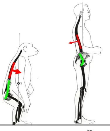
1.3.1 The challenge of modeling AIS in laboratory animals ... 27
1.3.2 AIS biomechanics ... 29
viii
2 CHAPTER II: ARTICLE ONE; IDENTIFICATION OF ELONGATED PRIMARY CILIA WITH IMPAIRED
MECHANOTRANSDUCTION IN IDIOPATHIC SCOLIOSIS PATIENTS ... 32
2.1 ABSTRACT... 35
2.2 INTRODUCTION ... 36
2.3 RESULTS ... 38
2.3.1 Osteoblasts of IS patients have longer cilia. ... 38
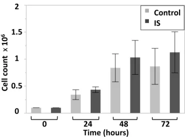
2.3.2 IS and control cells grow at the same rate. ... 39
2.3.3 Lithium Chloride (LiCl) increases the length of primary human osteoblasts. ... 39
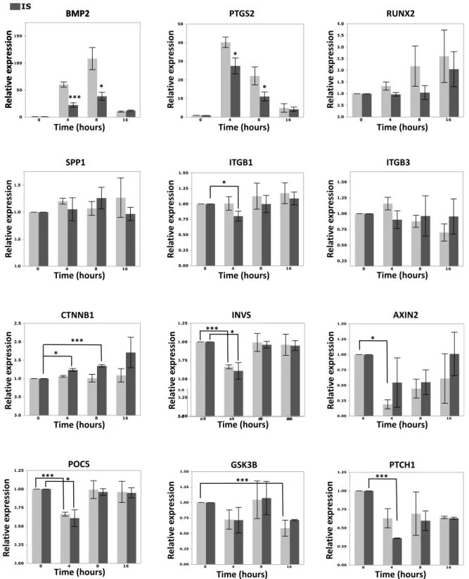
2.3.4 LiCl affects the expression of Wnt signaling indicators and osteogenic factors. ... 39
2.3.5 IS cells have impaired biomechanical response. ... 40
2.3.6 LiCl treated control cells also show impaired biomechanical response. ... 42
2.3.7 Whole Exome Sequencing (WES) results. ... 42
2.4 DISCUSSION ... 44 2.5 METHODS ... 50 2.5.1 Study cohort ... 50 2.5.2 Cell culture ... 50 2.5.3 Immunofluorescence ... 51 2.5.4 Proliferation assay ... 52
2.5.5 In vitro fluid flow stimulation ... 52
2.5.6 RNA extraction ... 54
2.5.7 Quantitative RT-PCR ... 54
2.5.8 Induced phenotype via Lithium Chloride (LiCl) ... 56
2.5.9 Exome and Sanger sequencing ... 56
2.5.10 Statistical analyses ... 57
2.5.11 Review of ciliary genes associated with spinal curvature... 58
2.6 ACKNOWLEDGEMENTS ... 59
2.7 FIGURE LEGENDS ... 60
2.8 FIGURES ... 63
2.9 TABLES ... 68
2.10 SUPPLEMENTARY MATERIALS ... 72
3 CHAPTER III: ARTICLE TWO; ALTERED MECHANOTRANSDUCTION IN ADOLESCENT IDIOPATHIC SCOLIOSIS: A PILOT STUDY ... 83
3.1 ABSTRACT... 85
3.2 INTRODUCTION ... 86
3.3 MATERIALS AND METHODS ... 88
3.3.1 Cell culture ... 88
3.3.2 In vitro fluid flow stimulation ... 88
3.3.3 Immunofluorescence staining ... 89
3.3.4 Scratch/wound healing test ... 89
3.3.5 Confocal microscopy and image analysis ... 90
3.3.6 Vascular endothelial growth factor (VEGF-A) measurement ... 90
3.4 RESULTS ... 91
3.4.1 Cilia length is differentially adjusted in AIS osteoblasts in response to fluid flow ... 91
3.4.2 Flow induced actin remodeling is impaired in AIS osteoblasts ... 91
ix
3.4.4 AIS cellular orientation in response to directional flow is impaired ... 93
3.4.5 Fluid flow does not induce VEGF secretion in the medium of cultured AIS osteoblasts. ... 94
3.5 DISCUSSION ... 94
3.6 ACKNOWLEDGMENTS ... 100
3.7 FIGURE LEGENDS ... 101
3.8 TABLES ... 110
4 CHAPTER IV: DISCUSSION ... 111
4.1 CILIARY ABNORMALITIES IN AIS OSTEOBLASTS ... 113
4.2 CYTOSKELETON ABNORMALITIES IN AIS OSTEOBLASTS ... 114
4.3 MOLECULAR ABNORMALITIES IN AIS OSTEOBLASTS ... 114
4.4 STUDY LIMITATIONS ... 116
4.5 POTENTIAL CLINICAL IMPACTS ... 117
5 CHAPTER V: FUTURE WORK & CONCLUSION ... 118
5.1 FUTURE WORKS ... 1137
5. CONCLUSIONS ... 113
6 REFERENCE LIST ... 122
7 ANNEX ... 144
7.1 REVIEW ARTICLE:GENETICS OF IDIOPATHIC SCOLIOSIS ... 145
7.1.1 Abstract ... 146
7.1.2 Introduction: ... 147
7.1.3 Hypothesis 1: Idiopathic scoliosis is a Mendelian disease ... 147
7.1.3.1 Support ... 147
7.1.3.2 Approach ... 149
7.1.3.3 Current perspectives ... 150
7.1.4 Hypothesis 2: Idiopathic scoliosis is caused by multiple genes of minor effect ... 151
7.1.4.1 Support ... 151
7.1.4.2 Approach ... 151
7.1.4.3 Current perspectives: ... 153
7.1.5 Hypothesis 3: Idiopathic scoliosis is caused by rare genetic variants of major effect ... 154
7.1.5.1 Support ... 154
7.1.5.2 Approach ... 155
7.1.5.3 Current perspectives ... 156
7.1.6 Untested hypotheses for idiopathic scoliosis ... 156
7.1.7 Other genetic approaches applied to idiopathic scoliosis ... 157
7.1.8 Conclusions ... 159
x
List of Tables
Table 1-I. Phenotypic consequences of ciliary protein disruption in bone cells. ... 19
Table 2-I. IS-associated genes in humans and/or animal models, which are also associated with cilia... 68
Table 2-II. Significantly longer cilia in IS-derived bone cells... 69
Table 2-III. The basal level of gene expression in LiCl-treated control and IS compared to non-treated control osteoblasts. ... 70
Table 2-IV. Clinical features of patients tested for ciliary morphology. ... 71
Table 3-I. The distribution of α angle in control and AIS osteoblast with or without flow application. ... 110
xi
List of Figures
Figure 1-1 Cobb angle measurement. ... 3
Figure 1-2 Levels of structural hierarchy in bone. ... 12
Figure 1-3 Human ciliopathies with skeletal deformities. ... 20
Figure 1-4 Bone cell mechanotransduction ... 26
Figure 1-5 Biomechanical differences of human with other bipeds. ... 27
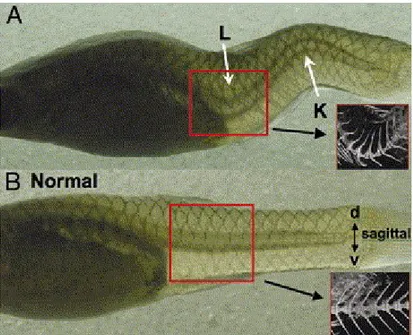
Figure 1-6 The Curveback guppy as a model for human idiopathic scoliosis. ... 28
Figure 2-1 Morphology of primary cilia in osteoblasts from IS and controls. ... 63
Figure 2-2. Similar growth rates among IS and control cells. ... 64
Figure 2-3. Biomechanical response profile of human primary osteoblast from IS patients with elongated cilia vs. control. ... 65
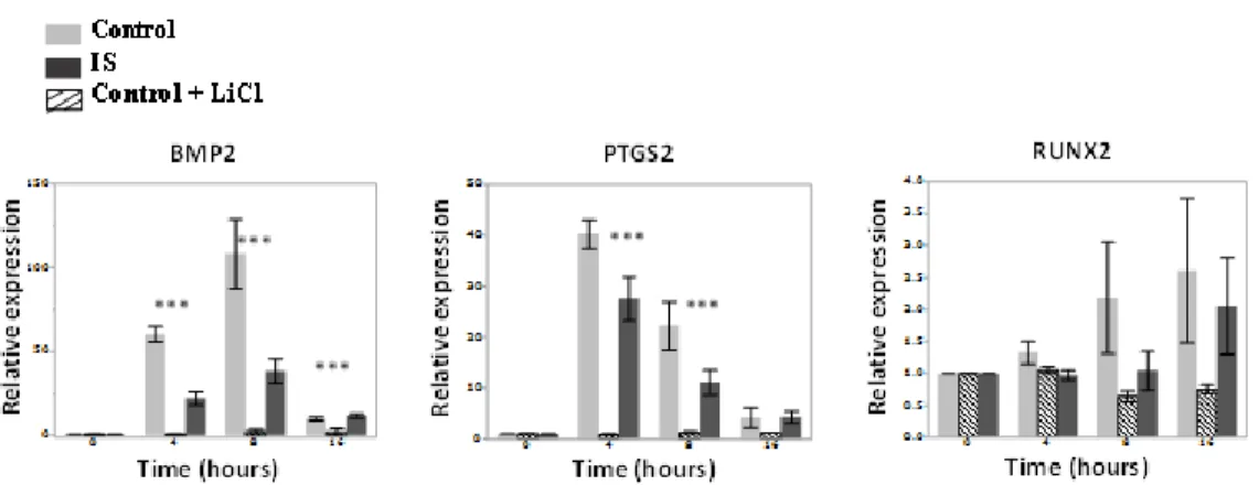
Figure 2-4. Osteogenic factors response profile of primary human osteoblasts from control, IS and LiCl treated controls under mechanical stimulation. ... 66
Figure 2-5. Molecules are connected through pathways linking ciliary mechanosensation to bone formation. ... 67
Figure 3-1. Flow induced primary cilia length adjustment is impaired in IS osteoblast independent of time... 104
Figure 3-2. Flow induces actin rearrangement in control osteoblasts but not IS. ... 105
Figure 3-3. Fluid flow does not increase the rate of Wound healing/cell migration in IS cells. ... 106
Figure 3-4. Cellular orientation adjustment relative to direction of flow varies between control and IS primary osteoblasts. ... 107
xii
Figure 3-5. Fluid flow does not induce VEGF secretion in IS osteoblast cells. ... 108
Figure 3-6. Annual distribution of publications on Idiopathic Scoliosis as were indexed in PubMed. ... 109
xiii
List of Supplementary Tables
Supplementary Table 2-I ... 77
Supplementary Table 2-II ... 81
List of Supplementary Figures
Supplementary Figure 2-1. Cilia Immunofluorescence co-staining. ... 72
Supplementary Figure 2-2. LiCl increases the length of cilium in primary human osteoblasts. ... 73
Supplementary Figure 2-3. Effect of 10 mM LiCl treatment on biomechanical response profile of control cells. ... 74
Supplementary Figure 2-4. Mutation profile of the tested IS patients. ... 75
xiv
Abbreviation list
AC: Adenylate cyclase
AIS: Adolescence idiopathic scoliosis ALP: Alkaline phosphatase
Alpha-MEM: Minimum Essential Medium Eagle – Alpha Modification ANOVA: Analysis of variance
ATP: Adenosine triphosphate BMD: Bone mineral density
BMP2: Bone morphogenetic protein 2 BSA: Bovine serum albumin
Ca: Calcium
CADD: Combined Annotation Dependent Depletion
CaM: Calmodulin
cAMP: Cyclic adenosin monophosphate
CLASP1: Cytoplasmic linker associated protein 1
COX: Cyclooxygenase
CTFC: Corrected total cell fluorescence CTNNB1: Catenin Beta 1
DNA: Deoxyribonucleic acid ECM: Extracellular matrix
ELISA: enzyme-linked immunosorbent assay ERK: Extracellular signal regulated kinases ERs: Estrogen receptors
FAK: Focal adhesion kinase FBS: Fetal bovine serum
FGF23: Fibroblast growth factor23
GH: Growth hormone
GPCRs: G protein-coupled receptors GSK3beta: Glycogen Synthase Kinase 3 beta GWAS: Genome-wide association study IFT: Intraflagellar transport
IP3: Inositol triphosphate 3 ITGB1: Integrin beta 1
KIF: Kinesin superfamily proteins LBX1: Ladybird Homeobox 1 LiCl: Lithium chloride
LRP: Lipoprotein receptor-related protein MAF: Minor allele frequency
xv
MATN1: Matrilin 1, cartilage Matrix Protein
mRNA: Messenger RNA
MSCs: Mesenchymal stem cells NIH: National institute of health
OPG: Osteoprotegrin
OPN: Osteopontin
PBS: Phosphate-buffered saline PCP: Planar cell polarity
PCR: Polymerase chain reaction
PFA: Paraformaldehyde
PGE2: Prostaglandin E2 PKA: Protein kinase A POC5: Protein of centriole 5
PSSE: Physiotherapeutic Scoliosis Specific Exercises PTGS2: Prostaglandin-Endoperoxide Synthase 2 PTK7: Protein Tyrosine Kinase 7
RANK: Receptor Activator of NF-KapaB RANKL: Receptor Activator of NF-KapaB ligand
RNA: Ribonucleic acid
RT-qPCR: Reverse transcriptase quantitative PCR RUNX2: Runt-related transcription factor 2
SKAT-O: Optimal SKAT
SKAT: Sequence kernel association test SNPs Single nucleotide polymorphisms SNVs: Single nucleotide variants
TGF1: Transforming growth factor type TRAP: Tartrate-resistant acid phosphatase VEGF: Vascular endothelial growth factor
xvi
To my mother: Azam
To my love: Alireza
xvii
Acknowledgments
I would like to express my deepest gratitude to my PhD supervisor Prof. Alain Moreau for accepting me as one of his doctoral students, and for his continues support throughout this journey. Alain generously shared his knowledge and time. He trusted me and gifted me with the freedom I needed to grow and develop my own scientific and academic expertise. This journey would not have been possible without him and his mentorship.
I am grateful to my doctoral committee members, Prof. Zoha Kibar and Prof. Muriel Aubry, for the scientific discussions and sincere supports that they provided when I needed the most. I also want to thank Prof. Carolina Alfieri, for her heartwarming presence and for hours of valuable discussions that I was privileged to enjoy with her learning about life and science.
Working with Alain’s group, I had the opportunity to meet and learn from a group of exceptional colleagues. Valuable friendships formed through these years which made everything much more fun. I learned critical thinking and scientific analysis from heated, enjoyable discussions with Prof. Kristen F. Gorman, who was a post doctorate fellow back then and is a dear friend now. I also learned a great deal of laboratory discipline and project management from Anita Franco. Her outstanding memory and organization made everything so much easier. I would also like to thank Dr. J. F. Lavoie, Dr. Cedric Julien, and other members of the lab, Lakshmi Suvarnan, Dr. Nancy Karam, Dr. Mohamed Elbakry and Dr. Dina Nada for their friendship and company.
I specially want to thank my mother for her continuous and unconditional support whenever I needed. Her brave scarifies helped me move forward. I experienced the joy and hardship of motherhood during last years of my PhD and if it was not for her warm presence and amazing support, I would not have made it. Also, many thanks to my father, who has always been proud of my achievements, small or big, since I was a child, encouraging me every step of the way.
Last but not least, my beloved family. My little son, Rodwin Leo, your big brown eyes overload me with joy and life, fueling me to push forward everyday. Alireza, thanks
xviii
for your love, support, encouragement, great patience and brutally frank criticisms throughout this journey.
I also would like to acknowledge the agencies who funded this research: Fondation CHU Sainte‐ Justine, Network of Applied Medical Genetics (RMGA), La Fondation Yves Cotrel de l’Institut de France, Paris, France, Paradigm Spine LLC/Fourth Dimension Spine LLC (NYC, USA), and Génome Québec (Montreal, Canada)
1
1 Chapter I
2
1.1 Adolescence idiopathic scoliosis (AIS)
1.1.1 Introduction and definition
The term “scoliosis” originates from Greek, meaning “crooked”, and was first used by Hippocrates (A.D. 460–370), to explain all kinds of malalignments in the human spine. Ambroise Paré (1510–1590), provided the first accurate description of Adolescent Idiopathic scoliosis (AIS) and tried to treat his patients with the historical well-known iron brace [1].
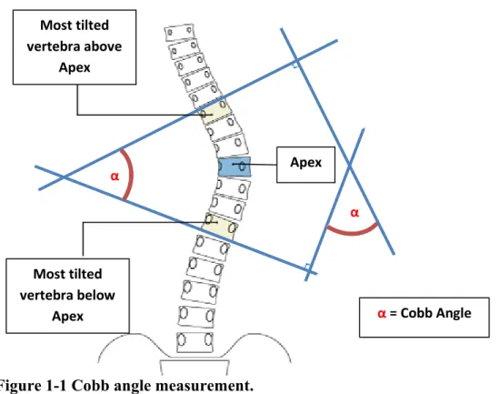
Today scoliosis is defined as a three-dimensional spinal deformity. The diagnosis is based on measuring the angle of the major spinal curve using the Cobb method. This method involves measuring the angle between the top surface of the most tilted upper vertebra and bottom surface of the most tilted lower vertebra in the spinal curve (Fig. 1-1). This angle is commonly referred to as the “Cobb angle”. A spinal curve with a Cobb angle greater than 10o is diagnosed with Scoliosis [2].
1.1.2 Epidemiology
Adolescent type of idiopathic scoliosis develops between 11 to 18 years of age and accounts for about 90% of idiopathic scoliosis. The remaining 10% include the Infantile and Juvenile types of idiopathic scoliosis, with age of onset being between 0-3 and 4-10 respectively. The prevalence of AIS varies for different ethnic and geographic groups. With a global prevalence of up to 5.2%, AIS is a common disease [3]. It represents a serious and
3
chronic health condition impacting individuals throughout their lives. Most patients are diagnosed with scoliosis between the ages of 10 and 15, and one out of every four cases would have a progressive curve requiring active treatment [4]. AIS prevalence is also sex and age dependent, with an increased ratio in girls. In fact, Luk KD. et al. [5] studied a large cohort of scoliotic children, from age 10 until skeletal maturity at 19, and found that AIS was more common in girls than boys for all curve severities. Asher et al. [6] have also shown that AIS patinates who are still undergoing spinal growth have a higher risk of curve progression by the end of skeletal maturity.
Most tilted vertebra above Apex Most tilted vertebra below Apex Apex α = Cobb Angle α α
Redrawn based on information on the “Spine Health” website https://www.spine-health.com/conditions/scoliosis/cobb-angle-used-measure-scoliosis-curves
4 1.1.3 Disease management
Due to the lack of specific biomarkers, AIS patients are usually referred to clinicians following screening exams. As a consequence of its persistently idiopathic nature, the standard care for AIS has not significantly changed in decades. Currently, diagnosed patients are generally observed for spinal deformity progression. Bracing is the chosen treatment in case of progression and spinal fusion surgery is used as a last resort. Because of patients’ discomfort and risk, the application of current treatments is delayed until a significant deformity or progression is detected. This results in suboptimal treatment, significant psychological negative effects, and significant economic burden for the families and healthcare systems.
Physiotherapy has been recommended as a first step in treatment of AIS while the patient is observed for curve progression. Whether physiotherapy is effective for AIS management is still debated among clinicians [7], [8]. While this conservative approach is routinely used in several European countries, North American physicians do not generally advocate its use. It is argued that generic physiotherapy exercises are less effective than Physiotherapeutic Scoliosis Specific Exercises (PSSE) protocols that are designed specifically and adapted individually to curve site, magnitude and clinical characteristics of each patient [9].
Curve progression is more probable in patients with immature skeletons. Cases with Cobb angles less that 20° are simply observed, while curvatures over this threshold are addressed by bracing (over 13 hours per day for a duration of 2.5 years) in hope of preventing the curve progression to the point of surgery [10]. The effectiveness of bracing has been controversial and subject to more heterogeneity based on the type of curvature, age of the patient, duration of brace wearing and the model of the bracing [11]–[14]. Last therapeutic option is spine fusion surgery that aims for preventing curve progression and long-term consequences of larger spine deformities such as pain, reduced pulmonary capacity and other functional consequences that profoundly reduce the quality of life in affected individuals. Like any other invasive surgical approaches, there is always some level of risk for complications, during or after surgery, such as loss of neurological response, early or late infections, implant failure and recurrence or additional deformity. Nevertheless, patients surveys and studies report satisfactory short-term outcomes for
5
scoliosis correction surgery, while not much data is available regarding its long-term outcomes [10].
1.1.4 Etiology
Despite decades of research, there is no consensus among the experts regarding the etiology of AIS nor its pathogenesis. Nevertheless, the existence of a genetic component using both familial and population-based approaches, is well recognized [2], [15]. Moreover, abnormalities in central nervous system, bone metabolism and spinal growth, metabolic pathways and biomechanics have also been proposed as the etiological factors of AIS [16]–[18]. Cheng J.C et al. [2] have recently published a comprehensive review of the current understanding of AIS etiology.
1.1.4.1 AIS complexity and clinical heterogeneity
Adolescent Idiopathic Scoliosis is a complex disease with a wide range of clinical heterogeneity in several levels including age at onset, curve phenotype and the progression of spinal curvature. This makes timely diagnosis and early interventions very difficult. The aforementioned female bias further adds to this complexity.
AIS heritability is widely accepted through genetic findings from classic familial linkage studies to vast population studies using relatively recent technology-oriented methods such as GWAS and whole exome sequencing. Genetic variations and mutation have been reported to be associated to the disease in certain populations or families that perhaps could explain the heritability in some patients. But these findings do not converge into a causative gene with putative role and a classic mode of Mendelian inheritance [15], [19]. The emerging view favours a complex polygenic model with considerable heterogeneity [15], [20]–[22].
1.1.4.2 Several tissues are involved in AIS
Due to association of scoliosis with neurological/neuromuscular disorders, neuropathological mechanisms have long been suggested to play a role in AIS. Surgically
6
induced small brain lesions in laboratory animals and the removal of the pineal gland from chicken, fish or bipedal rats reportedly caused AIS resembling curvatures [16]–[18]. Moreover, coexisting abnormalities in oculo-vestibular and proprioceptive functions in AIS patients were observed in several investigations [23]–[27].
It is not yet fully known how the primary changes in the nervous system can affect spinal curvature; however, it is believed that sensory nerves effects in bone homeostasis and cross talks between muscle, bone and connective tissue may be involved [28]. An interesting investigation by Fukuda et al. [29] showed that nerve specific knockout of the axon guidance gene, Sema3A, in mice caused low bone mass due to reduced bone formation. Removal of the same gene from osteoblasts did not affect the bone mass in the animal, suggesting the involvement of sensory nerve independent of the local alterations [29].
Fibers disarrangement in the ligamentum flavum [29] or reduced glycosaminoglycan content of intervertebral discs have also been reported in AIS patients [18]. AIS patients have shown relative hypertrophy and increased electromyographic signalling in the paraspinal muscles surrounding the scoliotic curves which are suggested to be the result of curve progression [27], [29]. Cartilage tissue abnormalities such as calcification of the cartilage end plate and the adjacent disc have also been reported in AIS [30]. A conditional gene targeting in the mouse showed loss of GRPR126 protein in cartilage affected normal spinal column development, resulting in a scoliosis phenotype [31].
Despite all the evidence on the involvement of nervous system, muscle, cartilage and bone in the development of AIS the existing data is insufficient to support a primary causal role for any of these tissues. In this research project we focus on bone.
1.1.5 Cellular-Molecular abnormalities in AIS 1.1.5.1 Bone metabolism factors
Investigating the cause of low bone mineral density (BMD) in AIS, Suh et al. [32] reported an elevation of RANKL and RANKL/OPG ratio in the serum of 72 AIS patients. This finding later was confirmed by others [33], [34]. Osteoblasts from 20 AIS patients
7
with low BMD showed an increase of RANKL in both mRNA and protein levels [35]. An association between a polymorphism in OPG gene and low BMD in lumbar spine has also been reported [34]. Antiosteoporosis treatments were suggested to help prevent AIS curve progression by restoring the balance of RANKL-OPG-RANK system and improving bone strength [36].
Osteocalcin is involved in both bone formation and resorption and is one of the hormones secreted from osteoblasts. Increased levels of osteocalcin in blood serum of AIS patients [33], [34] could be due to higher rate of bone remodeling and abnormal bone metabolism.
MATN1 protein forms filamentous structures in several tissues such as cartilage and helps in the organization of extracellular matrix (ECM). Several studies reported an association between SNPs in different regions of MATN1 and AIS [15], [19]. Recent meta-analysis concluded that some of these association might be restricted to specific ethnic backgrounds [37]. Reduced plasma levels of MATN1 protein are suggested to be a possible biomarker for disease progression [38]. Cartilage oligomeric matrix protein (COMP) is another ECM involved protein which is associated with skeletal disorders including scoliosis [39]. Reduced COMP levels in serum [40] and significant COMP mRNA downregulation in osteoblasts[41] were shown in AIS patients. It is of note that several recent studies showed interesting evidences supporting the contribution of rare variants enrichment in ECM genes to disease risk [42], [43].
1.1.5.2 Hematological factors (Platelets)
The similarity of platelet and muscle in their actin-myosin contractile apparatus, has led researchers to look for similar abnormalities in these two cell types in AIS. Since the early 1980s, a number of anomalies in platelets, calmodulin, and Ca transport in AIS have been documented in the literature (refer to [44] for a more comprehensive review). Calmodulin (CaM), binds to calcium, playing a role in platelet and muscle contraction through interactions with actin and myosin [44]. CaM concentration is increased in the platelet of AIS patients and this appears to be associated with disease progression. Non-progressive stabilized curves and healthy controls showed no difference in CaM
8
concentration. Also curve stabilization following therapeutic interventions reduce the increased CaM levels, suggesting a biomarker role for this protein [45], [46].
1.1.5.3 Systemic factors
G protein family is involved in transmitting extracellular signal to the cell, through interaction with a family of membranous receptors called GPCRs (G protein-coupled receptors). Based on the fact that melatonin receptors are GPCRs, Moreau et al. [47] studied the response of AIS osteoblasts to melatonin and found reduced response patterns compare to controls. They measured the cAMP concentration as a secondary messenger and reported an elevation of cAMP in AIS osteoblasts due to melatonin failure in reducing cAMP accumulation. Although this reduced response was shared among all tested AIS samples, the reduction pattern was not the same. The authors were able to classify AIS patients into three distinct groups based on their osteoblast response to melatonin [47], [48]. They were able to confirm their findings in another study using the cellular dielectric spectroscopy technique by direct measurement of the cellular response to Gi stimulation and reproducing three distinct patterns in AIS cells [49]. Reduced performance of Gi-coupled receptor signaling in AIS patients was later reported to be rather a systemic impairment and not restricted to downstream melatonin pathways. They found the same impairment in other cells (skeletal myoblasts and peripheral blood mononuclear cells) obtained from AIS patients which were also grouped in three differential groups or endophenotypes [49]. AIS complex nature and high levels of phenotypic and genotypic heterogeneity is one of the biggest challenges in understanding the disease. Endophenotypes can improve the stratification of the patients at a molecular level, potentially help with a better prognosis. The detailed procedure of the used methodology in AIS patients classification is available in JoVE visual journal [50].
Osteopontin (OPN) is another systemic factor that its association with idiopathic scoliosis is well documented. This multifunctional glycoprotein is expressed in all body fluids as well as in ECM of mineralized tissue [50]. Higher levels of circulating OPN in plasma of AIS patients is reported to correlate with curve severity [51]. Mice model studies
9
confirmed the increased OPN to be associated with disease development and severity. OPN depletion also showed to help improve the skeletal deformity [52]–[54].
1.1.5.4 Hormonal factors
Growth abnormalities are usually observed in AIS and they have been shown to specifically associate with the pubertal stage (reviewed in sub-section 1.1.6). Therefore growth hormone (GH) has been an interesting area to study for AIS researchers since the 1970s [52]. Elevated levels of GH [55]–[57] or its downstream players [58], [59] were showed to be associated with AIS. Genetic studies however failed so far to find a consensus correlation. The findings are conflictive and reported AIS associated SNPs concluded to be limited to an ethnic area or a proportion of the patients [60]–[64].
Estrogen has been assumed to play a role in AIS due to the biased prevalence in female and association of age at menarche with scoliosis development. Later age at menarche is parallel with higher AIS prevalence [65]. Estrogens influence growth, bone remodeling and bone gain, all of which are affected in AIS. There are some inconclusive data on blood estrogen levels [66]–[68], however its role in AIS is not concentration dependent, rather appears to be associated with the activity on their target cells [65], [69]. Estrogens suggested to have a key role in triggering the melatonin signaling dysfunction and bone remodeling in AIS patients [70]. A recent study on bipedal rats confirmed that estrogen increase the scoliosis incident and severity. Zheng et al. in this study suggested a loss of coupling of endochondral ossification between the anterior and posterior columns to be the underlying cause [71]. There has been extensive research trying to find an estrogen related genetic association with AIS in an effort to explain the higher prevalence in female. In result, multiple polymorphisms in estrogen receptors or other related genes have been reported, reviewed in [15]. Unfortunately, due to high discrepancy in the results, the estrogens genetic correlation with AIS remains debated. In essence estrogens may not be directly involved in AIS etiology, but still impact the disease through to its effects on many AIS etiopathological factors such as bone remodeling, growth factor and melatonin pathway [44].
10
Leptin is another hormone suggested to be involved in AIS. This energy regulating hormone plays also an important role in bone metabolism. Reduced levels of leptin in blood samples of AIS patients is reported in many studies [72]–[74]. Reduction in bone mass, bone strength and growth abnormalities have been listed to be associated to leptin bioavailability [75]. Hypothalamic hypersensitivity to circulating leptin levels, led to development of a double neuro-osseous theory that claims a role for both autonomic and somatic nervous systems in the development of IS [76]. AIS Osteoblast cells were shown to have significantly lower response to leptin stimulations compared to controls [75]. It is of note that melatonin [75] and cytosolic cAMP [75] can inhibit leptin synthesis. As mentioned above, there is an impairment in AIS cells involving cAMP and melatonin signaling pathways [47], [49].
1.1.6 Bone abnormalities in AIS 1.1.6.1 Growth and metabolism
Adolescence is a period of rapid skeletal growth, with skeletal mass doubling by the end of adolescence [77]. AIS occurs during pubertal growth spurt in children and curve progression is stabilized at skeletal maturity. Rapid growth phase is associated with the curve development and progression [78]. Taller and leaner children have been reported to show higher risk for AIS development [79], [80]. In addition to abnormal body height, asymmetries in the limb and segmental length were also reported [79], [80]. Whether these observed skeletal differences at the time of diagnosis will be resolved by skeletal maturity or remain present in adulthood is controversial [81], [82].
Since 1982, when Burner et al. [83] first reported lower BMD in patients with acquired back deformity, several studies have found a higher prevalence for osteoporosis in AIS compared to the general pediatric and adolescent population [84]–[86]. Lee et al. [87] reported an inverse relationship between curve severity and BMD in a cross-sectional study on 919 AIS girls. The causes of osteoporosis in AIS patients are unknown and whether poor bone quality is an etiological factor remains controversial. Bracing, due to partial immobilization of the body, has been postulated to cause a permanent loss of bone mineral mass, predisposing to adult osteoporosis, but this presumption has been refused in
11
several extensive studies[86]. The BMD measurement at the time of diagnosis might also serve as an additional objective indicator of curve progression, suggesting osteopenia to be an important risk factor with a prognostic value [88].
1.1.6.2 Structure and morphology
Structural factors, such as the cortical bone thickness and the trabecular bone micro-architecture, have been suggested to be of paramount importance for the determination of bone quality and strength [89]–[91]. Yu et al. [92] reported lower cortical thickness in AIS cases after the assessment of 214 AIS girls in comparison to 187 healthy age matched controls. They suggested AIS abnormal bone profile to be the result of altered endocortical modeling, deranged trabecular bone structure and disturbed bone mineralization.
Bone strength is a function of both BMD and bone quality. NIH reinforced this statement by defining osteoporosis as a skeletal disorder with compromised bone strength which features bone density and bone quality [93]. In 2017, Wang et al. (71) confirmed reduced mineralization and predominant trabecular changes in AIS patients. Interestingly, the tissue level of mRNA expression for RUNX2 was found to be lower in AIS bone biopsies, while SPP1 (OPN) and TRAP mRNA were found to be higher [94].
Despite decades of research, the etiology of AIS remains a mystery. Among all the studies on the etiology of AIS that have been carried out so far, cellular and molecular findings are among the least reported. Having access to a cellular bank from AIS patients we had the opportunity to investigate the etiology of AIS from a cellular-molecular perspective.
1.2 Bone cell mechanobiology
1.2.1 Introduction
Bone as a bio-ceramic composite features unique mechanical properties that have always attracted engineers. These mechanical features are mostly attributed to complex multilayer structures that are arranged in a hierarchical manner (Fig. 1-2).
12
Bone cells are tightly attached to their extracellular environment in a complex dynamic fashion. Environmental stimuli such as gravity, strains, shear, compression, stretch, and fluid flow have the ability to trigger cellular response and influence the whole bone tissue. In addition, components of these stimuli such as magnitude, frequency and strain rate also affect this cellular response [95]. The relationship between bone formation, remodeling and the surrounded mechanical environment is presented in Frost’s “mechanostat” theory [96]. This theory describes a homeostatic mechanism in bone that is responsible for sensing the applied mechanical changes and modifying the mass and conformation of bone to meet the mechanical demands in an optimal condition. He hypothesized that below a certain physiological threshold, bone reduces excess mass through increased remodeling. When strains exceed the upper physiological boundary, he argued, bone formation occurs through increased modeling to provide the needed strength [96]. Mechanostat defines a nonreplaceable role for mechanical forces to guide the effect
Trabecular bone
Cortical bone
Osteon and lamella Trabecular packet And lamella Mineralized collagen fibers Collagen/ Mineral composite Collagen alpha chains Deposited crystals 10-1 m 10- 4 m 10- 7 m 10- 9 m 10- 10 m
Organ and tissue Micro- and sub-microstructures Nano- and ultrastructures Molecular structures
The composite of bone includes a hard cortical shell that covers the spongy cancellous bone inside. Cortical bone microscopic structure is composed of many secondary haversian systems (osteons) that are the product of remodeling process. Both trabecular and cortical bone are lamellar with a different microstructure. Collagen fibers with plates of minerals compose the ultra- and nano-structural layers. Figure is inspired and redrawn from, Burr. D.B and Akkus O. Bone morphology and organization, 2014 (DOI: http://dx.doi.org/10.1016/B978-0-12-416015-6.00001-0). Some of the motifs are downloaded from smart servier medical art website. Figure 1-2 Levels of structural hierarchy in bone.
13
of physiological factors such as cellular interaction, hormones, etc. on bone adaptation mechanisms [95].
1.2.2 Bone cells
Bone tissue consists of three different cells, namely osteoblasts, osteoclasts and osteocytes. To carry out the diverse functions of skeletal maintenance, including bone formation, resorption, mineral homeostasis and bone repair, bone cells develop specialized morphology, function and characteristic locations. Some of them originate from the line of mesenchymal stem cells, which consist of pre-osteoblasts (undifferentiated cells), osteoblasts and bone lining cells. The rest rise from hematopoietic stem cell line including circulating or marrow monocytes, preosteoclasts and osteoclasts [97].
Cellular structure of the bone is mainly composed of osteocytes and osteoblasts as support cells plus osteoclasts as remodeling cells. Osteoblasts are responsible for osteogenesis through the deposition of bone matrix and regulation of bone resorption. Osteocytes are terminally differentiated osteoblasts that are trapped in their own bone matrix. They form a network through the extensions of their plasma membrane and are the most abundant cells in a bone [98]. The only cells that are capable of resorbing bone are Osteoclasts which are typically multinucleated cells.
1.2.2.1 Undifferentiated mesenchymal cells
Pre-osteoblasts or potent mesenchymal cells live inside the bone canals, endosteum, periosteum and bone marrow waiting for a stimulus to proliferate and differentiate into mature osteoblasts. They are mononuclear, irregular cells with minimum cytoplasm that respond to growth factors, hormonal changes, biomechanical stimulations among other factors. These cells have crucial role in healing a fracture or depositing more bone where needed [97], [99].
14 1.2.2.2 Osteoclasts
Osteoclasts are quite different from other bone cells in their morphology, function and origin. Unique in the entire body, these cells have impressive power for local digestion which makes bone resorption to be their primary role.
Unlike other bone cells, osteoclasts are derived from hematopoietic monocyte-macrophage lineage. The mononuclear precursor cells fuse to form the mature multinucleated osteoclasts. Multinucleation is an important feature of these cells that together with other adapted ultrastructural changes provide them with the bone resorption ability. The precursor cells are available in the marrow or blood stream and factors such as calcium gradient, secreted cytokines from osteoblasts and osteocytes, and matrix metalloproteinases can stimulate their recruitment to bone resorption sites [97].
The large multinucleated osteoclasts can have between three to twenty nuclei with numerous numbers of lysosomes and elongated mitochondria. The specific brush or ruffled border of osteoclasts adjacent the target area in the bone is perhaps the most complex and distinctive feature of these cells [100].
Beside bone resorption, osteoclasts also play a role in the regulation of minerals such as calcium and phosphate in the body alongside parathyroid and thyroid glands, osteocytes and renal hormones. Upon the discovery of osteoclasts activation by inflammatory stimuli, osteoimmunology was coined as a scientific term to explain the tight relationship between skeleton and immune system.
The most well-known regulators of osteoclastogenesis, the RANK/RANKL/OPG axis, were discovered through osteoimmunology studies. The Receptor Activator of NF-KapaB transcription Factor (RANK) is expressed by committed osteoclast precursors. Responding to its ligand (RANKL), RANK stimulates various intracellular pathways that lead to osteoclasts formation and activation. Secreted by osteocytes, Osteoprotegrin (OPG) on the other hand inhibits osteoclastogenesis by binding to RANK and competing with RANKL. In both osteocytes and osteoclasts, OPG secretion is regulated by the Wnt/beta-catenin pathways [100].
15 1.2.2.3 Osteoblasts
Although not terminally differentiated, osteoblast are mononucleated cells that are specialized mainly in bone matrix deposition and osteoclast regulation [101]. They coat the surface of bone packing tightly against each other [97]. Following bone formation completion, osteoblasts are destined for one of these three fates: some mature into their matrix and turn to entrapped osteocytes, some adapt a more flat form to cover the surface of the bone forming bone lining cells and the rest disappear from the site of bone formation by apoptosis [97]. Osteoblasts secrete large quantities of collagen type I and other specialized matrix proteins forming osteoid which later becomes a base for mineral deposition. They also express high levels of Alkaline phosphatase (ALP) and osteocalcin. In fact, these proteins are indicators of osteoblast existence [102].
Osteoblasts interact with each other, bone marrow and bone lining cells through adherent, tight and gap junctions. They also bind closely with extracellular matrix through their cytoskeleton and cellular junctions. In fact, osteoblast form a continuous network connecting the mineralized matrix to the bone surface, then to the bone marrow, and finally to the endothelial cells of the blood supplying vessels [102].
1.2.2.4 Osteocytes
Osteocytes are spider-shaped former osteoblasts that have been buried into the deposited bone matrix. They compose more than 90% of the bone material, and are among the most long-lived cells with up to 25 years life span [103]. The distinct positioning of the osteocytes into the bone tissue makes them easy to identify and observe, but it limits the functional studies due to difficulties in isolating them [104]. Osteocytes feature many long cytoplasmic projections (up to 50 per cell) that extend through the network of interconnecting bone canaliculi providing cell to cell communication that facilitate signal transduction. This network, filled with interstitial fluid that flows inside the osteocyte’s canaliculi, coordinating the response of bone to biological and mechanical signals [103]. During bone formation, some osteoblasts are transformed to osteocytes which result in dramatic changes in osteoblasts morphology and functions. Osteocytes are considered to be the mechanosensory cells of a mature bone. Following the translation of
16
mechanical sensation to chemical signal, osteocytes affect function and differentiation of osteoblasts and osteoclasts, leading to bone formation or resorption [105], [106].
Osteocytes effects on the regulating function of osteoblasts and osteoclasts are well documented [107]. Different hormonal and mechanical cues trigger osteocytes production of regulatory factors such as OPG, RANKL and sclerostin. These regulatory factors in turn can stimulate other bone cells through paracrine or autocrine mechanisms [107].
Given the access of osteocytes to the mineralized material, they play a prominent role in mineral hemostasis. Osteocytes-mediated osteolysis are stimulated by changes that need a quick release of minerals in a short span of time, such as lactation [104].
Similar to osteoblasts, the osteocytes also have hormone releasing ability. These cells also can secret the FGF23 hormone which regulates serum phosphate levels through affecting the metabolism in distant organs such as kidney. Endocrine roles have also been recently attributed to osteocytes such as regulating lymphopoiesis which in turn affects the immune system and fat metabolism [108].
1.2.3 Cellular Mechanotransduction
All bone cells directly or indirectly respond to their local strains. The process of translating the mechanical stimulus into biological signals is called mechanotransduction [95]. Osteocytes viability depends on mechanical stimulation [106], [109], [110]. Strain induced osteocyte’s apoptosis also has been found to correlate with percentage of bone resorption surfaces [111]–[113]. Osteocytes seem to be the main cells responsible for unloading-induced bone loss, as osteocyte-ablated mice are shown not to develop osteoporosis in the absence of mechanical stimuli [114]. Fluid flow is a well-known player in micronutrient and molecular transportation in bone as diffusion alone proven to be not sufficient to cover the entire lacunar-canalicular system [114].
The only other serious cellular candidate for mechanical sensation in bone is osteoblast. As the progenitors of osteocytes, it can be anticipated that osteoblast have some mechanosensory capacity. In fact, the major body of in vitro studies have been performed on osteoblasts with interesting findings that confirm osteoblasts’ sensitivity to mechanical
17
stimulation [95]. Compressive stress has been suggested to regulate osteoblastic differentiation [115]. Mechanical induction has been shown to affect osteoblasts proliferation, differentiation, migration and mineralization, in addition to triggering actin rearrangement and the Wnt signaling pathway [116]–[121].
It is important to note that there are strong evidence and scientific arguments that are in favor of osteocytes being the primary and main cellular recipient of bone mechanotransductory signals. The placement and distribution of osteocyte in the three-dimensional structure of the bone is architecturally well suited to sense the surrounding movements. In fact, these cells are extremely sensitive to microfluidic flow changes [122]. In some cases, osteoblasts and osteocytes have been found to show the same mechanical sensitivity [114]. Fortunately, the results of in vitro studies on these “bone derived cells” in culture have been in agreement with what is reported by in vivo results [122].
1.2.4 Cilia
The primary cilium is a non-motile microtubule-based organelle present on the surface of most mammalian cell types. Primary cilia transduce different types of extracellular stimuli into cellular responses through well-known signalling pathways such as Wnt, Hedgehog and platelet-derived growth factor. Through these pathways, cilia play a regulating role in cell differentiation, proliferation, migration and polarity, and are therefore essential in tissue morphogenesis [123].
The ciliary proteins are synthesized in the cytoplasm and must be transported to the cilium as an outward projected organelle. This tightly regulated task is performed through an active process called intraflagellar transport (IFT) that requires IFT complexes [124].
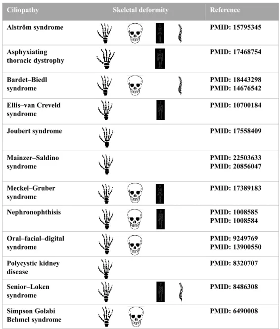
The importance of primary cilia has only been known for the last decade and ciliary machinery already have been implicated in several human disorders. Although ciliopathies share common clinical features they can target different organs or tissue including kidney, limbs, brain, retina, liver and bone [125]. Interestingly, skeletal anomalies are among common clinical features in most of human ciliopathies (Fig. 1-3).
18
The presence of cilia has been reported in most of bone cells including, osteocytes, osteoblasts and MSCs [123], [126]–[128]. Bone cell cilia are required for osteogenic differentiation, and mechanically induced bone remodelling and adaptation. Several studies showed that cilia disruption leads to impaired cellular bone functions such as osteoblasts differentiation, bone formation and even regulation of bone resorption through osteocytes [123], [129]–[134] (reviewed in Table 1-I). Disruption of an essential gene for cilia formation (Kif3A) led to the significant reduction of mechanically induced bone formation in mouse [135]. Espinha et al. [136] showed that oscillatory fluid flow induces an increase in microtubules around primary cilia in a time- and shear-rate-dependent manner. He showed also that primary cilia are essential for this load-induced cellular response.
The primary cilia are adaptive mechanosensors and possess a specific mechanism that can regulate the sensitivity of a cell to mechanical stimulation [95]. For example, cilium stiffness changes in response to mechanical and chemical stimuli. The sensory adaptation happens in a short span of time making cilia good candidates for targeted therapeutic approaches in disorders with impaired mechanosensitivity [137].
19
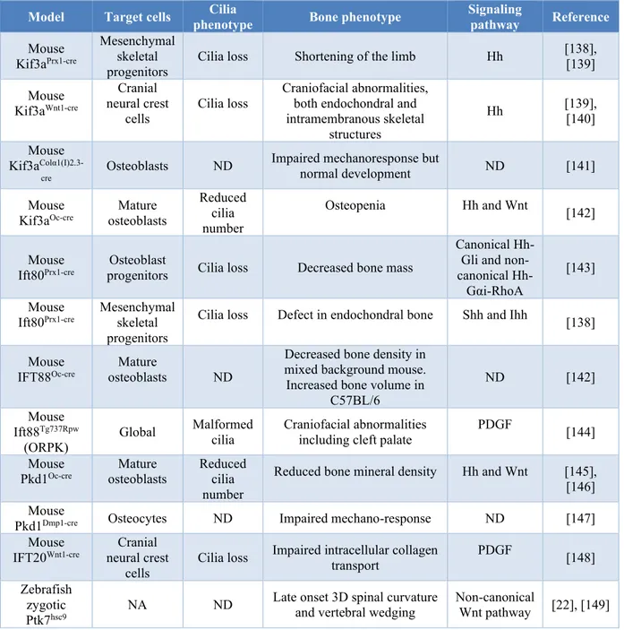
Table 1-I. Phenotypic consequences of ciliary protein disruption in bone cells.
Model Target cells phenotype Cilia Bone phenotype Signaling pathway Reference
Mouse Kif3aPrx1-cre
Mesenchymal skeletal
progenitors Cilia loss Shortening of the limb Hh
[138], [139] Mouse Kif3aWnt1-cre Cranial neural crest
cells Cilia loss
Craniofacial abnormalities, both endochondral and intramembranous skeletal structures Hh [139], [140] Mouse Kif3a Colα1(I)2.3-cre Osteoblasts ND
Impaired mechanoresponse but
normal development ND [141] Mouse
Kif3aOc-cre osteoblasts Mature
Reduced cilia number
Osteopenia Hh and Wnt [142]
Mouse
Ift80Prx1-cre progenitors Osteoblast Cilia loss Decreased bone mass
Canonical Hh-Gli and non-canonical
Hh-Gαi-RhoA
[143]
Mouse
Ift80Prx1-cre Mesenchymal skeletal progenitors
Cilia loss Defect in endochondral bone Shh and Ihh [138]
Mouse
IFT88Oc-cre osteoblasts Mature ND
Decreased bone density in mixed background mouse. Increased bone volume in
C57BL/6 ND [142] Mouse Ift88Tg737Rpw (ORPK) Global Malformed
cilia Craniofacial abnormalities including cleft palate PDGF [144] Mouse
Pkd1Oc-cre osteoblasts Mature Reduced cilia number
Reduced bone mineral density Hh and Wnt [145], [146] Mouse
Pkd1Dmp1-cre Osteocytes ND Impaired mechano-response ND [147] Mouse
IFT20Wnt1-cre neural crest Cranial
cells Cilia loss
Impaired intracellular collagen
transport PDGF [148]
Zebrafish zygotic
Ptk7hsc9 NA ND
Late onset 3D spinal curvature
and vertebral wedging Non-canonical Wnt pathway [22], [149]
20
Ciliopathy Skeletal deformity Reference
Alström syndrome PMID: 15795345
Asphyxiating thoracic dystrophy PMID: 17468754 Bardet–Biedl syndrome PMID: 18443298 PMID: 14676542 Ellis–van Creveld syndrome PMID: 10700184
Joubert syndrome PMID: 17558409
Mainzer–Saldino syndrome PMID: 22503633 PMID: 20856047 Meckel–Gruber syndrome PMID: 17389183 Nephronophthisis PMID: 1008585 PMID: 1008584 Oral–facial–digital syndrome PMID: 9249769 PMID: 13900550 Polycystic kidney disease PMID: 8320707 Senior–Loken syndrome PMID: 8486308 Simpson Golabi Behmel syndrome PMID: 6490008
The icons respectively represent abnormalities in the extremities, including polydactyly and phalangeal cone-shaped epiphyses, craniofacial defects, short stature or skeletal dysplasia and spinal abnormalities, including scoliosis and kyphosis. Reconstructed and adapted from Nguyen A.M., Jacobs C.R., Bone 54 (2013) 196–204.
21
1.2.5 Bone mechanotransduction involved pathways 1.2.5.1 Wnt signaling
The canonical Wnt or Wnt/catenin pathway is based on stabilizing beta-catenin. The Wnt proteins (such as Wnt10b, Wnt3a) attach to the LPR5/6 and Frizzled (FRZ) transmembrane receptors, activating a downstream signal that suppresses the activity of Glycogen Synthase Kinase 3 beta (GSK3beta), and inhibits beta-catenin phosphorylation [150]. A stable unphosphorylated beta-catenin molecule then translocate to the cell nucleus where it regulates the transcription of Wnt target genes. In the absence of Wnt initial signaling, GSK3beta remains active, destabilizing beta-catenin with phosphorylation, and eventually causing the ubiquitination and removal of this protein and thus Wnt signaling turns off [151], [152].
The canonical Wnt pathway is necessary for a healthy bone mass development and plays a major role in controlling skeletal patterning, development and homeostasis. This importance underscored by the number of human bone diseases associated with errors in this pathway [153]–[155]. Wnt pathway involvement in bone development starts at early stages by controlling the pattern in which bone material is laid.
Osteoblasts are one of the major cellular targets of the Wnt pathway in bone [151]. It has been reported that beta-catenin is an essential factor in determining whether mesenchymal progenitors become osteoblasts or chondrocytes, highlighting the role of Wnt signaling in osteoblast commitment [156], [157]. LRP5 loss- or gain-of-function mutations in humans or mice alter bone formation without affecting the resorption process [156]–[158]. This indicates that osteoblasts are indeed the main cellular targets of Wnt pathway in bone.
Loss and gain of functions in human Wnt co-receptor LRP5 are shown to lead to low and high bone mass syndromes, respectively [159], [160]. Wnt5a has also been implicated in bone, using an animal model study [161], which showed the reduction of bone mass following its genetic ablation, likely due to a decrease of osteoblast number.
In addition to osteoblast commitment and proliferation, Wnt signaling also affects osteoblast differentiation and function. Several Wnt signaling antagonists such as Sfrp2, Wif1, Dkk1 or FrzB, are strongly up-regulated during the late phase of osteoblast
22
differentiation. This fact suggests the involvement of a negative Wnt feedback loop throughout the last stages of osteoblast maturation. Expression of a constantly active mutant beta-catenin in osteoblasts led to an increase in expression of collagen in those cells [131], [151]. Wnt pathway also indirectly controls osteoclasts differentiation through its effects on osteoblasts and RANKL/OPG ratio.
In the case of transmitting mechanical signaling in bone, the wnt pathway plays an important role by activating or deactivating several signaling cascades [162]. Beta-catenin heterozygous deletion in mice osteocytes significantly mitigated the loading induced-anabolic response in bone [163]. Both Lrp5 and -6 play regulatory roles in loading-induced Wnt canonical signaling in bone. The loss of function mutation in Lrp5 have been shown to reduce or eliminate the load induced osteogenic response through reduction in mechanical-induced bone matrix deposition [164].
The positive effect of loading on bone formation could presumably be a consequence of an increase in Wnt stimulatory molecules or a reduction of Wnt inhibitory factors. For instance, the expression of Sclerostin (from Sost gene) as an Lrp5/Lrp6 antagonist is dramatically reduced in response to mechanical loading, and increased by skeletal disuse [165]. Despite similarities in structure and sequence between Lrp5 and -6, they seem to be different in their downstream load induced signalling. Lrp-5 affects bone formation while Lrp-6 regulates bone resorption. Lrp-6 effects are observed earlier in developmental phase than those of Lrp5 [150].
Beta-catenin is one of the major downstream targets of both Lrp5 and -6, and its inactivation in osteoblasts has been shown to cause osteopenia. The associated low bone mass phenotype in mice has been reported to be due to the increased number and activity of osteoclast thus affecting bone resorption rather than formation[166]. Interestingly, both beta-catenin alleles are proved to be necessary for mechanotransductory response in osteocyte and/or late stage osteoblasts [163].
1.2.5.2 Kinase signaling
Mitogen activated protein kinase (MAPK) pathways are activated in many cell types following an extracellular force induction. The MAPKs are among serine/threonine protein
23
kinases with fundamental roles in the differentiation, proliferation and cell survival. Physical forces can activate extracellular signal regulated kinases (ERK1/2) through a calcium dependent mechanism in bone cells, causing RANKL downregulation and decreased osteoclastic activity [167]. Akt kinase is another serine/threonine kinase which is activated by fluid shear stress in bone cells affecting MSCs differentiation process. This results in an increase in osteoblasts commitment and bone mass. Focal adhesion kinase (FAK) forms a network with other signaling proteins such as Src and the PIK3 kinases. This kinases network in response to mechanical activation of integrins stimulates downstream signals that lead to osteoblast proliferation. FAK also promotes the expression of osteopontin, osteocalcin, COX2, RUNX2 and Osterix (Osx) in mature osteoblasts which are among key osteogenesis regulators [168], [169].
1.2.5.3 Calcium signaling
Changes in intracellular levels of calcium affects cellular proliferation, differentiation and mobility. Mechanical stimulation of bone cells has been shown to follow up with a rapid rise in intracellular levels of calcium in bone cells through calcium channels [170]. Calcium movements can cause cytoskeleton rearrangement and activate various protein kinases such as PKA or MAPK, or trigger ATP and nitric oxide pathways in osteoblasts [171], [172].
1.2.5.4 G-Protein mediated signaling
G-protein coupled receptors are activated following mechanical induction causing rises in intracellular calcium, cAMP and cGMP levels[95]. Fluid shear stress application on osteocytes causes a temporary decrease in cAMP, which is catalyzed from ATP by adenylate cyclase (AC). AC isoform 6 has been shown to colocalize with cilium. Transient decrease in cAMP following shear stress is found to be AC6 dependent[173]. Primary cilium-mediated AC6 activation in osteocytes cells affects COX2 gene expression and PGE2 signaling [123]. Phospholipase C activation by fluid flow also initiates IP3 signaling, through a G-protein mediated mechanism, which in turn causes COX2 expression. RhoA GTPase is another important player in cellular response to mechanical strain. Mechanical
24
induced RhoA activity not only affects the MSC lineage commitment but also plays a role in the organization of cellular actin cytoskeleton to stress fibers. These contractile stress fibers are thicker structures that equip the cells to accommodate the applied mechanical challenge [119].
1.2.5.5 Prostaglandins
Prostaglandins are enzymatically derived from arachidonic acid that are implicated in bone remodeling. Therefore, they act as a vital element in healing processes and in bone tissue adaptation to stress responses [174]. Prostaglandin receptors are G-Protein coupled receptors. These receptors have a crucial role in regulating the functions of prostaglandins in bone [175]. Prostaglandin E2 (PGE2), in particular, plays a regulatory role in various processes of inflammation and bone metabolism [174]. Different types of mechanical induction have been reported to increase PGE2 levels in skeleton, such as hypergravity [175], fluid flow [176] and weight bearing [177].
Arachidonic acid transformation to prostaglandins is regulated by two cyclooxygenase (COX) isoenzymes, COX-1 and COX-2. They both affect the bone tissue but COX2 is the dominant one in osteoblasts [174]. COX2 protein is among the osteogenic factors that their expression in osteoblasts are induced by mechanical induction [127], [178], [179].
1.2.5.6 Estrogens
Declining levels of circulating estrogens in aging females and its direct correlation with post-menopausal osteoporosis suggest that estrogen receptors (ERs) are involved in bone mechanical induced adaptation. Both alpha and beta forms of ERs are facilitators of pathways functions in strain stimulated bone cells (osteoblasts and osteocytes) proliferation and osteogenesis. ER-alpha mostly contributes to the load-induced osteogenic response through the promotion of anabolic influences in osteoblasts and their progenitors. ER-beta is more involved in the strain-related responses induced in resident cells such as osteocytes. Activation of TGF1 receptors and COX2 expression or severe down regulation of sclerostin are among downstream effects of the ER stimulation following a mechanical
25
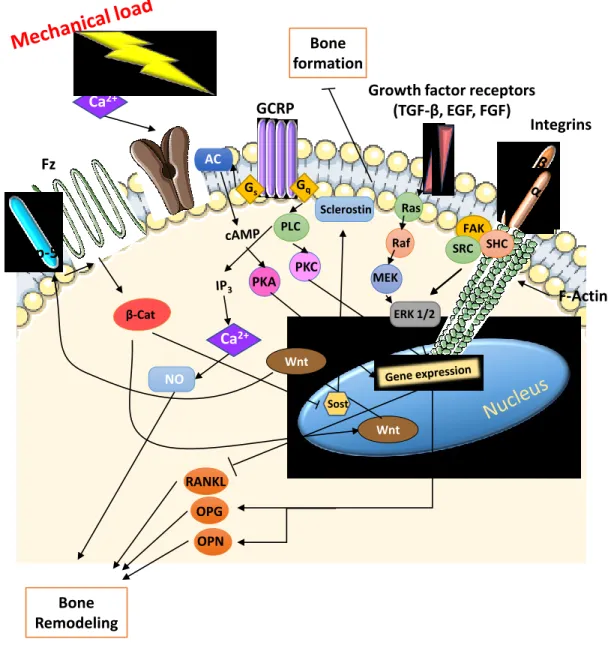
signal[180], [181]. Bone cell mechanotransduction pathways are summerized in figure 1-4.
26 Bone formation Integrins F-Actin FAK SHC SRC ERK 1/2 Fz GCRP
Growth factor receptors (TGF-β, EGF, FGF) Lrp-5 β-Cat Wnt Wnt Sost Sclerostin Ras Raf MEK Gs Gq AC PKA PKC PLC NO RANKL OPG OPN Ca2+ Ca2+ Bone Remodeling
Mechanical loading induces a complex cascade of events through several membrane bound sensors in bone cells. Here we show some of the more important players in osteoblasts and osteocytes. Inspired and redrawn from J Musculoskeletal Neuronal Interact 2016; 16(3):221-236.