Université de Montréal
Identification of Genomic Variants Associated with
Adolescent Idiopathic Scoliosis (AIS) in French-Canadian
Population
par: Qi Lin TANG
Département de Biochimie et Médecine Moléculaire Faculté de Médecine
Mémoire présenté à la Faculté des études supérieures et postdoctorales en vue de l’obtention du grade de Maîtrise en Sciences (M.Sc)
en Biochimie
option Génomique Humaine
December 2014
Université de Montréal
Faculté des études supérieures et postdoctorales
Ce mémoire intitulé :
Identification of Genomic Variants Associated with
Adolescent Idiopathic Scoliosis (AIS) in French-Canadian
Population
présenté par : Qi Lin TANGa été évalué par un jury composé des personnes suivantes :
Muriel Aubry, président-rapporteur René St-Arnaud, membre du jury Alain Moreau, directeur de recherche
i
Résumé
La scoliose idiopathique est une déformation tridimensionnelle de la colonne vertébrale dont la pathogenèse reste obscure. Cette maladie affecte 2-4% des adolescents de 10-18 ans parmi les garçons et les filles. Il est à noter que les filles sont plus sévèrement affectées et ce en plus grand nombre que les garçons. Les études de jumeaux ont montré que les facteurs génétiques jouent un rôle important dans la scoliose idiopathique de l'adolescent (SIA).
Depuis 2010, les études d'association pan génomiques ont été multipliées dans les recherches, visant à trouver des gènes candidats impliqués dans la SIA à travers des examens des polymorphismes nucléotidiques (SNPs). Un test génétique nommé "ScoliScore" a été publié pour essayer de prédire la progression de courbure dans la population caucasienne. Cependant, l'association n'a pas été reproduite dans une grande étude japonaise, soulignant l'importance d'une étude de réplication dans une population caucasienne indépendante.
Dans ce contexte, mon projet de maîtrise a permis de génotyper plus de 1,4 millions de SNPs dans une cohorte canadienne-française dans le but: 1) de valider l'association de ScoliScoreTM; et 2) d’identifier les variants génomiques associées à la SIA dans la population québécoise.
Notre étude a montré qu’aucun des variants constituant le test ScoliScoreTM n’était associé à la SIA. Ceci suggère que l'absence d'association dans une cohorte japonaise n'est pas due à l'appartenance ethnique. Aussi, nous avons identifié des variants génomiques associés significativement à l’initiation et/ou la progression de SIA dans la population québécoise, suggérant des gènes candidats impliqués dans la pathogenèse de SIA.
Mots-Clés
scoliose idiopathique de l'adolescent, polymorphisme d'un seul nucléotide, variant génomique, étude d'association pan génomique, ScoliScoreTM, progression de la courbe de colonne vertébrale, population caucasienne, canadienne-française, analyse de l'association, génotypage
ii
Abstract
Idiopathic scoliosis is a common spinal deformation occurring without clear reason. This disease affects 2-4% adolescents aging from 10-18 years old in both genders. Of note, girls are more affected in number and severity than boys. Twin studies demonstrated that genetic factors play an important role in adolescent idiopathic scoliosis (AIS).
Since 2010, Genome-wide association studies (GWAS) have been multiplied in AIS researches, aiming to find out candidate genes involved in the disease by an examination of single nucleotide polymorphisms (SNPs) throughout the entire genome. A genetic test named “ScoliScore” was released for the prediction of curvature progression in Caucasian AIS population using 53 SNPs. However, such association was not replicated in a larger Japanese-population study. Such a discrepancy could be explained by ethnicity, raising the importance of a replication study in an independent Caucasian population of European descent.
In that context, we genotyped over 1.4 million SNPs in a French-Canadian cohort: 1) to validate the association in ScoliScoreTM test; and 2) to identify genomic variants associated with AIS in the population of Quebec.
As a result, the association of ScoliScoreTM genomic markers could not be reproduced in French-Canadian AIS patients, suggesting that the lack of association of these SNPs in a Japanese cohort is not due to ethnicity. Meanwhile, we identified genome-wide significant variants associated with spinal curve initiation and/or progression in French-Canadian population, suggesting candidate genes involved in AIS pathogenesis.
Keywords
adolescent idiopathic scoliosis, single nucleotide polymorphism, genomic variant, genome-wide association study, ScoliScoreTM, spinal curve progression, Caucasian, French-Canadian, association analysis, genotype
iii
Table of Contents
Résumé ... i
Abstract ... ii
Table of Contents ... iii
List of Figures ... v
List of Tables ... vi
List of Abbreviations ... vii
Acknowledgements ... x
CHAPTER 1. REVIEW OF LITERATURE ... 1
1.1 Introduction of idiopathic scoliosis ... 2
1.1.1 Sub-groups of IS by age at disease onset ... 2
1.1.2 Prevalence of IS in adolescents ... 3
1.2 Scoliosis detection and screening ... 4
1.3 Scoliosis management ... 6 1.3.1 Observation ... 6 1.3.2 Bracing... 6 1.3.3 Surgery... 7 1.4 Etiopathogenesis of scoliosis ... 9 1.4.1 Genetic theory ... 9
1.4.1.1 Linkage studies in pedigrees... 10
1.4.1.2 Genome-wide association studies ... 11
1.4.1.3 Whole exome sequencing ... 13
1.4.2 Neurological theory ... 14
1.4.3 Muscular theory ... 14
1.4.4 Connective tissue theory ... 15
1.4.5 Bone growth mismatch theory ... 15
1.4.6 Endocrine abnormality theory ... 16
1.5 Hypothesis and objectives... 18
1.5.1 Hypothesis ... 18
1.5.2 Objectives ... 18
CHAPTER 2. ARTICLE I ... 19
A Replication Study for Association of 53 Single Nucleotide Polymorphisms in ScoliScoreTM Test with Adolescent Idiopathic Scoliosis in French-Canadian Population ... 20
Authors’ contribution ... 20
Structured abstract ... 23
Mini abstract ... 24
Introduction ... 25
Materials and methods ... 26
iv
Discussion ... 30
Acknowledgements ... 33
Tables and figures ... 34
References ... 44
CHAPTER 3. ARTICLE II ... 46
A Genome-Wide Association Study of Adolescent Idiopathic Scoliosis in French-Canadian Population ... 47
Authors’ contribution ... 47
Structured abstract ... 50
Mini abstract ... 51
Introduction ... 52
Materials and methods ... 53
Results ... 55
Discussion ... 56
Acknowledgements ... 59
Tables and figures ... 60
References ... 63
CHAPTER 4. DISCUSSION ... 65
4.1 Future work in GWAS approach ... 68
4.2 Other genetic hypotheses and relative approaches in AIS study ... 69
4.2.1 Common disease-rare variant hypothesis and whole exome sequencing ... 69
4.2.2 Gene expression studies on epigenetic modifications ... 71
4.2.3 Functional group classification among AIS patients ... 72
CHAPTER 5. CONCLUSION ... 73
v
List of Figures
Figure 1. The Adam’s forward bending test by scoliometer. ... 5
Figure 2. The Cobb method to quantify spinal curve severity. ... 5
Figure 3. A corset worn in brace treatment for scoliosis. ... 8
Figure 4. Spinal fusion surgery for severe scoliosis case. ... 8
vi
List of Tables
Table I. Demographic and clinical characteristics of severe patients and non-severe patients with AIS. ... 34 Table II. Twenty-five SNPs included both in ScoliScoreTM and in Illumina genotyping microarray. ... 35 Table III. Twenty-seven SNPs in ScoliScoreTM and their proxy SNPs in Illumina genotyping microarray. ... 36 Table IV. Association of 25 ScoliScoreTM SNPs with AIS in French-Canadian population. .. 37 Table V. Association of 27 Proxy SNPs with AIS in French-Canadian population. ... 38 Table VI. Association of 25 ScoliScoreTM SNPs with AIS progression in French-Canadian population. ... 39 Table VII. Association of 27 Proxy SNPs with AIS progression in French-Canadian population. ... 40 Table VIII. Association of 25 ScoliScoreTM SNPs with AIS progression in French-Canadian population. ... 41 Table IX. Association of 27 Proxy SNPs with AIS progression in French-Canadian population. ... 42 Table X. Statistical power calculations for each association study in R software. ... 43 Table XI. Demographic and clinical characteristics of severe patients and non-severe patients with AIS at the last visit. ... 61 Table XII. Statistical power calculations for each association study in R software. ... 61 Table XIII. Significant SNPs and candidate genes identified by GWAS approach in French-Canadian population. ... 62
vii
List of Abbreviations
AIS: Adolescent Idiopathic ScoliosisALGGEN: Algorithmics and Genetics Group CaM: Calmodulin
CDCV: Common Disease-Common Variant CDRV: Common Disease-Rare Variant
CELF2: CUGBP- and Elav-like family member 2 ChIP-seq: chromatin immunoprecipitation sequencing CHL1: cell adhesion molecule with homology to L1CAM CI: Confidence Interval
CNTNAP2: contactin associated protein-like 2 DSCAM: Down syndrome cell adhesion molecule ENCODE: Encyclopedia of DNA Elements FBN1: fibrillin 1
FBN2: fibrillin 2
FDA: U.S Food and Drug Administration
GCFC2: GC-rich sequence DNA-binding factor 2 GPR126: G protein-coupled receptor 126
GWAS: Genome-Wide Association Study IBD: Identity-By-Descent
IS: Idiopathic Scoliosis
KCNJ2: potassium inwardly-rectifying channel, subfamily J, member 2 KLC4: kinesin light chain 4
LBX1: ladybird homeobox 1 LD: Linkage Disequilibrium
LRRTM4: leucine rich repeat transmembrane neuronal 4 MAF: Minor Allele Frequency
miRNA: micro ribonucleic acid MRI: Magnetic Resonance Imaging
viii mRNA: messenger ribonucleic acid
MT2: melatonin receptor 1B or MTNR1B OMIM: Online Mendelian Inheritance in Man OR: Odds Ratio
PBMC: Peripheral Blood Mononuclear Cell QC: Quality Control
qPCR: quantitative Polymerase Chain Reaction SNP: Single Nucleotide Polymorphism
SOX9: sex determining region Y-box 9 SRS: Scoliosis Research Society
TGF-β: transforming growth factor beta YY1: Yin and yang 1
ix
x
Acknowledgements
Firstly, I would like to express my sincere gratitude towards Dr. Alain Moreau, my research director, for giving me this opportunity to join in his wonderful lab, for his patience and encouragement during my master study. He is a wonderful role model and has provided me with many exciting opportunities over the last two years. I am deeply obliged.
Secondly, I would like to thank Dr. Cedric Julien, the research supervisor, for his guidance and support on my project.
I also wish to appreciate Dr. Kristen Fay Gorman for her ideas and advices which are very valuable for me to improve the project.
Special thanks to all of my colleagues for their help and encouragement during my study at university of Montreal: Anita Franco, DaShen Wang, Ginette Lacroix, Saadallah Bouhanik, Jean-François Lavoie, Niaz Olizadeh, Lakshmi Suvranan, Dina Nada, Nancy Karam, Mohamed Elbakry.
Finally, I cannot express enough my gratitude towards my parents and husband for their constant encouragement and support.
2
1.1 Introduction of idiopathic scoliosis
Scoliosis was first documented in 400 B.C. by Hippocrates in Greece, characterized by a lateral spinal curvature, usually accompanied by vertebral rotation. It is a three dimensional spinal deformation in the frontal (lateral curvature), sagittal (thoracic lordosis) and transversal plane (vertebral rotation). The Scoliosis Research Society (SRS) has defined scoliosis as a lateral curvature of the spine exceeding 10 degrees as measured using the Cobb method on a standing radiograph [Kane 1977].
There are four categories of scoliosis: 1) idiopathic scoliosis (IS) is the most common type of scoliosis. It occurs in 80% of scoliosis patients without clear reason; 2) congenital
scoliosis is a rare type of scoliosis. It is often due to abnormal formation of the bones of the
spine; 3) neuromuscular scoliosis is a lateral curvature of the spine due to loss of control of the nerves or muscles that support the spine; 4) degenerative scoliosis occurs in adults and is due to degeneration of the spine that occurs with aging.
1.1.1 Sub-groups of IS by age at disease onset
Idiopathic scoliosis (IS, OMIM 181800) can be observed at any age. Traditionally, it is categorized by patient’s age when the scoliosis is first identified. Infantile idiopathic
scoliosis is defined by the age at disease onset as younger than 3 years and accounts for fewer
than 1% of all IS cases in the United States. Juvenile idiopathic scoliosis is defined as scoliosis detected between ages 3 and 10. Adolescent idiopathic scoliosis is detected between the age of 10 years and skeletal maturity. Idiopathic scoliosis is more common in juveniles and adolescents when children are growing rapidly. Juvenile represents 12-21% of patients with IS, whereas adolescent makes up approximately 80% of all IS cases [Dobbs and Weinstein 1999; James 1954; Riseborough and Wynne-Davies 1973] .
In my project, we focus on scoliosis presenting in adolescents without clear undergoing cause, termed as adolescent idiopathic scoliosis (AIS), because AIS constitutes the majority of the IS cases.
3
1.1.2 Prevalence of IS in adolescents
The scoliosis affects 2% to 4% of adolescents in the world with unknown reason. Of adolescents diagnosed with scoliosis, only 10% have curve progression requiring medical intervention. The ratio of girls to boys with small curves around 10 degrees is equal. But the ratio increases, among cases with curves greater than 30 degrees, to an impressive 10 to 1. Scoliosis in girls tends to progress more frequently. Therefore, girls need treatment more commonly than boys do. Patient gender is one of the main factors to be taken under consideration in the estimation of curve progression risk by clinicians. Besides this, patient’s age and the curve magnitude at the time of diagnosis need to be taken into account while estimating the risk. Younger patients having greater growth potential are at high risk of curve progression. The larger the initial curve, the greater the likelihood of curve progression [Miller 1999; Roach 1999].
Several studies supported that AIS clusters in families. There is a higher incidence of AIS within the families of affected patients than in the general population. First-degree relatives of the affected individuals are at the highest risk and third-degree relatives are at the lowest risk [Riseborough and Davies 1973; Ward, Ogilvie, Argyle et al. 2010; Wynne-Davies 1968].
4
1.2 Scoliosis detection and screening
Currently, there is no diagnostic tool to predict the occurrence of idiopathic scoliosis among asymptomatic adolescents. Patients’ family members are usually the first to notice the physical symptoms indicating scoliosis, such as one shoulder higher than the other or uneven leg lengths.
School-based scoliosis screening is recommended as a valuable tool to identify suspected cases which are sent for diagnostic confirmation. This screening allows the identification of scoliosis at an earlier stage. Given the statements of the SRS International Task Force on Scoliosis screening, supported by the SRS Board of Directors, females should be screened twice, at age 10 and 12, and boys once, at age 13 or 14.
At present, the scoliometer is a good tool in terms of reliability and validity to identify suspect individuals with spinal deformity in scoliosis screening. It is small and non-invasive that is placed over the spine while the person being measured is in a forward bending position (Adam’s forward bend test, Figure 1). The scoliometer is a good indicator for trunk asymmetry, but should not be used as a diagnostic tool. The scolimeter measurement may underestimate the actual curve. An adolescent with positive screening results may be referred for a spinal x-ray. If so, the Cobb angle of the spinal curve(s) would be reported [Cote, Kreitz, Cassidy et al. 1998; Kotwicki, Chowanska, Kinel et al. 2013].
The Cobb angle was first described in 1948 by Dr. John Robert Cobb (1903-1967), where he outlined how to measure the angle of the spinal curve. The Cobb angle measurement is used as the standard measurement to quantify and track the progression of scoliosis (Figure
2). Today, it is the “gold standard” of scoliosis evaluation endorsed by the Scoliosis Research
Society (SRS). The Cobb angle degree is also an important parameter in our study for quantification of the severity of scoliosis deformation.
5
Figure 1. The Adam’s forward bending test by scoliometer.
Clinicians identify the suspected adolescents with scoliosis by this screening, in which the individual bends from the waist as if touching the toes.
Figure adapted from http://www.posturetek.com/en/scoliometer.html
Figure 2. The Cobb method to quantify spinal curve severity.
Step 1. Identify the upper and lower end vertebrae Step 2. Draw lines extending along the vertebral borders Step 3. Measure the Cobb angle directly or geometrically Figure adapted from e-radiography.net and core concepts
6
1.3 Scoliosis management
In clinics, scoliosis is defined when Cobb angle is greater than 10 degrees. At this time, there is no cure program for scoliosis but treatment options for scoliosis patients based on their severity of the curves, including observation, bracing, and surgical treatment [Kotwicki, Chowanska, Kinel et al. 2013].
1.3.1 Observation
Patients with a spinal curve less than 25 degrees take routine x-ray testing periodically to observe the tendency of curve progression. In x-ray exams, two radiologic pictures are usually taken in a standing position, one from the back (postero-anterior or PA view) and one from the side (lateral view). The scoliosis patients might be asked to repeat the radiologic testing at regular intervals, sometimes every 3-12 months, to monitor the curve progression. If the curve remains below 25 degrees, no treatment is needed.
Although the amount of radiation used in an x-ray testing is small to minimize radiation hazards, adolescents in the growth stage are more vulnerable to radioactive harm. Thus, greater care is recommended in deciding which adolescents need further x-ray tests in their future.
1.3.2 Bracing
If the curve is between 25 and 45 degrees and the patients are still growing, adolescents need to wear a corset until their growth finish (Figure 3). More recently, Weinstein et al. reported a significant improvement on treatment success rate after bracing (72%) compared to the rate after observation (48%) among high-risk patients given references for bracing treatment. They revealed a positive association of average hours of daily brace wear with the treatment’s success rate [Weinstein, Dolan, Wright et al. 2013].
It is important to note that bracing does not correct scoliotic curvature, but may help slow or halt the spinal curve from getting worse until skeletal maturity. Patients reaching
7
skeletal maturity are unlikely to benefit from the use of a brace [Weinstein, Dolan, Wright et al. 2013].
1.3.3 Surgery
Once the curve is greater than 45 degrees, it will probably continue getting worse for the rest of patients’ life. It leads very much likely to lung or heart problems. As the last resort, a spinal fusion surgery is called, in which bone grafts combined with metal screws and rods are used to prevent further curvature in specific parts of the spine (Figure 4). In most cases, there is no need to remove the metal screws and rods from the spine. The goal of fusion surgery is to correct and stabilize the spinal curve.
The treatment cost for scoliosis varies by region in the world. Typically, it costs $1,000 or more per year for observation, including periodic x-rays and doctor visits, about $2,000-$6,000 for initial bracing, and about $100,000-$150,000 or more for surgery. For example, according to a study of hospital charges to more than 76,000 patients, the average cost to the patient for scoliosis surgery was about $113,000 [Daffner, Beimesch and Wang 2010]. Such expensive costs of scoliosis treatment raise the importance of developing a genetic test in the prediction of curve progression. Effective diagnostic/prognostic tool would help the AIS patients to be treated as soon as possible, notably with new fusionless devices and minimally invasive surgical approaches.
8
Figure 3. A corset worn in brace treatment for scoliosis.
The goal of brace treatment is to prevent the spinal curve from getting worse. Figure adapted from http://www.ncbi.nlm.nih.gov/pubmedhealth/PMH0002221/
Figure 4. Spinal fusion surgery for severe scoliosis case.
The goal of fusion surgery is to correct and stabilize the spinal curve.
9
1.4 Etiopathogenesis of scoliosis
Despite considerable advances made in the scoliosis management in the past decades, the etiopathogenesis of AIS has not been clarified. Etiologic hypotheses and concepts of AIS etiopathogenesis have been proposed, most including genetic theory, neurological theory, muscular theory, connective tissue theory, bone growth mismatch theory and endocrine abnormality theory [Burwell and Dangerfield 2012; Burwell, Dangerfield, Moulton et al. 2011; Dayer, Haumont, Belaieff et al. 2013; Kouwenhoven and Castelein 2008; Wang, Yeung, Chu et al. 2011; Yagi, Machida and Asazuma 2014].
1.4.1 Genetic theory
AIS is sometimes abounded in certain families with multiple members affected, suggesting that AIS is inherited within families and that relatives of AIS patients have a greater risk than general populations [Wynne-Davies 1968].
More evidence of a genetic contribution to AIS was revealed by studies in twins. Monozygotic twins have identical genetic information while dizygotic twins share half of their genetic information. A concordance rate is defined as the proportion of a certain condition’s occurrence in both twins among total twin pairs that at least one of the twins has the condition. If the genetic contribution exists, this rate in monozygotic twins will be significantly different to that in dizygotic twins. By a meta-analysis of studies in twins, Kesling and Reinker reported a concordance rate of AIS at 73% in 37 pairs of monozygotic twins and at 36% in 31 pairs of dizygotic twins [Kesling and Reinker 1997].
Recently, using the Danish Twin Registry, one of the most comprehensive registers of twins in the world, Andersen et al. reported concordance rates for AIS in 110 sets of twins, in which one or both of the twins were considered to have AIS. In their findings, 6 out of total 44 monozygotic pairs were affected by AIS in both twins. They did not find one pair that was both affected among 91 dizygotic twins. The concordance rates were 13% and zero for monozygotic and dizygotic twins, respectively [Andersen, Thomsen and Kyvik 2007].
Both twin studies showed statistically significant concordance rates in monozygotic twins and in dizygotic twins, supporting the evidence of genetic contribution to AIS.
10
Nevertheless, within families of 207 AIS patients, Riseborough and Wynne-Davies reported the disease risk at 11%, 2.4% and 1.4% in first-, second- and third-degree relatives, respectively, suggesting a multifactorial mode of AIS inheritance which is distinct from single gene disease [Riseborough and Wynne-Davies 1973]. In addition, a heritability study of 69 extended Utah families with a history of AIS indicated that this disease is a polygenic and multifactorial condition, demonstrating the genetic and phenotypic complexity for AIS [Ward, Ogilvie, Argyle et al. 2010].
Focusing on different hypotheses of genetic contribution to human complex diseases like AIS, researchers performed various approaches in genetic studies, including family-based linkage studies, population-based association studies and whole exome sequencing studies [Gorman, Julien, Oliazadeh et al. 2014].
1.4.1.1 Linkage studies in pedigrees
Distinct observation of AIS aggregation within families suggested heritability of the disease, leading linkage studies in multiplex families. Linkage study is a statistical approach in a hypothesis-driven fashion, in which polymorphic markers are tested for linkage with disease. This approach has been successful in the discovery of Mendelian disease genes. But the majority has failed to identify causative genes for complex disease, such as AIS. The failure could possibly come from genetic and phenotypic heterogeneity [Dawn Teare and Barrett 2005].
In AIS research field, candidate genes from clinical observation were examined in early linkage studies. The findings were limited by the studied sample size in pedigrees and uncertain gene functions at that time. Since 2000, through non-biased whole-genome linkage studies, several loci have been reported significant under different modes of inheritance: 3q12.1, 5q13.3, 9q31.2-34.2, 12p, 17p11, 19p13.3, Xq22.3-27.2, 6q15-q21, 10q23-q25.3 and 19p13.3, supporting that AIS is genetically heterogeneous and multifactorial disease [Gorman, Julien and Moreau 2012].
11
1.4.1.2 Genome-wide association studies
Genome-wide association study (GWAS) is an effective and non-hypothesis based approach to discover risk variants associated with a trait through a large-scale genomic screening. It is an approach designed to identify common genetic variants with minor effect. Currently, only two genome-wide association studies have been documented in AIS field, one in the Caucasian population, the other in the Japanese population.
In 2011, the first GWAS study was conducted in Caucasian population. AIS-associated variants were first identified in 419 trio-families in Utah. Two most significant variants in the same gene were replicated in other three independent cohorts. Their findings demonstrated the most significant SNP (rs10510181, p-value=8.22×10⁻⁷, odds ratio: OR=1.37, 95% confidence interval: CI=1.20-1.58) in the gene CHL1 (cell adhesion molecule with homology to L1CAM), suggesting the involvement of the axon guidance pathway in AIS susceptibility in the Caucasian population [Sharma, Gao, Londono et al. 2011]. Furthermore, they suggested another two genes, DSCAM (Down syndrome cell adhesion molecule) and CNTNAP2 (contactin associated protein-like 2), as candidate genes in AIS pathogenesis, which are involved in the axon guidance pathway. However, there was no statistical association between the polymorphisms and AIS susceptibility in Chinese populations [Qiu, Lv, Zhu et al. 2014; Zhou, Zhu, Qiu et al. 2012].
The other GWAS study was conducted in a Japanese female population composed of 1033 AIS-affected patients and 1473 healthy individuals. Based on the genotype data from their GWAS and then combined with a replication study in a total of 11000 Japanese female cohort, they reported that three risk variants located near the gene LBX1 (ladybird homeobox 1) were significantly associated with AIS susceptibility [Takahashi, Kou, Takahashi et al. 2011]. The most significant association in Japanese population (rs11190870, combined p-value=1.24×10⁻¹⁹, OR=1.56, 95% CI=1.41-1.71) was successfully replicated in three independent Chinese populations, suggesting that the abnormal somatosensory function was implicated in the etiology of spinal deformity in East Asia population [Fan, Song, Chan et al. 2012; Gao, Peng, Liang et al. 2013; Jiang, Qiu, Dai et al. 2013; Liang, Xing, Li et al. 2014].
Likewise, another significant genetic association with AIS was identified through the above GWAS, and then followed by three replication studies using Japanese, Chinese and
12
Europe-ancestry populations (rs6570507, combined p-value=1.27×10⁻¹⁴, OR=1.27, 95% CI=1.20-1.35). This time, the variant locates in the intron region of the GPR126 gene (G protein-coupled receptor 126), which is involved in the growth and ossification of developing spine and in neurological development. The variant reached sufficient significance level in East Asia and Europe-ancestry populations, suggesting the involvement of the GPR126 gene in AIS occurrence [Kou, Takahashi, Johnson et al. 2013].
With a definition of severe curvature if the Cobb angle was above 40°, genotype data from the above GWAS in Japanese females was used to find risk variants associated with severe curves compared with control subjects. The association of rs12946942, located between two genes (SOX9 and KCNJ2), was identified with severe curves in females and followed by replication studies in Japanese and Chinese populations (combined p value=6.43×10⁻¹², OR=2.21, 95% CI=1.76-2.77). Although the variant rs12946942 was located in a region without clear effect yet, their findings suggested closest genes SOX9 (sex determining region Y-box 9) and KCNJ2 (potassium inwardly-rectifying channel, subfamily J, member 2) as promising candidate genes that played a role in AIS onset and/or progression in Japanese and Chinese female patients [Miyake, Kou, Takahashi et al. 2013].
There were other independent GWA studies in AIS field presented in seminars and conferences, suggesting chromosome 3p25.3, 9p21.1, 10q24.3 and 12q12 as AIS susceptibility loci [Dormans, Grant, Sampson et al. 2011; Nelson, Chettier, Ogilvie et al. 2011].
From another unpublished GWAS, 53 SNPs have been reported associated with AIS curve progression among Caucasian female patients in the United States. These genotyping data gave birth to an AIS progression prognostic tool [Ward, Ogilvie, Singleton et al. 2010]. Incorporating patients’ initial Cobb angle measured between 9 and 13 years, this tool was built to quantify risk of spinal curve progression for Caucasian patients with a Cobb angle <25º, which was then commercialized under the name of ScoliScoreTM. Although not yet approved by the FDA (the U.S Food and Drug Administration), ScoliScoreTM is the only DNA-based test developed to identify patients with mild AIS in Caucasian population who have a low risk of spinal curve progression. However, for some academic and/or commercial reasons, the authors did not describe enough details in their study design, leading to hesitation and consideration about the scientific foundation of ScoliScoreTM [Dobbs and Gurnett 2011; Grant and Dormans 2011]. In addition, a recent study in an independent 85 Caucasian AIS
13
patients failed to replicate any genetic association between the 53 SNPs of ScoliScoreTM and spinal curve progression [Roye, Wright, Williams et al. 2012]. Another study in a Japanese population did not yield any result supporting the presumed genetic associations by ScoliScoreTM [Ogura, Takahashi, Kou et al. 2013].
1.4.1.3 Whole exome sequencing
Most of the associated variants found in GWAS were located in non-protein-coding region with unexplained biological function.
Lately, the first study of rare variants was published in AIS field. Buchan et al. reported rare variants (defined as absent from the dbSNP database build 137) in the genes FBN1 (fibrillin 1) and FBN2 (fibrillin 2) that were concentrated in AIS patients with severe curve. Identified in an exome sequencing screen among 91 severe AIS cases (Cobb angle ≥40° or surgically treated) and 337 controls, frequency of rare variants in FBN1 among severe cases was significantly different from that among controls (p-value=3.17×10⁻⁴, OR=10.4, 95% CI=2.7-39.5). Meanwhile the related gene FBN2 demonstrated a weak association to severe AIS (p-value=0.04). Verified in a larger cohort of European ancestry (323 severe cases versus 493 controls), the frequency of FBN1 and FBN2 rare variants in severe AIS was over 3 times the frequencies in two independent control cohorts (7.6% versus 2.4% and 2.3%, respectively). Moreover, FBN1 and FBN2 rare variants were not significantly associated with non-severe AIS cases compared to control cohorts (p-value=0.47 and 0.42, respectively). Similar results were observed in a replication study using 370 Chinese AIS patients (p-value=0.048) [Buchan, Alvarado, Haller et al. 2014].
Of course, one of the limitations of this study is the fact that it remains to be proven that these variants have a pathological contribution by measuring changes in the expression of genes located in the vicinity of these variants. Furthermore, functional analysis in animal models will be required to further understand their contribution. We expect that in the next few years there will be more studies of rare variants (unknown or/and with low frequency) that can shed light on our understanding of AIS pathogenesis.
14
1.4.2 Neurological theory
The nervous system has been studied to explore potential factors playing a role in the etiopathogenesis of AIS. Children with AIS demonstrated abnormalities in electroencephalographic activity, postural balance, vestibular, somatosensory function equilibrium [Beaulieu, Toulotte, Gatto et al. 2009; Cheng, Guo, Sher et al. 1999; Guo, Chau, Hui-Chan et al. 2006; Petersen, Sahlstrand and Sellden 1979; Sahlstrand and Petruson 1979; Sahlstrand, Petruson and Ortengren 1979; Simoneau, Richer, Mercier et al. 2006]. Regional brain volume differences, examined via magnetic resonance imaging (MRI), were revealed among children with AIS when compared with age-matched healthy control individuals [Liu, Chu, Young et al. 2008]. Evidence in other MRI studies also revealed an uncoupled growth between the skeleton and the neural system in AIS cases. Mismatch of bone growth and spinal cord growth could induce stretching-tethering forces on the spine which result in spinal deformation with the continuing growth of the vertebral bodies [Chu, Lam, Chan et al. 2006; Chu, Man, Lam et al. 2008; Porter 2001a; 2000; 2001b], proposing the asynchronous spinal neuro-osseous growth theory for AIS etiopathogenesis.
1.4.3 Muscular theory
The paraspinal muscles have been suggested as a possible causative factor in AIS etiology. Several electromyographic studies showed an increased activity of the paraspinal muscles on the convex side of the spine [Alexander and Season 1978; Cheung, Halbertsma, Veldhuizen et al. 2005; Zetterberg, Bjork, Ortengren et al. 1984]. However, interpretations of the electromyographic findings are quite different. It remains an argument whether the increased muscle activity is a causative factor to initiate the spinal curve initiation or a secondary consequence due to the curvature of spine.
15
1.4.4 Connective tissue theory
Because scoliosis is sometimes associated with connective tissue diseases, such as osteogenesis imperfecta and Marfan’s syndrome [Sponseller, Hobbs, Riley et al. 1995], connective tissues could also have implicated in the AIS pathogenesis. Hadley-Miller et al. reported that a high proportion (82%) of AIS patients exhibited disarrangement of elastic fibers in the ligamentum flavum. Moreover, 23% of AIS patients showed a marked decrease in fiber density. Seventeen percent of patients demonstrated a defect of fibrillin in the metabolism of its incorporation into the extracellular, suggesting the potential role of the elastic fiber system as a component in the pathogenesis of some AIS patients [Hadley-Miller, Mims and Milewicz 1994]. However, this could be secondary to the physical changes associated with the spinal deformity.
Most recently, through an exome sequencing study, a burden of rare variants in fibrillin genes, FBN1 (fibrillin 1) and FBN2 (fibrillin 2), was found in severely affected AIS cases [Buchan, Alvarado, Haller et al. 2014]. Previous studies have demonstrated that mutations in FBN1 are associated with Marfan’s syndrome [Dietz, Loeys, Carta et al. 2005; Kainulainen, Karttunen, Puhakka et al. 1994]. Mutations in FBN2 are associated with Beals syndrome, a rare congenital connective tissue disorder [Gupta, Putnam, Carmical et al. 2002; Putnam, Zhang, Ramirez et al. 1995]. Although further studies are needed to prove the pathological contribution of these variants, this study suggests the role of fibrillin-related genes involved in AIS etiopathogenesis.
1.4.5 Bone growth mismatch theory
Idiopathic scoliosis occurs more often in adolescents when their skeletons are growing rapidly, proposing abnormal spinal growth as a contributing factor in the etiology of idiopathic scoliosis. A simple model of the spine shaped a scoliosis as a result of overgrowth of the anterior spine relative to the posterior spinal growth. The greater the overgrowth, the more pronounced the deformity [Murray and Bulstrode 1996]. However, the cause of this imbalance of the anterior and posterior structures of the spine has not been reported yet.
16
Factors inducing skeletal growth mismatch could play a role in the initiation and progression of a scoliosis.
1.4.6 Endocrine abnormality theory
Several endocrine abnormalities, such as calmodulin and melatonin, have been described associated with AIS disease.
Calmodulin (CaM), a calcium receptor protein modulating intracellular calcium activity, regulates the contractile properties of skeletal muscle and platelets through its interaction with actin-myosin system. Increased CaM levels over time in platelet have been shown in association with the curve progression of AIS patients. But these levels usually decreased in patients undergoing curve stabilization by bracing or spinal fusion [Kindsfater, Lowe, Lawellin et al. 1994; Lowe, Lawellin, Smith et al. 2002]. However, there was no establishment of the normal range for platelet CaM because of a large inexplicable discrepancy between baseline levels of different patients, necessitating the use of the AIS subjects as their own controls. Dr. Lowe considered the platelet as a “mini” skeletal muscle with a similar actin-myosin contractile system, suggesting the muscle hypothesis in the AIS etiology [Lowe, Burwell and Dangerfield 2004]. Furthermore, elevated CaM is a feature of activated platelets, which release growth factors as well, such as transforming growth factor beta (TGF-β), suggesting a skeletal hypothesis [Geoffrey Burwell and Dangerfield 2003].
Idiopathic scoliosis-like changes were induced by experimental pinealectomy in chickens and bipedal rats, but not in quadrupedal rats, suggesting the importance of melatonin in the bipedal animal models [Machida, Murai, Miyashita et al. 1999; Thillard 1959]. Melatonin, also known as N-acetyl-5-methoxytryptamine, is a hormone secreted from the pineal gland. There were lower blood melatonin concentrations in pinealectomized chickens with scoliosis. Furthermore, melatonin administration may prevent the progression of scoliosis in the pinealectomized chickens model and in AIS patients [Machida, Dubousset, Imamura et al. 1995; Machida, Dubousset, Yamada et al. 2009]. However, there are no significant differences in circulating melatonin levels among AIS patients and healthy controls,
17
suggesting the role of other components in the melatonin signaling pathway [Girardo, Bettini, Dema et al. 2011].
Dr. Moreau demonstrated several years ago the occurrence of a melatonin signaling impairment in AIS patients using their osteoblasts and PBMCs (peripheral blood mononuclear cells) [Akoume, Azeddine, Turgeon et al. 2010; Moreau, Wang, Forget et al. 2004]. Recently, a significantly lower expression of MT2 (or MTNR1B, melatonin receptor 1B) was found in AIS patients and was also correlated with abnormal systemic skeletal growth [Yim, Yeung, Sun et al. 2013]. Although the mechanism of melatonin signaling pathway in skeletal bone growth is not completely understood, the findings mentioned above suggest the important role of melatonin and its receptors and signaling pathway to the etiopathogenesis of AIS.
18
1.5 Hypothesis and objectives
1.5.1 Hypothesis
The contribution of genetic factors to the pathogenesis of Adolescent Idiopathic Scoliosis (AIS) has been revealed by twin studies. The identification of genetic variants associated with the susceptibility or severity of spinal curvature would facilitate the development of diagnostic/prognostic tools. The population in Quebec is unique because it is more isolated than the rest of North America and the incidence of AIS is higher than average here, leading to a valuable founder population with low genetic variability to medical genetic research. Thus, there is strong potential to identify variants aggregated in this population due to founder effects.
We assumed that AIS is a consequence of a moderate to large number of common genetic variants, each of which contributes to several percent of the risk for curvature and/or progression. For complex common diseases with an apparent polygenic inheritance, the common disease-common variant hypothesis (CDCV hypothesis) has motivated the pursuit of genome-wide association studies (GWAS). The goal of GWAS is to identify the causative variants that are underlying genomic markers associated with a disease, and then to characterize their functional effects.
1.5.2 Objectives
There have been a number of loci identified through genome-wide association studies in many populations. Here with a French-Canadian cohort, we performed a GWAS approach to: 1) verify the AIS-associated genetic loci previously identified by ScoliScoreTM research team through GWAS; 2) identify and validate the genetic variants associated with the development or/and the progression of adolescent idiopathic scoliosis, in order to determine their values in clinical practice and in further etiopathogenesis research.
20
A Replication Study for Association of 53 Single Nucleotide
Polymorphisms in ScoliScore
TMTest with Adolescent Idiopathic
Scoliosis in French-Canadian Population
Authors’ contribution
Qi Lin Tang performed association analyses and drafted the initial manuscript.
Alain Moreau is the research director of the study. He conceptualized and supervised study
design. He revised the manuscript and approved the final manuscript.
Cedric Julien supervised and designed the study. He provided support and guidance in
association analyses.
Robert Eveleigh and Guillaume Bourque participated in quality control measurement of
genomic data.
Kristen Fay Gorman critically revised the manuscript.
Anita Franco contributed to sample preparation for genotyping and revised the manuscript. Hubert Labelle, Benoit Poitras, Guy Grimard, Stephan Parent, Jean Ouellet, and Jean-Marc Mac-Thiong participated in the study population screening and recorded patients’
medical conditions.
All authors read and approved the final manuscript.
The manuscript has been submitted to Spine journal in July 2014 and is considered acceptable with minor revisions.
21
A Replication Study for Association of 53 Single Nucleotide Polymorphisms
in ScoliScore
TMTest with Adolescent Idiopathic Scoliosis in
French-Canadian Population
Qi Lin Tang1,2 BSc, Cedric Julien1 PhD, Robert Eveleigh3 BSc, Guillaume Bourque3 PhD, Anita Franco1 MSc, Hubert Labelle4,5 MD, Benoit Poitras5 MD, Guy Grimard5 MD,
Stephan Parent5 MD, Jean Ouellet6 MD, Jean-Marc Mac-Thiong5 MD, Kristen F. Gorman1,2 PhD, and Alain Moreau1,2,7 PhD
1Viscogliosi Laboratory in Molecular Genetics of Musculoskeletal Diseases, Sainte-Justine University Hospital Research Center, Montréal, Qc, Canada;
2Department of Biochemistry, Faculty of Medicine, Université de Montréal, Montréal, Qc, Canada;
3Genome Quebec Innovation Center, McGill University, Montréal, Qc, Canada;
4LIS3D Laboratory, Sainte-Justine University Hospital Research Center, Montréal, Qc, Canada;
5Orthopedic Division, Sainte-Justine University Hospital and Department of Surgery, Faculty of Medicine, Université de Montréal, Montréal, Qc, Canada;
6Orthopedic Division, The Montreal Children’s Hospital, Department of Surgery, McGill University, Montréal, Qc, Canada;
7Department of Stomatology, Faculty of Dentistry, Université de Montréal, Montréal, Qc, Canada.
*Corresponding author: Prof. Alain Moreau, Sainte-Justine University Hospital Research Center, Director of Viscogliosi Laboratory in Molecular Genetics of Musculoskeletal Diseases, 3175 Cote-Sainte-Catherine Road, Montreal, Quebec, H3T 1C5, Canada. Phone: 514-345-4931; Fax: 514-345-4801
22
This work was supported by grants from La Fondation Yves Cotrel de l’Institut de France, Paris, France (to Dr. Moreau), and Génome Québec (to Dr. Moreau).
The institutional review boards of The Sainte-Justine University Hospital, The Montreal Children’s Hospital, The Shriners Hospital for Children in Montreal and McGill University approved the study.
23
Structured abstract
Study Design: A replication association study that used genomic data generated from
French-Canadian case and control cohorts.
Objectives: To determine whether the 53 single nucleotide polymorphisms (SNPs) that were
previously associated with spinal deformity progression in an American Caucasian cohort, are similarly associated in the French-Canadian population.
Summary of Background Data: It is widely accepted that genetic factors contribute to AIS.
The identification of genetic variants associated with the predisposition or progression of curvature could facilitate diagnostic/prognostic tool development. Although 53 SNPs have been associated with spinal curve progression in Caucasian cohorts in the USA, these associations were not replicated in a large Japanese-population study, arguing that such a discrepancy could be explained by ethnicity, thus raising the importance of a replication study in an independent Caucasian population of European descent.
Methods: Genomic data were collected from the French-Canadian population, using the
Illumina HumanOmni 2.5M BeadChip. Fifty-two SNPs, tested in ScoliScoreTM or in high linkage disequilibrium (LD) with SNPs in the test, were selected to assess their association with scoliosis generally, and with spinal curve progression. One SNP in ScoliScoreTM, rs16909285, could not be evaluated in our GWAS.
Results: None of the SNPs used in ScoliScoreTM were associated with AIS curve progression or curve occurrence in the French-Canadian population. We evaluated 52 SNPs in severe patients by comparing risk allele frequencies with those in non-severe patients and with those in control individuals. There was no significant difference between the severe group and the non-severe group or between the severe group and the control group.
Conclusions: Although the 52 SNPs studied here were previously associated with curve
progression in an American population of European descent, we found no association in French-Canadian AIS patients. This second replication cohort suggests that the lack of association of these SNPs in a Japanese cohort is not due to ethnicity.
24
KEYWORDS: adolescent idiopathic scoliosis, single nucleotide polymorphism, ScoliScoreTM, spinal curve progression, French-Canadian, Caucasian, genetic test, genotype, association analysis, statistical power
Key Points
--Previously reported association of 53 SNPs with curve progression in white AIS patients was evaluated in a French-Canadian cohort.
--The association is not statistical significant in the first replication study in Caucasians. --The lack of association of these SNPs in a previous Japanese cohort is not due to ethnicity.
Mini abstract
The association of 53 SNPs with scoliosis progression has been reported in a Caucasian population, generating a commercial product (ScoliScoreTM) that evaluates risk of curve progression. A previous study using a Japanese population failed to replicate the association. Our study indicates no genetic association between these SNPs and AIS among French-Canadian population.
25
Introduction
Adolescent idiopathic scoliosis (AIS) is the most common spinal deformity, affecting an average of about 4% of children globally, from 10 to 18 years old [Kane 1977; Lonstein 1994; Weinstein, Dolan, Cheng et al. 2008]. Among those affected, only 10% have curve progression so that medical intervention is required [Miller 1999]. It is observed that girls tend to develop progressive curves more often than boys, and the reason for this is unknown [Roach 1999]. In pedigrees, AIS tends to cluster so that the disease incidence in patients’ relatives is much higher than in the general population, indicating a genetic basis [Riseborough and Wynne-Davies 1973; Ward, Ogilvie, Argyle et al. 2010; Wynne-Davies 1968]. Furthermore, there is strong evidence from twin studies showing that genetic factors contribute to AIS [Andersen, Thomsen and Kyvik 2007; Kesling and Reinker 1997]. Identification of genetic factors that are associated with AIS could facilitate screening for risk of curve onset and/or progression.
In 2010, from an unpublished Genome-Wide association study (GWAS), Ward et al. selected 53 single nucleotide polymorphisms (SNPs) associated with AIS curve progression among Caucasian female patients in the United States. Based on the genotype data for these 53 SNPs, as well as the patients’ initial Cobb angle measured between 9 and 13 years, they built an algorithm to quantify risk of spinal curve progression for Caucasian patients with a Cobb angle <25º, which was then commercialized under the name of ScoliScoreTM. Although not yet approved by the FDA (the U.S Food and Drug Administration), ScoliScoreTM is the only DNA-based test developed to identify patients with mild AIS in Caucasian population who have low risk of spinal curve progression [Ward, Ogilvie, Singleton et al. 2010].
However, in an independent study of Caucasian AIS patients who received ScoliScoreTM testing, no significant difference was found in risk scores between patients at low risk and at high risk, both being evaluated by traditional clinical estimates [Roye, Wright, Williams et al. 2012]. In addition, a recent study failed to replicate any genetic association between the 53 SNPs (of ScoliScoreTM) and spinal curve progression in a Japanese population [Ogura, Takahashi, Kou et al. 2013]. To determine whether this association is exclusive to the
26
Caucasian population, we conducted a replication study in a French-Canadian cohort using genomic data.
Materials and methods
Study population and data source
This study has the approval from the institutional review boards of The Sainte-Justine University Hospital, The Montreal Children’s Hospital, The Shriners Hospital for Children in Montreal and McGill University, as well as the Affluent and Montreal English School Boards. We recruited 1056 individuals from schools. Additional genomic control data of 750 individuals were from the CARTaGENE project [Awadalla, Boileau, Payette et al. 2013; Godard, Marshall and Laberge 2007]. To rule out the presence of scoliosis among study populations, school screening was conducted by one of the orthopedic surgeons at Sainte-Justine Hospital in Montreal, Quebec, Canada, and the CARTaGENE adult phenotype records were checked.
Genomic DNA samples were extracted from the peripheral blood for the subjects at the hospital and schools and then genotyped by the Illumina HumanOmni 2.5-8 BeadChip. Control data from CARTaGENE was merged into the microarray outcome to generate files in the appropriate format to be analyzed.
Quality control for genomic data
Quality control (QC) measure was applied to genomic data following previously outlined standards [Turner, Armstrong, Bradford et al. 2011; Weale 2010]. PLINK [Purcell, Neale, Todd-Brown et al. 2007] and R [Team 2012] software packages were utilized to 1) filter gender mismatches, 2) filter missingness at both the sample- (< 2%) and SNP-level (< 2%), 3) assessment of sample heterozygosity, 4) filter SNPs with a minor allele frequency (MAF) less than 1%, and 5) filter SNPs in Hardy-Weinberg disequilibrium [Neale and Purcell 2008; Samani, Erdmann, Hall et al. 2007]. Linkage disequilibrium (LD) thinning was performed on the filtered genomic data prior to ancestral and relatedness testing using EIGENSTRAT [Price, Patterson, Plenge et al. 2006] and PLINK [Purcell, Neale,
Todd-27
Brown et al. 2007] identity-by-descent (IBD), respectively. Ancestral outliers and related samples (pi_hat >0.1875) were removed. These QC procedures retained over 1.4 million SNPs among 667 AIS patients (545 females and 122 males) and 901 controls (476 females and 425 males, 170 individuals from schools and 731 individuals from the CARTaGENE project).
Definition of severe and non-severe cases
Curve severity was defined by the Cobb angle that was recorded at the last clinic visit. As scoliosis curvatures vary from a single type to quadruple type, the worst curve or the major curve for each individual was used to determine AIS patients’ severity. Severe patients were defined by a major curve Cobb angle ≥40º. Two patients were included even though their major curve Cobb angles were less than 40º at their last visits. One female with a major curve of 39º was considered as a severe case because she was less than 12 years old at the last recorded visit. The second exception was a patient with a curvature of 37º at her last visit, since her highest Cobb angle in prior records reached 41º.
Non-severe AIS patients were defined as their major curve Cobb angle between 10º and 39º by skeletal maturity. To simplify the concept “skeletal maturity”, we fixed the cutoff age as 14 years for girls and 16 years for boys. Because generally AIS has an unclear curvature progression tendency, patients having major curve Cobb angle 10º-39º, younger than 14 for girls and younger than 16 for boys, were excluded from non-severe group, but were still kept in case group. The non-severe group consisted of skeletally mature patients only. Certain individuals, whose major curve Cobb angles were less than 10º at their last visit, were included in non-severe group if their prior spinal curve degrees had been ≥10º and were reduced because of bracing impact or unclear reason.
As a result of the criteria we define here, 148 patients were classified as severe and 302 patients were in a non-severe group. Clinical characteristics of two groups are shown in Table
28
Association study
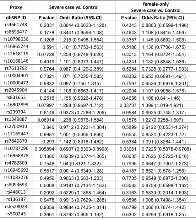
Among the 53 SNPs previously associated with curve progression among Caucasians [Ward, Ogilvie, Singleton et al. 2010], 25 were included in our genomic data (Table II). For the remaining 28 SNPs, we searched for proxy SNPs that were in high linkage disequilibrium (LD). Conceptually, LD is when an allele of one SNP is often observed with an allele of another SNP within a population. Thus, the allele of the one SNP is able to represent the allele of the other SNP. We queried the 28 SNPs using SNAP (www.broadinstiture.org/mpg/snap/), an online tool for SNP Annotation and Proxy, based on genotype data from the International HapMap Project and the 1000 Genomes Project [Johnson, Handsaker, Pulit et al. 2008]. We restricted our search to SNPs represented on the Illumina OmniChip 2.5M array, and used an r2>0.8 as a cutoff for a proxy in European ancestry population. In the genetic analysis, LD is reported in terms of D’ (D-prime) and r2 (r-square). Both are statistical measures of linkage disequilibrium scaled from 0 to 1. The case D’=1 is referred to as complete LD, indicating no recombination between the two SNPs within the population. The case r2=1 happens exclusively if 2 of the 4 possible haplotypes are present in the population and the two SNPs have the same allele frequencies, which is referred to as perfect LD. SNPs in perfect LD are necessarily in complete LD, but SNPs in complete LD may have low r2 value if the alleles at two loci are not correlated. These values are represented for each SNP in Table III. We found 27 SNPs in our genomic dataset that are in high LD with their relative SNPs in ScoliScoreTM. However, no SNP matched our query criteria to represent the rs16909285 SNP in ScoliScoreTM.
Using PLINK software, we evaluated the association of 52 SNPs with AIS in French-Canadian population by chi-square test. Considering that the 53 SNPs in the original study were associated among Caucasian female AIS patients only, we conducted our association analyses in all French-Canadian samples as well as in females only. For all SNPs, we evaluated associations among totals and among female case versus female control for: 1) presence of scoliosis versus controls; 2) severe scoliosis versus controls; and 3) severe scoliosis versus non-severe scoliosis.
For statistical significance, we used a conservative Bonferroni correction to adjust the p-value depicting probable association. We adjusted the probability of the false positive results from 0.05 to (0.05/k) where k is the number of SNPs tested in each independent association
29
test (k=52 in our study). Therefore, SNPs with a p-value <1×10⁻³ demonstrate significant association in statistics [Bush and Moore 2012].
Statistical power calculation
The pwr package in R software (http://www.statmethods.net/stats/power.html) [Champely and Champely 2007] was used in statistical power calculations for each association study while effect size was defined as small, medium and large, respectively, as outlined by Cohen [Cohen 1988].
Results
Disease-associated study
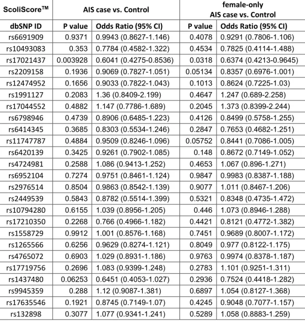
To evaluate whether the 52 SNPs are associated with the occurrence of spinal curvature, we compared the frequency of each SNP among all AIS patients to those of all controls. We applied the same comparison in female samples as well (545 cases vs. 476 controls). As shown in Table IV-V, none of the 52 SNPs were significantly associated (p-value<1×10⁻³) in either cohort.
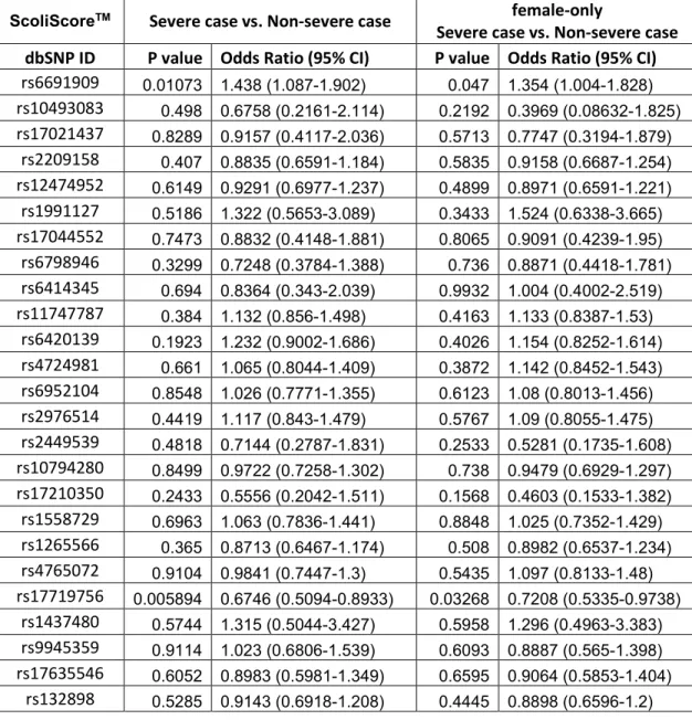
Progression-associated study
To detect the genetic association with AIS progression, we conducted two association analyses independently: one between 148 severe AIS patients and 302 non-severe patients, the other between 148 severe patients and 901 healthy controls, in both genders as well as in females. All the association analysis results are shown in Tables VI-IX. There is no association between the SNPs and severe AIS, in total samples or in female samples in French-Canadian population.
Statistic power analysis
Power calculations for each association study are listed in Table X. We concluded that the statistical power for each association analysis is strong enough for medium genetic effect.
30
Discussion
In this study, we first attempted to replicate the association between AIS and the 53 SNPs from an unpublished GWAS, and then we attempted to replicate the association of these SNPs to severe curvature. Although the genome chip used in our study contained only 25 of the SNPs published by Ward et al. [Ward, Ogilvie, Singleton et al. 2010], we identified 27 SNPs in high linkage disequilibrium with the remaining SNPs of the original study. One SNP was completely unavailable for us to study. We were unable to replicate any association between these 52 SNPs and AIS in our French-Canadian population.
This is the second study that has not replicated the association between AIS and the 53 SNPs used in the algorithm that the ScoliScoreTM test employs for its prediction of risk of curve progression. Recently, Y. Ogura et al. genotyped Japanese AIS patients, of which 600 individuals were divided into a progression group and 1114 individuals were divided into a non-progression group. With power greater than 80% in 24 out of 53 SNPs, no association with curve severity was found [Ogura, Takahashi, Kou et al. 2013]. However, it was possible that this lack of replication in the Japanese study population came from the ethnic admixture in Japanese and Caucasian cohorts between the two studies. Populations having distinct migration sources are likely to have a different disease penetrance due to varying degrees of genetic contributions, resulting in population stratification. Thus, our study sought to ascertain association of the SNPs in a Caucasian population of European descent, similar to that of the original study. Furthermore, an earlier study found no significant correlation in risk prediction of curve progression between ScoliScoreTM results and common clinical estimates in 83 Caucasians [Roye, Wright, Williams et al. 2012], emphasizing the need for replication of the original genetic association in a Caucasian cohort.
That the original GWAS that produced the association between AIS and the 53 SNPs has not been published brought hesitation and consideration about the scientific foundation of ScoliScoreTM [Dobbs and Gurnett 2011; Grant and Dormans 2011]. To evaluate the statistical power of the initial study, we lack important details in the study design such as: control cohort definition, quality control criteria for SNPs and subjects, quantity of testing markers and adjusted significance level. In light of this missing information and assuming that the original GWAS was a classical case/control design, we first tested the association of the 53 SNPs to
31
AIS, and then tested for association to curve severity. With 100% power to detect a moderate to strong genetic effect and 75% power to detect a minor effect, we did not find an association between the 52 tested SNPs and AIS. Using two approaches, we did not find an association between curve severity and the 52 SNPs. By our calculations, we had sufficient power to detect a large and moderate effect, although we had reduced power to detect a minor genetic effect. However, with a study sample composed of 450 patients classified into a severe group and a non-severe group, the latter test was similar in size to that in the ScoliScoreTM validation study.
Possible reasons for the irreproducibility of genetic associations lie in various factors that affect the statistical power in association studies [Hirschhorn, Lohmueller, Byrne et al. 2002; McClellan and King 2010; Sham and Purcell 2014]. One important determinant of statistical power in association studies is variable LD between studied markers and the true causal variants [Hirschhorn, Lohmueller, Byrne et al. 2002]. For the proxy SNPs that we used in our study that are not in perfect LD with the query SNPs of the original study, it is possible that recombination events among individuals caused disassociation. Sixteen out of 27 proxy SNPs were in absolute LD (r2=1, D’=1) with query SNPs. In 2 cases where D’<1, there was still chance in French-Canadian population that query SNPs have been separated from proxy SNPs by recombination events. Even no recombination happened in 9 cases (D’=1, r2<1), proxy SNPs could not substitute completely query ones as the allele frequencies at two loci were not exactly the same. Still with r2 ≥0.8, we had a quite small number of subjects carrying mismatched alleles from SNPs in LD [Wray 2005]. Therefore, it is quite reasonable to expect that the negative result of genetic association of SNPs in ScoliScoreTM with spinal curve progression in French-Canadian population was not entirely due to our employment of proxy SNPs. Importantly, there was no evidence that the 53 identified SNPs were causal variants in AIS progression. They might have a correlation with the causal variants because of linkage disequilibrium in the initial study. Therefore, an increased sample size was required in the replication study to reach the same level of statistical power in the initial study [Hirschhorn, Lohmueller, Byrne et al. 2002; Sham and Purcell 2014], which reduced the probability to reproduce the prior association results in our study.
Another important consideration for the discrepancy between our results and the original study is the criteria used to define the phenotype. Firstly, skeletal maturity was