REGULAR ARTICLE
Soluble GPVI is elevated in injured patients: shedding is mediated by
fibrin
activation of GPVI
Samantha J. Montague,1-3,* C ´eline Delierneux,4,* Christelle Lecut,5Nathalie Layios,4,6Robert J. Dinsdale,7,8Christine S.-M. Lee,2 Natalie S. Poulter,1,9Robert K. Andrews,10Peter Hampson,7,8Christopher M. Wearn,7,8Nathalie Maes,11Jonathan Bishop,12
Amy Bamford,7Chris Gardiner,13Woei Ming Lee,2,3Tariq Iqbal,14Naiem Moiemen,7Steve P. Watson,1,9C ´ecile Oury,4,* Paul Harrison,7,8,* and Elizabeth E. Gardiner2,*
1Institute of Cardiovascular Sciences, University of Birmingham, Birmingham, United Kingdom;2Australian Cancer Research Foundation Department of Cancer Biology and Therapeutics,
John Curtin School of Medical Research, and3Research School of Engineering, College of Engineering and Computer Science, The Australian National University, Canberra, ACT,
Australia;4Groupe Interdisciplinaire de G ´enoprot ´eomique Appliqu ´ee–Cardiovascular Sciences, University of Li`ege, Li`ege, Belgium;5Laboratory of Hematology and6Department of
General Intensive Care, University Hospital of Li `ege, Li `ege, Belgium;7Scar Free Foundation for Burns Research, Queen Elizabeth Hospital Birmingham, University Hospitals Birmingham
National Health Service (NHS) Foundation Trust, Birmingham, United Kingdom;8Institute of Inflammation and Ageing, University of Birmingham, Birmingham, United Kingdom;9Centre of
Membrane Proteins and Receptors, Universities of Birmingham and Nottingham, Midlands, United Kingdom;10Australian Centre for Blood Diseases, Monash University, Melbourne, VIC,
Australia;11Department of Biostatics and Medico-Economic Information, University Hospital of Li`ege, Li `ege, Belgium;12National Institute for Health Research Surgical Reconstruction
and Microbiology Centre (Trauma Research), University of Birmingham, Birmingham, United Kingdom;13Department of Haematology, University College London, London, United
Kingdom; and14Queen Elizabeth Hospital Birmingham, University Hospitals Birmingham NHS Foundation Trust, Birmingham, United Kingdom.
Key Points
• Soluble GPVI is ele-vated in patients with thermal injury with sep-sis, and sGPVI levels augment severity score prediction of mortality. • The GPVI ligand, fibrin,
induces GPVI shedding without requirement for platelet activation or signaling
Soluble glycoprotein VI (sGPVI) is shed from the platelet surface and is a marker of platelet activation in thrombotic conditions. We assessed sGPVI levels together with patient and clinical parameters in acute and chronic inflammatory conditions, including patients with thermal injury and inflammatory bowel disease and patients admitted to the intensive care unit (ICU) for elective cardiac surgery, trauma, acute brain injury, or prolonged ventilation. Plasma sGPVI was measured by enzyme-linked immunosorbent assay and was elevated on day 14 after thermal injury, and was higher in patients who developed sepsis. sGPVI levels were associated with sepsis, and the value for predicting sepsis was increased in combination with platelet count and Abbreviated Burn Severity Index. sGPVI levels positively correlated with levels of D-dimer (afibrin degradation product) in ICU patients and patients with thermal injury. sGPVI levels in ICU patients at admission were significantly associated with 28- and 90-day mortality
independent of platelet count. sGPVI levels in patients with thermal injury were associated with 28-day mortality at days 1, 14, and 21 when adjusting for platelet count. In both cohorts, sGPVI associations with mortality were stronger than D-dimer levels. Mechanistically, release of GPVI was triggered by exposure of platelets to polymerizedfibrin, but not by engagement of G protein-coupled receptors by thrombin, adenosine 59-diphosphate, or thromboxane mimetics. Enhancedfibrin production in these patients may therefore contribute to the observed elevated sGPVI levels. sGPVI is an important platelet-specific marker for platelet activation that predicts sepsis progression and mortality in injured patients.
Introduction
Beyond its primary role in hemostasis, fibrin also contributes to thrombotic and inflammatory conditions. Excessive fibrin formation occurs in acute and severe inflammation, including trauma,1-3 sepsis,4 disseminated intravascular coagulation (DIC),5and deep vein thrombosis.6Furthermore, fibrin deposition, Submitted 31 July 2017; accepted 13 December 2017. DOI 10.1182/
bloodadvances.2017011171.
*S.J.M., C.D., C.O., P.H., and E.E.G. contributed equally to this study.
The full-text version of this article contains a data supplement. © 2018 by The American Society of Hematology
ablated fibrinolysis, and hypofibrinogenemia are associated with multiple organ failure and mortality.7,8Together with thrombocytopenia, coagulation factor consumption, and acute coagulopathy,9,10 fibrin formation and platelet activation are potentially linked in inflammatory settings.
Glycoprotein (GP) VI is the major platelet signaling receptor for collagen and fibrin,11,12 and GPVI-fibrin engagement may under-pin fibrin-related disease pathology. GPVI is expressed only on megakaryocytes and platelets13in association with the Fc receptor g-chain containing an immunoreceptor tyrosine-based activation motif (ITAM).14 On resting platelets, GPVI is predominantly monomeric, but clusters and dimerizes on activation,15-17triggering ITAM signaling involving Src family kinases.17,18In hemostasis and thrombosis, GPVI supports platelet adhesion and aggrega-tion. GPVI is critical for the maintenance of vessel wall integrity in inflammation19 by inhibiting neutrophil-induced vascular damage.20,21
GPVI levels are stable on circulating platelets, but GPVI undergoes rapid metalloproteolytic cleavage, and in some cases internalization, on activation.22-25 GPVI shedding is induced by GPVI ligands including collagen, collagen-related peptide (CRP), and venom toxins,23,24,26,27 and by engagement of other platelet ITAM receptors FcgRIIA and CLEC-2.28,29 Elevated fluid shear stress and active factor X (FXa) also trigger GPVI shedding, quantified by detection of the soluble ectodomain fragment (sGPVI) in plasma by an enzyme-linked immunosorbent assay.30,31Plasma sGPVI reflects platelet activation in thrombotic conditions including microangiop-athy,32stroke,33DIC,33and Alzheimer’s disease34and rheumatoid arthritis.28,35 However, elevated plasma sGPVI levels in these patient groups is surprising, as only a fraction of platelets would be exposed to collagen, FXa, or elevated shear, and neither FcgRIIA or CLEC-2 plays major roles in hemostasis/thrombosis. The recent finding that fibrin activates GPVI provides a plausible explanation for this increase.
Here, we report elevated sGPVI in inflammatory patient cohorts including patients with injury, active inflammatory bowel disease (IBD), and sepsis. sGPVI correlated with D-dimer levels in both acute inflammatory patients admitted to an intensive care unit (ICU) and patients with thermal injury. sGPVI was associated with sepsis progression and mortality, and sGPVI levels enhanced APACHE (Acute Physiology and Chronic Health Evaluation) II score pre-diction of mortality. Furthermore, we show that fibrin induces GPVI shedding in vitro, possibly explaining sGPVI elevation in patients with acute injury.
Methods
ReagentsRefer to supplemental Material.
Blood collection
Healthy controls. Venous blood was collected from con-senting, healthy volunteers into sodium citrate (4%) or 3.2% trisodium citrate vacutainers (Becton Dickinson, Oxford, United Kingdom). Ethical approval was granted by Birmingham University Internal Ethical Review (ERN_11-0175).
ICU patients. Citrated blood was collected from 83 con-senting patients admitted to tertiary ICU who underwent cardiac
surgery, trauma, invasive ventilation for more than 48 hours, or acute brain injury at University Hospital of Li `ege, Li `ege, Belgium. (The experimental protocol was approved by the ethics committee of the University Hospital of Li `ege [Centre Hospitalier Universitaire; reference number B707201111981].). Sepsis diagnosis based on previous sepsis definitions.36
Thermal injury patients. Ninety-nine patients with injury afflicting up to 95% total body surface area (TBSA) were recruited to the Scientific Investigation of the Biological Pathways Following Thermal Injury Study (SIFTI: REC-12/EM/0432) at the Queen Elizabeth Hospital, Birmingham, United Kingdom.37,38 Citrated blood was collected at intervals after injury (day [D] 1, D3, D7, D14, D21, and D28 and month 2, 3, 6, and 12 postinjury). Sepsis diagnosis was made when more than 3 American Burn Association criteria39were met, plus positive bacterial culture and/or evidence of antibiotic response.37Platelet impedance counts were measured using a Beckman-Coulter UniCel DxH-800 (High Wycombe, United Kingdom) or a Sysmex XN-1000 Analyzer (Milton Keynes, United Kingdom).
Patients with IBD. Citrated blood was collected from 42 consented patients diagnosed with inactive or active Crohn’s disease and/or ulcerative colitis (UC; Ethics: REC:13/NE/0249). Patient details can be found in the supplemental Material.
Plasma preparation
Platelet-free plasma was isolated from blood by serial centrifu-gation: 2000g for 20 minutes and then 13 000g for 20 minutes at 4°C for samples from patients with thermal injury and two 15-minute 2500g centrifugations at 37°C for samples from other patient groups and healthy controls (HCs).
Washed platelet preparation and experiments
Washed platelets were prepared as described40 and washed twice with calcium-free Modified Tyrode’s buffer (134 mM NaCl, 0.34 mM Na2HPO4, 2.9 mM KCl, 12 mM NaHCO3, 20 mM
HEPES, 5 mM glucose, and 1 mM MgCl2 at pH 7.3) by
centrifugation at 1000g for 10 minutes and resuspended to 5003 109platelets/L. Washed platelets were preincubated for 5 minutes with 1 mM CaCl2and with/without inhibitors (2 mM GI254023, 25.7 mM GM6001, 10 mM PRT060318, or 10 mM dasatinib) before addition of 9 mM eptifibatide (aIIbb3 inhibitor). Platelet suspensions were stirred at 1200 rpm/37°C for 1 minute before agonist addition. For fibrin treatment, fibrinogen (100 mg/mL) was added 3 minutes before thrombin (1 U/mL) stimulation. Next, 10 mM Gly-Pro-Arg-Pro peptide (polymeriza-tion inhibitor) was added to fibrinogen to achieve monomeric fibrin conditions. Samples were stirred for 1 hour. Levels of intact and proteolyzed GPVI were assessed by western blot, using 1 mg/mL rabbit anti-human GPVI cytoplasmic tail antibody, detecting intact and cleaved GPVI.23 Densitometry measure-ments were made using Li-cor Image Studio software.
sGPVI Enzyme-Linked Immunosorbent Assay
sGPVI levels were measured by sandwich enzyme-linked immuno-sorbent assay,28,30and concentrations extrapolated from standard curves generated by serial dilution of GPVI ectodomain into 5% vol/vol GPVI-depleted plasma.28
D-dimer measurements
D-dimer levels were measured in plasma using the Innovance D-dimer immunoturbidimetric assay (Siemens Healthcare, Eschborn, Germany).
Statistical analysis
Results are reported as mean6 standard deviation, unless stated. D’Agostino-Pearson normality tests determined normality. Student t-tests were performed for normally distributed data; otherwise, Mann-Whitney U tests were performed. For multiple groups, Kruskal-Wallis tests with Dunn’s posttests were performed. Univariate and multivariate logistic regression was used to analyze variable association with 28- and 90-day mortality. Spearman’s rank correlation coefficients assessed associations between sGPVI levels and clinical parameters. Longitudinal analysis of sGPVI levels on sepsis, multiple-organ failure, and mortality was performed using linear mixed-effects models (supplemental Material). Statistical analyses were performed using GraphPad Prism (versions 5, 7), SPSS (IBM), and R (version 3.0.3).
Results
We assessed the utility of sGPVI as a biomarker of disease pathology and progression in patients with injury, sepsis, and inflammation by measuring sGPVI in samples from ICU patients, patients with thermal injury, and patients with IBD.
sGPVI levels correlate with clinical and platelet-specific parameters in ICU patients
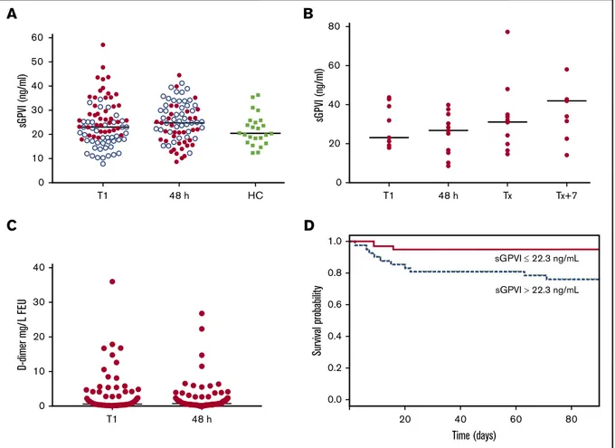
Plasma sGPVI levels were measured in 83 ICU patients with acute brain injury (n5 12), trauma (n 5 13), elective cardiac surgery (n 5 53), or prolonged ventilation (n5 5). At T1 (day of ICU admission), sGPVI levels were not significantly elevated above age-matched HCs or at 48 hours postadmission (Figure 1A), although wide ranges of sGPVI levels were observed at both points (5th-95th percentiles: T1, 11.1-43.5 ng/mL; 48 hours, 12.1-39.8 ng/mL). sGPVI at 48 hours increased in 42/83 patients (51%; Figure 1A). In the 15 ICU patients who developed sepsis, sGPVI levels were not elevated above T1 samples, both on day of sepsis diagnosis (Tx) and 7 days after (Tx17), suggesting no elevations with sepsis onset
A
60 50 40 30 20 10 0 sG PV I (ng/ml) T1 48 h HC 0 20 40 60 80 sG PV I (ng/ml) T1 48 h Tx Tx+7B
0 10 20 30 40 D-dimer mg/ L F EU T1 48 hC
1.0 0.8 0.6 0.4 0.2 0.0 Sur vival probabilit y 20 sGPVI 22.3 ng/mL sGPVI 22.3 ng/mL 40 60 Time (days) 80D
Figure 1.sGPVI is detectable in patients admitted to ICU and associated with patient mortality.(A) sGPVI levels were measured in plasma from patients admitted to ICU in samples obtained at day 1 (T1) and 48 hours after admission (n5 83) compared with HCs (n 5 24). Blue dots represent sGPVI levels in patients that increased after 48 hours. Median shown. (B) sGPVI levels measured in ICU patients who developed sepsis at T1, 48 hours after admission, Tx (day of sepsis diagnosis), and Tx17. (C) D-dimer levels of ICU patients at day 1 (T1) and 48 hours after admission to ICU. Levels below 0.5 mg/L FEU were considered normal, levels from 0.5 to 4.0 mg/L FEU moderately increased, and levels more than 5.0 mg/L FEU strongly increased.41(D) Association of sGPVI levels and patient mortality. Kaplan-Meier curve based on sGPVI
levels at T1. Solid line represents low sGPVI levels below the median (sGPVI low;#22.3 ng/mL), and the dotted line represents high sGPVI levels above the median (sGPVI high;.22.3 ng/mL).
or treatment phases of ICU patients (Figure 1B). sGPVI levels at T1 did not associate with sepsis occurrence, using a simple logistic regression model (P5 .085).
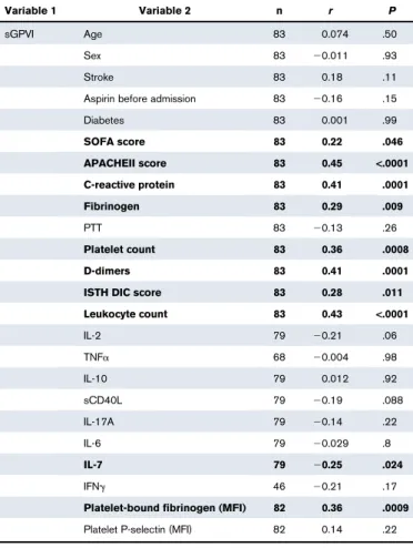
We compared sGPVI levels in ICU T1 patient samples against biological and clinical parameters (Table 1). As expected, positive correlations were seen with C-reactive protein, white blood cell count, and APACHEII and Sequential Organ Failure Assessment (SOFA) scores. Interestingly, platelet-related parameters, including platelet count and fibrin(ogen)-binding, as well as D-dimer and fibrinogen levels, also correlated significantly with sGPVI. After correction for platelet count, D-dimer levels and platelet fibrin(ogen)-binding remained significantly correlative at T1. D-dimer levels did not increase at 48 hours (Figure 1C). In some patients, D-dimer levels were elevated above 5 mg/L fibrinogen equivalent units (FEU), representing a strong increase.41
sGPVI levels in ICU patients are associated with mortality
sGPVI association with ICU patient mortality at T1 was assessed by simple logistic regression, using a sGPVI cutoff based on median levels found in healthy donors to stratify this cohort into low/normal
(#22.3 ng/mL) and elevated (.22.3 ng/mL) sGPVI groups. The elevated sGPVI group showed reduced survival probability (Figure 1D). Significant associations between T1 sGPVI with 28-and 90-day mortality were also observed (supplemental Table 4). This significant association remained when using multiple logistic regression alongside platelet count (P 5 .036 and P 5 .014). SOFA and APACHE(II) scores formed significant associations with mortality (supplemental Table 4); however, sGPVI levels no longer predicted mortality in patients with sepsis from this ICU cohort after correction for disease severity. Interestingly, D-dimer levels and platelet count formed no significant associations with 28- and 90-day mortality at T1 (P5 .059 and .058 and P 5 .220 and .077, respectively; supplemental Table 4).
sGPVI is raised in thermal injury patients with sepsis
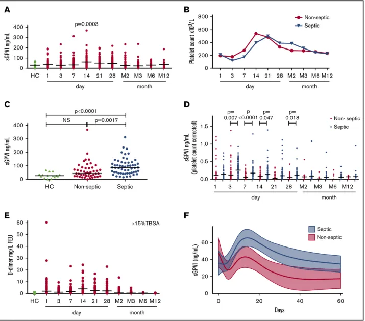
Although the range of sGPVI levels in HCs is tightly maintained, sGPVI in longitudinal samples from 99 patients with thermal injury with burns afflicting up to 95% TBSA burn (mean, 24%; IQR, 8%-38%) showed a broad distribution (Figure 2A). sGPVI levels increased from D1 postinjury, with a significant peak observed at around D14 before slowly returning to HC levels by month 2 postinjury (Figure 2A).
sGPVI levels weakly correlated with platelet count in this cohort (Spearman’s rank coefficient r 5 0.119; P 5 .015). Consistent with other reports of significant platelet count reductions at D3 followed by rebound thrombocytosis at D14 postinjury,42 here platelet counts within septic and nonseptic patients reached a nadir at D3, followed by rebound thrombocytosis peaking at D14 for nonseptic patients and D21 for septic patients (Figure 2B).
A 58% incidence of sepsis occurred in this cohort, with onset between D4 and D8 (median, 5.5 days).37 Septic patients had higher peak sGPVI levels compared with nonseptic patients and HCs (Figure 2C). After normalization for platelet count, sGPVI was significantly elevated in septic patients compared with nonseptic patients at D3, D7, D14, and D28 (Figure 2D), coincident with sepsis onset and progression.
D-dimer levels in patients with thermal injury with burns of 15% TBSA or more (n5 61) increased from D1 postthermal injury to a peak at D14, gradually normalizing during M3 (Figure 2E), echoing sGPVI levels (Figure 2A). Significant positive correlation with D-dimer and sGPVI levels were observed when assessing samples from all points (r 5 0.35; Table 2). Alongside this, significant positive correlations were seen at D14, D28, and month 2, with D14 giving the strongest correlation (r5 0.46; Table 2, sGPVI). D-dimer and sGPVI correlations also strengthened after correction for platelet count (Table 2, normalized sGPVI).
sGPVI levels in patients with thermal injury are associated with sepsis and mortality
sGPVI associations with sepsis, multiple-organ failure, and mortality were assessed in patients with thermal injury. x2statistical analysis demonstrated significant associations with sGPVI and the pro-portion of patients developing sepsis and patient outcome (P, .001), with overrepresentation of patients bearing high sGPVI developing sepsis across all times (supplemental Table 5). Signif-icant sGPVI associations with sepsis patient outcome at D7 and D14 postinjury were observed when examining set times (P5 .023 and .043, respectively). Interestingly, sGPVI association with sepsis
Table 1.sGPVI correlations with clinical and biological parameters of ICU patients
Variable 1 Variable 2 n r P
sGPVI Age 83 0.074 .50
Sex 83 20.011 .93
Stroke 83 0.18 .11
Aspirin before admission 83 20.16 .15
Diabetes 83 0.001 .99 SOFA score 83 0.22 .046 APACHEII score 83 0.45 <.0001 C-reactive protein 83 0.41 .0001 Fibrinogen 83 0.29 .009 PTT 83 20.13 .26 Platelet count 83 0.36 .0008 D-dimers 83 0.41 .0001
ISTH DIC score 83 0.28 .011
Leukocyte count 83 0.43 <.0001 IL-2 79 20.21 .06 TNFa 68 20.004 .98 IL-10 79 0.012 .92 sCD40L 79 20.19 .088 IL-17A 79 20.14 .22 IL-6 79 20.029 .8 IL-7 79 20.25 .024 IFNg 46 20.21 .17
Platelet-bound fibrinogen (MFI) 82 0.36 .0009
Platelet P-selectin (MFI) 82 0.14 .22
Spearman’s correlation coefficients, represented as r values, between sGPVI levels and
clinical/biological parameters measured from samples taken on day 1 (T1) on admission to
ICU. Significance observed when P, .05. Bold indicates significant correlation.
at D7 and D14 was strengthened when corrected for platelet count (supplemental Table 5).
Longitudinal statistical prediction models were used to evaluate sGPVI as a predictive marker of sepsis (Figure 2F). The sGPVI discriminatory value for predicting sepsis in patients with thermal injury, represented as an area under the receiver operating characteristic (AUROC) curve value, was determined using a linear mixed-effects model of sGPVI and time and identified that D14 provided the strongest predictive value for sepsis (AUROC, 0.73) and multiple-organ failure (AUROC, 0.77; supplemental Table 8).
Multivariate analysis of sGPVI with platelet count strengthened the sepsis predictive value at D3, D7, and D14 (P 5 .75-.78; supplemental Table 9).
Logistic regression was performed to assess sGPVI associations with mortality in the patients with thermal injury with 15% or more TBSA burns and compared with D-dimer levels. Significant associations of sGPVI levels at D1 and 28-day mortality were observed (P , .05; supplemental Table 6). After correcting for platelet count, D1 sGPVI mortality association was not significant, but it was at D14 and D21 (supplemental Table 7). Interestingly,
HC 1 3 7 14 21 28 M2 M3 M6 M12 0 200 300 100 400
A
B
C
D
E
F
p=0.0003 day sG PV I ng/mL month 0 200 400 600 800 Non-septic Septic Platelet count x1 0 9/L day month 1 3 7 14 21 28 M2 M3 M6 M12 0 100 200 300 400 HC Non-septic p0.0001 NS p=0.0017 Septic sG PV I ng/mL 0.0 0.5 1.0 1.5 Non- septic Septic p= 0.007 p 0.00010.047p= p= 0.018 sG PV I ng/mL(platelet count corrected)
day month 1 3 7 14 21 28 M2 M3 M6 M12 0 20 40 60 Non-septic Septic sG PV I (ng/mL) Days 0 20 40 60 HC 0 10 20 30 40 50 60 15%TBSA 1 3 7 day month D-dimer mg/ L F EU 14 21 28 M2 M3 M6 M12
Figure 2.sGPVI is detectable in patients with thermal injury and raised in patients who develop sepsis.(A) sGPVI levels of patients with thermal injury (n5 99) during the full time course, from day 1 to month 12 (M12) compared with HCs (n5 15). Mann-Whitney U test was performed to compare each patient time to HC (median shown). (B) Platelet counts of patients with thermal injury split into nonseptic and septic groups, measured using the Sysmex XN-1000-Hematology Analyzer, during the full-time course (median shown). (C) Peak sGPVI levels in patients with thermal injury of septic and nonseptic patients and HCs. Patients with thermal injury who developed sepsis are defined as meeting at least 3 of the American Burn Association sepsis criteria and a positive culture. Mann-Whitney U test was performed to compare sGPVI levels of septic patients compared with nonseptic burn patients and HCs (median shown). (D) Comparisons of sGPVI levels after platelet count normalization in patients with thermal injury who developed sepsis compared with nonseptic patients during the time course. Mann-Whitney U test was performed to compare sGPVI levels of septic patients and nonseptic burns patients (median shown). (E) D-dimer levels of patients with thermal injury higher than 15% TBSA (n5 61) over time compared with HCs (n 5 12). (F) Longitudinal analysis of sGPVI as a moderate predictive marker of sepsis progression in patients with thermal injury. A linear mixed-effects model examining the relationship between sGPVI and time according to sepsis status. Line represents mean predicted effects, and shaded area represents 95% confidence interval.
D-dimer association with 28-day mortality was not significant (supple-mental Table 7). D7, D14, and D28 samples postinjury gave the best sGPVI discriminatory predictive values for predicting mortality at 1 year (represented as AUROC, 0.68-0.75; supplemental Table 8). Mortality predictive values of sGPVI were also improved when adjusted for platelet count (supplemental Table 7). Multivariate analysis of sGPVI with platelet count improved the predictive value at D3, D7, and D14 for predicting mortality (P5 .71-.83; supplemental Table 9). sGPVI and ABSI values on D1 enhanced or maintained predictive power for sepsis (AUROC, 0.80; compared to AUROC, 0.77 for ABSI alone) and mortality (AUROC, 0.80; unchanged from ABSI alone). sGPVI and APACHE(II) together demonstrated improved prediction of mortality from D3 to D28 (supplemental Table 10). Together, these data suggest sGPVI could be a useful addition to other clinical parameters for predicting sepsis and mortality.
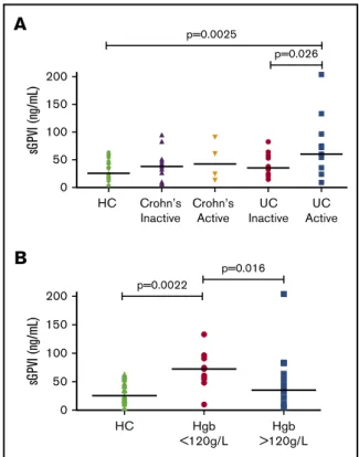
sGPVI is elevated in patients with IBD with active UC
We assessed plasma sGPVI levels in 42 patients with IBD to compare sGPVI levels in patients with chronic inflammation. sGPVI was elevated in patients with active UC compared with patients with inactive UC and HCs, and was distributed more broadly, likely because of disparate levels of inflammation in this cohort (Figure 3A). Plasma sGPVI levels in active and inactive Crohn’s disease were not significantly elevated above HC values, possibly indicating a reduced inflammatory status compared with patients with active UC. There was no significant correlation between sGPVI and platelet count (supplemental Table 11) in these patient groups. Iron deficiency anemia is commonly associated with IBD. We investigated whether there was a link between platelet activation, IBD inflammatory status observed, and iron deficiency. When stratifying patients with IBD on the basis of hemoglobin levels, an indicator of iron deficiency, patients with low hemoglobin (,120 g/L) showed highly elevated sGPVI levels compared with patients with hemoglobin levels above 120 g/L and HCs (Figure 3B). Hemoglobin levels
negatively correlated with plasma sGPVI levels (supplemental Table 11). Furthermore, ferritin levels, another indicator of iron deficiency, negatively correlated with sGPVI in patients with active IBD with C-reactive protein levels greater than 5 mg/mL.
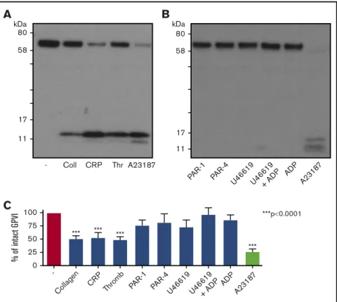
Exposure of platelets to fibrin, but not GPCR stimulation, induces GPVI shedding
Engagement of GPVI by collagen, CRP, or convulxin leads to ITAM-signaling dependent metalloproteolytic release of a 55-kDa sGPVI fragment, leaving a 10- to 15-kDa membrane-bound remnant.23,24,26,27,43To ascertain whether fibrin could induce GPVI shedding, we first assessed the effect of thrombin or other GPCR ligands on GPVI levels. GPVI shedding was calculated as a percentage of GPVI detected in unstimulated samples compared with GPVI levels after stimulation. Figure 4A shows that 1-hour treatment of washed platelets with thrombin induced a loss of full-length GPVI that was comparable to shedding achieved by collagen or CRP and similar loss to the potent GPVI shedder, A23187 (Figure 4C). These treatments induced the appearance of the 10- to 15-kDa remnant in;50% of donors. In contrast, treatment with adenosine 59-diphosphate (ADP), the PAR-1 or PAR-4 peptide agonists, or thromboxane mimetic U46619 did not induce loss of intact GPVI (Figure 4B-C).
As engagement of PAR-1 or PAR-4 via activating peptides did not induce GPVI cleavage, and GPVI does not carry a recognized
Table 2.Correlations of sGPVI with D-dimers in patients with thermal injury
Time
sGPVI Normalized sGPVI
n r P n r P HC 12 0.03 .943 — — — D1 49 0.17 .242 40 0.297 .062 D3 75 20.15 .190 64 20.315 .011* D7 35 0.04 .808 30 0.127 .503 D14 44 0.46 .002** 40 0.643 <.001*** D21 42 0.32 .042 33 0.438 .011* D28 35 0.39 .021* 29 0.540 .003** M2 33 0.44 .011* 23 0.250 .250 M3 20 0.28 .239 11 0.041 .908 M6 18 0.35 .188 8 0.333 .428 M12 12 0.27 .387 7 0.714 .088 All times 341 0.349 <.001*** 286 0.155 .009**
Spearman’s correlation coefficients, represented as r values, between sGPVI levels and normalized sGPVI corrected for platelet count and D-dimer levels in patients with thermal injury at different points at injury. Bold indicates significant correlation.
*P, .05. **P, .01. ***P, .005. 200 150 100 50 0 sG PV I (ng/mL) HC
A
p=0.0025 p=0.026 Crohn’s Inactive Crohn’s Active UC Inactive UC Active 200 150 100 50 0 sG PV I (ng/mL)B
p=0.0022 p=0.016 HC Hgb <120g/L Hgb >120g/LFigure 3.sGPVI is detectable in patients with IBD.(A) sGPVI levels of patients with IBD with inactive and active Crohn’s disease (n 5 13 and n 5 4) and inactive and active UC (n5 12 and n 5 13) compared with HC (n 5 20). Mann-Whitney U test was performed to compare sGPVI levels of different IBD patient groups and HC (median shown). (B) sGPVI levels of patients with IBD with hemoglobin (Hgb) levels of above or below 120 g/L (n5 28, n 5 13, respectively) compared with HC (n5 20). A Kruskal-Wallis statistical test with Dunn’s multiple comparisons was performed to compare the 3 groups of patients (median shown).
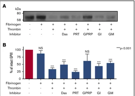
thrombin-cleavage site, we considered whether GPVI shedding was secondary to GPVI/fibrin engagement. Platelets were treated with thrombin in the presence of fibrinogen to produce fibrin. This treatment triggered loss of intact GPVI (Figure 5A) and was comparable to A23187-induced shedding. Fibrinogen alone did not induce GPVI shedding (Figure 5A), and shedding required fibrin polymerization as inclusion of GPRP ablated GPVI proteolysis (Figure 5A-B), demonstrating that fibrin polymers were more effective than monomers at inducing shedding.
To assess whether fibrin-induced GPVI shedding required ITAM signaling, platelet suspensions were pretreated with maximally effective concentrations of Src and Syk inhibitors, dasatinib, and PRT060318. GPVI shedding induced by thrombin-fibrinogen in the presence of either inhibitor was not different to fibrin alone (Figure 5), indicating that an active ITAM signaling pathway was not crucial for fibrin-induced GPVI shedding. The role of multiple metalloproteinases in fibrin-induced GPVI shedding was de-termined by inclusion of GM6001, a broad metalloproteinase inhibitor, or GI254023, an inhibitor of A Disintegrin and Metal-loproteinase (ADAM) 10, to platelet suspensions, with both having minimal effect on reducing fibrin-mediated shedding (Figure 5B).
Discussion
The incidence, management, and knowledge of pathophysiological processes relating to sepsis have improved during the last 20 years; however, sepsis remains a significant public health problem across the world, with more than 31 million cases presenting annually and sepsis-related fatality occurring in 1 in 5 cases.44,45Biomarkers with high sensitivity and specificity that rapidly and accurately differen-tiate sepsis from noninfectious conditions including systemic inflammatory response syndrome are therefore in demand to help implement the correct therapeutic regime. Diagnosis of sepsis is a subjective clinical judgment focused on assessing the roots of sepsis, using tools such as the SOFA or APACHE scoring systems,46-48 with both systems including evaluation of patient platelet count. Blood rheology and platelet function are pro-gressively and severely altered in patients with severe sepsis.49,50 Evidence suggests a prominent role for inappropriate platelet activation and aggregation during sepsis,51 and platelet indices beyond platelet count are useful to evaluate illness severity and prospectively identify critically ill patients.52,53In this regard, sGPVI represents an excellent candidate marker of pathological platelet activation in the setting of sepsis, as it is a platelet/megakaryocyte-specific membrane protein that is stable on circulating platelets but
-Collagen CR
P
Thromb PAR-1 PAR-4U46619
C
***p0.0001 % of int act G PV I 100 75 50 25 0 U46619+ AD P ADP A2318 7 *** *** *** *** -11 17 58 80 kDaA
Coll CRP Thr A23187 11 17 58 80 kDaB
PAR-1 PAR-4 U4 6619 U4 6619 + AD P ADP A2318 7Figure 4.Stimulation of platelets with GPCR agonists do not induce GPVI shedding.(A) Western blot for GPVI after platelet stimulation by GPVI agonists, thrombin, or A23187. Washed platelets (5003 109/L) were stimulated with collagen (30 mg/mL), CRP (30 mg/mL), thrombin (1 U/mL), and calcium ionophore (A23187: 10 mM), a
positive control for GPVI shedding, in suspension under stirring conditions for 1 h at 37°C, in the presence of eptifibatide (9 mM) and CaCl2(1 mM). Representative figure of
data from at least 12 individual donors. Membranes were blotted with an anti-GPVI antibody for GPVI (60-65 kDa) and the GPVI remnant band (10-17 kDa) observed after shedding. (B) Western blot for GPVI after platelet stimulation with GPCR agonists, PAR-1 peptide (SFLLRN: 100 mM), PAR-4 peptide (AYPGKF: 100 mM), U46619 (10 mM), U46619 (10 mM)1ADP (10 mM), and ADP (10 mM) in the presence of eptifibatide (9 mM) and CaCl2(1 mM) under the same conditions as before. Membranes were blotted
with an anti-GPVI antibody for GPVI, as stated earlier. (C) Quantitation analysis of GPVI shedding after platelet stimulation with various GPVI and GPCR agonists. GPVI shedding represented as percentage of intact GPVI remaining compared with unstimulated GPVI levels. Results are shown as mean6 standard error of the mean. A 1-way ANOVA with Tukey’s posttest was performed to compare shedding with unstimulated platelets. n 5 81 donors.
released on ligand engagement of ITAM receptors, exposure to FXa, or abnormal fluid shear rates.54sGPVI levels are not elevated as a consequence of ablated platelet production.55
We have investigated the utility of sGPVI as a marker of platelet activation in these pathological settings and explored the value of sGPVI to aid diagnosis and predict patient outcomes in conjunction with standard clinical parameters, including platelet count and injury severity scores. We have also investigated mechanisms that may drive release of sGPVI in trauma/inflammation patients. Elevated sGPVI levels were detected in the plasma of patients with thermal injury and patients with IBD with active disease. Although there were no significant sGPVI elevations in patients with ICU, we found significant correlations with sGPVI levels and other biological parameters including ISTH DIC score, C-reactive protein, and injury severity scores (APACHEII, SOFA). sGPVI levels no longer predicted mortality in ICU sepsis patients after correction for disease severity.
sGPVI was not elevated during the acute thermal injury phase but became significantly elevated with sepsis development. Dissemi-nated intravascular coagulation is commonly observed in septic patients. Platelets are activated and form fibrin-rich thrombi, leading to further platelet activation. Fibrin accumulates during acute inflammation and tissue injury and underlies sepsis pathology.4,5 D-dimers, a fibrin byproduct of coagulation and clot resolution produced by the action of plasmin, are an indirect index of fibrin formed in these patients. D-dimer levels correlated with sGPVI levels in ICU patients and patients with thermal injury, even when correcting for platelet count, suggesting a potential link between
sGPVI and fibrin in multiple patient cohorts. In support of this, sGPVI levels significantly correlated with platelet-bound fibrin(ogen) in samples from ICU patients, indicating both fibrin formation and platelet activation. Synergy between D-dimer and sGPVI levels in both ICU and thermal injury patient cohorts implies a role for fibrin in GPVI shedding observed in these patients.
Spontaneous aggregation of platelets and hyperfibrinogenemia are enhanced in the acute period (,48 hours) after thermal trauma.56 We found that sGPVI levels were significantly elevated in patients with burn injuries at D14 postinjury. Low sGPVI levels were observed at earlier points, suggesting that only a fraction of platelets were activated by the injury and related collagen exposure, or that low sGPVI levels reflected low platelet counts. When sGPVI levels were adjusted for platelet count, there was significant elevation at D3 postinjury in septic patients and sGPVI remained elevated at D7 and D14. As minimal collagen exposure is expected at these times, an alternative mechanism for platelet activation and GPVI shedding is likely.
We observed associations between sepsis and high sGPVI levels in patients with thermal injury at all points when stratifying sGPVI levels into low (#12.7 ng/mL), medium (.12.7 to #37.7 ng/mL), and high (.37.7 ng/mL) levels. These associations were improved when sGPVI levels were corrected for platelet count. Significant associations with sGPVI and sepsis were observed at D7, potentially correlating to sepsis onset. A longitudinal statistical prediction of sGPVI levels in septic and nonseptic patients over time indicated sGPVI having moderate value for predicting sepsis at early points with a score of 0.68 at D7 and 0.73 at D14, which improved
B
0 25 50 75 100 - + NS NS *** *** *** *** *** ***p0.001 % of int act G PV I + + + + + + - - + + + + + + - - - Das PRT GPRP GI GM Fibrinogen Thrombin InhibitorA
58 80 kDa Fibrinogen - + + + + + + + - - + + + + + + - - - Das PRT GPRP GI GM Thrombin InhibitorFigure 5.Fibrin stimulation of platelets induces GPVI shedding.(A) Western blot for GPVI after stimulation of platelets in suspension by fibrinogen, fibrin, or fibrin in the presence of inhibitors. Washed platelets (5003 109/L) were stimulated with fibrinogen (100 mg/mL) alone, thrombin (1 U/mL) in the presence of fibrinogen (polymerized fibrin), fibrin in the presence of Src and Syk inhibitors (dasatinib, 10 mM; PRT-060318, 10 mM), fibrin in the presence of GI254023 (2 mM, ADAM10 inhibitor) and GM6001 (25.7 mM, broad matrix metalloproteinase inhibitor), and monomeric fibrin in the presence of GPRP (10 mM) under stirring conditions for 1 h at 37°C in the presence of eptifibatide (9 mM) and CaCl2(1 mM). GPRP was added with fibrinogen 3 minutes before thrombin stimulation to prevent fibrin polymerization. GI254023 and GM6001 were
added 5 minutes before fibrinogen and thrombin stimulation. Membranes were blotted for GPVI and GPVI remnant after shedding. (B) Quantitation analysis of GPVI shedding induced by different forms of fibrin and fibrin-induced shedding in the presence of inhibitors. Percentage of GPVI represents levels calculated as a percentage of GPVI detected in unstimulated samples compared with GPVI levels after stimulation. Results are shown as mean6 SEM. A 1-way ANOVA was performed, with Tukey’s posttest, to compare fibrin-induced shedding in the presence of GPRP and the presence of inhibitors and fibrinogen alone to unstimulated samples. N.S., nonsignificant; n5 7 donors.
when correcting for platelet count (0.76 and 0.80, respectively). The discriminatory power of sGPVI for predicting sepsis was also improved after multivariate analysis, where sGPVI and platelet count were combined (0.76 and 0.78, respectively). Furthermore, the discriminatory power for sepsis prediction at D1 improved when sGPVI was combined with ABSI score, or at D3 with APACHE(II). sGPVI discriminatory power may further increase if used in combination with other strong sepsis predictive markers such as cell-free DNA and immature granulocytes.37,38 This inflammatory trauma cohort presumably contained an abundance of healthy people who were essentially well and unencumbered before receiving their injury. Measurement of sGPVI on day 1 could potentially enable clinicians to stratify patients based on sepsis risk and aid clinical management of patients.
A longitudinal statistical prediction of sGPVI levels over time with all patients with thermal injury gave a moderate value for mortality at D14 and strengthened when corrected for platelet count or used in combination with ABSI. In both cohorts, sGPVI associations with mortality exceeded D-dimer associations with mortality. Together, these data provide rationale for the combined analysis of sGPVI levels in combination with other markers of sepsis onset and injury severity scores and suggests sGPVI is a useful marker of platelet activation and indicator for mortality risk and sepsis progression. In the chronic IBD inflammatory cohort, sGPVI levels were elevated in patients with active UC and correlated inversely with ferritin and hemoglobin levels. Iron deficiency anemia is commonly associated with IBD, with around 17% of patients with IBD having iron deficiency anemia, increasing in prevalence to around 60% when studying hospitalized patients.57,58Although the molecular basis is unclear, there are numerous links between iron deficiency and platelet activation.59 sGPVI levels may reflect platelet activation in iron-deficient patients with IBD, which could also aid clinical management of these patients. Fibrin was recently reported to activate platelets via GPVI, with the interaction contributing to thrombus stabilization.11,12 Here we demonstrate for the first time that fibrin-exposed platelets shed GPVI to an extent comparable to GPVI ligands. GPVI shedding was not a result of PAR engagement, as neither PAR-1- nor PAR-4-activating peptides induced GPVI shedding. Treatment with ADP and U46619 also failed to induce shedding, suggesting activation of platelets via GPCRs is not sufficient to induce GPVI shedding. Thrombin treatment variably stimulated GPVI shedding between donors, consistent with other studies,23,26which may depend on sufficient fibrinogen storage and release from platelets and amount of fibrin formed. GPVI lacks a recognized cleavage site for thrombin, arguing against direct GPVI cleavage by the protease.
Fibrin generation is a potential consequence of disease for most patient cohorts studied here, and as fibrin engages GPVI,11,12we assessed whether fibrin stimulation induced GPVI shedding. Thrombin stimulation in the presence of fibrinogen consistently induced GPVI shedding, independent of fibrinogen interaction with integrin aIIbb3, as shedding occurred in the presence of eptifibatide. ADAM10 plays a major role in GPVI shedding, with ADAM17 contributing, at least in mice.60,61Fibrin-induced shedding was reduced but not blocked by GM6001 or by ADAM10-specific inhibitor GI254023, suggesting roles for multiple sheddases. Unlike collagen-induced shedding,26fibrin-induced GPVI shedding did not require active ITAM signaling. Exposure to elevated shear or FXa induces shedding that is also independent of platelet activation.62,63Fibrin binds to multiple proteins on the platelet surface; 1 or more of these interactions may
contribute to GPVI shedding, possibly by clustering and juxtaposing GPVI to ADAM10 and/or other metalloproteinases. Further, soluble fibrin can also reduce GPVI ligand-induced function in platelets, presumably by blockade or steric hindrance of the ligand binding site, as the effect was not blocked by GM6001.64
Monomeric fibrin, the predominant species in the presence of GPRP, induced only minimal GPVI shedding, as determined by the inhibitory effect of GPRP. This is consistent with fibrin-mediated activation of GPVI in platelet suspensions requiring fibrin polymerization,11,12 although monomeric fibrin and D-dimer can activate GPVI when immobilized.11,65 Targeting the conformational state of fibrin, or blocking the fibrin-GPVI interaction,65may permit modulation of fibrin-mediated GPVI shedding and GPVI responsiveness.64
In conclusion, sGPVI and D-dimer levels fluctuate coincidentally in injured patient cohorts where there is ongoing coagulopathy. We have shown that sGPVI levels are predictive of patient outcomes and enhance the predictive power of the APACHE and ABSI clinical evaluation tools in a thermal injury cohort. The prognostic value of measuring sGPVI should therefore be prospectively evaluated in large cohorts of patients in whom sepsis is an expected outcome. Our data support a role for fibrin-inducing GPVI shedding, potentially explaining elevated sGPVI levels where there is minimal exposure to collagen. Plasma sGPVI strongly associates with onset of sepsis and with mortality, supporting the relevance of sGPVI as a clinical platelet activation marker.
Acknowledgments
The authors thank the nursing team at the Birmingham Burns Centre and staff at the gastrointestinal medicine department at the Queen Elizabeth Hospital (Birmingham, United Kingdom) for their assistance with patient recruitment and sample collection. The authors would also like to thank Julie Rayes for helpful discussions.
The research was part funded by the National Institute for Health Research (NIHR) Surgical Reconstruction and Microbiology Re-search Centre. The authors also thank the Scar Free Foundation and the British Heart Foundation for funding. S.P.W. holds a BHF Chair (CH/03/003/15571). This project was initiated during a sabbatical visit by E.E.G. supported by the Institute for Advanced Studies at the University of Birmingham. The project was also supported by funding from National Health and Medical Research Council Australia and the Fonds National pour la Recherche Scientifique Belgium (FNRS-FRSM 3.4611.11, CDR J.0043.13) and by the French Community of Belgium (FSRC-12/13, ARC-SF 12/14-05). C.L. was a postdoctoral researcher at the FNRS. C.D. was supported by a“Fond pour la recherche industrielle et agricole” fellowship. C.O. is a senior research associate at the Fonds National pour la Recherche Scientifique.
S.J.M. completed this study as part of her candidature in the laboratory of S.P.W.
The views expressed are those of the authors and not necessarily those of the National Health Service, the NIHR, or the Department of Health.
Authorship
Contribution: S.J.M designed and performed research; col-lected, analyzed, and interpreted data; made the figures; and wrote the manuscript. R.J.D. and C.D. collected, analyzed, and interpreted data and processed samples. C.S.-M.L., J.B., and N.M., analyzed and interpreted data. C.L. N.L., P. Hampson, C.M.W., A.B.,
and C.G. collected data and processed patient samples. N.S.P. and R.K.A. interpreted data and supplied reagents. N.M. and W.M.L. inter-preted data. T.I. designed research and interinter-preted data. P. Harrison, S.P.W., and C.O. designed research, interpreted data, and wrote the manuscript. E.E.G designed, analyzed, and interpreted data; supplied reagents; and wrote the manuscript.
Conflict-of-interest disclosure: The authors declare no compet-ing financial interests.
ORCID profiles: S.J.M., 0002-8934-3901; C.D., 0000-0003-2638-0104; C.S.-M.L., 0000-0002-3851-9364; N.S.P., 0002-3187-2130; J.B., 0003-1789-5886; A.B., 0002-8887-484X; C.G., 0002-2318-0062; S.P.W., 0000-0002-7846-7423; C.O., 0000-0002-7561-0132; P. Harrison, 0000-0003-4610-8909; E.E.G., 0000-0001-9453-9688.
Correspondence: Elizabeth E. Gardiner, ACRF Department of Cancer Biology and Therapeutics, John Curtin School of Medical Research, The Australian National University, 131 Garran Rd, Can-berra, ACT 2601, Australia; e-mail: elizabeth.gardiner@anu.edu.au; and Paul Harrison, Institute of Inflammation and Ageing, College of Medical and Dental Sciences, University of Birmingham, Edgbaston, Birmingham B15 2TT, United Kingdom; e-mail: p.harrison.1@bham. ac.uk.
References
1. Hayakawa M, Sawamura A, Gando S, et al. Disseminated intravascular coagulation at an early phase of trauma is associated with consumption coagulopathy and excessive fibrinolysis both by plasmin and neutrophil elastase. Surgery. 2011;149(2):221-230.
2. Dunbar NM, Chandler WL. Thrombin generation in trauma patients. Transfusion. 2009;49(12):2652-2660.
3. Frith D, Goslings JC, Gaarder C, et al. Definition and drivers of acute traumatic coagulopathy: clinical and experimental investigations. J Thromb Haemost. 2010;8(9):1919-1925.
4. Levi M, van der Poll T. Coagulation and sepsis. Thromb Res. 2017;149:38-44.
5. Semeraro N, Ammollo CT, Semeraro F, Colucci M. Sepsis-associated disseminated intravascular coagulation and thromboembolic disease. Mediterr J Hematol Infect Dis. 2010;2(3):e2010024.
6. Davis RP, Miller-Dorey S, Jenne CN. Platelets and coagulation in infection. Clin Transl Immunology. 2016;5(7):e89.
7. Mitra B, Cameron PA, Mori A, Fitzgerald M. Acute coagulopathy and early deaths post major trauma. Injury. 2012;43(1):22-25. 8. Winearls J, Campbell D, Hurn C, et al. Fibrinogen in traumatic haemorrhage: A narrative review. Injury. 2017;48(2):230-242. 9. Brohi K, Singh J, Heron M, Coats T. Acute traumatic coagulopathy. J Trauma. 2003;54(6):1127-1130.
10. Hayakawa M. Pathophysiology of trauma-induced coagulopathy: disseminated intravascular coagulation with the fibrinolytic phenotype. J Intensive Care. 2017;5(14):14.
11. Alshehri OM, Hughes CE, Montague S, et al. Fibrin activates GPVI in human and mouse platelets. Blood. 2015;126(13):1601-1608.
12. Mammadova-Bach E, Ollivier V, Loyau S, et al. Platelet glycoprotein VI binds to polymerized fibrin and promotes thrombin generation. Blood. 2015; 126(5):683-691.
13. Clemetson JM, Polgar J, Magnenat E, Wells TN, Clemetson KJ. The platelet collagen receptor glycoprotein VI is a member of the immunoglobulin superfamily closely related to FcalphaR and the natural killer receptors. J Biol Chem. 1999;274(41):29019-29024.
14. Gibbins JM, Okuma M, Farndale R, Barnes M, Watson SP. Glycoprotein VI is the collagen receptor in platelets which underlies tyrosine phosphorylation of the Fc receptor g-chain. FEBS Lett. 1997;413(2):255-259.
15. Jung SM, Moroi M, Soejima K, et al. Constitutive dimerization of glycoprotein VI (GPVI) in resting platelets is essential for binding to collagen and activation in flowing blood. J Biol Chem. 2012;287(35):30000-30013.
16. Loyau S, Dumont B, Ollivier V, et al. Platelet glycoprotein VI dimerization, an active process inducing receptor competence, is an indicator of platelet reactivity. Arterioscler Thromb Vasc Biol. 2012;32(3):778-785.
17. Poulter NS, Pollitt AY, Owen DM, et al. Clustering of glycoprotein VI (GPVI) dimers upon adhesion to collagen as a mechanism to regulate GPVI signaling in platelets. J Thromb Haemost. 2017;15(3):549-564.
18. S ´everin S, Nash CA, Mori J, et al. Distinct and overlapping functional roles of Src family kinases in mouse platelets. J Thromb Haemost. 2012;10(8): 1631-1645.
19. Lee RH, Bergmeier W. Platelet immunoreceptor tyrosine-based activation motif (ITAM) and hemITAM signaling and vascular integrity in inflammation and development. J Thromb Haemost. 2016;14(4):645-654.
20. Boulaftali Y, Hess PR, Getz TM, et al. Platelet ITAM signaling is critical for vascular integrity in inflammation. J Clin Invest. 2013;123(2):908-916. 21. Gros A, Syvannarath V, Lamrani L, et al. Single platelets seal neutrophil-induced vascular breaches via GPVI during immune-complex-mediated
inflammation in mice. Blood. 2015;126(8):1017-1026.
22. Qiao JL, Shen Y, Gardiner EE, Andrews RK. Proteolysis of platelet receptors in humans and other species. Biol Chem. 2010;391(8):893-900. 23. Gardiner EE, Arthur JF, Kahn ML, Berndt MC, Andrews RK. Regulation of platelet membrane levels of glycoprotein VI by a platelet-derived
metalloproteinase. Blood. 2004;104(12):3611-3617.
24. Bergmeier W, Rabie T, Strehl A, et al. GPVI down-regulation in murine platelets through metalloproteinase-dependent shedding. Thromb Haemost. 2004;91(5):951-958.
25. Rabie T, Varga-Szabo D, Bender M, et al. Diverging signaling events control the pathway of GPVI down-regulation in vivo. Blood. 2007;110(2):529-535. 26. Stephens G, Yan Y, Jandrot-Perrus M, Villeval JL, Clemetson KJ, Phillips DR. Platelet activation induces metalloproteinase-dependent GP VI cleavage to
down-regulate platelet reactivity to collagen. Blood. 2005;105(1):186-191.
27. Wijeyewickrema LC, Gardiner EE, Moroi M, Berndt MC, Andrews RK. Snake venom metalloproteinases, crotarhagin and alborhagin, induce ectodomain shedding of the platelet collagen receptor, glycoprotein VI. Thromb Haemost. 2007;98(6):1285-1290.
28. Gitz E, Pollitt AY, Gitz-Francois JJ, et al. CLEC-2 expression is maintained on activated platelets and on platelet microparticles. Blood. 2014;124(14): 2262-2270.
29. Gardiner EE, Karunakaran D, Arthur JF, et al. Dual ITAM-mediated proteolytic pathways for irreversible inactivation of platelet receptors: de-ITAM-izing FcgammaRIIa. Blood. 2008;111(1):165-174.
30. Al-Tamimi M, Mu FT, Moroi M, Gardiner EE, Berndt MC, Andrews RK. Measuring soluble platelet glycoprotein VI in human plasma by ELISA. Platelets. 2009;20(3):143-149.
31. Bigalke B, P ¨otz O, Kremmer E, et al. Sandwich immunoassay for soluble glycoprotein VI in patients with symptomatic coronary artery disease. Clin Chem. 2011;57(6):898-904.
32. Yamashita Y, Naitoh K, Wada H, et al. Elevated plasma levels of soluble platelet glycoprotein VI (GPVI) in patients with thrombotic microangiopathy. Thromb Res. 2014;133(3):440-444.
33. Al-Tamimi M, Gardiner EE, Thom JY, et al. Soluble glycoprotein VI is raised in the plasma of patients with acute ischemic stroke. Stroke. 2011;42(2): 498-500.
34. Laske C, Leyhe T, Stransky E, et al. Association of platelet-derived soluble glycoprotein VI in plasma with Alzheimer’s disease. J Psychiatr Res. 2008; 42(9):746-751.
35. Stack JR, Madigan A, Helbert L, et al. Soluble glycoprotein VI, a specific marker of platelet activation is increased in the plasma of subjects with seropositive rheumatoid arthritis. PLoS One. 2017;12(11):e0188027.
36. Singer M, Deutschman CS, Seymour CW, et al. The Third International Consensus Definitions for Sepsis and Septic Shock (Sepsis-3). JAMA. 2016; 315(8):801-810.
37. Hampson P, Dinsdale RJ, Wearn CM, et al. Neutrophil dysfunction, immature granulocytes, and cell-free DNA are early biomarkers of sepsis in burn-injured patients: a prospective observational cohort study. Ann Surg. 2017;265(6):1241-1249.
38. Dinsdale RJ, Devi A, Hampson P, et al. Changes in novel haematological parameters following thermal injury: A prospective observational cohort study. Sci Rep. 2017;7(1):3211.
39. Greenhalgh DG, Saffle JR.Holmes JH 4th, et al. American Burn Association consensus conference to define sepsis and infection in burns. J Burn Care Res. 2007;28(6):776-790.
40. Suzuki-Inoue K, Inoue O, Frampton J, Watson SP. Murine GPVI stimulates weak integrin activation in PLCgamma2-/- platelets: involvement of PLCgamma1 and PI3-kinase. Blood. 2003;102(4):1367-1373.
41. Bakhtiari K, Meijers JC, de Jonge E, Levi M. Prospective validation of the International Society of Thrombosis and Haemostasis scoring system for disseminated intravascular coagulation. Crit Care Med. 2004;32(12):2416-2421.
42. Marck RE, Montagne HL, Tuinebreijer WE, Breederveld RS. Time course of thrombocytes in burn patients and its predictive value for outcome. Burns. 2013;39(4):714-722.
43. Andrews RK, Karunakaran D, Gardiner EE, Berndt MC. Platelet receptor proteolysis: a mechanism for downregulating platelet reactivity. Arterioscler Thromb Vasc Biol. 2007;27(7):1511-1520.
44. Kaukonen KM, Bailey M, Suzuki S, Pilcher D, Bellomo R. Mortality related to severe sepsis and septic shock among critically ill patients in Australia and New Zealand, 2000-2012. JAMA. 2014;311(13):1308-1316.
45. Fleischmann C, Scherag A, Adhikari NK, et al; International Forum of Acute Care Trialists. Assessment of global incidence and mortality of hospital-treated sepsis. Current estimates and limitations. Am J Respir Crit Care Med. 2016;193(3):259-272.
46. Vincent JL, Moreno R, Takala J, et al. The SOFA (Sepsis-related Organ Failure Assessment) score to describe organ dysfunction/failure. On behalf of the Working Group on Sepsis-Related Problems of the European Society of Intensive Care Medicine. Intensive Care Med. 1996;22(7):707-710. 47. Knaus WA, Draper EA, Wagner DP, Zimmerman JE. APACHE II: a severity of disease classification system. Crit Care Med. 1985;13(10):818-829. 48. Vincent JL, Mira JP, Antonelli M. Sepsis: older and newer concepts. Lancet Respir Med. 2016;4(3):237-240.
49. Reddi BA, Iannella SM, O’Connor SN, Deane AM, Willoughby SR, Wilson DP. Attenuated platelet aggregation in patients with septic shock is independent from the activity state of myosin light chain phosphorylation or a reduction in Rho kinase-dependent inhibition of myosin light chain phosphatase. Intensive Care Med Exp. 2015;3(1):37.
50. Yaguchi A, Lobo FL, Vincent JL, Pradier O. Platelet function in sepsis. J Thromb Haemost. 2004;2(12):2096-2102. 51. Katz JN, Kolappa KP, Becker RC. Beyond thrombosis: the versatile platelet in critical illness. Chest. 2011;139(3):658-668.
52. Zhang S, Cui YL, Diao MY, Chen DC, Lin ZF. Use of platelet indices for determining illness severity and predicting prognosis in critically ill patients. Chin Med J. 2015;128(15):2012-2018.
53. Layios N, Delierneux C, Hego A, et al. Sepsis prediction in critically ill patients by platelet activation markers on ICU admission: a prospective pilot study. Intensive Care Med Exp. 2017;5(1):32.
55. Qiao J, Schoenwaelder SM, Mason KD, et al. Low adhesion receptor levels on circulating platelets in patients with lymphoproliferative diseases before receiving Navitoclax (ABT-263). Blood. 2013;121(8):1479-1481.
56. Levin GY, Egorihina MN. The role of fibrinogen in aggregation of platelets in burn injury. Burns. 2010;36(6):806-810.
57. Guagnozzi D, Lucendo AJ. Anemia in inflammatory bowel disease: a neglected issue with relevant effects. World J Gastroenterol. 2014;20(13): 3542-3551.
58. Bergamaschi G, Di Sabatino A, Albertini R, et al. Prevalence and pathogenesis of anemia in inflammatory bowel disease. Influence of anti-tumor necrosis factor-alpha treatment. Haematologica. 2010;95(2):199-205.
59. Byrnes JR, Wolberg AS. Red blood cells in thrombosis. Blood. 2017;130(16):1795-1799.
60. Bender M, Hofmann S, Stegner D, et al. Differentially regulated GPVI ectodomain shedding by multiple platelet-expressed proteinases. Blood. 2010; 116(17):3347-3355.
61. Facey A, Pinar I, Arthur JF, et al. A-disintegrin-and-metalloproteinase (ADAM) 10 activity on resting and activated platelets. Biochemistry. 2016;55(8): 1187-1194.
62. Al-Tamimi M, Grigoriadis G, Tran H, et al. Coagulation-induced shedding of platelet glycoprotein VI mediated by factor Xa. Blood. 2011;117(14): 3912-3920.
63. Al-Tamimi M, Tan CW, Qiao J, et al. Pathologic shear triggers shedding of vascular receptors: a novel mechanism for down-regulation of platelet glycoprotein VI in stenosed coronary vessels. Blood. 2012;119(18):4311-4320.
64. Lee MY, Verni CC, Herbig BA, Diamond SL. Soluble fibrin causes an acquired platelet glycoprotein VI signaling defect: implications for coagulopathy. J Thromb Haemost. 2017;15(12):2396-2407.