Review
Role for estradiol in female-typical brain and behavioral
sexual differentiation
Julie Bakker
a,*, Michael J. Baum
baCenter for Cellular & Molecular Neurobiology, University of Lie`ge, Belgium bDepartment of Biology, Boston University, USA
Available online 26 July 2007
Abstract
The importance of estrogens in controlling brain and behavioral sexual differentiation in female rodents is an unresolved issue in the field of behavioral neuroendocrinology. Whereas, the current dogma states that the female brain develops independently of estradiol, many studies have hinted at possible roles of estrogen in female sexual differentiation. Accordingly, it has been proposed that a-fetopro-tein, a fetal plasma protein that binds estrogens with high affinity, has more than a neuroprotective role and specifically delivers estrogens to target brain cells to ensure female differentiation. Here, we review new results obtained in aromatase and a-fetoprotein knockout mice showing that estrogens can have both feminizing and defeminizing effects on the developing neural mechanisms that control sexual behavior. We propose that the defeminizing action of estradiol normally occurs prenatally in males and is avoided in fetal females because of the protective actions of a-fetoprotein, whereas the feminizing action of estradiol normally occurs postnatally in genetic females.
Ó 2007 Elsevier Inc. All rights reserved.
Keywords: Sexual differentiation; Brain; Estrogens; Aromatase; a-Fetoprotein; Sexual behavior
1. Introduction
The classic view of sexual differentiation in mammalian species holds that sex differences in the brain develop under the influence of testosterone and/or estradiol derived from neural aromatization of testosterone: the brain develops as male in the presence of these steroid hormones, and as female in their absence. In agreement with this view, it
has been proposed by McEwen et al.[64] that the female
rodent brain needs to be protected from estrogens pro-duced by the placenta or male siblings, and that a-fetopro-tein (AFP)—an important fetal plasma proa-fetopro-tein present in many developing vertebrate species and produced tran-siently in great quantities by the hepatocytes of the fetal liver [3,94]—is the most likely candidate to achieve this protection because of its estrogen-binding capacity. How-ever, the idea that the female brain develops in the absence
of estrogens as well as the role of AFP in protecting the brain against the differentiating action of estrogens have been challenged. First, there is evidence that the normal development of the female brain might actually require the presence of estrogens (e.g. [29,35]). Second, the pres-ence of AFP within neurons in the abspres-ence of any evidpres-ence for local AFP synthesis suggests that AFP is transported from the periphery into the brain. It was therefore
pro-posed by Toran-Allerand [99] that AFP acts as a carrier,
which actively transports estrogens into target brain cells and, by doing so, has an active role in the development of the female brain. Thus, two clearly opposing views exist on the function of AFP in the sexual differentiation of the rodent brain, as well as on the role of estrogens in the development of the female brain. In this review, we re-examine the role of perinatal estrogens and consequently the role of AFP in the development of the female brain by discussing results obtained in two different knockout mouse models, i.e. the aromatase knockout (ArKO) mouse
[44] and the AFP-KO mouse [34]. Behavioral evidence
from these mouse models suggests that estrogens can have
0091-3022/$ - see front matter Ó 2007 Elsevier Inc. All rights reserved. doi:10.1016/j.yfrne.2007.06.001
*
Corresponding author. Fax: +32 4 366 5971. E-mail address:[email protected](J. Bakker).
www.elsevier.com/locate/yfrne
Frontiers in
Neuroendocrinology
both feminizing and defeminizing effects on the developing brain mechanisms that control sexual behavior. We there-fore suggest here that the defeminizing action of estradiol normally occurs prenatally in males and is avoided in fetal females because of the protective actions of AFP. Further-more, the feminizing action of estradiol normally occurs in genetic females between birth and the age of puberty, when the ovaries start to produce estrogens and AFP no longer plays a significant role.
2. Classical theory of brain and behavioral sexual differentiation
In male mammals, the presence of the Sry gene on the Y-chromosome causes the undifferentiated gonads to develop into testes instead of ovaries [50]. Testosterone secreted by the testicular Leydig cells promotes the devel-opment of the Wolffian ducts into the internal male genital structures whereas anti-Mu¨llerian hormone secreted by tes-ticular Sertoli cells causes regression of the female-typical Mu¨llerian ducts. The penis and scrotum develop under the influence of dihydrotestosterone which is formed from testosterone by the enzyme, type II 5a-reductase. In normal female differentiation, the Mu¨llerian ducts develop without any apparent hormonal input into the uterus, the fallopian tubes, and the distal portion of the vagina. The Wolffian ducts regress and disappear in the absence of any
andro-genic stimulation. Phoenix and co-workers [75] provided
the first evidence that the capacity to display sex-specific behaviors in adulthood (and by inference, the sexual differ-entiation of the brain) follows the same pattern as that of the genitals. Thus female guinea pigs treated with testoster-one propionate in utero showed elevated levels of male-typ-ical mounting behavior together with reduced levels of female-typical lordosis behavior in adulthood [75]. Sup-portive evidence for a role of perinatal testosterone in the development of the male brain came from subsequent stud-ies by Feder and Whalen[31], and Grady et al. [40], and many others (reviewed in[10]) showing that removal of tes-tosterone by neonatal castration reduced males’ later capacity to show male sexual behaviors while enhancing their ability to show female sexual behaviors. Additional evidence suggested that testosterone secreted by the testes acts perinatally, either directly via androgen receptors or after being aromatized into estradiol and stimulating estra-diol receptors ([58,68]) to masculinize (enhance male-typi-cal sexual responses) and/or defeminize (suppress female-typical responses) the neural substrate that controls sexual behavior (Fig. 1). The results of these early studies also implied that the neural mechanisms which control later female-typical sexual behavior normally develop perina-tally in females ‘‘by default’’, i.e. without the need for any sex steroid stimulation. Consistent with this view is the observation that the female ovaries are quiescent dur-ing perinatal development, i.e. the rodent ovary does not secrete significant amounts of estradiol before postnatal day 7 [53]. Accordingly, the fetal rodent ovary does not
seem to express at least 3 of the enzymes (P450scc, P450c17, and p450arom) that are necessary for estrogen production whereas these enzymes are present in the fetal rodent testis [41]. Finally, any estrogens secreted by the mother during gestation are thought to be not available to the fetal (male or female) rodent brain because they are bound with high affinity and capacity to a-fetoprotein (AFP), a plasma glycoprotein produced in high quantities by the fetal liver[3,64,94].
3. Role of estradiol in female-typical brain and behavioral sexual differentiation
Whereas, the concept of the male sexual differentiation of the brain depending on the presence of testosterone and/or estradiol has been based on the results of a large number of studies (reviewed in [10,18,27,38,39,78]), the concept of the female sexual differentiation of the brain proceeding in the absence of these hormones has been pri-marily based on assumptions. For example, the finding that neonatally castrated male rats show lordosis behavior when primed with ovarian hormones in adulthood cer-tainly suggests that gonadal hormones may not be neces-sary to develop the potential to show lordosis behavior in adulthood; however, it does not prove that it is the case. In fact, several studies have suggested that ovarian secre-tions are necessary for a normal development of the female brain, thereby challenging the concept of a default develop-mental program for the female brain. However, it has been proven difficult to provide solid experimental evidence of a
Fig. 1. Sexual differentiation of the brain. In male rodents, testosterone (T) secreted by the testes enters the brain where it is aromatized to estradiol (E), which subsequently binds to the estradiol receptor to promote gene expression that masculinize and defeminize the neural mechanisms controlling sexual behavior.
role for ovarian hormones in the development of the female brain, mainly because of technical difficulties in manipulat-ing estrogen levels (or neural actions) durmanipulat-ing early develop-ment in females. Here, we will give a short overview of some of these studies.
3.1. The role of the ovary in female sexual differentiation In a first approach to investigate the role of ovarian hormones in female sexual differentiation, ovariectomy was used as method to clear the developing female of
cir-culating estrogens. Thus, early studies by Jost [45] and
Pfeiffer [74] showed that fetal or neonatal gonadectomy
did not interfere with the female differentiation of the genitals thereby setting the basis for the concept of a default developmental program in the female. Estrogen levels are shown to be very high during fetal develop-ment [104] as well as during early postnatal life [65] in females. It was thus assumed that ovariectomy would render the developing female free from circulating estro-gens. However, the ovaries are probably not the primary source of these estrogens since they do not secrete any detectable levels of estrogens before day 7 after birth
[53]. Thus any estrogens circulating in the fetal and new-born female rodent must be derived from extra-ovarian
sources, such as the adrenal glands [36], the mother
[116] or male siblings that are adjacent to the female in utero[104]. Therefore, there is little reason to believe that fetal or neonatal ovariectomy would actually render the female fetus free from estrogens and thus that the sub-jects in the Jost and Pfeiffer studies were not exposed to any estrogens.
In a second approach to address the role of the ovary in sexual differentiation, female rats were ovariectomized on the day of birth and subsequently re-implanted (or not) with ovaries until the age of puberty in order to determine whether ovaries are needed to be present to develop the potential to show lordosis behavior in adult-hood. Thus, both Lisk[56] and Gerall et al. [35]reported that female rats which were ovariectomized on the day of birth had lower lordosis quotients after adult treatment with estradiol and progesterone than females which either kept their ovaries[56]or were ovariectomized at birth and subsequently implanted with ovaries from the day of birth until day 60 [35]. In addition, neonatally castrated male rats implanted with ovaries at several different periods between birth and day 60 showed higher lordosis quo-tients than neonatally castrated males which were not
given ovarian implants [29,35]. In another study [16],
the possession of ovaries beyond the age of puberty atten-uated the ability of an injection of testosterone propionate on postnatal day 4 to reduce later lordotic responses of female rats to ovarian hormones. Also, subcutaneous administration of a silastic capsule containing estradiol over postnatal days 30–40 enhanced their later lordotic responsiveness in both male and female rats that were gonadectomized on day one after birth whereas estradiol
treatment over postnatal days 10–20 was less effective in
feminizing this capacity [93]. These early behavioral
results thus suggest that exposure to a (low) level of estro-genic stimulation over a postnatal interval between birth and the age of puberty facilitates the later capacity to dis-play female sexual behavior; however, they do not pro-vide incontrovertible epro-vidence that estradiol normally contributes to the development of female sexual behavior in female mammals. First, the effects of neonatal ovariec-tomy on the potential to show lordosis behavior later in life were only transient since any differences in female sex-ual behavior disappeared following repeated testing [35].
Second, the study by Whalen and Edwards [113] actually
showed no effect of neonatal ovariectomy on later lordo-sis behavior. Male and female rats which were gonadecto-mized on the day of birth, showed equivalent levels of receptivity as gonadally intact female rats. Finally, it was never experimentally established that these ovarian implants actually secreted estradiol.
Therefore, in a third approach to investigate the potential role for estrogens in the development of the female brain, estrogen action was inhibited pharmacolog-ically by either treating newborn female rats with the
estrogen receptor antagonist tamoxifen [26] or
adminis-tering the aromatase inhibitor
1,4,6-androstatriene-3,17-dione (ATD; [12]) to female ferrets during embryonic
development. Dohler et al. [26] showed that neonatal
treatment of female rats with tamoxifen decreased their later capacity to show lordosis behavior whereas concur-rent neonatal administration of a low dose of estradiol benzoate prevented this effect. Likewise, Baum and Tobet
[12] found that female ferrets which were treated
prena-tally with ATD and then treated in adulthood with a low or moderate dose of estradiol benzoate displayed decreased acceptance quotients when paired with a stim-ulus male. However, these later two studies also did not provide conclusive evidence of a role of estrogen in female development. First, in addition to its anti-estro-genic actions, tamoxifen can exert estrogen-like agonist actions in the brain [61]. Therefore, the observed reduc-tion in lordosis behavior induced by administering
tamoxifen neonatally to female rats [26] may actually
have resulted from a partial defeminization of the brain by the estradiol-like actions of tamoxifen on neural estra-diol receptors. Finally, prenatally ATD-treated female ferrets and control females displayed equivalent, high, acceptance quotients when tested after receiving a high
dose of estradiol benzoate in adulthood [12] again
indi-cating that ATD-treated females were capable of display-ing full-blown receptive behavior.
Thus, there are certainly indications for a role of estro-gens in the development of the female brain; however, it has been difficult to provide conclusive evidence for such a role. As a result, the hypothesis languished since the mid-1980s due to the absence of a suitable animal model in which rigorously to assess the possible contribution of estradiol to the development of the female brain.
3.2. New approach to investigate the role of estrogens in the development of the female brain
The introduction of the aromatase knockout (ArKO) mouse [33,44,97], which is deficient in aromatase activity due to a targeted mutation in the Cyp19 gene, has provided a new model in which to study the role of estradiol in the development of the brain and behavior. The ArKO model provides unique research opportunities in that it allows the study of animals that are devoid of endogenous estrogen production but whose genetic deficiency can be corrected at the phenotypic level by simply administering exogenous estrogens since they have functional estrogen receptors. This model is therefore more amenable to the experimental testing of the effects of estrogen on the development of the brain than the models in which estrogen action was inhib-ited pharmacologically[12,26]or those in which the estra-diol receptor has been disrupted (ERKO). In the latter case, the phenotypic correction is difficult if not impossible. Thus by administering estradiol to adult ArKO mice, one can assess the consequences of the absence of estradiol bio-synthesis and cellular action earlier in life and thus distin-guish between organizational and activational effects of estradiol on the brain and behavior. This makes the ArKO mouse an excellent model to readdress the question whether the normal female-typical differentiation of the
brain and behavior requires perinatal exposure to
estrogens.
3.2.1. Reduced female sexual behavior in female ArKO mice In a first experiment, we determined whether lordosis
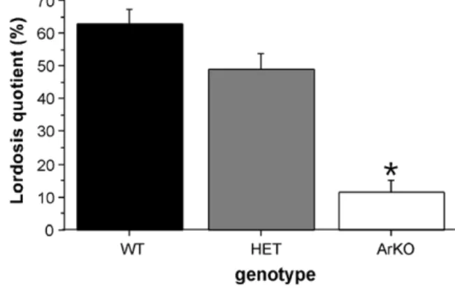
behavior was affected in female ArKO mice [8]. If the
female brain really develops by default, i.e. in the absence of any estrogens, lordosis behavior of female ArKO mice should be indistinguishable of that of normal, wild-type (WT) female mice. Thus, female mice of three genotypes, i.e. wild-type (WT), heterozygous (HET), and homozygous ArKO, were ovariectomized in adulthood and subse-quently implanted with a silastic capsule containing crys-talline estradiol (diluted 1:1 with cholesterol). All females were injected subcutaneously with 500 lg progesterone 2– 4 h before each lordosis test with a sexually active male. We found that the display of lordosis in response to the mounts of the stimulus male was severely impaired in
ArKO females (Fig. 2). The same hormone treatment
was, however, effective in inducing lordosis behavior in WT and HET females. In contrast with previous studies
[12,35], the reduction in lordosis behavior observed in ArKO females did not disappear with repeated testing or prolonged (>6 weeks) of estradiol treatment in adulthood suggesting permanent effects of early estradiol deprivation on the later potential to show female sexual behavior.
The absence of lordosis behavior in ArKO females could have been caused by a partial defeminization of their brains due to the presence of phytoestrogens in the food. In con-trast with endogenous estrogens, phytoestrogens are gener-ally non-steroidal in nature and have lower affinities for
estrogen-binding plasma proteins such as AFP[69]. They
may thus evade the protective actions of AFP and freely enter the brain where they could interfere with brain sexual differentiation [55,85,114]. This suggests that phytoestro-gens could be a potential source of estrogen action in the ArKO female brain, in particularly in the absence of any local competition with endogenous estrogens for binding to neural estradiol receptors. Furthermore, Kudwa et al.
[51] showed that ArKO females whose mothers were fed
a phytoestrogen-free diet showed higher lordosis quotients (equivalent to those of WT females) than ArKO females whose mothers were fed a phytoestrogen-rich diet suggest-ing that estrogens present in diet can attenuate the display of female sexual behavior in ArKO mice. Furthermore, Kudwa et al.[51]proposed that these phytoestrogens prob-ably act via the ERb receptor since male ERbKO mice were capable of showing lordosis behavior in adulthood following castration and subsequent treatment with estra-diol and progesterone[51], and phytoestrogens have been
shown to preferentially bind to the ERb receptor [52].
However, it is unlikely that the effects observed in our ArKO mice[8]relate to an estrogenic action by phytoestro-gens since (i) our mice were fed a mouse chow (UAR 03, Epinay sur Orge, France) that does not appear to have any biologically active estrogens, as revealed by its lack of effect on uterine growth and on the growth of estro-gen-dependent cell lines (M. Huard, UAR, personal com-munication), and (ii) we recently observed very low, almost non-detectable, progesterone receptor (PR) levels in the medial preoptic (MPN) and ventromedial nuclei (VMN) of gonadally intact ArKO mice of both sexes at several postnatal ages (days 10, 20, 30, and 40; Fig. 4; unpublished results). The latter animals were derived of
mothers fed the same diet as in our previous study [8].
The results on PR expression provide the best evidence for the lack of any significant estrogenic action in the ArKO brain during development since previous studies
by Wagner and colleagues [80–82,105,106] have clearly
Fig. 2. Lordosis quotients of female wild-type (WT), heterozygous (HET), and aromatase knockout (ArKO) mice. All females were ovariectomized in adulthood and subsequently treated with estradiol and progesterone prior to each behavioral test with a sexually active male. *p < 0.05
compare to WT and HET females. Data shown are means (±SEM) of a total of five tests.
shown that the sex difference in PR levels in the MPN and VMN, with fetal and neonatal males showing a higher PR expression than females, depends on the production of estradiol acting via the ERa receptor in the developing male brain. For instance, maternal administration of the aromatase inhibitor ATD significantly decreased hypotha-lamic PR expression in male rat fetuses[82], whereas 3 days of administering testosterone propionate or the synthetic estrogen DES (but not dihydrotestosterone) to pregnant female rats over embryonic days 19–22 stimulated hypo-thalamic PR levels in female offspring when they were killed on E22[82]. The sex difference in PR expression in the MPN, which also exists in mice, is abolished in trans-genic mice lacking the functional ERa gene [105]. In the rat, the female-typical profile of PR expression develops after birth. Thus ovariectomy on postnatal day 4 prevented the female-typical increase in PR levels observed between day 10 and 28 in the MPN[80]. Interestingly, preliminary results from our laboratory also suggest a role for postna-tal estrogens in the development of PR in the female mouse. We observed a similar increase in PR levels in WT female mice between days 10 and 30 after birth, whereas this increase was completely absent in ArKO females, confirming the lack of any estrogen action in this mouse model.
It seems unlikely that the deficit in lordosis behavior can be attributed to excessive androgen action during develop-ment leading to a masculinization and defeminization of the brain in ArKO females. Previous work on another
strain of ArKO mice [33] has indicated that ArKO mice
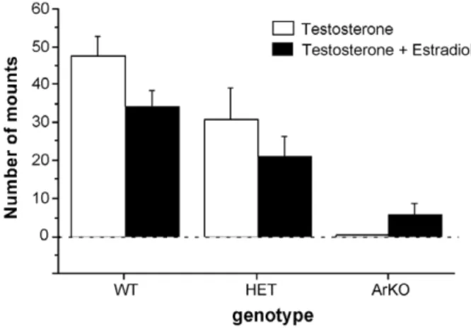
of both sexes are exposed to increased plasma levels of tes-tosterone during adulthood. These increased levels of androgens probably result from the interruption of the ste-roid feedback on the gonadotropin secretion, which is known to be mediated by estrogens, as suggested by the increased levels of circulating luteinizing hormone and fol-licle-stimulating hormone in these mice. Alternatively, the increase in plasma testosterone could be caused, at least in part, by the accumulation of the androgenic substrate, which can no longer be transformed into an estrogen by the ovaries because of the disruption of the aromatase gene. Because the fetal and neonatal ovaries are not very active[53,108,109], it is unlikely that this increase in andro-gen levels actually takes place during early development, but since no data are available to evaluate this question, it could be speculated that increased levels of androgens contribute to the development of the behavioral phenotype of ArKO mice. Thus to determine whether the absence of lordosis behavior in ArKO females did not result from masculinization and defeminization of their brains by excessive androgen action during development, new groups of female WT, HET, and ArKO mice were tested for their potential to show male-typical sexual behavior with an estrous female. Female subjects were ovariectomized and implanted subcutaneously with a silastic capsule containing crystalline testosterone. Following several (>3) weeks of testosterone treatment, female ArKO mice showed no
mounting behavior whereas WT and, to a lesser extent, HET females readily displayed mounting and intromis-sion-like behaviors (Fig. 3). The addition of estradiol (5 lg/mouse/day) to the testosterone treatment stimulated a little mounting and intromission-like behavior in ArKO females, but not to the levels shown by WT and HET females (Fig. 3). Such an outcome would not have occurred had ArKO females been exposed to high levels of testoster-one perinatally capable of masculinizing their coital capac-ity. These results also suggest that estrogens organize mounting behavior in mice to some extent since ArKO females showed no or very little mounting behavior and HET females showed intermediate levels of male sexual behavior, thereby indicating a dose-dependent effect of estrogens on this behavior. By contrast, results obtained in male ArKO mice strongly suggest that male sexual behavior is organized by androgens and not estrogens since almost normal levels of male sexual behavior could be induced in male ArKO mice when treated in adulthood with estrogens in combination with dihydrotestosterone
[9]. In addition, work from the group by Sato et al. [86]
using androgen receptor knockout mice (ARKO), also pro-vided evidence of the differentiation of male sex behavior by androgens. Male ARKO showed very little mounting behavior, whereas female mice treated with DHT perina-tally showed high levels of mounting behavior, but this effect of perinatal DHT treatment was not observed in
female ARKO mice [86]. Finally, to make matters even
more confusing, recent data obtained in mice carrying the testicular feminization (Tfm) mutation of the androgen receptor suggest that male sexual behavior is not organized in the mouse since estrogen-treated male and female mice including Tfm mice, showed equivalent, high levels of
mounting behavior [17]. Perhaps the sex difference in
mounting behavior lies in the sensitivity to adult steroid treatment, i.e. larger doses of testosterone or estradiol are
Fig. 3. Mounting behavior of female wild-type (WT), heterozygous (HET), and aromatase knockout (ArKO) mice. All females were ovari-ectomized in adulthood and tested once for mounting behavior with an estrous female when treated with testosterone and then once more when treated with both testosterone and estradiol (5 lg/mouse/day).*p < 0.05
compared to WT and HET females,#p < 0.05 compared to WT females. Data shown are means ± SEM.
needed to stimulate mounting behavior in female compared
to male mice. In the Bodo and Rissman study [17], mice
were tested using adult estradiol treatment by silastic implant (5 mm of 17b-estradiol diluted 1:1 with choles-terol). This treatment has been shown to lead to very high levels of estradiol[110]and has been used to induce lordo-sis behavior in female mice in our laboratory (e.g.
[8,46,47]). Thus, this treatment did not allow for the detec-tion of any possible genotype differences in sensitivity to estradiol in activating male sexual behavior. Clearly more studies are needed to determine whether male sexual behavior is organized or not by steroids in the mouse and if so, by which steroids. It is possible that in mice, as in fer-rets[95], fetal exposure to estrogens followed by neonatal exposure to testosterone is required for complete masculin-ization of male-typical mating behavior. It should be noted that even though ArKO male mice readily mounted and intromitted an estrous female when treated with estrogens and DHT in adulthood[9], they rarely showed any ejacula-tory behavior, suggesting a possible contribution of estro-gens to the organization of male sexual behavior.
Taken together, the results obtained in female ArKO mice [8]are best explained by assigning an active role for estradiol in females in the development of female sexual behavior. At present, these results provide the best avail-able evidence affirming a role of estradiol in female brain sexual differentiation.
3.2.2. Neural mechanisms potentially affected in female ArKO mice
The reduction in lordosis behavior observed in female ArKO mice[8]may reflect deficits in (i) the neural circuitry regulating the lordosis reflex and/or (ii) the neural mecha-nisms regulating sexual motivation and sexual partner pref-erence. Classically, studies of female sexual behavior have concentrated on the neural and hormonal control of lordo-sis behavior in the female rat (for a detailed description, see
[73]). Briefly, male mounting stimulates pressure receptors on the flanks, posterior rump, tail base, and perineum of the female. Axons of these receptors form a sensory nerve that projects to the dorsal root of the ganglion of the spinal cord. Then, the signal is transmitted to the reticular forma-tion in the brain stem and the midbrain central gray area. When the female is in estrus, i.e. when estradiol concentra-tions are high, several brain regions including the ventro-lateral portion of the VMN (a brain region rich in estrogen receptors) and the MPN, activate via the midbrain central gray, medullary reticular formation, and medial geniculate body, the spinal motoneurons innervating the back muscles critical to the display of lordosis [23]. Thus, the VMN plays a critical role in integrating hormonal and sensory information necessary for the display of lordo-sis in female rodents. Accordingly, lesions of the VMN, or destruction of its afferent and efferent fibers, typically reduce the frequency of lordosis behavior in female rats
[20,60,72]and hamsters[59], whereas implants of estradiol into the VMN induce lordosis behavior in ovariectomized
female rats[84]. Our preliminary results (unpublished) indi-cated that gonadally intact ArKO females, which were not supplemented with any estrogens, show much lower levels of PR in the VMN than WT females. Whether the estradiol treatment used to induce female sexual receptivity in our
previous study [8] also normalized PR expression in the
VMN of ArKO females has not yet been investigated. However, preliminary results from Kudwa et al. (Kudwa, Schank, Honda, and Rissman, SBN abstracts, 2001) sug-gest that adult treatment with estradiol failed to induce normal, WT female, levels of PR expression in the VMN of ArKO females indicating a contribution of postnatal estradiol to the development of PR receptors, as was already suggested by the work of Quadros et al.[80]. Fur-ther studies are needed to determine wheFur-ther the hor-monal–neural circuitry of lordosis behavior has not been feminized in ArKO females and thus whether there is an active contribution of postnatal estradiol to the develop-ment of this circuit.
In contrast with the neural and hormonal control of lor-dosis behavior, very few studies have concentrated on other aspects of female sexual behavior, including proceptivity (i.e. sexual motivation) and sexual partner preference. Nev-ertheless, the ability to seek out and identify potential mates is as critical to female sexual behavior as is the capacity to display the lordosis reflex when mounted by a male. Rodent species use primarily odors to identify indi-viduals of their own species and accordingly, release to their environment a wide variety of volatile and non-vola-tile odors via extraorbital lacrimal glands[49], skin glands, urine, and feces. Two different olfactory systems have evolved to detect these odors. It is generally thought that the main olfactory system is used to detect a wide variety of volatile odorants derived from food and potential preda-tors, among many sources, whereas the accessory olfactory system evolved to detect and process a subset of non-vola-tile odors that influence a variety of reproductive and
aggressive behaviors in mammalian species [32,48].
How-ever, recent evidence points to an important role for the main olfactory system in detecting and processing olfactory cues used for mate recognition[46,47,89]. In female mice, olfactory cues have been shown to facilitate the display of lordosis behavior. Peripherally induced anosmia by intranasal application of zinc sulfate solution attenuated
lordosis behavior in hormone-primed female mice [30,46]
whereas removal of the vomeronasal organ actually com-pletely abolished lordosis behavior in estrogen and
proges-terone-treated female mice [47] thereby emphasizing the
importance of the accessory olfactory system in the control of female sexual behavior. Thus the reduction in lordosis behavior observed in female ArKO mice may reflect deficits in the detection and processing of olfactory cues. For instance, it is possible that ArKO female mice did not rec-ognize the stimulus male on the basis of his odors and as a result did not become sexually receptive. If so, then this would suggest a role for estrogens in the activation and/ organization of olfactory function. Indeed, sex differences
have been reported in olfactory sensitivity with females being better able than males to detect male-derived odors. For instance, sows are significantly better than boars at using decreasing concentrations of the volatile male pig pheromone, androstenone, as a discriminative stimulus in operant tests for a sucrose award[28]. These sex differences may not only be restricted to the detection of opposite-sex odors, but may also involve same-sex odors. Using habitu-ation/dishabituation tests to determine odor attraction thresholds, female mice responded more reliably than male mice to low concentrations of volatile urinary odors from either sex [11]. The greater olfactory sensitivity observed in female mice probably reflects perinatal actions of gona-dal hormones since these sex differences were already observed in long-term gonadectomized mice suggesting that gonadal hormones are not necessary to activate this behavior[11]. Furthermore, Dorries et al.[28]showed that the olfactory performance of neonatally castrated male pigs falls between those of sows and boars indicating a con-tribution of perinatal androgens and/or estrogens in the ability to detect androstenone. Thus, we hypothesized that olfactory investigation of conspecific odors is affected in ArKO females due to them being deprived of estrogens during perinatal development. Indeed, in our initial study ([8];Fig. 4) we observed that female ArKO mice displayed reduced levels of olfactory investigation directed towards volatile odors emitted from either estrous female or male conspecifics when provided with a choice between both odor sources in a Y-maze[8]. Female subjects were ovari-ectomized in adulthood and treated over a prolonged (>3 weeks) period of time with estradiol indicating that the reduction in olfactory investigation could not be corrected by supplementing ArKO females with estradiol (Fig. 4). By contrast, no differences were observed between ArKO and WT females in olfactory investigation of soiled bedding
which contains primarily non-volatile odors and are thought to be detected and processed by the accessory olfactory system. These results thus suggest in particular, deficits in main olfactory function in ArKO females. There-fore, in a follow-up study[76], we determined whether this reduction in olfactory investigation reflect deficits in the ability of the main olfactory system to detect and/or discriminate volatile odors derived from male as opposed to female conspecifics. Using habituation/dishabituation tests, we found that gonadectomized ArKO and WT mice, which were tested without any sex hormone replacement, reliably distinguished between undiluted volatile urinary odors of either adult males or estrous females versus deionized water as well as between these two urinary odors themselves (Fig. 5). Thus, the previously observed deficits
[8] in the preference of female ArKO mice to approach
volatile body odors from conspecifics of either sex cannot be attributed to an inability of ArKO mice to detect or discriminate volatile urinary odors from males versus females.
Next, we compared the ability of ArKO and WT mice to detect decreasing concentrations of either male or female urinary odors [76]. We found a clear-cut sex difference in urinary odor attraction thresholds among WT mice: WT females responded to higher urine dilutions than male mice, thereby confirming previous results obtained by
Baum and Keverne [11]. Interestingly, male ArKO mice
resembled WT females in their ability to detect lower con-centrations of urinary odors, raising the possibility that the observed sex difference among WT mice in urine attraction thresholds results from the perinatal actions of estrogen in the male nervous system. Female ArKO mice failed to respond to some of the female urine dilutions, suggesting olfactory-perceptual deficits. Therefore, the ability of ArKO and WT mice to discriminate low concentrations
Fig. 4. Total time spent investigating volatile odors by female wild-type (WT), heterozygous (HET), and aromatase knockout (ArKO) mice when given the choice between volatile body odors from an intact male versus those from an estrous female in a Y-maze. All female subjects were ovariectomized in adulthood and first tested for their odor preferences when receiving testosterone and then when receiving estradiol.*p < 0.05
compared to WT and HET females. Data shown are means ± SEM of two successive tests.
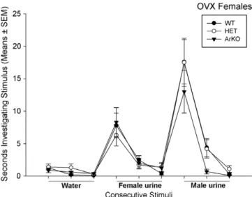
Fig. 5. Time spent by ovariectomized female wild-type (WT), heterozy-gous (HET), and aromatase knockout (ArKO) mice investigating deion-ized water or volatile urinary stimuli.*p < 0.05 between the time spent
investigating the third presentation of a particular stimulus and the first presentation of the subsequent stimulus.
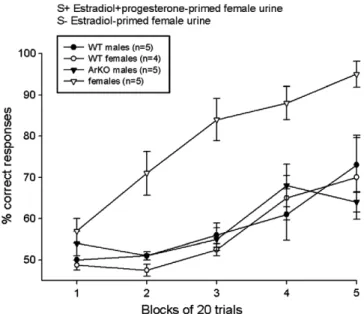
of different volatile urinary odors was also assessed using a food-motivated operant (olfactometer) task[112]. All mice were gonadectomized in adulthood and subsequently trea-ted with a low (1 lg) dose of estradiol benzoate. All groups of animals, regardless the sex or the genotype, eventually learned to distinguish between intact male urine and estrous female urine. Among the four groups of subjects, WT females performed the worst and ArKO females the best in discriminating several pairs of urinary and non-bio-logical (amyl versus butyl acetate) odors. This was most apparent when animals had to discriminate volatile urinary odors from ovariectomized female mice treated with estra-diol sequenced with progesterone versus estraestra-diol alone (Fig. 6): ArKO females quickly learned this discrimination whereas WT females and males as well as ArKO males failed to do so. Thus these results suggest that the weak performance of ArKO females in the habituation/dishabit-uation tests of odor detection[76] cannot be attributed to deficits in the function of the main olfactory system per se. It is possible that the previously observed [8]decrease in olfactory investigation of volatile body odors in Y-maze tests actually reflected an increased olfactory sensitivity in ArKO females, i.e. less proximal investigation of the source of urinary odors was needed for ArKO females to distin-guish the different odors presented in the two arms of the Y-maze. The superior olfactory performance of ArKO females over WT females may have resulted from an increased estradiol-induced olfactory neurogenesis in these females.
Taken together, the deficits observed in lordosis behav-ior of female ArKO mice[8]are not due to deficits in their capacity to recognize the stimulus male on the basis of his
odors. There are certainly indices of a reduced motivation in ArKO females to investigate conspecific odors[8,76], but when appropriately motivated (by food-depriving them for instance), they are capable and even better than WT females in discriminating between different pairs of biolog-ical and non-biologbiolog-ical odors [112]. Thus, the neural sys-tems underlying the reduced display of lordosis behavior in ArKO females do most likely not include the olfactory systems.
4. Role of a-fetoprotein in the sexual differentiation of the rodent brain
The role of a-fetoprotein (AFP) in brain sexual differen-tiation has been another topic of debate in the field of behavioral neuroendocrinology during the 1970–1980s
[26,64,99]. AFP was discovered about half a century ago
to be the major serum fetal protein in mammals [1,15].
AFP is produced in great quantities during fetal life by the endodermal cells of the visceral yolk sac, by the hepa-tocytes, and in lesser amounts, by the gastrointestinal tract
[3,90,94]. The protein produced by the embryo is trans-ferred into the maternal blood circulation, and levels of AFP in the maternal serum are commonly used as a diag-nostic marker to reveal developmental anomalies of the
fetus [19,42,70]. Abnormally high levels of AFP in the
maternal serum indicate an elevated risk of neural tube defects of the fetus such as spina bifida or anencephaly
[54], whereas abnormally low levels indicates an elevated
risk of Down’s syndrome [22]. The synthesis of AFP
decreases rapidly after birth and only trace amounts are detected in adults[3,83].
Until recently, the physiological function of AFP during embryonic development remained largely unidentified. The observation that AFP is able to bind estrogens with high affinity in rats and mice has led to the suggestion that AFP may play a role in the sexual differentiation of the brain, in particularly in protecting the female brain from excessive exposure to estrogens that are circulating in high concentrations during the critical period of sexual differen-tiation ([64,83]; Fig. 7a). However, in addition to binding estradiol, AFP, like albumin, is able to bind other steroids as well as endogenous and exogenous substances such as fatty acids, bilirubin, and various pharmaceutical agents, suggesting that AFP may play a transportation role in gen-eral (for review see[37]). In fact, an intracellular pool of AFP of unknown function has been detected in the brain cytosol during fetal life in various vertebrate species, including rats, mice, sheep, pig, and humans (reviewed in
[99]). AFP is present within neurons of both sexes at all stages of development of the central nervous system from the postmitotic neuroblast to more differentiated neurons. However AFP is not present in the adult mouse brain underlying that AFP may have important functions within the developing central nervous system. Furthermore, although some reports exist of local synthesis (e.g.[2,57], there is no convincing evidence that intraneuronal AFP is
Fig. 6. Ability of wild-type (WT) and aromatase knockout (ArKO) male and female mice to learn to discriminate between two female urine stimuli in an olfactometer test. S+ = rewarded stimulus; S = non-rewarded stimulus. Data expressed as means ± SEM.*p < 0.05 compared to WT
synthesized locally since no messenger RNA for AFP could be detected in the fetal, newborn, or adult mouse brain[88]
suggesting that the observed high levels of AFP immunore-activity in the brain must be derived from external sources, perhaps by receptor-mediated endocytosis [99,100]. These latter observations suggest that AFP may have more than the attributed ‘‘protective’’ role [64] and may serve as a
transport carrier for estrogens into the brain [99];
Fig. 7b). In particular, the discrete intracytoplasmic locali-zation of rodent AFP suggests its possible active involve-ment in estrogen-sensitive neurons during the critical period of sexual differentiation. Since there is a difference of several orders of magnitude between the affinity
con-stants for estradiol binding by AFP (KD10 8M; [87])
and by the estrogen receptor (KD10 11
M) the subsequent intracytoplasmic dissocation of the AFP/estradiol complex in estrogen-receptor containing neurons could liberate the steroid and lead directly to receptor binding. By doing so, intraneuronal AFP could thus provide target neurons of both sexes with low levels of estrogen and thus serve as an intracellular reservoir of estrogen (Fig. 7b). It should be noted, however, that although a widespread intraneuro-nal localization of immunoreactive AFP is observed in numerous brain regions in rodents of both sexes, estro-gen-receptor containing regions of the brain, such as the diagonal band of broca, the medial preoptic area, the arcu-ate, and the ventral premammillary nuclei of the hypothal-amus, and the medial and cortical amygdaloid nuclei are characterized by a complete absence of AFP-immunoreac-tivity[98]. This latter observation clearly questions the
pos-sible role of AFP as transporter of estrogens to estrogen-sensitive neural targets during the sexual differentiation of the brain. While the estrogen-binding capacity of the rodent AFP emphasizes its potential importance for brain sexual differentiation, its possible role with respect to its capacity to bind substances other than estrogens, such as teratogens and polyunsaturated free fatty acids such as ara-chidonic, docosahexaenoic, and docosatetraenoic acides, of which their importance has been shown with regard to
neu-ral development in mammals, including humans [21,77],
should not be overlooked, in particularly since these
ligands are bound to AFP in all species [103]. Taken
together, clearly opposing views exist on the function of AFP during development and in particularly during sexual differentiation of the brain (Fig. 7). These different hypoth-eses have not been experimentally tested due to the absence of a suitable animal model. Some indirect evidence for a
protective role of AFP comes from a study [62] showing
that the addition of neonatal serum to [3H] estradiol
strongly reduced its uptake into the brain of adult female rats. Furthermore, the study by Mizejewski and Vonnegut
[67]in which neonatal male and female mice were injected intracranially with anti-AFP immunoglobulin, showed an androgenization of the female mouse, suggesting a protec-tive role of AFP. However, these mice also had gross neu-rological lesions, i.e. external hydrocephaly, as a result of the intracranial injection with anti-AFP thereby making it difficult to interpret the data.
The recent introduction of an AFP-KO mouse[34]has
now made it possible to test these opposing hypotheses
Fig. 7. Two competing hypotheses on the role of a-fetoprotein (AFP), a fetal plasma protein that binds estrogens with high affinity, in female sexual differentiation: (a) AFP may serve to keep estrogens from entering the brain (hypothesis proposed by[64]) and (b) AFP may deliver estrogens to specific brain regions to promote feminization of the neural mechanisms controlling female sexual behavior (hypothesis proposed by[99]).
and thus to determine the function of AFP in brain sexual differentiation.
4.1. The a-fetoprotein knockout mouse
4.1.1. a-Fetoprotein knockout mice are infertile
Afp knockout mice were generated by replacing an Afp genomic fragment extending from exon 1 to intron 3 KO1), or extending from exon 2 to intron 3
(AFP-KO2), by a IRES-LacZ-neo selection cassette [34]. The
AFP-KO1 allele was generated in two different genetic backgrounds, the outbred CD-1 and the inbred C57Bl/6j strain, whereas the AFP-KO2 allele was generated in only the CD-1 background. Both invalidations gave rise to via-ble homozygous animals. AFP-KO animals are apparently normal with males being fertile, but females are not, owing
to a complete absence of ovulation[34]. AFP-KO ovaries
contain follicles at different stages of maturation, including the last Grafiaan follicle stage, but no corpora lutea, indic-ative of ovulation, could be detected, which is in accor-dance with the low levels of progesterone in the serum. Reciprocal ovarian transplantation experiments demon-strated that AFP-KO ovaries are functional: AFP-KO ova-ries transplanted in normal mice were able to ovulate and the transplanted females generated pups from the mutated parental oocytes. By contrast, WT ovaries implanted into female AFP-KO mice did not show any ovulation. How-ever, ovulation in AFP-KO females could be induced by injecting gonadotropins indicating that their ovaries are responsive to any signals from the hypothalamus–pituitary. These results thus suggest that the infertility observed in AFP-KO females is not due to any defects in the ovaries, but is most likely caused by a deficit in hypothalamic–pitu-itary–gonadal (HPG) axis. Indeed, microarray studies [24]
showed that several genes implicated in female fertility, such as Egr1, Cish2, Ptprf, Psa, and Tkt, were down-regu-lated in the pituitary of AFP-KO females. Furthermore, several genes implicated in the GnRH pathway, such as the GnRH receptor gene, and several genes activated by the GnRH receptor (cFos, Egr2, Tgfb li4, and Ptp4a1)
are down-regulated in female AFP-KO mice [24]. In the
hypothalamus, the gene encoding the hypothalamic GnRH decapeptide is itself down-regulated, suggesting a dysfunc-tion of the GnRH pathway in AFP-KO females.
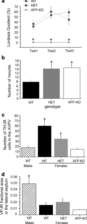
4.1.2. Brain and behavior of AFP knockout mice are defeminized
In order to determine whether their infertility reflected anomalies in the sexual differentiation of the brain, we first tested female AFP-KO mice for their ability to show lordo-sis behavior when paired with a sexually active male [6]. Groups of female WT, HET, and AFP-KO mice were ovariectomized in adulthood and subsequently implanted with a silastic capsule containing crystalline estradiol (diluted 1:1 with cholesterol). All female subjects received 500 lg of progesterone subcutaneously 2–4 h before test-ing. AFP-KO females never showed any lordosis behavior
in any of the three behavioral tests, whereas WT and HET females showed substantial levels of lordosis behavior (Fig. 8a). Very similar results were obtained in all AFP-KO mouse models generated[6]. These results suggest that AFP-KO females have lost their ability to show lordosis behavior. As discussed previously, olfaction plays a pivotal role in the expression of lordosis behavior in female mice
[30,46,47]. The complete absence of lordosis behavior in female AFP-KO mice may be explained by a reduced moti-vation to investigate male-derived odors. Therefore we investigated odor preferences in AFP-KO mice of the two
different background strains [7]. AFP-KO females always
preferred to investigate male over female odors when given the choice between these two odor stimuli in a Y-maze, and thus remained very female-like in this regard. The absence of lordosis behavior in these females can thus not be explained by a reduced motivation of AFP-KO females to investigate male-derived odors.
We also determined whether AFP-KO females have an increased capacity to display male-typical sexual behaviors. Normal female mice can show substantial levels of mount-ing and intromission-like behaviors when ovariectomized in adulthood and treated with high doses of either testos-terone or estradiol [8,111]suggesting that this behavior is less sexually differentiated than the ability to show lordosis behavior. Nevertheless, mount frequencies were higher in AFP-KO females compared to WT females and this effect was already visible in HET females ([6];Fig. 8b). The latter have probably been exposed to increased concentrations of estrogens due to having decreased levels of AFP[34]. The binding affinity of estradiol for AFP is lower than for its own receptor stressing the importance of having excess lev-els of AFP during fetal development[5]. These results also point to a role of fetal estrogens in the organization of male sexual behavior in the mouse and thus further support the notion of a possible synergistic role of estradiol and andro-gen receptor activation in masculinization of mating behavior capacity ([9,86,95]; see discussion in previous section).
4.1.3. Reduced female sexual behavior in female ArKO mice This behavioral phenotype in AFP-KO females was
associated with neurochemical changes [6]. The sexually
dimorphic expression of tyrosine hydroxylase (TH) and vasopressin (VP) were used as indices of brain sexual differ-entiation. The number of TH-expressing neurons in the anteroventral preoptic area (AVPv) is sexually dimorphic
with females having greater numbers than males
[91,92,96]. The AVPv plays a critical role in female repro-ductive function by transducing hormonal feedback con-trolling hypothalamic GnRH release and consequent pituitary luteinizing hormone secretion, as well as that it is needed for hormonally induced ovulation[115]. By con-trast, males show a denser expression of VP particularly in the lateral septum than females[25]. We confirmed the sex differences in the number TH-expressing neurons in the AVPV as well as in VP expression in the lateral septum.
Female AFP-KO mice had decreased, male-like numbers of tyrosine hydroxylase (TH) neurons in the anteroventral
preoptic area (AVPV; Fig. 8c) which may be related to
their infertility [24,34]. By contrast, female AFP-KO mice showed female-typical densities of VP-immunoreactive fibers in the lateral septum (Fig. 8d). The latter result was rather surprising in light of earlier observations showing an important role for perinatal estrogens in the sexual dif-ferentiation of the vasopressin system in the rat [43]. It is possible, however, that the development of sex differences in the vasopressin system relies more on the perinatal action of androgens than on that of estrogens in the mouse, an idea supported by recent findings in male ArKO mice ([79]; unpublished results). Furthermore, sex chromosomes may also contribute directly to the development of the sex-ually dimorphic vasopressin system in mice [25]. Indeed, XY males and XY female mice (i.e. females with a deletion of the Sry gene) are more masculine than XX mice with regard to the density of vasopressin-expressing fibers in the lateral septum [25]. So perhaps there is an interaction between estrogens and sex chromosomes in the sexual dif-ferentiation of this neuropeptide system.
4.1.4. AFP protects the developing female brain from estrogens
Our observations of a complete absence of female sexual behavior and male-like numbers of TH neurons in the AVPV in female AFP-KO mice did not allow us to discrim-inate between the two opposing theories about the role of AFP in brain sexual differentiation (Fig. 7). Both hypothe-ses would predict that male AFP-KO mice would be more or less normal but that the females would be affected. It could be argued that the brains of AFP-KO females were defeminized because they were no longer protected from the estrogens produced by their mother or male siblings. This argument would support the hypothesis that AFP serves primarily to protect the female brain from excessive exposure to estrogens [64]. By contrast, it could also be argued that the brains of AFP-KO females were not femi-nized because they were lacking the AFP to transport small quantities of estrogens to target brain areas and thus to promote the differentiation of these structures in a female direction. This argument would favor the hypothesis that AFP is needed to transport estrogens into the brain and thus has an active role in female sexual differentiation[99]. To discriminate between these competing hypotheses, we blocked estrogen production during prenatal develop-ment by treating pregnant female mice heterozygous for
the Afp mutation with the aromatase inhibitor ATD [6].
If AFP protects the female brain from being exposed to estrogens, AFP-KO offspring of ATD-treated mothers should not be defeminized, as the ATD treatment would prevent the formation, during prenatal development, of high defeminizing levels of estradiol. However, if AFP actually acts as an estrogen carrier and is thus necessary for the feminizing the female brain, then the AFP-KO off-spring of ATD-treated mothers should not show normal
Fig. 8. Behavioral and neurochemical phenotype of female a-fetoprotein knockout (AFP-KO) mice. (a) Lordosis quotients of female wild-type (WT), heterozygous (HET), and AFP-KO mice. All female subjects were ovari-ectomized in adulthood and subsequently tested with estradiol and progesterone prior to each behavioral test.*p < 0.05 compared to WT and
HET females. (b) Mounting behavior of female WT, HET, and AFP-KO females when paired with an estrous female. Female subjects were ovariectomized in adulthood and subsequently treated with estradiol.
*p < 0.05 compared to WT females. (c) Numbers of tyrosine
hydroxylase-immunoreactive (TH-IR) neurons in the anteroventral preoptic area (AVPv). Female subjects were ovariectomized in adulthood and treated with estradiol.*p < 0.05 compared to WT males, and HET and AFP-KO
females, #p < 0.05 compared to WT males and females and AFP-KO
females. (d) Brain vasopressin expression assessed by the fractional areas covered by vasopressin-IR structures in the posterior lateral septum. Female subjects were ovariectomized in adulthood and treated with estradiol.
lordosis behavior because of a lack of feminization by estrogens (absence of the steroid and its carrier).
Prenatal treatment with ATD completely rescued
lordo-sis behavior in AFP-KO females (Fig. 9a). Furthermore,
such ATD-treated AFP-KO females had female-like num-bers of TH neurons in the AVPv (Fig. 9b). Accordingly, the fertility of AFP-KO females was also restored as well as gene expression in the HPG[24]. These results clearly dem-onstrate that AFP serves to protect the female brain from becoming masculinized and defeminized by estrogens cir-culating during embryonic development[6].
These findings do not explain, however, why AFP is found inside neurons without being locally produced[88]. Little or no intraneuronal AFP is found in limbic, hypotha-lamic, and amygdaloid areas, whereas large amounts are present in adjacent regions [98]. This could indicate that AFP protects from estrogens those brain regions involved in reproductive function, such as the hypothalamus, but
may deliver estrogens to other brain regions, and thereby may influence the sexual differentiation of various func-tions, including non-reproductive behaviors such as
learn-ing and memory capacity [63]. However, it should be
noted that the mechanisms controlling the differentiation of sex differences in learning and memory capacity remain to be elucidated. Perhaps a closer look at the neurons that contain AFP will suggest a function for this protein in these cells. In addition, it will be interesting to determine whether non-reproductive behaviors have been altered in AFP-KO mice and if so, whether this can be corrected or not by blocking estrogen synthesis during fetal development.
Whether AFP plays a similar protective role in the sex-ual differentiation of the human brain is unclear. There are diverging views in the literature as to whether human AFP
has any estrogen-binding capacities. Nunez et al. [71]
reported a low-binding affinity of human serum for steroid hormones during embryonic and early postnatal
develop-ment. By contrast, both Uriel et al. [101] and Arnon
et al. [4] showed that human AFP could actually bind
estrogens. In either case, human AFP-derived peptides
are able to display some anti-estrogenic activity
[14,66,102]. These AFP-derived peptides are currently under investigation as potential agents for treating
estro-gen-dependent breast cancers and other tumors [13,66].
Finally, it is not known if a protection from maternal estro-gens is actually required since the reigning idea is that in humans, androgens, not estrogens, are the primary cause of brain masculinization (reviewed in[107]). Furthermore, primates do not undergo receptive defeminization, i.e. males will accept repeated mounting by other males by dis-playing the posture normally associated with female recep-tivity. As a result, primates may not need a protective role of AFP against a defeminizing action of fetal estradiol exposure[10].
5. Conclusions and future directions
The results obtained in AFP-KO mice show that the principal action of prenatal estrogen exposure, regardless of whether it occurs in female or male mice, is to defeminize and, to some extent, masculinize brain and behavior. Fur-thermore, AFP, which binds estradiol circulating in the female fetus with affinity and capacity, protects the devel-oping brain from a male-typical organization by this ste-roid. So at first glance, these findings are at odds with
the results[8]obtained in female ArKO mice implying an
active contribution of estrogens to the development of the female brain. However, such a dual role of estradiol in brain sexual differentiation was earlier suggested by Dohler et al. [26]. Based on their results, they proposed the ‘‘progressive hypothesis of brain sexual differentiation’’ which asserts that (i) the embryonic brain is not differenti-ated in either males or females, and (ii) under the influence of moderate levels of estrogen, female-typical neural and behavioral traits develop whereas under the influence of high levels of estrogen, male-typical neural and behavioral
Fig. 9. Female phenotype of AFP-KO females is rescued by prenatal treatment with the aromatase inhibitor ATD. (a) Lordosis quotients of wild-type (WT), AFP-KO, and heterozygous (HET-ATD) and AFP-KO females treated prenatally with ATD (AFP-KO-ATD). All female subjects were ovariectomized in adulthood and treated subsequently with estradiol and progesterone.*p < 0.05 compared to WT, HET-ATD, and
AFP-KO-ATD females. (b) Numbers of tyrosine hydroxylase-immunoreactive (TH-IR) neurons in the anteroventral preoptic region (AVPv). Female subjects were ovariectomized in adulthood and treated with estradiol.*p < 0.05
traits develop. However, we now know that any prenatal estrogen action is blocked by AFP in the developing female, thus if estrogens normally contributes to the devel-opment of the female brain, they most likely act postnatally when the amount of AFP has decreased substantially and AFP no longer plays a protective role. AFP levels are the highest at birth, after which they decrease by about 50% during the first 24 h. Only trace levels (about 0.01% of fetal levels) are detected at 3 weeks of age in rats[83]. Thus pre-sumably when the ovaries start to secrete estrogens at day 7 after birth [53], AFP no longer plays a significant role. Consistent with a postnatal role of estradiol in feminization of the brain is the observation by Steward and Cygan[93]
of an enhanced of female receptivity in neonatally gonadec-tomized male and female rats treated with estradiol over postnatal days 30–40. We propose thus that the defeminiz-ing action of estradiol normally occurs prenatally in males and is avoided in fetal females because of the protective actions of AFP. We further propose that the feminizing action of estradiol normally occurs in genetic females between birth and the age of puberty (postnatal days 40– 50). Accordingly, several studies in which estrogen expo-sure was manipulated in both sexes during the perinatal developmental period have suggested that different critical periods exit for male- and female-typical organization of
the brain [80,82]. Thus, prenatal exposure to estrogens
induces the male-typical pattern of PR expression in the MPN and VMN, whereas postnatal exposure to estrogens leads to the female pattern of PR expression in these brain regions. Future studies should take advantage of the ArKO mouse model to determine the contribution of postnatal estradiol to the development of female sexual behavior. If normal levels of lordosis behavior can be induced in female ArKO mice by treating them with estradiol postnatally, then these results would provide the best evidence for a normal role of postnatal estradiol in promoting female-typ-ical brain and behavioral sexual differentiation.
Acknowledgments
This work was supported by NICHD Grant No. HD044897 to M.J.B. and J.B., two grants from the Fonds National de la Recherche Scientifique (No. 1.5.082.04 and 1.5.104.06), and one grant from the University of Lie`ge (No. C-06/89), to J.B. J. Bakker is a research associate from the Fonds National de la Recherche Scientifique. References
[1] G.I. Abelev, S.D. Perova, N.I. Khramkova, Z.A. Postnikova, I.S. Irlin, Production of embryonal alpha-globulin by transplantable mouse hepatomas, Transplantation 1 (1963) 174–180.
[2] M. Ali, K. Mujoo, M.K. Sahib, Synthesis and secretion of alpha-fetoprotein and albumin by newborn rat brain cells in culture, Brain Res. 282 (1982) 47–55.
[3] G.K. Andrews, M. Dziadek, T. Tamaoki, Expression and methyl-ation of the mouse alpha-fetoprotein gene in embryonic, adult, and neoplastic tissues, J. Biol. Chem. 257 (1982) 5148–5153.
[4] R. Arnon, E. Teicher, M. Bustin, M. Sela, Preparation of antisera to alpha-fetoprotein making use of estradiol affinity column, FEBS Lett. 32 (1973) 335–338.
[5] C. Aussel, R. Masseyeff, Rat alpha-fetoprotein-estrogen interaction, J. Steroid Biochem. 9 (1978) 547–551.
[6] J. Bakker, C. De Mees, Q. Douhard, J. Balthazart, P. Gabant, J. Szpirer, C. Szpirer, Alpha-fetoprotein protects the developing female mouse brain from masculinization and defeminization by estrogens, Nat. Neurosci. 9 (2006) 220–226.
[7] J. Bakker, C. De Mees, J. Szpirer, C. Szpirer, J. Balthazart, Exposure to oestrogen prenatally does not interfere with the normal female-typical development of odour preferences, J. Neuroendocri-nol. 19 (2007) 329–334.
[8] J. Bakker, S. Honda, N. Harada, J. Balthazart, The aromatase knock-out mouse provides new evidence that estradiol is required during development in the female for the expression of sociosexual behaviors in adulthood, J. Neurosci. 22 (2002) 9104–9112. [9] J. Bakker, S. Honda, N. Harada, J. Balthazart, Restoration of male
sexual behavior by adult exogenous estrogens in male aromatase knockout mice, Horm. Behav. 46 (2004) 1–10.
[10] M.J. Baum, Differentiation of coital behavior in mammals: a comparative analysis, Neurosci. Biobehav. Rev. 3 (1979) 265–284. [11] M.J. Baum, E.B. Keverne, Sex difference in attraction thresholds for
volatile odors from male and estrous female mouse urine, Horm. Behav. 41 (2002) 213–219.
[12] M.J. Baum, S.A. Tobet, Effect of prenatal exposure to aromatase inhibitor, testosterone, or antiandrogen on the development of feminine sexual behavior in ferrets of both sexes, Physiol. Behav. 37 (1986) 111–118.
[13] J.A. Bennett, L. DeFreest, I. Anaka, H. Saadati, S. Balulad, H.I. Jacobson, T.T. Andersen, AFPep: an anti-breast cancer peptide that is orally active, Breast Cancer Res. Treat. 98 (2006) 133–141. [14] J.A. Bennett, F.B. Mesfin, T.T. Andersen, J.F. Gierthy, H.I.
Jacobson, A peptide derived from alpha-fetoprotein prevents the growth of estrogen-dependent human breast cancers sensitive and resistant to tamoxifen, Proc. Natl. Acad. Sci. USA 99 (2002) 2211– 2215.
[15] C.G. Bergstrand, B. Czar, Demonstration of a new protein fraction in serum from the human fetus, Scand. J. Clin. Lab. Invest. 8 (1956) 174.
[16] D. Blizard, C. Denef, Neonatal androgen effects on open-field activity and sexual behavior in the female rat: the modifying influence of ovarian secretions during development, Physiol. Behav. 11 (1973) 65–69.
[17] C. Bodo, E.F. Rissman, Androgen receptor is essential for sexual differentiation of responses to olfactory cues in mice, Eur. J. Neurosci. 25 (2007) 2182–2190.
[18] J.E. Booth, Sexual differentiation of the brain, in: C.A. Finn (Ed.), Oxford Reviews of Reproductive, vol. I, Clarendon Press, Oxford, 1979, pp. 58–158, Ref Type: Serial (Book,Monograph).
[19] P. Brownbill, D. Edwards, C. Jones, D. Mahendran, D. Owen, C. Sibley, R. Johnson, P. Swanson, D.M. Nelson, Mechanisms of alphafetoprotein transfer in the perfused human placental cotyledon from uncomplicated pregnancy, J. Clin. Invest. 96 (1995) 2220–2226. [20] A.S. Clark, J.K. Pfeifle, D.A. Edwards, Ventromedial hypothalamic damage and sexual proceptivity in female rats, Physiol. Behav. 27 (1981) 597–602.
[21] M.A. Crawford, A.J. Sinclair, Nutritional influences in the evolution of the mammalian brain, in: CIBA Foundation Symposium, Churchill London, Malnutrition and the Developing Brain, 1972, pp. 267–287.
[22] H.S. Cuckle, N.J. Wald, R.H. Lindenbaum, Maternal serum alpha-fetoprotein measurement: a screening test for Down syndrome, Lancet 1 (1984) 926–929.
[23] D. Daniels, R.R. Miselis, L.M. Flanagan-Cato, Central neuronal circuit innervating the lordosis-producing muscles defined by trans-neuronal transport of pseudorabies virus, J. Neurosci. 19 (1999) 2823–2833.