HAL Id: hal-01832952
https://hal.archives-ouvertes.fr/hal-01832952
Submitted on 13 Jul 2018HAL is a multi-disciplinary open access archive for the deposit and dissemination of sci-entific research documents, whether they are pub-lished or not. The documents may come from teaching and research institutions in France or abroad, or from public or private research centers.
L’archive ouverte pluridisciplinaire HAL, est destinée au dépôt et à la diffusion de documents scientifiques de niveau recherche, publiés ou non, émanant des établissements d’enseignement et de recherche français ou étrangers, des laboratoires publics ou privés.
mouse heart
Rianne Nederlof, Simone Denis, Benjamin Lauzier, Christine Des Rosiers,
Markku Laakso, Jacob Hagen, Carmen Argmann, Ronald Wanders, Riekelt H.
Houtkooper, Markus W. Hollmann, et al.
To cite this version:
Rianne Nederlof, Simone Denis, Benjamin Lauzier, Christine Des Rosiers, Markku Laakso, et al.. Acute detachment of hexokinase II from mitochondria modestly increases oxygen consumption of the intact mouse heart. Metabolism: Clinical and Experimental, 2017, Equipe IIb, 72, pp.66–74. �10.1016/j.metabol.2017.04.008�. �hal-01832952�
Artikkelit Terveystieteiden tiedekunta
2017
Acute detachment of hexokinase II
from mitochondria modestly increases
oxygen consumption of the intact
mouse heart
Nederlof R
Elsevier BV info:eu-repo/semantics/article info:eu-repo/semantics/acceptedVersion © Elsevier B.V CC BY-NC-ND https://creativecommons.org/licenses/by-nc-nd/4.0/ http://dx.doi.org/10.1016/j.metabol.2017.04.008 https://erepo.uef.fi/handle/123456789/4287Rianne Nederlof, Simone Denis, Benjamin Lauzier, Christine Des Rosiers, Markku Laakso, Jacob Hagen, Carmen Argmann, Ronald Wanders, Riekelt H Houtkooper, M.W. Hollmann, Sander M Houten, Coert J Zuurbier
PII: S0026-0495(17)30116-6
DOI: doi:10.1016/j.metabol.2017.04.008
Reference: YMETA 53591 To appear in: Metabolism
Received date: 4 January 2017 Accepted date: 17 April 2017
Please cite this article as: Nederlof Rianne, Denis Simone, Lauzier Benjamin, Rosiers Christine Des, Laakso Markku, Hagen Jacob, Argmann Carmen, Wanders Ronald, Houtkooper Riekelt H, Hollmann MW, Houten Sander M, Zuurbier Coert J, Acute de-tachment of hexokinase II from mitochondria modestly increases oxygen consumption of the intact mouse heart, Metabolism (2017), doi: 10.1016/j.metabol.2017.04.008
This is a PDF file of an unedited manuscript that has been accepted for publication. As a service to our customers we are providing this early version of the manuscript. The manuscript will undergo copyediting, typesetting, and review of the resulting proof before it is published in its final form. Please note that during the production process errors may be discovered which could affect the content, and all legal disclaimers that apply to the journal pertain.
ACCEPTED MANUSCRIPT
1
Acute detachment of hexokinase II from mitochondria modestly increases
oxygen consumption of the intact mouse heart
Rianne Nederlof,1 Simone Denis,2 Benjamin Lauzier,3 Christine Des Rosiers,4 Markku Laakso,5 Jacob Hagen,6 Carmen Argmann,6 Ronald Wanders,2 Riekelt H Houtkooper,2 M.W. Hollmann,1 Sander M Houten,6* Coert J Zuurbier1*
1
Laboratory of Experimental Intensive Care and Anesthesiology, Department of Anesthesiology, Academic Medical Center, Amsterdam, The Netherlands; 2Laboratory of Genetic Metabolic Diseases, Academic Medical Center, Amsterdam, The Netherlands; 3l’institut du thorax, INSERM, CNRS, Université de Nantes, Nantes, France; 4Montreal Heart Institute Research Center and Department of Nutrition, Université de Montréal, Montréal, Québec, Canada; 5Institute of Clinical Medicine, Internal Medicine, University of Eastern Finland and Kuopio University Hospital, Finland; 6Department of Genetics and Genomic Sciences, Icahn School of Medicine at Mount Sinai, New York, USA.
*contributed equally
Running title: Mitochondrial hexokinase II and cardiac metabolism
Address of correspondence:
Dr. C.J. Zuurbier, Department of Anesthesiology Academic Medical Center, University of Amsterdam Meibergdreef 9, 1105 AZ Amsterdam, The Netherlands Phone: + 31 (20) 566 5259; Fax: + 31 (20) 6979004 E-mail: c.j.zuurbier@amc.uva.nl
ACCEPTED MANUSCRIPT
2 Abstract
Objective. Cardiac hexokinase II (HKII) can translocate between cytosol and mitochondria and change its cellular expression with pathologies such as ischemia-reperfusion, diabetes and heart failure. The cardiac metabolic consequences of these changes are unknown. Here we measured energy substrate utilization in cytosol and mitochondria using stabile isotopes and oxygen consumption of the intact perfused heart for 1) an acute decrease in mitochondrial HKII (mtHKII), and 2) a chronic decrease in total cellular HKII.
Methods/Results. We first examined effects of 200 nM TAT (Trans-Activator of Transcription)-HKII peptide treatment, which was previously shown to acutely decrease mtHKII by ~30%. In Langendorff-perfused hearts TAT-HKII resulted in a modest, but significant, increased oxygen consumption, while cardiac performance was unchanged. At the metabolic level, there was a nonsignificant (p=0.076) ~40% decrease in glucose contribution to pyruvate and lactate formation through glycolysis and to mitochondrial citrate synthase flux (6.6±1.1 vs 11.2±2.2%), and an 35% increase in tissue pyruvate (27±2 vs 20±2 pmol/mg; p=0.033). Secondly, we compared WT and HKII+/- hearts (50% chronic decrease in total HKII). RNA sequencing revealed no differential gene expression between WT and HKII+/- hearts indicating an absence of metabolic reprogramming at the transcriptional level. Langendorff-perfused hearts showed no significant differences in glycolysis (0.34±0.03 µmol/min), glucose contribution to citrate synthase flux (35±2.3%), palmitate contribution to citrate synthase flux (20±1.1%), oxygen consumption or mechanical performance between WT and HKII+/- hearts.
Conclusions. These results indicate that acute albeit not chronic changes in mitochondrial HKII modestly affect cardiac oxygen consumption and energy substrate metabolism.
ACCEPTED MANUSCRIPT
3 1. Introduction
The glycolytic enzyme hexokinase (HK) facilitates the immediate phosphorylation of glucose upon entering the cell, thereby trapping glucose within the cell. The heart mainly contains the isoforms HK I and II, in approximately equal amounts [1]. HKI is present in almost all cells, mainly localized at the outer mitochondrial membrane, and hardly regulated by hormonal or metabolic signals. In contrast, HKII is mostly present in insulin-sensitive tissue (skeletal muscle, adipose tissue, heart) with its activity, expression and cellular localization (cytosolic or mitochondrial) highly regulated by hormonal or metabolic signals, and responsive to pathological conditions such as diabetes, heart failure, cancer and ischemia-reperfusion (IR) [2-7].
It remains unclear, however, what the metabolic consequences are of decreased mitochondrial HKII (mtHKII) or decreased total cardiac HKII on energy substrate selection and oxygen consumption within the intact heart. Fueger et al [8, 9] did show a reduced cardiac glucose uptake during exercise or an insulin clamp in in vivo HKII+/- as compared to wild-type hearts, but cellular glucose metabolism was not further studied. Cellular studies have suggested increased activity of HK when bound to mitochondria, due to privileged access to mitochondrially-produced ATP [10]. Binding of HKII to mitochondria resulted in increased glycolysis in isolated cell cultures provided with glucose-only substrate, whereas cytosolic HKII did not contribute to glycolysis [11, 12]. Whether these data would directly translate to similar effects (i.e. decreased glycolysis and/or glucose oxidation with decreased mtHKII) in the intact heart with high energy turnover and supplied with several substrates remains to be examined.
In addition, it is now well established that reductions in mtHKII makes the heart sensitive to IR injury [5, 13, 14]. Several mechanisms have been proposed to explain how diminished mtHKII in tissues or organs can increase sensitivity to IR damage [5, 12, 14]. One putative mechanism figuring
prominently in IR injury, is the activity of the respiratory chain, activation of which often results in increased IR injury [15, 16]. No information is currently available whether decreased cardiac mtHKII,
ACCEPTED MANUSCRIPT
4
either acutely or chronically, actually results in increased respiratory activity (oxygen consumption) within the intact heart. Accordingly, herein we examine the cardiac metabolic consequences of alterations in expression and cellular localization of HKII. Cardiac oxygen consumption and energy substrate metabolism in cytosol and mitochondria were determined in isolated heart models during perfusion conditions that were previously shown to have reduced mtHKII, i.e. 1) acutely through peptide treatment disrupting mtHKII binding in the presence of glucose, lactate and pyruvate [17], or 2) chronically through deletion of one HKII copy in the presence of glucose, lactate, pyruvate, insulin and fatty acids (FFA) [17, 18]. Cardiac metabolism of both heart models was determined using either stable isotope labeled glucose or stable isotope labeled palmitate to assess relevant metabolic fluxes using mass isotopomer distribution analysis [19]. To further examine whether cardiac metabolism is sensitive to the total amount of HKII chronically present, metabolic programming at the
transcriptional level was compared between HKII+/- and WT hearts using RNA sequencing.
In the present study we hypothesize that within the intact heart 1) acute decreases in mtHKII are associated with decreased glucose breakdown and increased oxygen consumption, 2) chronic decreases in total and mtHKII result in altered cardiac metabolism (glucose and fatty acid breakdown), oxygen consumption, and differential (metabolic) gene transcription.
ACCEPTED MANUSCRIPT
5 2. Methods
2.1. Animals
C57BL/6J HKII+/- [20] and their wild-type (WT) littermates were obtained from the in house breeding facility, as reported before [17]. The wild-type (WT) mice in the TAT-HKII series were all C57BL/6J and obtained from Charles River. Experiments were performed with male mice between 2-4 months old. All experiments were approved by the animal ethics committee of the Academic Medical Center, Amsterdam, The Netherlands. Although the experimenter at the day of experiment was aware of genotype and peptide treatment (this was inherent to the execution of the experiment because the experimenter had to select the animals from the breeding facility and make the peptide solution), all results assessed (metabolic, mechanical) were performed in a blinded fashion.
2.2 Heart Perfusion
Mice were heparinized (15 IU) and anesthetized with pentobarbital (80 mg kg-1). Following tracheotomy, the mice were mechanically ventilated and a thoracotomy performed. The hearts were cannulated in situ with perfusion started before excision of the heart. Hearts were Langendorff-perfused at a constant flow (initial perfusion pressure 80 mm Hg) at 37C with non-recirculating Krebs-Henseleit solution containing (mmol l-1) NaCl 118, KCl 4.7, CaCl2 2.25 (2.5 in the 3% albumin
series to ensure equal free calcium), MgSO4 1.2, NaHCO3 25, KH2PO4 1.2, EDTA 0.5. and gassed with
95% O2/5% CO2. The perfusate was in-line filtered by a 0.45-µm filter. End-diastolic pressure (EDP)
was set at ~4-8 mmHg using a water-filled polyethylene balloon inserted into the left ventricular (LV) cavity via the mitral valve. The hearts were continuously submerged in 37C perfusate. LV developed pressure was calculated as the systolic pressure (Psys) minus EDP. The rate-pressure product (RPP, index of mechanical performance) was the product of the developed LV pressure and the heart rate. Following stabilization of LV pressure, all hearts were perfused for 30 min, starting at baseline (t=0
ACCEPTED MANUSCRIPT
6
min). For the TAT-HKII peptide series, we used the identical substrate composition of our previous study demonstrating the TAT-HKII induced ~ 30% decrease in mitochondrial HKII [17], i.e. 11 mM glucose, 1 mM lactate, 0.1 mM pyruvate and 0.5 mM glutamine. For the HKII+/- series with ~ 50% less cardiac HKII in the in vivo condition [18], the isolated hearts were also perfused with in vivo-like substrates: 5.5 mM glucose, 1 mM lactate, 0.1 mM pyruvate, 0.5 mM glutamine, 100 mU/L insulin, 0.05 mM L-carnitine and 0.3 mM palmitate bound to 3% albumin. Throughout perfusion, effluent samples were collected for oxygen partial pressures (at baseline and 25 min perfusion for the TAT-HKII peptide series, and at 25 min perfusion for the TAT-HKII+/- series) and lactate and pyruvate
concentration (at 25 min perfusion for both series). For the TAT-HKII peptide series an estimate of glucose uptake was derived from glucose measurements in the influent and effluent samples using a bloodgas analyser (RapidPoint400, Siemens). Influent samples were collected at the end of each experiment. TAT and TATHKII peptide were dissolved in perfusate Krebs-Henseleit solution and administered at 1% of total perfusion flow through a side-arm connected to a custom-made mixing chamber. Peptide administration was started at baseline (t= 0 min) perfusion and continued to the end of the 30 min perfusion period. At the end hearts were immediately frozen in liquid nitrogen, weighted and stored at -80 °C until further analysis.
For the TATHKII peptide series we restricted ourselves to similar substrate composition for which we had been able to demonstrate previously, with the elaborate and demanding technique of immunogold labeling of HKII in hundreds of EM images (Smeele Km et al, CircRes 2011), that the used dosage of 200 nm TATHKII reduced mtHKII by about 30%. For the partial KO we were not restricted to a specific substrate composition, because we had shown previously that the difference in HKII occurs both in isolated hearts perfused with or without insulin, or in the in vivo condition with fatty acids present (Wu R, Smeele KM et al, Circ Res 2011). Therefore, we choose for the most physiological composition of the perfusate, i.e. next to glucose-lactate-pyruvate, also the addition of
ACCEPTED MANUSCRIPT
7
insulin and fatty acids. This allowed evaluation of the role of HKII for cardiac metabolism within a high metabolic contextance.
2.3 Mass isotopomer distribution analysis
The isotope distribution of lactate, pyruvate and citric acid cycle intermediates was determined in heart tissue freeze clamped after Langendorff perfusion. Heart tissue (~100 mg wet weight) was homogenized in 1 mL 8% (w/v) sulfosalicylic acid and 100 μL of internal standard (0.2 mM 2-phenylbutyric acid) using an Ultra-Turrax and sonication or the Tissuelyser (Qiagen). The resulting suspension was centrifuged (10 minutes, 12000 g), after which the pH of the supernatant was adjusted to 7 by adding 30% NaOH dropwise. The sample was then split in two. In half of the extract the keto groups were directly oximated by addition of 50 μL 5M hydroxylamine hydrochloride and incubation at 60°C for 30 minutes. The other half of the extract was first reduced by 50 μL 200 mM NaBH4, and incubated at room temperature for 30 minutes. After inactivation and neutralization
using hydrochloric acid and KOH/MOPS, respectively, the sample was further treated with citrate lyase and hydroxylamine, to convert citrate into oximated oxaloacetate (750 μL of 500 mM triethylamine pH 7.4, 100 mM MgSO4, 50 mM EDTA, 10 μL 5M hydroxylamine hydrochloride and
2.5U citrate lyase). The mixture was sonicated and incubated for 5 minutes at 37°C after which the sample was acidified by adding 100 μL saturated sulfosalicylic acid and centrifuged (10 minutes, 1200 g). Next, both samples were reacidified by adding 100 μL 37% HCl, and saturated with salt by adding ~0.2 g NaCl. The organic acids were extracted twice by adding 3 mL ethylacetate. Both ethylacetate extracts were pooled and dried. The residue was derivatized by addition of 75 μL
N-tert-butyldimethylsilyl-N-methyltrifluoroacetamide, and incubation at 80°C for 60 minutes. Within 24 hours, the sample (1 μL) was analyzed by gas chromatography-mass spectrometry (GC-MS). Peak intensities were integrated, corrected for natural abundance, and used for the calculation of
ACCEPTED MANUSCRIPT
8
contribution of glucose and palmitate to acetyl-CoA formation for citrate synthesis [19].
Concentrations of citric acid, 2-ketoglutaric acid, malic acid, lactic acid, succinic acid, pyruvic acid and fumaric acid were determined using 0.2 mM 2-phenylbutyric acid as internal standard.
All hearts in the TAT series were perfused with [U-13C6] glucose (initial molar percent enrichment
(MPE): 99%; Cambridge Isotope Laboratories, Andover, USA ); for the HKII+/- hearts unlabelled glucose or palmitate were replaced by either [U-13C6] glucose or [U-13C16] palmitate (MPE:98%; Cambridge
Isotope Laboratories, Andover, USA), respectively, to probe both carbohydrates (CHO) and long-chain fatty acid metabolism (PAL). Using the stable isotopes analysis we determined: 1) anaerobic
glycolysis from the efflux rate of [U-13C3]-labeled lactate and [U-13C3]-labeled pyruvate derived from
the breakdown of exogenous [U-13C6] glucose; 2) fractional enrichment of tissue pyruvate by 13C
glucose, using the molar percent enrichment of pyruvate for all 3 C atoms of pyruvate (MPE pyruvate (M3))[21]; 3) fractional contribution of CHO (glucose and lactate-pyruvate) to acetyl-CoA formation for citrate synthesis (CS; from the 13C enrichment of the acetyl and oxaloacetate moiety of citrate), this contribution is defined as pyruvate decarboxylation to citrate (PDC/CS); and 4) fractional contribution of PAL to acetyl-CoA formation for citrate synthesis (PAL/CS).
2.4. RNA isolation and RNA sequencing analysis
RNA was isolated from 6 WT and 6 HKII+/- hearts using QIAzol lysis reagent followed by cleaned up using the RNeasy kit. RNA samples were submitted to the Genomics Core Facility at the Icahn
Institute and Department of Genetics and Genomic Sciences. cDNA libraries were prepared using the Illumina TruSeq RNA Library Preparation kit (#RS-122-2001). Samples were run on Illumina HiSeq 2000 at a read length of 100nt single end, and a depth of about 35 million per sample. Count files were generated by aligning the fastq files to the mouse genome mm10 (UCSC) with STAR and then using featureCounts to count overlaps with exons. Final counts were grouped at the gene level [22].
ACCEPTED MANUSCRIPT
9
Outlier detection identified 2 samples (1 WT and 1 HKII+/-), which were removed from subsequent analysis. Differential expression analysis was conducted with R the packages limma (with voom transformation) [23] and DESeq2 [24](Love et al, 2014). Low count genes were removed in the limma analysis, genes were kept if they had at least one count per million in at least 5 samples. A false discovery rate (Benjamini-Hochberg) of 0.05 was chosen as the cut off.
2.5. Statistics
Values are presented as mean ± SEM. Differences between TAT and TATHKII groups for mechanical performance and oxygen consumption were analysed by a two-way ANOVA for repeated measurements followed by contrast comparison (SPSS version 23). Differences between groups were analyzed with paired or unpaired Student’s t-test when applicable. Differences were considered statistically significant at P<0.05.
ACCEPTED MANUSCRIPT
10 3. Results
3.1. Functional effects of acute detachment of HKII using TATHKII peptide treatment in ex vivo Langendorff hearts
The impact of 25 min TATHKII peptide treatment was compared with that of control, the TAT peptide (Figure 1). Peak systolic ventricular pressure (Fig. 1A) and RPP (Fig. 1C) showed a moderate decrease over time in the control group, probably as a result of deterioration of the preparation over time. No changes in heart rate over time were observed in the control group (Fig. 1B). TATHKII also significantly reduced RPP during 25 min peptide perfusion. However, TATHKII was without significant effect on these changes in mechanical performance when compared to vehicle. In contrast, TATHKII treatment resulted in a significant, albeit modest, increase in oxygen consumption as compared to vehicle (Figure 1D). No differences in flow or heart weight were present between groups (Table 1). These data demonstrate that acute detachment of HKII from mitochondria in the intact heart increased oxygen consumption with no direct effect on mechanical performance.
3.2. Metabolic effects of acute detachment of HKII using TATHKII peptide treatment in ex vivo Langendorff hearts
Next we evaluated whether acute detachment of HKII from mitochondria affected glucose metabolism. Lactate and pyruvate efflux rate (reflecting glycolysis from exogenous labeled glucose) was low in all hearts, probably due to the lack of insulin in these conditions (Figure 2A). TATHKII treatment was without significant effect on the rate of anaerobic glycolysis, albeit the rate was reduced by 43% (p=0.28). From the MPE of pyruvate it can be concluded that exogenous glucose contributes 38% to endogenous pyruvate formation, the remainder probably coming from exogenous lactate and pyruvate (table 1). TATHKII peptide treatment was without effect on the MPE of pyruvate, i.e. the pyruvate formation from glucose. The relative contribution of carbohydrates to
ACCEPTED MANUSCRIPT
11
citrate synthesis is depicted in Figure 2B. Carbohydrates contributed approximately 28% to the citrate synthase flux, with the contribution of lactate being 18% and that of glucose 10%. TATHKII treatment resulted in a trend towards a decrease of glucose contribution to citrate synthase flux (p=0.076).
To further explore possible mechanisms for the increased oxygen consumption and reduced glucose metabolism in the TAT-HKII treated hearts, we examined glucose uptake and 13C intermediates in the two groups of hearts (table 2). TAT-HKII treatment was associated with a non-significant trend (p=0.131) in decreased glucose uptake, and a significant increase in tissue pyruvate concentration.
3.3. Functional effects of chronic partial HKII knockout in ex vivo Langendorff hearts
Mechanical performance and oxygen consumption for WT and HKII+/- hearts are given in Figure 3. Peak systolic pressure, heart rate and RPP are not different between the genotypes. Flow and heart weight were also not different between the genotypes (Table 1). Cardiac oxygen consumption is also not affected by deletion of one HKII copy.
In these insulin and FFA perfused hearts (in addition to glucose, lactate, pyruvate and glutamine) mechanical performance is similar to hearts perfused without insulin and FFA (compare to Fig. 1), when comparing baseline conditions between WT and TAT groups. However, adding 100 mU/L insulin and 0.3 mM FFA show a non-significant trend (p=0.084) in lowering cardiac oxygen consumption (Fig.1D vs Fig. 3D; 7.6 ± 0.6 vs 6.3 ± 0.4 µmol/min/gww, respectively).
3.4. Metabolic effects of partial HKII knockout in ex vivo Langendorff hearts
Anaerobic glycolysis rates in these hearts perfused with additional insulin and FFA was much higher (0.36 ± 0.02 µmol/min; Fig. 4A) than in the hearts perfused without insulin and FFA (0.07 ± 0.03 µmol/min; Fig. 2a), likely due to the presence of the relative high insulin concentration. Partial deletion of HKII was, however, without effect on anaerobic glycolysis rate. Glucose enrichment of
ACCEPTED MANUSCRIPT
12
tissue pyruvate (table 1) was not significantly higher than in the hearts perfused without insulin and FFA, and was not affected by genotype (p=0.22). The relative contribution of glucose and palmitate to acetyl-CoA formation for citrate synthesis in TCA cycle is shown in Fig. 4B and 4C, respectively. Despite the presence of FFA (known to inhibit glucose metabolism), carbohydrate contribution to citrate synthesis was again much higher (61.4 ± 6.9%) in these insulin-perfused hearts as compared to hearts perfused without insulin (28.4 ± 4.5%; Fig 2B). However, the contribution of glucose, and lactate and pyruvate, to citrate synthesis was not affected by the partial deletion of HKII. Correspondingly, palmitate contribution to acetyl-CoA formation for citrate synthesis was also similar between the two genotypes (Fig. 4C).
3.5. Consequences of partial HKII knockout on the cardiac transcriptome
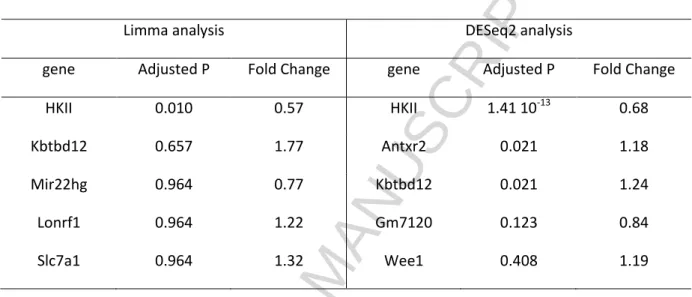
In order to study the consequences of deletion of one HKII copy at the transcriptome level, we performed RNAseq analysis of WT and HKII+/--hearts. Analysis of differentially expressed genes revealed a small impact of the partial HKII deletion, with only the decrease in HKII expression reaching the FDR cut off of 0.05 in both limma and DESeq2 (Table 3, showing the first 5 genes with largest tendencies of being differentially expressed between the genotypes). In DESeq2, two more genes, Antxr2 and Kbtbd12, were differentially expressed with small log2 fold changes (0.24 and 0.31, respectively). Given the lack of previous association of these genes with cardiac metabolism and the overall small changes in gene expression in the HKII+/--hearts, the identification of these transcripts as differentially expressed is likely spurious.
ACCEPTED MANUSCRIPT
13 4. Discussion
Although it is well recognized that the degree of mitochondrion-HKII association is sensitive to many cardiac pathologies and interventions (IR, cardioprotection, insulin treatment, diabetes, hypertrophy, heart failure [5]), the metabolic consequences for the intact heart are, surprisingly, to a large extent unknown. We now report that 1) acute disruption of HKII from mitochondria in intact hearts is associated with an increase in oxygen consumption and a nonsignificant trend of decreased glucose contribution to citrate synthase flux; 2) chronically reduced HKII (including reduced mtHKII) is not associated with alterations in cardiac oxygen consumption,metabolism, or metabolic reprogramming at the transcriptional level of the intact heart not cause.
4.1. Acute decreases in mitochondrial hexokinase II and cardiac metabolism
We have demonstrated that acute detachment of HKII from mitochondria increases cardiac oxygen consumption. We are unaware of other studies that have specifically examined the effects of mitochondria-HKII association on oxygen consumption. Our data indicates that HKII attachment to the mitochondria slows down mitochondrial respiration in a functional, working organ. This is commensurate with our previous observations that short periods of ischemia, able to induce cardioprotection and associated with increased mtHKII [25-27], result in slower mitochondrial activation of oxygen consumption [28, 29]. This slowing down was only observed with glucose, and not with lactate or pyruvate substrates, suggesting a glycolytic-origin, possibly HKII, of this effect. Since cardioprotection is associated with increased glucose metabolism [30], it is then suggested that increased glucose metabolism results in attenuated mitochondrial function. Indeed, in the present study increased binding of HKII to mitochondria was associated with a trend for increased glucose metabolism. It is generally believed that the physical binding of HK to mitochondria increases HK activity through decreasing its Km for ATP, relieve of allosteric inhibition by G6P and
glucose-1,6-ACCEPTED MANUSCRIPT
14
biphosphate, and preferential access to mitochondrial produced ATP [2, 31, 32]. HKII binding to mitochondria promoted glycolysis in CHO cells [11]. Increased glucose metabolism could potentially decrease mitochondrial oxygen consumption because of the higher P/O ratio for glucose relative to other substrates (less oxygen needed per ATP produced), or because of increased transport of cytosolic NADH into the mitochondria, which transport may impair oxygen consumption [33]. Alternatively, the increased oxygen consumption with similar cardiac function could also suggest a lower cardiac efficiency. Although care should be taken for over interpretation, the observed non-significant trend for decreased glucose uptake and increased tissue pyruvate concentration with less mtHKII, indicates that the decrease in glucose metabolism with detachment of mtHKII is controlled by impairment of glucose uptake and pyruvate oxidation within the intact heart. The increased oxygen consumption is then likely a result of a decrease in glucose metabolism, which, because no changes in mechanical work were observed, is then compensated with increased FA oxidation with its consequently higher oxygen need to provide similar ATP.
4.2. Chronic decreases in (mitochondrial) hexokinase II and cardiac metabolism
In contrast to the acute manipulation of mitochondrial HKII, we observed no changes in oxygen consumption and metabolism in the partial HKII knockout heart, which we previously showed to have 50% less total and mitochondrial HKII [18]. Since chronic reductions in proteins are notorious for inducing compensatory alterations in other genes/proteins, possibly explaining the lack of changes in cardiac metabolism and oxygen consumption with chronic reduction in HKII, we
characterized the cardiac transcriptome through RNA sequenching. The absence of differential gene expression demonstrated that compensation at the level of gene expression was absent, negating a role for gene expression adaptation in this model. However, at this stage, we cannot exclude that adaptation did occur posttranscriptonally (proteins and its posttranslational modifications). That cardiac glucose metabolism was unaffected in these partial HKII knockout hearts, is in support of
ACCEPTED MANUSCRIPT
15
previous findings using the similar partial HKII knockout and reporting unaltered cardiac glucose uptake during sedentary conditions in these hearts [8, 34].
4.3. No insulin versus high-dose insulin and cardiac glucose metabolism
In the present study we used perfusion conditions with and without insulin, as dictated by our previous experimental conditions where effects of the HKII manipulations were already determined [17, 18]. The absence of insulin, despite glucose being present at 11 mM, resulted in a rather low rate of anaerobic glycolysis (0.07 ± 0.03 µmol/min) and glucose contribution to acetyl-CoA formation for citrate synthesis (11.2 ± 2.2%), even when compared to other heart studies using glucose-only perfusates. Total carbohydrates (glucose, lactate, pyruvate) contribution to acetyl-CoA formation for citrate synthesis amounted to near 30%, suggesting that a major part of citrate synthesis was derived from endogenous FA oxidation from stored triglycerides. It was shown before that in conditions of glucose-only perfusate myocardial triglyceride turnover contributes significantly to energy
production in isolated hearts [35]. The low rates of glucose metabolism are likely due to the presence of 1 mM lactate; physiological concentrations of lactate decrease glucose metabolism to a large extent [36]. The data suggest that in perfusion conditions with glucose and lactate both being present, insulin is needed to facilitate glucose metabolism in the isolated heart. In the presence of insulin and FFA in the perfusate, carbohydrates and FFA acids contributed for 60% and 15%, respectively, to citrate synthesis in the intact hearts. These values are commensurate with values reported before in the isolated working rat heart [21], although much higher contributions of fatty acids have also commonly been observed. The relatively low fatty acid contribution could be due to the relatively high insulin concentration (100mU/L), the presence of 1 mM lactate (physiological concentration, and often neglected in isolated heart studies), knowing that lactate inhibits fatty acid oxidation [37], and the low, but physiologically-relevant for the fed condition [38], fatty acid
ACCEPTED MANUSCRIPT
16 4.4. Methodological considerations
In the current study the effects of modest changes (30-50% decrease) in HKII on cardiac metabolism were examined. These modest HKII changes probably partly explain why only modest or even no changes in cardiac metabolism were observed. Importantly, these modest changes in HKII mimic the changes in HKII that occur during pathophysiological interventions such as IR and ischemic
preconditioning [24 -26], type I and type II diabetes [4], insulin treatment [24, 39], or cardiac
remodeling during hypertrophy and heart failure [6, 40]. Were we to use a complete knockout of HKII or transgenic overexpression of HKII by more than a factor of 5, larger changes in metabolism would probably have occurred. However, we believe the imposed manipulations of HKII and their metabolic consequences in the current study are more relevant to the real life pathophysiological conditions.
In the present work effects of acutely reduced mtHKII on cardiac metabolism were examined during perfusion with a specific substrate composition of 11 mM glucose, 1 mM lactate and 0.1 mM
pyruvate. It is known that this mixture results in only small contributions of glucose to ATP production [36], as supported by our low rates of glycolysis and glucose contribution to citrate formation,
possibly explaining that we observed only non-significant trends for TAT-HKII effects on glucose metabolism. Therefore, it would be of interest to examine glucose contribution to citrate in the absence of lactate, or in the presence of insulin in the TATHKII series. Under these metabolic substrate conditions, glycolysis and glucose contribution to citrate would be more prominent making it perhaps easier to obtain significant effects of partly decreasing mt-HKII. Further research is needed to examine these questions. Finally, myocardial oxygen consumption was increased with acute detachment of HKII from mitochondria, but not with chronic reductions in mtHKII. New research should be directed to examine whether the acute increase in oxygen consumption is only a reversible phenomenon at maintained lower levels of mtHKII, possibly explaining that in longer-term oxygen consumption normalizes again to pre-treatment levels.
ACCEPTED MANUSCRIPT
17
In conclusion, our data suggest that an acute, but not chronic, decrease in HKII binding to
mitochondria increases cardiac oxygen consumption that is associated with a trend for decreased glucose contribution towards citrate synthesis in the mitochondria.
Authors Contributions
RN, SMH and CJZ contributed to the design and conduct of the study, data collection and analysis, data interpretation and manuscript writing; BL contributed to design and conduct of the study, data interpretation and manuscript writing; SD, JH, CA contributed to data collection and analysis, data interpretation and manuscript writing; CDR, ML, RW, RHH and MWH contributed to data
interpretation and manuscript writing. All authors approved the final version of the manuscript.
Funding:
This work was supported by a research grant (NHS2010B011) of the Dutch Heart Foundation.
Disclosure statement:
ACCEPTED MANUSCRIPT
18
Table 1. Physiological parameters of isolated mouse hearts and 13C enrichments of tissue pyruvate isolated from Langendorff-perfused mouse hearts perfused with [U-13C6]glucose.
TAT TATHKII WT HKII
+/-Heart weight (mg) 165±5 178±5 198±8 210±8
Coronary flow (ml/min/gww)
11.6±0.8 11.8±0.9 10.3±0.5 10.5±0.9
MPE pyruvate (M3) 38.2±2.9 34.8±1.9 44.4±5.1 37.0±3.0
Values are means ± SE. (n=8/9 for TAT/TATHKII hearts and n=16/17 for WT/HKII+/- hearts)
Table 2. Glucose uptake and 13C intermediates of metabolism derived from 13C enrichment
TAT TATHKII P value
Glucose uptake (µmol/min/Gww) 1.20 ± 0.59 0.27 ± 0.18 0.131 Pyruvic acid 20 ± 2 27 ± 2 0.033* Lactic acid 8301 ± 1251 6208 ± 691 0.135 Citrate 778 ± 58 843 ± 53 0.424 2-ketoglutaric acid 5 ± 0 6 ± 0 0.087 Malic acid 232 ± 37 183 ± 20 0.235 Succinic acid 566 ± 35 479 ± 28 0.068 Fumaric acid 29 ± 6 27 ± 4 0.761
ACCEPTED MANUSCRIPT
19
Table 3. RNA sequence and differential expression gene analysis results for two different analysis approaches (LImma and DESseq2). The first five genes with the largest differences in expression between genotypes are displayed.
Limma analysis DESeq2 analysis
gene Adjusted P Fold Change gene Adjusted P Fold Change
HKII 0.010 0.57 HKII 1.41 10-13 0.68
Kbtbd12 0.657 1.77 Antxr2 0.021 1.18
Mir22hg 0.964 0.77 Kbtbd12 0.021 1.24
Lonrf1 0.964 1.22 Gm7120 0.123 0.84
Slc7a1 0.964 1.32 Wee1 0.408 1.19
ACCEPTED MANUSCRIPT
20
Legends
Figure 1: TATHKII peptide effects on mechanical performance and oxygen consumption of isolated hearts. Langendorff-perfused hearts were treated for 25 min, starting from baseline, with 200 nM TAT (control group) or 200 nM TATHKII peptide, and effects on peak systolic left ventricular pressure (Peaksys; 1A), heart rate (1B), rate-pressure
product (1C) and oxygen consumption (1D). (n=8/9 per group). *P < 0.05 vs baseline or TAT.
Figure 2: TATHKII peptide effects on cardiac metabolism in isolated hearts perfused with 13C
glucose, lactate and pyruvate. TATHKII effects on anaerobic glycolysis (2A) and the contribution of carbohydrates to citrate synthesis, as assessed by the PDC/CS ratio for carbohydrates, lactate and glucose (2C), are given. (n=8/9 per group).
Figure 3: Mechanical performance and oxygen consumption of HKII+/- and WT isolated hearts. Peak systolic left ventricular pressure (Peaksys; 1A), heart rate (1B), rate-pressure
product (1C) and oxygen consumption (1D) for HKII+/- and WT hearts. (n=16/17 per group).
Figure 4: Cardiac metabolism in isolated HKII+/- and WT hearts perfused with 13C glucose or 13C palmitate, and lactate, pyruvate and insulin. Effects of HKII+/- on anaerobic glycolysis (4A), on the contribution of carbohydrates to citrate synthesis as assessed by the PDC/CS ratio for carbohydrates, lactate and glucose (4B), and on palmitate contribution to citrate synthesis (4C) are given. (n=7/8 per group).
ACCEPTED MANUSCRIPT
21 References
1. Smeele KM, ter Horst LH, Koeman A, Heikkinen S, Laakso M, Weber NC, Hollmann MW, Zuurbier CJ. The effect of standard chow and reduced hexokinase II on growth, cardiac and skeletal muscle hexokinase and low-flow cardiac ischaemia-reperfusion injury. Lab Anim 45: 160-166, 2011.
2. Wilson JE (1995). Hexokinases. Rev Physiol Biochem Pharmacol 126: 65-198
3. Mathupala SP, Ko YH, Pedersen PL. Hexokinase-2 bound to mitochondria: Cancer’s stygian link to the “Warburg effect” and a pivotal target for effective therapy. Semin Cancer Biol 19: 17-24, 2009.
4. Gürel E, Ustunova S, Kapucu A, Yilmazer N, Eerbeek O, Nederlof R, Hollmann MW, Demirci_Tansel C, Zuurbier CJ. Hexokinase cellular trafficking in ischemia-reperfusion and ischemic preconditioning is altered in type I diabetic heart. Mol Biol Rep 40: 4153-4160, 2013.
5. Nederlof R, Eerbeek O, Hollmann MW, Southworth R, Zuurbier CJ. Targeting hexokinase II to mitochondria to modulate energy metabolism and reduce ischaemia-reperfusion injury in the heart. Br J Pharmacol 171: 2067-2079, 2014.
6. Balestra GM, Mik EG, Eerbeek O, Specht PA, van der Laarse WJ, Zuurbier CJ. Increased in vivo mitochondrial oxygenation with right ventricular failure induced by pulmonary arterial hypertension: mitochondrial inhibition as driver of cardiac failure? Respir Res 16: 6, 2015. 7. Roberts DJ, Miyamoto S. Hexokinase II integrates energy metabolism and cellular protection:
Akting on mitochondria and TORCing to autophagy. Cell Death Differ 22: 248-257, 2015. 8. Fueger PT, Heikkinen S, bracey DP, Malabanan CM, Pencek RR, Laakso M, Wasserman DH.
Hexokinase II partial knockout impairs exercise-stimulated glucose uptake in oxidative muscles of mice. Am J Physiol Endocrinol Metab 285: E958-E963, 2003.
9. Fueger PT, Lee-Young RS, Shearer J, Bracy DP, Heikkinen S, Laakso M, Rottman JN,
Wasserman DH. Phosphorylation barriers to skeletal and cardiac muscle glucose uptakes in high-fat-fed-mice. Diabetes 56:2476-2484, 2007.
10. Wilson JE (2003). Isozymes of mammalian hexokinase: structure, subcellular localization and metabolic function. J Exp Biol 206:2049-2057.
11. John S, Weiss JN, Ribalet B. Subcellular localization of hexokinase I and II directs the metabolic fate of glucose. PLoSOne 6: e17674, 2011.
12. Calmettes G, Ribalet B, John S, Korge P, Ping P, Weiss JN. Hexokinase and cardioprotection. J Mol Cell Cardiol 78: 107-115, 2015.
ACCEPTED MANUSCRIPT
22
13. Zuurbier CJ, Smeele KM, Eerbeek O. Mitochondrial hexokinase and cardioprotection of the intact heart. J Bioeng Biomembr 41: 181-185, 2009.
14. Halestrap AP, Pereira GC, Pasdois P. The role of hexokinase in cardioprotection-mechanism and potential for translation. Br J Pharmacol 172: 2085-2100, 2015.
15. Burwell LS, Nadtochiy SM, Brookes PS. Cardioprotection by metabolic shut-down and gradual wake-up. J Mol Cell Cardiol 46: 804-810, 2009.
16. Pell VR, Chouchani ET, Murphy MP, Brookes PS, Krieg T. Moving forwards by blocking back-flow. The Yin and Yang of MI therapy. Circ Res 118: 989-906, 2016.
17. Smeele KM, Soutworth R, Wu R, Xie R, Nederlof R, Warley A, nelson JK, van Horssen P, van den Wijngaard JP, Heikkinen S, Laakso M, Koeman A, Siebes M, Eerbeek O, Akar FG, Ardehali H, Hollmann MW, Zuurbier CJ. Disruption of hexokinase II-mitochondrial binding blocks ischemic preconditioning and causes rapid cardiac necrosis. Circ Res 108:1165-1169, 2011.
18. Wu R, Smeele KM, Wyatt E, Ichikawa Y, Eerbeek O, Sun L, Chawla K, Hollmann MW, Nagpal V, Heikkinen S, Laakso M, Jujo K, Wasserstrom JA, Zuurbier CJ, Ardehali H. Reduction in hexokinase II levels results in decreased cardiac function and altered remodelling after ischemia-reperfusion injury. Circ Res 108: 60-69, 2011.
19. Ruiz M, Gelinas R, Vaillant F, Lauzier B, Des Rosiers C. Metabolic tracing using isotope-labeled substrates and mass in the perfused mouse heart. In: Methods in Enzymology, edited by Metallo CM. Amsterdam, The Netherlands: Elsevier, 2015, p.107-147.
20. Heikkinen S, Pietila M, Halmekyto M, Suppola S, Pirinen E, Deeb SS, Janne J, Laakso M. Hexokinase II-deficient mice: prenatal death of homozygotes without disturbances in glucose tolerance in heterozygotes. J Biol Chem 274: 22517-22523, 1999.
21. Vincent G, Bouchard B, Khairallah M, Des Rosiers C. Differential modulation of citrate synthesis and release by fatty acids in perfused working hearts. Am J Physiol Heart Circ Physiol 286:H257-H266, 2004.
22. Dobin A, Davis CA, Schlesinger F, Drenkow J, Zaleski C, Jha S, et al. STAR: ultrafast universal RNA-seq aligner. Bioinformatics. 2013;29(1):15-21. doi: 10.1093/bioinformatics/bts635. PubMed PMID: 23104886; PubMed Central PMCID: PMCPMC3530905.
23. Smyth GK. Limma: Linear Models for Microarray Data. In: Gentleman R, Carey VJ, Huber W, Irizarry RA, Dudoit S, editors. Bioinformatics and Computational Biology Solutions Using R and Bioconductor. New York, NY: Springer New York; 2005. p. 397-420.
24. Love MI, Huber W, Anders S. Moderated estimation of fold change and dispersion for RNA-seq data with DESeq2. Genome Biol. 2014;15(12):550. doi: 10.1186/s13059-014-0550-8. PubMed PMID: 25516281; PubMed Central PMCID: PMCPMC4302049.
25. Zuurbier CJ, Eerbeek O, Meijer AJ. Ischemic preconditioning, insulin, and morphine all cause hexokinase redistribution. Am J Physiol Heart Circ Physiol 289: H496-499, 2005.
ACCEPTED MANUSCRIPT
23
26. Gürel E, Smeele KM, Eerbeek O, Koeman A, Demirci C, Hollmann MW, Zuurbier CJ. Ischemic preconditioning affects hexokinase activity and HKII in different subcellular compartments throughout cardiac ischemia-reperfusion. J Appl Physiol 106: 1909-1916, 2009.
27. Pasdois P, Parker JE, Halestrap AP. Extent of mitochondrial hexokinase II dissociation during ischemia correlates with mitochondrial cytochrome c release, reactive oxygen species production, and infarct size on reperfusion. J Am Heart Assoc 2: e005645, 2012.
28. Zuurbier CJ, Ince C. Post-ischaemic changes in the response time of oxygen consumption to demand in the isolated rat heart are mediated partly by calcium and glycolysis. Pflügers Arch 443: 908-916, 2002.
29. Zuurbier CJ, van Beek JHGM. Mitochondrial response to heart rate steps in isolated rabbit heart is slowed after myocardial stunning. Circ Res 81: 69-75, 1997.
30. Ji L, Zhang X, Liu W, Huang Q, Yang W, Fu F, Ma H, Su H, Wang H, Wang J, Zhang H, Gao F. AMPK-regulated and Akt-dependent enhancement of glucose uptake is essential in ischemic preconditioning-alleviated reperfusion injury. PloSOne 8: e69910, 2013.
31. Beitner R, Lilling G. Inhibition of mitochondrial and soluble hexokinase from various rat tissues by glucose 1,6-biphosphate. Int J Biochem 16: 991-996, 1984.
32. Lynch RM, Carrington W, Fogarty KE, Fay FS. Metabolic modulation of hexokinase association with mitochondria in living smooth muscle cells. Am J Physiol Cell Physiol 270: C488-C499, 1996.
33. Kobayashi K, Neely JR. Control of maximum rates of glycolysis in rat cardiac muscle. Circ Res 44: 166-175, 1979.
34. Heikkinen S, Pietila M, Halmekyto M, Suppola S, Pirinen E, Deeb SS, Janne J, Laakso M. Hexokinase II-deficient mice. Prenatal death of homozygotes without disturbances in glucose tolerance in heterozygotes. J Biol Chem 274: 22517-22523, 1999.
35. Saddik M, Lopaschuk GD. Myocardial triglyceride turnover and contribution to energy substrate utilization in isolated working rat hearts. J Biol Chem 266:8162-8170, 1991. 36. Chatham JC, Gao ZP, Bonen A, Forder JR. Preferential inhibition of lactate oxidation relative
to glucose oxidation in the rat heart following diabetes. Cardiovasc Res 43: 96-106, 1999. 37. Van der Vusse GJ and de Groot MJ. Interrelationship between lactate and cardiac fatty acid
metabolism. Mol Cell Biochem 116:11-17, 1992.
38. Ruiz M, Coderre L, Lachance D, Houde V, Martel C, Thompson Legault J, Gillis MA, Bouchard B, Daneault C, Carpentier AC, Gaestel M, Allen BG, Des Rosiers C. MK2 deletion in mice prevents diabetes-induced perturbations in lipid metabolism and cardiac dysfunction. Diabetes 65: 381-392, 2016.
39. Southworth R, Davey KA, Warley A, Garlick PB. A reevaluation of the roles of hexokinase I and hexokinase II in the heart. Am J Physiol Heart Circ Physiol 292: H378-H386, 2007.
ACCEPTED MANUSCRIPT
24
40. Riehle C, Wende AR, Zaha VG, Pires KM, Wayment B, Olsen C et al. PCG-1β deficiency accelerates the transition to heart failure in pressure overload hypertrophy. Circ Res 109: 783-793, 2011.
ACCEPTED MANUSCRIPT
25
Figure 1
ACCEPTED MANUSCRIPT
26
Figure 2
ACCEPTED MANUSCRIPT
27
Figure 3
ACCEPTED MANUSCRIPT
28
Figure 4
ACCEPTED MANUSCRIPT
29 Highlights
Acute detachment of Hexokinase II from mitochondria increases cardiac oxygen consumption
Acute detachment of hexokinase II from mitochondria trends to decrease glucose metabolism
Chronic decrease in hexokinase II is without effect on cardiac metabolism
![Table 1. Physiological parameters of isolated mouse hearts and 13 C enrichments of tissue pyruvate isolated from Langendorff-perfused mouse hearts perfused with [U- 13 C 6 ]glucose](https://thumb-eu.123doks.com/thumbv2/123doknet/11499788.293570/21.892.105.680.586.939/physiological-parameters-isolated-enrichments-pyruvate-isolated-langendorff-perfused.webp)