HAL Id: hal-03228637
https://hal.sorbonne-universite.fr/hal-03228637
Submitted on 18 May 2021
HAL is a multi-disciplinary open access
archive for the deposit and dissemination of
sci-entific research documents, whether they are
pub-lished or not. The documents may come from
teaching and research institutions in France or
abroad, or from public or private research centers.
L’archive ouverte pluridisciplinaire HAL, est
destinée au dépôt et à la diffusion de documents
scientifiques de niveau recherche, publiés ou non,
émanant des établissements d’enseignement et de
recherche français ou étrangers, des laboratoires
publics ou privés.
development of pathological features in a rat model of
Charcot-Marie-Tooth disease 1 A
Benoit Gautier, Helene Hajjar, Sylvia Soares, Jade Berthelot, Marie Deck,
Scarlette Abbou, Graham Campbell, Maria Ceprian, Sergio Gonzalez,
Claire-Maëlle Fovet, et al.
To cite this version:
Benoit Gautier, Helene Hajjar, Sylvia Soares, Jade Berthelot, Marie Deck, et al.. AAV2/9-mediated
silencing of PMP22 prevents the development of pathological features in a rat model of
Charcot-Marie-Tooth disease 1 A. Nature Communications, Nature Publishing Group, 2021, 12 (1), pp.2356.
�10.1038/s41467-021-22593-3�. �hal-03228637�
AAV2/9-mediated silencing of PMP22 prevents
the development of pathological features in a rat
model of Charcot-Marie-Tooth disease 1 A
Benoit Gautier
1
✉
, Helene Hajjar
1,10
, Sylvia Soares
2
, Jade Berthelot
1
, Marie Deck
1
, Scarlette Abbou
1
,
Graham Campbell
1
, Maria Ceprian
1,11
, Sergio Gonzalez
1,11
, Claire-Maëlle Fovet
3
, Vlad Schütza
4
,
Antoine Jouvenel
1
, Cyril Rivat
1
, Michel Zerah
5
, Virginie François
6
, Caroline Le Guiner
6
, Patrick Aubourg
7,8
,
Robert Fledrich
4
✉
& Nicolas Tricaud
1,9,11
✉
Charcot-Marie-Tooth disease 1 A (CMT1A) results from a duplication of the PMP22 gene in
Schwann cells and a de
ficit of myelination in peripheral nerves. Patients with CMT1A have
reduced nerve conduction velocity, muscle wasting, hand and foot deformations and foot
drop walking. Here, we evaluate the safety and ef
ficacy of recombinant adeno-associated
viral vector serotype 9 (AAV2/9) expressing GFP and shRNAs targeting Pmp22 mRNA in
animal models of Charcot-Marie-Tooth disease 1 A. Intra-nerve delivery of AAV2/9 in the
sciatic nerve allowed widespread transgene expression in resident myelinating Schwann cells
in mice, rats and non-human primates. A bilateral treatment restore expression levels of
PMP22 comparable to wild-type conditions, resulting in increased myelination and prevention
of motor and sensory impairments over a twelve-months period in a rat model of CMT1A.
We observed limited off-target transduction and immune response using the intra-nerve
delivery route. A combination of previously characterized human skin biomarkers is able to
discriminate between treated and untreated animals, indicating their potential use as part of
outcome measures.
https://doi.org/10.1038/s41467-021-22593-3
OPEN
1INM, Univ. Montpellier, INSERM, Montpellier, France.2Sorbonne Université, CNRS, INSERM, IBPS, Neuroscience Paris Seine, Paris, France.3INSERM U1184,
Immunology of Viral, Auto-immune, Hematological and Bacterial Diseases (ImVA-HB), IDMIT Department, CEA, Fontenay-Aux-Roses, France.4Institute of Anatomy, Leipzig University, Leipzig, Germany.5Paediatric Neurosurgery Department, Université Paris Descartes and Assistance Publique-Hôpitaux de Paris, Hôpital Universitaire Necker, Paris, France.6INSERM UMR 1089, Université de Nantes, CHU de Nantes, Nantes, France.7Department of Paediatric Neurology, Centre Hospitalier Universitaire de Bicêtre, Le Kremlin-Bicètre, France.8INSERM U1169, Université Paris-Sud, Orsay, France.9I-Stem, UEVE/UPS U861,
INSERM U861, AFM, Corbeil-Essonnes, France.10Present address: Institute of Regenerative Medicine and Biotherapies (IRMB), University of Montpellier,
INSERM, Montpellier, France.11Present address: Laboratory of Pathogen-Host Interactions (LPHI), University of Montpellier, Montpellier, France.
✉email:benoit.gautier@inserm.fr;robert.fledrich@medizin.uni-leipzig.de;nicolas.tricaud@inserm.fr
123456789
P
eripheral nerves that bundle axons emanating from
neu-ronal cell bodies are found throughout the body forming
the peripheral nervous system (PNS). These axons are
covered with Schwann cells and those of large caliber are wrapped
in a myelin sheath made by myelinating Schwann cells (mSC) to
assure an extremely fast nerve conduction (up to 100 m/s). The
myelin forms several successive segments named internodes,
which electrically isolate the axonal membrane except at
unmyelinated nodes of Ranvier where action potentials are
propagated
1.
This myelin sheath is critical for both motor and sensory
functions in humans. Indeed, several peripheral nerve diseases
impair the myelin sheath leading to reduced nerve conduction
velocity, nerve dysfunction, muscle wasting, limb extremities
deformations and walking and sensory problems
2. The large
majority of patients suffering from hereditary diseases of
per-ipheral nerves, namely Charcot-Marie-Tooth diseases (CMT),
have defects in the myelin sheath formation, function or
maintenance
3. The most common of these myelin-related CMT
diseases is CMT1A (prevalence: 0.5–1.5/10,000)
4,5. This disease,
resulting from the duplication of the PMP22 gene, is
character-ized by a set of heterogeneous symptoms, the most serious being
feet and hands deformation and walking impairment
4. PMP22
protein is a transmembrane glycoprotein located in the myelin
sheath. In CMT1A, PMP22 protein overexpression in Schwann
cells leads to defects in the number of myelinated segments that
are formed, short internodes, myelin sheath defects, myelin
degeneration (demyelination) and
finally axonal loss
6.
No cure exists for this disease, however, pharmacological
treatments have been recently investigated
4. Some preclinical
studies in rodents demonstrated that antisense oligonucleotides
targeting Pmp22 mRNA expression significantly improved the
phenotype
7–9, suggesting PMP22 silencing is an effective way to
tackle the disease. Feeding a phospholipid enriched diet to a rat
model of CMT1A increased myelination, prevented axonal loss
and dramatically delayed the occurrence of the disease
10.
How-ever, the benefit only lasted as long as the treatment was
administrated.
Gene therapy therefore constitutes an alternative therapy
allowing for long term benefits for patients
11. Proofs of concept
for gene therapy using lentiviral vectors injected in the spinal cord
of myelin-related CMT mouse models have been reported
12–14.
However, currently, in vivo gene therapy assays mostly use
adeno-associated virus (AAV)-based strategies as these vectors do
not integrate the genome of transduced cells, spread more easily
in the tissues and display a limited immunogenicity
15. AAV
serotypes 2/9 and 2/rh10 are commonly used to transduce the
central nervous system and several clinical trials are ongoing with
promising results
11,16. Moreover, an AAV2/9-based therapy
recently obtained market authorization from FDA to treat spinal
motor atrophy in infants
17. Gene therapy may therefore represent
an efficient and safe way to treat CMT1A in the long term.
However, the pattern of transduction of Schwann cells with
AAV2/9 or rh10 remains unclear.
Recent clinical trials for peripheral neuropathies, and in
par-ticular for CMT1A, have shown that the chronicity of the disease
makes the evaluation of the treatment outcome very difficult.
Composite scores have been shown to be the most reliable
out-come measure, but they remain poorly discriminant
18–20.
Mole-cular biomarkers have been characterized for CMT1A and some
of them allow discrimination on the basis of disease severity
21.
However, none of them has been validated yet as outcome
measure for a therapy.
Here we show that mSC of mouse, rat and non-human primate
sciatic nerves are widely and specifically transduced by AAV2/9
when injected directly into the nerve. Using an AAV2/9
expressing a small hairpin inhibitory RNA (shRNA) directed
against Pmp22 mRNA, we treated a rat model of CMT1A when
myelination begins. A single injection treatment in both sciatic
nerves prevented the disease symptoms for at least one year. The
combined expressions of human biomarkers in the paw skin of
rats correlated with the phenotype and allowed discrimination
between treated animals and their sham-treated littermates,
indicating that these markers can be used as an outcome measure
of the treatment. In addition, the dispersion of the vector
remained limited to the injected nerves and the humoral immune
response generated against the vector remained barely detectable
in injected animals. Taken together, this work suggests that an
intra-nerve AAV2/9-mediated gene therapy represents an
effec-tive and attraceffec-tive therapy for myelin-related CMT diseases.
Results
Broad and specific transduction of mSC is reached after AAV2/
9 injection in sciatic nerves of mammals. As the literature
reported a limited transduction efficiency with several AAV
serotypes
22,23, we investigated the transduction efficiency of
recombinant single-stranded AAV2/9 and AAV2/rh10 vectors
expressing enhanced GFP under a CAG promoter after injection
into the sciatic nerves of mice and rats. These injections were
done using a non-traumatic microinjection protocol previously
described
24. Briefly, fine glass needles containing a viral solution
stained with Fast Green were introduced in the sciatic nerve and
the solution was slowly injected using multiple short-time
pres-sure pulses
24. In these conditions, we observed a high amount of
transduced GFP-expressing cells along the nerve of both species
(Fig.
1
a and Supplementary Fig. S1). To analyze the nature of
these cells, we
first imaged injected sciatic nerves using Coherent
Antistokes Raman Scattering (CARS) non-linear microscopy
technique
25. This allows myelin sheath imaging without any
labeling
25. Using this technique on intact nerves in a longitudinal
view, we observed that the majority of transduced cells were
myelinated cells (Fig.
1
b) and therefore mSC. In addition, typical
morphological characteristics of mSC were seen through GFP
labeling and/or subcellular markers: Schmidt-Lanterman
inci-sures (Supplementary Fig. S2a and b, white arrows), Cajal’s bands
(Supplementary Fig. S2a, blue arrows) and paranodal loops
sur-rounding the node of Ranvier (Supplementary Fig. S2a and c,
arrowheads). To go further, sciatic nerve cross-sections were
immunostained for axonal (Tuj1) and myelin (MBP) markers
allowing to definitively identify mSC as GFP-labeled
MBP-posi-tive cells surrounding axons (Fig.
1
c). As some non-myelinated
cells (nmc) located in the nerve, such as non-myelinating
Schwann cells and
fibroblasts, are not easily seen in
cross-sec-tions, we teased sciatic nerve
fibers on a glass slide and identified
GFP-positive mSC, axons and nmc using their morphology and/
or specific markers (Supplementary Fig. S3). On 100 cells
trans-duced with AAV2/9-CAG-GFP vector after intra-nerve injection
in rat pup, 95 were mSC, 4 nmc and 1 was an axon (Table
1
). The
same specificity was observed after intra-nerve injection in adult
and pup rat and mouse for AAV2/9-CAG-GFP and in adult
mouse and rat pup for AAV2/rh10-CAG-GFP (Table
1
). Taken
together the data highlight the specificity of AAV2/9 and AAV2/
rh10 for mSC after intra-nerve injection.
Next, as each large axon is surrounded by mSC in nerves, we
used nerve cross-sections immunostained for MBP and Tuj1 to
measure the number of transduced mSC (GFP and MBP positive)
surrounding axons (Tuj1 positive), as a measure of the
transduction rate of mSC. Injections of 5 × 10
10vg (vector
genome)/nerve and 1.8 × 10
11vg/nerve of AAV2/9-CAG-GFP in
adult mouse and rat resulted in the transduction of 93% and 80%
transduction efficiencies were also obtained after intra-nerve
injection of AAV2/9-CAG-GFP in newborn mouse and rat nerves
when myelination starts (Table
2
). The injection of the same
amounts of AAV2/rh10-CAG-GFP vector in adult mouse nerves
and newborn rat nerves resulted in a lower transduction
efficiency (Table
2
).
In order to document the vectors ability to transduce primate
mSC in vivo, we used a regular 22 G syringe to inject 5 × 10
12vg/
nerve of each vector into the sciatic nerve of an adult non-human
primate (NHP). As described in rodents, immunostaining of
nerve cross-sections indicated that both vector transduced mSC
in these conditions (Supplementary Fig. S4). Calculating the
transduction rate as described previously, a high transduction rate
for mSC was observed using AAV2/9-CAG-GFP but not using
AAV2/rh10-CAG-GFP (Supplementary Fig. S4, Supplementary
Table S1).
We next evaluated the diffusion of the vectors along mouse and
rat sciatic nerves collecting injected nerve samples proximally and
distally from the injection site in order to cover the full length of
the nerve (Supplementary Fig. S5). This diffusion was very
significant for AAV2/9-CAG-GFP as the average transduction
rate was 73% at these two distant points in both mice and rats
(Table
2
). In the NHP sciatic nerve injected with
AAV2/9-CAG-GFP vector, the diffusion was evaluated at three distant points
distally and proximally of the injection point covering 30 to 50%
of the nerve length. The transduction rate was 68% at the
injection site, 69% 2 cm distally, 55% 2 cm proximally and 21%
4 cm proximally (Supplementary Table S1).
Designing and validating a small hairpin inhibitory RNA to
downregulate
Pmp22 expression in CMT1A rat model. The
high transduction rate of mSC in mouse, rat and NHP by AAV2/
Table 1 Speci
ficity of the transduction pattern after
intra-nerve injection of AAV2/9 and AAV2/rh10-CAG-GFP in
rodents.
Cellular specificity of the transduction pattern after intra-nerve injection (%)
AAV2/9-CAG-GFP AAV2/rh10-CAG-GFP Mouse Rat Mouse Rat Adult Pup Adult Pup Adult Pup mSC 97 ± 3 87 ± 12 89 ± 9 95 ± 2 82 ± 17 95 ± 1 nmc 3 ± 3 2 ± 2 7 ± 5 4 ± 1 4 ± 4 4 ± 1 Axons 0 11 ± 11 4 ± 4 1 ± 1 14 ± 13 1 ± 1
Animal groups were composed of three animals. Mouse and rat pups were injected at P2-P3 and P6-P7 respectively. Adult mice and rats were injected at 2-3 months old. All animals were sacrificed one month post-injection. Sciatic nerves were then teased and analyzed. The specificity is the ratio of mSC (GFP and MBP positive cells), nmc (GFP and glial fibrillary acidic protein, GFAP, positive cells) or axons (GFP and Neurofilament, NF, positive cells) transduced on the overall number of transduced cells (GFP positive cells). The results are expressed as the mean ± SD. Source data are provided as a Source Datafile.
Table 2 Quanti
fication of the transduction pattern after
intra-nerve injection of AAV2/9 and AAV2/rh10-CAG-GFP
in rodents.
Transduction rate after intra-nerve injection (%) AAV2/9-CAG-GFP
AAV2/rh10-CAG-GFP
Mouse Rat Mouse Rat Adult Pup Adult Pup Adult Pup Proximally 63 ± 24 74 ± 7 NA 81 ± 7 42 ± 22 53 ± 3 Injection site 93 ± 2 85 ± 15 80 ± 14 87 ± 1 51 ± 11 46 ± 5 Distally 91 ± 2 74 ± 14 NA 50 ± 24 42 ± 16 5 ± 6
Animal groups were composed of three animals. Mouse and rat pups were injected at P2-P3 and P6-P7 respectively. Adult mice and rats were injected at 2-3 months old. All animals were sacrificed one month post-injection. The transduction rate is the percentage of transduced mSC (GFP and MBP positive cells surrounding Tuj1 positive axons) on the overall number of mSC (MBP positive cells surrounding Tuj1 positive axons) per section. Proximal distances of the injection site were 2 cm for mice and 3 cm for rats. Distal distances of the injection site were 0.5 cm for mice and 1 cm for rats. The results are expressed as the mean ± SD. NA, Not available (not done). Source data are provided as a Source Datafile.
Fig. 1 AAV2/9 and AAV2/rh10-CAG-GFP transduce mSC after intra-nerve injection in mouse and rat. a Representative images of sciatic intra-nerve cross-sections showing GFP protein expression at the injection site after intra-nerve injection of AAV2/9 and AAV2/rh10-CAG-GFP in rat pup (P6-P7, 1 × 10¹¹vg/nerve in 8µl, n = 3 animals per group). Control represents a
rat pup sciatic nerve injected with Fast Green alone. All animals were sacrificed one month post-injection. Inserts show the circular shape of transduced cells. Scale bars: 50µm and 10 µm for the inserts. b GFP (green) and CARS (red) imaging largely overlap in a sciatic nerve injected with AAV2/9-CAG-GFP in rat pup (P6-P7, 1 × 10¹¹vg/nerve in 8µl, n = 3
animals) twelve months post-injection. Scale bar: 100µm. c Mouse and rat sciatic nerves cross-sections immunostained for myelin MBP (red) and axonal Tuj1 (blue) after AAV2/9-CAG-GFP injection (adult mouse injected at 2–3 months old with 5 × 10¹⁰vg/nerve in 8µl, rat pup injected at P6-P7
with 1 × 10¹¹vg/nerve in 8µl, n = 3 animals for each species). GFP (green)
partially colocalizes with MBP and both surround axons. Animals were sacrificed one month post-injection. Scale bar: 4 µm (mouse) and 2 µm (rat).
9 prompted us to evaluate its use as a vector to carry a therapeutic
tool into defective mSC in CMT1A disease. As CMT1A results
from PMP22 overexpression, we looked for shRNAs targeting
human PMP22 mRNA in order to decrease PMP22 expression in
CMT1A mSC. Two independent shRNAs or a control shRNA
with no target were cloned in a pAAV plasmid under a U6
promoter followed by a CMV-GFP reporter cassette. Both
shRNAs were found to be effective in reducing human PMP22 in
HEK293 cells (Supplementary Fig. S6a), showing that decreasing
PMP22 expression in mSC may represent a relevant therapeutic
approach for CMT1A disease.
To go further and perform a proof of concept, we chose the rat
model of CMT1A in which mouse Pmp22 mRNA is
overexpressed
26. Indeed, this model mimics the clinical aspects
of the human disease more closely than any others: immediately
after peripheral nerve myelination begins, nerves show
hypo-myelination, shorter internodal lengths, deficit in large fibers,
hypermyelination of small
fibers, myelin sheath defects and
demyelination
27–29. Two independent shRNAs (sh1 and sh2),
cloned in pAAV as described previously, were found to
significantly decrease mouse PMP22 protein level in a mouse
Schwann cell line in a dose-dependent manner (Supplementary
Fig. S6b). Only sh1 was effective on rat PMP22 level
(Supplementary Fig. S6c) and both were ineffective to
down-regulate human PMP22 level (Supplementary Fig. S6a).
AAV2/9 vectors expressing sh1 or sh2 (AAV2/9-sh1 and
AAV2/9-sh2) were then manufactured and injected bilaterally
(1 × 10
11vg/nerve) in the sciatic nerves of CMT1A rat pups
(P6-P7) (groups CMT1A sh1 and CMT1A sh2 respectively). WT
littermates and CMT1A rat pups injected with AAV2/9 carrying a
control shRNA (AAV2/9-ctr.sh) were used as controls (groups
WT ctr.sh and CMT1A ctr.sh respectively). Treated and control
animals were followed for as long as twelve months after injection.
We
first investigated the efficiency of both AAV2/9-sh1 and
AAV2/9-sh2 vectors to reduce PMP22 expression in mSC in vivo.
At the mRNA level, mouse but not rat Pmp22 mRNA was
upregulated relative to myelin marker Mpz in control CMT1A
rats, resulting in an overall higher Pmp22 mRNA expression
(Supplementary Fig. S7). However, neither AAV2/9-sh1 nor
AAV2/9-sh2 treatment downregulated mouse or rat Pmp22
mRNAs expression (Supplementary Fig. S7). At the protein level,
Western blot analysis showed that PMP22 level normalized on
the myelin marker MPZ was increased in CMT1A ctr.sh
compared to WT ctr.sh (Fig.
2
a, b). The transduction of mSC
by AAV2/9-sh1 and AAV2/9-sh2 decreased PMP22 level back to
control WT ctr.sh (Fig.
2
a, b). No downregulation beyond that of
control levels was observed in treated CMT1A animals. Sciatic
nerve sections immunostained for PMP22 also showed a
decreased PMP22 protein expression in mSC of treated animals
(Supplementary Fig. S8). Taken together these data showed that
AAV2/9 vectors carrying shRNAs targeting Pmp22 were able to
prevent PMP22 overexpression in mSC of CMT1A rats through a
mechanism independent of the mRNA stability.
AAV2/9-sh1 and -sh2 reduce myelinated
fiber defects in the
sciatic nerve of CMT1A rats. As CMT1A disease affects mSC
number and therefore myelin amount in nerves, we used Western
blot to measure the amount of myelin marker MPZ over the total
amount of protein after AAV2/9-sh1 and -sh2 treatment (Fig.
3
a).
While CMT1A rats showed a lower level of MPZ than WT ctr.sh
rats, MPZ levels in CMT1A sh1 and sh2 rats were not statistically
different from WT ctr.sh rats (Fig.
3
b).
Second, we focused on the specific myelin sheath defects
observed in the CMT1A rat model using CARS imaging on
freshly
fixed nerves. As described in detail previously
25, the
CMT1A myelin sheath showed a high heterogeneity compared to
WT rat littermates: thin myelin sheath or demyelination (Fig.
4
a,
white arrows), focal hypermyelination (Fig.
4
a, blue arrows), and
myelin degeneration with ovoids formation (Fig.
4
a, stars). While
still present, all these defective features were less abundant in
CMT1A rats treated with AAV2/9-sh1 or
–sh2 (Fig.
4
a). A typical
feature of CMT1A nerves is the shorter internodal distance
29,30.
Using CARS, we found that the number of nodes of Ranvier
(Fig.
4
a, b, arrowheads) per
fiber increased in CMT1A ctr.sh
nerves compared to WT ctr.sh nerves (Fig.
4
b, c), indicating
shorter internodes. When CMT1A rats were treated with AAV2/
9-sh1 or -sh2, the number of nodes of Ranvier per
fiber decreased
significantly, showing that internodes were longer (Fig.
4
c).
Fig. 2 Intra-nerve injections of AAV2/9-sh1 and sh2 prevent PMP22 overexpression in CMT1A sciatic nerves. a Representative image of Western blot showing PMP22 and MPZ protein levels (upper panel) and total protein as loading control (lower panel) in rat sciatic nerve lysates from WT ctr.sh, CMT1A ctr.sh, CMT1A sh1 or CMT1A sh2 three months after injection.b Graph shows the average level of PMP22 / MPZ ratio in sciatic nerve lysates normalized to total protein loaded as loading control (n= 7 animals per group). Statistical test shows one-way ANOVA followed by Tukey’s post hoc, two-sided. **p = 0.0027 between WT ctr.sh and CMT1A ctr.sh, **p= 0.0056 between CMT1A ctr.sh and CMT1A sh1, **p = 0.008 between CMT1A ctr.sh and CMT1A sh2; ns, not significant; arb. units, arbitrary unit. All error bars show SEM. Source data are provided as a Source Datafile.
Finally, we investigated the myelin sheath structure using
electron microscopy semi-thin nerve sections. Light microscopy
imaging of these sections showed typical features of rat CMT1A
disease: lower density of myelinated
fibers, hypomyelination
(Fig.
4
d, green arrows) or demyelination of large axons (Fig.
4
d,
orange stars) and hypermyelination of small axons (Fig.
4
d, blue
stars). These features were still present in the nerve of treated
animals but at lesser extent (Fig.
4
d). We found that AAV2/9-sh1
and -sh2 treatment significantly increased the myelinated fibers
density (Fig.
4
e), the number of large myelinated axons and
proportionally reduced the number of small myelinated axons
(Fig.
4
f). Finally, following treatment, the g-ratio (axon diameter/
myelinated
fiber diameter) was increased for small caliber axons
and decreased for large caliber axons (Fig.
4
g), which corrected
values toward control WT ctr.sh values. Taking together, these
data indicate that the treatment with AAV2/9-sh1 or -sh2
prevents myelin loss and the occurrence of myelinated
fiber
defects in CMT1A rats.
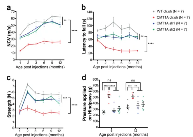
AAV2/9-sh1 and -sh2 treatments prevent motor and sensitive
defects on the long term in CMT1A rats. As CMT1A is a
myelin-related disease, one of the
first symptoms to occur is the
decrease of the nerve conduction velocity (NCV). Accordingly,
NCV was significantly reduced in CMT1A ctr.sh animals as soon
as one month after birth compared to control WT ctr.sh animals
(Fig.
5
a). When CMT1A animals were treated with AAV2/9-sh1
or-sh2, the NCV remained close to WT ctr.sh values at all-time
points for at least twelve months (Fig.
5
a).
The strong decrease of NCV in diseased animals correlated
with a clumsy behavior when animals crossed a narrow beam and
the treatment prevented this defect (Supplementary Videos 1
and 2). Motor behavior tests of rotarod and grip strength also
showed reduced performances of CMT1A ctr.sh animals
compared to control WT ctr.sh animals, starting two months
after birth and these deficiencies were largely prevented by the
treatment with AAV2/9-sh1 and
–sh2 (Fig.
5
b, c). No significant
evidence of a reduction in the effectiveness of the treatment could
be observed even twelve months after the treatment.
CMT1A disease shows a reduced sensory perception due to
both a defect in myelinated sensory
fibers and a loss of sensory
axons on the long term
31. Consequently, we performed
Randall-Selitto test to assess the effects of the therapeutic vectors on the
mechanical pain sensitivity of lower limbs at six months and
twelve months post-injection (Fig.
5
d). We
first observed an
increase in the mechanical nociceptive threshold of CMT1A ctr.
sh animals as compared to the WT ctr.sh group indicating
hypoalgesia. Treatment with both AAV2/9-sh1 and
–sh2
completely prevented this sensory deficit (Fig.
5
d). Consequently,
treatment with AAV2/9 vectors that reduces PMP22 levels in
mSC constitutes an efficient and long-term preventive treatment
for CMT1A symptoms in rats.
Human skin biomarkers reliably and robustly discriminate
treated from sham and wild-type animals. Recent clinical trials
for drugs targeting CMT diseases have illustrated the lack of
outcome measures sensitive enough for such chronic peripheral
neuropathies
32,33. We therefore tested whether recently
dis-covered human skin mRNA biomarkers
21could be used as an
outcome measure for our gene therapy efficiency. Forepaw
glabrous skin was collected at 12 months in all cohorts and the
expression of nine identified biomarkers was quantified using
RT-qPCR. Some of these biomarker expressions were significantly
differentially expressed in the skin of CMT1A sh1 and CMT1A
sh2 animals versus CMT1A ctr.sh animals (Supplementary
Fig. S9). However, the expression levels remained highly
hetero-geneous among animals, reflecting the variability of CMT1A
disease expression that is recapitulated in transgenic rats
28. We
therefore performed a multi-variated principle component
ana-lysis (PCA), including the entire battery of nine validated human
biomarkers. In this analysis, biomarkers allowed a significant
segregation of WT ctr.sh animals from CMT1A ctr.sh animals,
and of CMT1A sh1 and CMT1A sh2 animals from CMT1A ctr.sh
animals (Fig.
6
a). These data indicated that biomarkers
expres-sion in the paw skin allows for detecting the outcome of the gene
therapy treatment in CMT1A rats.
To go further, we performed a correlation analysis including
both functional phenotypes (Rotarod, grip strength test, Nerve
Conduction Velocity and Randall-Selitto test) and biomarkers
expression at 12 months. While correlations were more
significant (stars and large disk size) within the phenotypic test
group or more numerous within the biomarkers group (11/27),
significant correlations occurred between functional phenotypes
and biomarkers groups (10/27) (Fig.
6
b). Nrg1.1 (3 correlations),
Gria1 (2), Cda (2), Gstt2 (1) Anpep (1) and Enpp1 (1) were the
genes whose skin expression correlated the best with the
phenotype improvement of CMT1A rats following gene therapy.
Fig. 3 Intra-nerve injections of AAV2/9-sh1 and -sh2 in CMT1A rats normalize MPZ expression back to WT rat level. a Representative images of Western blot showing MPZ protein levels (upper panel) and total protein as loading control (lower panel) in sciatic nerve lysates from WT ctr.sh, CMT1A ctr.sh, CMT1A sh1 or CMT1A sh2 three months after injection.b Graph shows mean MPZ level in sciatic nerve lysates normalized to total protein level loaded as loading control (n= 7 animals per group). Statistical test shows one-way ANOVA followed by Tukey’s post hoc, two-sided. *p = 0.0122 between WT ctr.sh and CMT1A ctr.sh, **p= 0.0028 between CMT1A ctr.sh and CMT1A sh1, **p = 0.0022 between CMT1A ctr.sh and CMT1A sh2; ns, not significant; arb. units, arbitrary unit. All error bars show SEM. Source data are provided as a Source Data file.
Indeed, when the 3 most relevant biomarkers (Nrg1.1, Gria1 and
Cda) were selected for the PCA analysis, the segregation between
CMT1A ctr.sh and CMT1A sh1 and CMT1A sh2 animals was
clear (Fig.
6
c). According to the correlation matrix, the grip
strength test (4 correlations) was slightly more relevant than
Rotarod (3), Randall-Selitto (2) and NCV (1) tests. However,
Rotarod (3) and Randall-Selitto (2) tests were sufficient to
robustly cover the 3 most relevant biomarkers (Fig.
6
b). Taken
together these data indicate that a combinative score grouping
functional tests and skin biomarkers analysis constitute a reliable
and robust measure of CMT1A gene therapy outcome in
CMT1A rats.
Fig. 4 Intra-nerve injections of AAV2/9-sh1 and sh2 prevent the myelin sheath defects in CMT1A rats. a, b Representative images of CARS imaging on WT ctr.sh, CMT1A ctr.sh, CMT1A sh1 and CMT1A sh2 rat sciatic nerves twelve months post-injection (n= 6 animals for WT ctr.sh, n = 8 animals for CMT1A ctr.sh, n= 5 animals for CMT1A sh1 and n = 5 animals for CMT1A sh2). a CMT1A ctr.sh nerve shows typical myelin sheath defects such as thin myelin sheath or demyelination (white arrows), focal hypermyelination (blue arrows) and myelin degeneration with myelin ovoids (stars). These defects are less abundant in CMT1A sh2 rat sciatic nerves. The insert represents a zoom of a node of Ranvier (arrowheads). Scale bars: 10µm and 2 µm for the insert.b Nodes of Ranvier were labeled with arrowheads. Nodes of Ranvier are more abundant in CMT1A ctr.sh sciatic nerves compared to WT ctr.sh nerves indicating shorter internodes. CMT1A sh1 and CMT1A sh2 rat sciatic nerves showed less nodes than CMT1A ctr.sh nerves. Scale bars: 50µm. c Graph showing the mean number of nodes of Ranvier on the total number of myelinatedfibers per field (n = 6 animals for WT ctr.sh, n = 8 animals for CMT1A ctr.sh, n= 5 animals for CMT1A sh1 and n = 5 animals for CMT1A sh2). Statistical test shows one-way ANOVA followed by Tukey’s post hoc, two-sided. ****p < 0.0001 between WT ctr.sh and CMT1A ctr.sh, *p= 0.0271 between CMT1A ctr.sh and CMT1A sh1, **p = 0.0043 between CMT1A ctr.sh and CMT1A sh2.d Representative images of electron microscopy semi thin sections on WT ctr.sh, CMT1A ctr.sh, CMT1A sh1 and CMT1A sh2 rat sciatic nerves twelve months post-injection. CMT1A ctr.sh nerve shows typical myelinatedfiber defects such as large demyelinated axons (orange stars), large hypomyelinated axons (green arrowheads) and small hypermyelinated axons (blue arrows). These defects are less abundant in CMT1A sh1 and CMT1A sh2 rat sciatic nerve. Scale bars: 10µm. Graphs showing e the mean number of myelinated fibers per area unit (µm2),f the mean percentage of axon caliber
distribution per axon diameter andg g-ratio relative to axon diameter. (n= 6 animals for WT ctr.sh, n = 8 animals for CMT1A ctr.sh, n = 5 animals for CMT1A and n= 6 animals for CMT1A sh2). Statistical tests show one-way ANOVA followed by Tukey’s post hoc, two-sided (e), ****p < 0.0001 between WT ctr.sh and CMT1A ctr.sh, **p= 0.0082 between CMT1A ctr.sh and CMT1A sh1, **p = 0.002 between CMT1A ctr.sh and CMT1A sh2) or two-way ANOVA followed by Dunnett’s post hoc, two-sided (f), *p < 0.05, ***p < 0.001, ****p < 0.0001 (all p-values of two-sided multiple comparison tests are available in the Source Data File); ns, not significant). All error bars represent SEM. Source data are provided as a Source Data file.
Fig. 5 Intra-nerve injections of AAV2/9-sh1 and -sh2 prevent motor and sensory defects on the long term in CMT1A rats. Graphs showing a NCV (meter/second),b Rotarod test (second), c grip test (Newton) one to twelve months after injection and d Randall Selitto test (gram) six and twelve months after injection in WT ctr.sh, CMT1A ctr.sh, CMT1A sh1 and CMT1A sh2 animals (n= 7 animals per group). Statistical analysis shows two-way ANOVA followed by Tukey’s post hoc, sided, (a, b and c) comparing all groups paired two by two (**p < 0.01, ***p < 0.001, ****p < 0.0001, all p-values of two-sided multiple comparison tests are available in Table S2) or one-way ANOVA followed by Tukey’s post hoc, two-sided, for six and twelve months post-injection ind. At six months post-injection, ****p < 0.0001 between WT ctr.sh and CMT1A ctr.sh, between CMT1A ctr.sh and CMT1A sh1, between CMT1A ctr.sh and CMT1A sh2; at twelve months post-injection, ***p= 0.0008 between WT ctr.sh and CMT1A ctr.sh, **p = 0.0033 between CMT1A ctr.sh and CMT1A sh1, **p= 0.0091 between CMT1A ctr.sh and CMT1A sh2; ns, not significant. Results are expressed as mean ± SEM. Source data are provided as a Source Datafile.
Off-target transduction and immune response to the vector are
limited after intra-nerve injections. Regarding a future clinical
trial for this gene therapy approach, we investigated the
biodis-tribution of the AAV2/9 vector after its injection in the sciatic
nerve of rat pups three months post-injection. Tissues were
col-lected in AAV2/9 injected animals and vector genome copies
were analyzed using normalized and standardized qPCR
(Table
3
). Among the 32 analyzed animals, 29 showed vector
genome copies in the sciatic nerves indicating that the injection
technique was highly reliable with a success rate of 91%. Among
all groups, the average amount of vector genome relative to
diploid genome (vg/dg) in the sciatic nerve was 0.54 ± 0.09
(Table
3
). Four out of 32 animals displayed very low levels of
vector genome copies in the liver (0.005 ± 0.003 vg/dg), three in
the heart (0.004 ± 0.001 vg/dg) and
five in lumbar dorsal root
ganglia L4 L5 (0.004 ± 0.001 vg/dg). One out of 7 animals
dis-played vector genome copy in the blood (0.005 vg/dg). No vector
genome copy was detected in other tested organs (brainstem,
spinal cord, kidney, spleen).
This limited distribution of the vector prompted us to measure
the immune response against the vector capsid. A validated and
standardized ELISA approach was used to measure AAV2/9
neutralizing factors in sera of ten injected animals (5 WT ctr.sh
and 5 CMT1A ctr.sh) and four non-injected controls (2 WT and
2 CMT1A rats). Among these samples, only two showed
neutralizing factors at low titer (1/500) (Table
4
). Taken together
these data show that intra-nerve injections of AAV2/9 vector
results in a restricted transduction of the injected nerves with
limited off-target tissues and a weak humoral immune response
toward the vector.
Fig. 6 Multi-variated analysis of skin biomarkers and sensory-motor phenotypes allows the detection of the therapy outcome. a Principal component analyses (PCA) of all nine transcriptional biomarkers in forepaw skin biopsies on twelve-month-old animals. Note little to no overlap between WT ctr.sh (gray, n= 7) and CMT1A ctr.sh (red, n = 8), whereas the treated groups, CMT1A sh1 (blue, n = 7) and CMT1A sh2 (green, n = 7), show more overlap with the WT ctr.sh group than with the CMT1A ctr.sh group. The mean of each group is given as a center point including the confidence interval (95%) given as an ellipse.b Correlation matrix from all animals (total n= 28 with n = 7 per group) including the expression levels of the skin biomarkers (green labels) and the four functional phenotypic analyses (purple labels): GS, grip strength; NCV, nerve conduction velocity; ROD, Rotarod; RST, Randall-Selitto test). Shown is data from a two-sided Pearson’s correlation analyses with graphical representation of the correlation coefficients, from red (−1) to blue (+1) (indicated by circle size and color), and the respective p-values (asterisks indicate p < 0.05, all the exact p-values are available in the Source Data File).c Principal component analyses (PCA) of the three best biomarkers (Nrg1-I, Gria1, Cda; see correlation matrix inb) in forepaw skin biopsies on twelve-month-old animals (same analysis as ina). Source data are provided as a Source Datafile.
Discussion
CMT1A is the main hereditary peripheral neuropathy
repre-senting more than 50% of all these significantly disabling
diseases
34. Since 2004, several therapeutic strategies have been
proposed and tested preclinically and clinically but as of yet no
treatment is available for this disease
4. At the present time, the
most advanced strategy is a pharmacological treatment that
reached the clinical phase III (NCT02579759). Another
phar-macological
treatment
is
reaching
the
clinical
phase
I
(NCT03610334)
and
several
others
are
in
preclinical
phases
8–10,27,35–37. As all these pharmacological treatments
require a regular and permanent treatment with potential side
effects on the long term, gene therapy represents an attractive
alternative. Indeed, an indirect gene therapy approach involving
the transduction of muscle cells to increase their production of
neurotrophin-3 and promote axon survival is in clinical trial
phase I/IIa (NCT03520751)
38,39. Here, we investigated the
con-ditions for a successful gene therapy approach directly targeting
mSC, the defective cells in the disease, through an intra-nerve
delivery.
Our
first goal was to evaluate the transduction efficiency and
the specificity of AAV2/9 and 2/rh10 serotypes, which are the
main serotypes used to transduce the nervous system, regarding
mSC when injected directly in the nerve. In 2015, Tanguy et al.
40observed a cell transduction in the sciatic nerve of mice
intra-venously injected with AAV2/9 one day after birth but no detail
on the cell type was provided. Hoyng et al. described in 2015 the
transduction efficiency of AAV2/1 to 9 in rat and human
Schwann cells in vitro and in sectioned mouse nerve segments
undergoing demyelination ex vivo
23. As the existing data were
inconclusive, we tested AAV2/9 and AAV2/rh10 serotypes in
mouse, rat and NHP in vivo after intra-nerve injections. We
found that both serotypes were able to transduce mSC in all these
species and at different ages (newborn and adult). AAV2/9 was
significantly more efficient than AAV2/rh10 regarding the
transduction rate at the injection site (83% vs 32% respectively on
average). This high transduction rate is clearly correlated to the
injection protocol as an intrathecal injection of the same vectors
of newborn or adult mice resulted in the transduction of a large
amount of neurons and glial cells
41–44but no mSC in sciatic
nerves. While we cannot rule out a transduction of mSC in nerve
roots close to the spinal cord after intrathecal injection,
intra-nerve injection appears as the most efficient way to transduce
these cells in vivo.
The AAV2/9 specificity for mSC was found to be slightly
superior to that of AAV2/rh10 serotype in these conditions (on
average 92% vs 88% respectively of transduced cells are mSC). As
very few axons were found positive for GFP after AAV2/9
injection in the nerve, further restricting the transgene expression
in mSC through a specific promoter appeared redundant.
We found that the intra-nerve injection of 1 × 10
11vg AAV2/9
in the rat pup sciatic nerve resulted in an average of 0.54 vg/dg.
As AAV2/9 is 95% specific for mSC in our conditions, 87% of
mSC are transduced by this vector and knowing that mSC
represent 51% of the cells in a sciatic nerve
45, this indicated that
Table
3
AAV2/9
biodistribution
in
sciatic
nerves
and
different
organs
three
months
after
intra-nerve
injection.
WT ctr.s h CMT1A ctr.sh CMT1 Ash1 CMT 1Ash2 Ove rall Positi ve/total vg /dg Positi ve/total vg /d g Pos itive/tot al vg /dg Pos itive/total vg /dg Positi ve/total vg /dg Sciat ic ne rve 8/8 0.40 ± 0.31 7/8 0. 21 ± 0 .1 1 7/8 0.55 ± 0.22 7/8 1.03 ± 0.8 6 29/32 0. 54 ± 0 .054 Drg L4 –L5 2/8 0.003 ± 0.001 2/8 0. 006 ± 0 .004 1/8 0.003 0/8 NA 5/32 0. 004 ± 0.002 Lumb ar spinal cord 0/8 NA 0/4 NA 0/ 2 N A 0/2 NA 0/16 N A Heart 2/8 0.004 ± 0.001 0/8 NA 1/8 0.003 0/8 NA 3/32 0. 004 ± 0.001 Liver 1/8 0.003 1/8 0. 008 2/8 0.004 ± 0.002 0/8 NA 4/32 0. 005 ± 0.003 Sple en 0/4 NA 0/2 NA 0/ 1 N A 0/1 NA 0/8 N A Kidne y 0/4 NA 0/2 NA 0/ 1 N A 0/1 NA 0/8 N A Brai nstem 0/4 NA 0/2 NA 0/ 1 N A 0/1 NA 0/8 N A Whol e blood 0/3 NA 1/2 0. 005 0/ 1 N A 0/1 NA 1/7 0. 005 Rats were injected at P6 –P7 and sacri fi ced three months later. We collected 0.5 cm long sciatic nerves lo cated 0.5 cm pro ximally to the injection site and the other organs. The fi rst column shows the number of positive sample s (> LOQ ) over all tested sample s. The second column sh ows the transduction rate expressed in vector genome/diploid genome (vg /dg ). Resul ts are express ed as the mean ± SD. NA, Not Applicable (< LO Q ). Source data are provided as a Sourc e Data fi le.Table 4 AAV2/9 neutralizing factors titration three months
after intra-nerve injection.
Positive sera/total Titer
WT ctr.sh 1/5 1/500
CMT1Actr.sh 1/5 1/500
Thefirst column shows the number of positive sera over all tested samples. The second column shows the titer of positive sera
nearly all mSC of the nerve are transduced with one vector copy.
This was consistent with the high transduction rate of mSC that
we observed using immunolabelling. This also indicated that the
vector dose we used to transduce mSC of the rat pups is sufficient.
Moreover, the high transduction rate that we obtained in the
other species, and in particular in NHP, suggested that we also
reached the sufficient dose in these conditions. Further
optimi-zations of the treatment are therefore to be found in the extent of
the vector diffusion along the nerve.
We evaluated the diffusion of the vectors when injected in the
nerve. Both vectors diffused similarly (at least over 2.5 cm) in the
adult mouse sciatic nerve. However, 5 × 10
10vg of AAV2/9
dis-tributed in 8 µl was able to transduce on average 83% of mSC
along 2.5 cm-length of the entire sciatic nerve, while AAV2/rh10
only transduced on average 45% of mSCs along the same length
in the same conditions. This large diffusion of AAV2/9 vector in
injected nerves was significantly superior to that of AAV2/8
vector reported by Homs et al. (0.8 cm)
22. Similar results were
obtained for AAV2/9 in the rat pup nerve (1 × 10
11vg in 8 µl
transduced on average 73% of mSC over 4 cm) and in NHP nerve
(5 × 10
12vg in 416 µl transduced on average 53% of mSC over 6
cm). This represented the total length of rat pups and adult mice
sciatic nerves and 30–50% of the adult NHP sciatic nerve. The
injection technique that we set up to inject viral vectors in
per-ipheral nerves of adult mice and rat pups
24is essential in this
significant diffusion ability. Indeed, injections are done with a
very
fine needle under 1.5 to 2 bars pressure by successive small
pulses of few nanoliters each. The
fine needle allows for
perfor-ating the sheath that surrounds the
fibers (epineurium and
perineurium) while limiting
fibers injury. The short and
moder-ated pressure pulses allow injecting a relatively large volume
without inducing pressure injury in the nerve. In addition, it
prevents the leak of the viral solution out of the nerve due to the
immediate tissue resistance to large volume increase. Adapting
and using this technique for NHP intra-nerve injection will most
likely increase the spread of the vector in this species also. In
patients, while transdermal intra-nerve injections are not
uncommon in particular during regional anesthesia
46, the direct
injection in the nerve is presently undesired because of the toxic
nature of concentrated anesthetics and the risk of
fiber damages
due to the large needles used and the high pressure applied
47.
Therefore, adapting our non-traumatic injection technique for
injection of non-toxic vector solution in human nerves via the
NHP model would probably be an essential step toward a clinical
application of this gene therapy.
The AAV2/9 vector was used to introduce shRNAs into mSC
of CMT1A rat sciatic nerves bilaterally in order to decrease
PMP22 overexpression. CMT1A rats are transgenic animals that
overexpress additional copies of mouse Pmp22 in mSC in
addi-tion to two endogenous copies of rat Pmp22
26. While several
mouse models overexpressing human PMP22 are available to
mimic the disease in animals
48, the rat CMT1A was chosen here
because it mimics more closely the clinical aspect of the disease
28.
Indeed, notwithstanding the discovery of two shRNAs specifically
targeting human PMP22 protein expression, our goal was less to
characterize a therapeutic product than to evaluate the functional
benefit of such a gene therapy directly targeting the molecular and
cellular causes of the disease. In
fine the success of a therapy for
CMT1A patients will be less based on the product itself than on
the benefit (vs risk) for the patient and on the way we measure
this benefit. In this regard, the validation of skin biomarkers as
markers of the gene therapy efficiency in CMT1A rats constitutes
a progress.
We designed two different shRNAs: sh1 significantly decreases
both mouse and rat PMP22 expression while sh2 targets only
mouse PMP22 expression. Both shRNAs efficiently and similarly
reduced the relative amount of PMP22 protein in CMT1A rat
nerves down to that of WT ctr.sh level. As we were unable to
distinguish between the mouse and the rat PMP22 proteins, we
do not know whether sh1 had a different impact on the mouse or
the rat protein expression. However, our data suggest that the
reduction of relative PMP22 protein level is not correlated to the
species specificity but to the general amount of active shRNA that
is expressed in cells. This indicates that the intensity of the
downregulation is positively correlated with the amount of vector
injected in the tissue. As PMP22 haploinsufficiency and hence
under expression is responsible for peripheral neuropathy with
liability to pressure palsy (HNPP), the control of the
down-regulation represents one of the challenges of this gene therapy.
Thus, the definition of the maximal safe dose to be injected is
another step toward a clinical application.
While we observed a decrease of PMP22 protein expression, we
failed to record any decrease in the expression of mouse or rat
Pmp22 mRNA in treated CMT1A rats. The operating mechanism
of shRNA that are processed into siRNA by Dicer in cells has
been reported to be dual: the interaction of some of these
inhi-bitory molecules with the target mRNA leads to the degradation
of the target or to the block of the translation machinery
49. Our
data suggest that both sh1 and sh2 act through the second
mechanism.
We treated young CMT1A animals six to seven days postnatal
when peripheral nerve myelination is most active because a large
amount of nerve defects and of motor impairments result from
the alteration of the initial phase of nerve myelination. Indeed,
early nerve defects and impairments already occur in young
CMT1A rats
27–29. This is consistent with the disease onset
occurring in the
first decade in 75% of CMT1A patients
50.
Moreover, the treatment of young CMT1A rats (P6 to P18) with
soluble Neuregulin-1 was sufficient to halt disease progression at
least until 9 weeks of age, while treatment of adult animals has
only a limited impact on the disease
27. We found that
AAV2/9-sh1 and -sh2 treatments significantly increased MPZ protein
expression in treated CMT1A nerves suggesting that myelin
production is increased. Indeed, morphological analysis indicated
that significantly more axons were myelinated and the myelin
thickness is slightly increased (g-ratio decrease) in treated
CMT1A nerves. Moreover, CARS analysis showed that
inter-nodes, the myelinated part of the axon between two nodes of
Ranvier, were longer in treated CMT1A animals. As the number
of myelinated segments and the length of internodes are
deter-mined early during myelination
51, this indicates that our gene
therapy prevents the deficit of myelination occurring early on in
CMT1A rats. This is confirmed by the NCV analysis: at one
month CMT1A ctr.sh rats already have a reduced NCV compared
to WT ctr.sh rats due to the deficit of myelinated segment at early
stages of postnatal development. The gene therapy is able to
prevent this defect as soon as one month, well before
impair-ments appear at the motor behavior level, indicating that the
benefit of the therapy occurs through an improved myelination at
early stages. While our CARS analysis suggest that AAV2/9-sh1
and -sh2 treatments also prevents late-occurring defects such as
focal hypermyelination and segmental demyelination, a benefit of
the gene therapy for older diseased animals remains to be shown.
Regarding potential future clinical studies, these data suggest that
the treatment of CMT1A through gene therapy could entail
modulating the expression of PMP22 as early as possible. The
possibility to treat CMT1A children in the long term using a gene
therapy approach could constitute a major change as all existing
pharmacological strategies target adult patients.
Over the past decade AAV-based therapies have shown
numerous successes from proof of concept to clinical trials in
transduction and humoral immune response against the vector as
the two main serious obstacles that hinder successful AAV-based
therapies in patients
52–54. We therefore evaluated the
biodis-tribution of AAV2/9 vector throughout the rat body three months
after injection in sciatic nerves. While 91% of the injected nerves
showed vector expression, very few animals had this vector in
their liver (12%), heart (9%), kidney (0%), spinal cord (0%),
spleen (0%) and blood (14%). Moreover, vector genome copies
were detected in dorsal root ganglia L4 and L5 for only few
animals (16%) despite the fact that these ganglia are located in the
close vicinity of the sciatic nerve. As AAV9 are efficiently
transported retrogradely when injected in muscles
55, the low
amount of dorsal root ganglia and spinal cords positive for GFP
mRNA in our experiments suggests that intranerve injections
limits the transduction of axons. This biodistribution pattern is
unusually limited for an AAV2/9-based gene therapy treatment.
Indeed, different delivery routes of AAV2/9 in mammals (mouse,
rat, dog and NHP), such as intravascular, intracerebroventricular,
intrathecal and intraparenchymal routes resulted in a large
amount of transduction in the liver and the heart in the majority
of injected animals
44,56–60. The intra-nerve injection clearly limits
the spread of the vector throughout the body probably through
the several layers of cells that compose the nerve surrounding
sheath
61. This restricted biodistribution of the vector will
potentially prevent off-target side effects that usually plague gene
therapy approaches.
Another probable effect of this limited spread of the vector
throughout the body is the low immune response that we
observed following the treatment. This response, often directed
against the AAV capsid, can block AAV transduction if
neu-tralizing factors are pre-existing. Therefore, the immune response
toward the injected therapeutic vector is a serious hurdle for gene
therapy treatments when considering vector administration or
re-administration
52,53. Fortunately, among all serotypes, AAV
ser-otype 9 exhibits one of the lowest seroprevalence in humans with
less than 50% of the population
62. In this study, 2 out of 10
injected animals presented neutralizing factors against AAV2/9
capsid in their blood. Moreover, the titers of these factors were
low suggesting they were not abundant. Whether these factors
pre-existed the treatment is not known. In any case, we found no
correlation between the presence of these factors and a reduced
benefit of the therapy suggesting these low-titer neutralizing
factors had no impact on the therapy. In addition, these data
suggest that a re-injection of the vector in other nerves of treated
animals will not generate an acquired immune response to the
vector. This opens the possibility for successive treatments of
several nerves in a potential gene therapy strategy for CMT1A.
A clinical trial of this gene therapy approach will require the
measure of the outcome of the treatment. Several clinical scores
based on functional tests and assessments such as ONLS
63,
CMTNS
19,20or CMTPedS for children
64exist. However, several
clinical trials have shown that these scores remain weakly
dis-criminative regarding a slowly progressive disease such as
CMT1A
32,33. While most of these trials have involved adult
patients for whom the disease may progress more slowly than for
children, it remains to be seen whether other means are required
to measure outcomes. We focused here on skin biomarkers
expression described by R. Fledrich and M. Sereda
21,28. The
expression of several genes in skin biopsies of CMT1A rats were
used to identify prognostic and disease severity biomarkers,
which correlate with clinical impairment. Nine of these genes
were then selected as biomarkers of the disease severity in 46
patients
28. More recently, these biomarkers were further validated
in 266 clinically well-characterized genetically proven patients
with CMT1A and their use as markers of the disease progression
was validated on a 2–3 years interval
21. We found that, while
individually none of these biomarkers was robust enough, taken
together they were able to discriminate between sham-treated and
PMP22 shRNA-treated animals. Furthermore, we showed that a
multi-variated analysis including functional tests (Rotarod, grip
strength, NCV and Randall-Selitto) and biomarkers analysis
provide a list of three biomarkers that are sufficient to measure
the outcome of the treatment. A combination of two functional
tests is also sufficient to cover the full scale of relevant
bio-markers. Therefore, we provide here a toolbox to reliably measure
the outcome of a treatment in a preclinical study in CMT1A rats.
The variable number of significant correlations between
bio-markers and functional tests also suggests the scalability of the
proposed outcome measure. However, this remains to be
con-firmed in a protocol using different doses of the treatment. In
addition, as these events that are linked and promoted by our
treatment, it remains unclear whether the biomarkers changes
reflected a higher myelin amount or a higher maintenance of
axons. Finally, as we tested animals twelve months after the
treatment, it would be useful to know the robustness of the
outcome measure in a less favorable situation such as at one or
two months post-treatment. In any case, if one considers that our
functional tests in CMT1A rats are similar to clinical scores, our
data suggest that combining a clinical score with a transcriptional
analysis of the three most relevant skin biomarkers will provide a
reliable, robust and probably scalable outcome measure of the
gene therapy in CMT1A patients.
Methods
Study design. The goal of this study was tofirst assess the transduction pattern of AAV vectors serotype 2/9 and 2/rh10 in rodents and NHP after intra-nerve injection, and then the efficiency and the safety of a gene therapy approach based on AAV serotype 9 viral vectors expressing shRNA directed against Pmp22 mRNA in CMT1A rats. The therapeutic readouts analyzed were: downregulation of Pmp22 mRNA and PMP22 protein levels in sciatic nerves using RT-qPCR and Western blot; upregulation of myelin amount trough the measure of MPZ protein level in sciatic nerves using Western blot; nervefibers morphological evaluation using CARS, immunohistology and thin section electron microscopy; nerve electro-physiological analysis; motor and sensory behavioral performances; skin mRNA biomarkers analysis using RT-qPCR; multi-variated analysis including skin bio-markers expression and functional phenotypes data; biodistribution of the vectors in several organs using qPCR; vector neutralizing factors tittering in the blood. Experimental groups were sized according to the literature to allow for statistical analysis. No outliers were excluded from the study. Behavioral data originating from animals that died or had physical disabilities unrelated to CMT1A disease during the study were not used. Sample collection, tissue processing and treatment are included and described in the Results and Methods. Rats were randomly assigned to the different experimental groups after genotyping. Scientists who performed the experiments and analysis were blinded to the group’s identity. Data were analyzed by those carrying out the experiments and verified by the supervisors.
Cloning and vector production. Cloning of the enhanced GFP, mouse and control shRNAs in pAAV and AAV vector productions were provided by the CPV Vector Core of INSERM UMR 1089, Université de Nantes (France). Briefly, ssAAV2/9-CAG-GFP and ssAAV2/rh10-ssAAV2/9-CAG-GFP vectors were obtained from pAAV-CAG-GFP plasmid containing AAV2 inverted terminal sequences, CAG promoter and the enhanced GFP. These vectors were used to determine the transduction pattern following a direct intra-nerve injection. ShRNA sh1 recognizes both Rattus Nor-vegicus and Mus Musculus Pmp22 mRNAs while shRNA sh2 only recognizes Mus Musculus Pmp22 mRNA. Both shRNAs were cloned under the control of U6 promoter in a pAAV plasmid expressing enhanced GFP protein under a CMV promoter (pAAV-sh1 and pAAV-sh2 respectively). These two plasmids were used to generate AAV2/9-sh1 and AAV2/9-sh2 vectors. These two vectors were used to evaluate their efficiency in the CMT1A rat model. A control vector AAV2/9-U6-ctrl.sh-CMV-GFP expressing a shRNA with no target in mammals served as a control (named AAV2/9-ctr.sh).
Vector production was performed following the CPV facility protocol65. Briefly,
recombinant AAVs were manufactured by co-transfection of HEK293 cells and purified by cesium chloride density gradients followed by extensive dialysis against phosphate-buffered saline (PBS). Vector titers were determined by qPCR, the target amplicons correspond to the inverted terminal repeat (ITR) sequences, ITR-2. ShRNAs targeting human PMP22 mRNA (shA and shB) were also cloned in pAAV vector as described above (pAAV-shA and pAAV-shB respectively). All primer sequences can be found in Supplementary Table S3.