UNIVERSITÉ DE MONTRÉAL
FREEZE-DRIED CHITOSAN PLATELET-RICH PLASMA MIXTURES FOR KNEE MENISCUS REPAIR
LEILI GHAZI ZADEH
INSTITUT DE GÉNIE BIOMÉDICAL ÉCOLE POLYTECHNIQUE DE MONTRÉAL
THÈSE PRÉSENTÉE EN VUE DE L’OBTENTION DU DIPLÔME DE PHILOSOPHIAE DOCTOR
(GÉNIE BIOMÉDICAL) DÉCEMBRE 2018
ÉCOLE POLYTECHNIQUE DE MONTRÉAL
Cette thèse intitulée :
FREEZE-DRIED CHITOSAN PLATELET-RICH PLASMA MIXTURES FOR KNEE MENISCUS REPAIR
présentée par : GHAZI ZADEH Leili
en vue de l’obtention du diplôme de : Philosophiae Doctor a été dûment acceptée par le jury d’examen constitué de :
M. GERVAIS Thomas, Ph. D, président
M. LAVERTU Marc, Ph. D, membre et directeur de recherche
M. BUSCHMANN Michael, Ph. D, membre et codirecteur de recherche Mme HOEMANN Caroline, Ph. D, membre et codirectrice de recherche M. LI Jianyu, Ph. D, membre
DEDICATION
TO THE MEMORY OF MY FATHER, WHO ALWAYS SUPPORTED ME, MY MOTHER, AND MY BROTHER: I OWE IT ALL TO YOU.
ACKNOWLEDGEMENTS
It all started in winter 2015 when I received an offer to pursue my Ph.D. in Biomaterials and Cartilage Lab (BCL), Department of Chemical Engineering, Biomedical Engineering Institute, Polytechnique Montreal, Quebec, Canada. At that time, I was a master student in the Netherlands studying Regenerative Medicine and Technology at Utrecht University with my interest in cartilage repair. I was genuinely excited and happy to start my Ph.D. on a project I really wanted. I was so passionate and determined to be accepted and work on meniscus repair with using biomaterials in large animal models in another country, Canada. My Ph.D. journey was a great experience and opportunity not only I grow up in science and academic life, but also, I learned how to turn the challenges into a solution and success, how to deal with turning points in my personal life, and how to be resilient in everyday life.
There are many people that have earned my gratitude for their contribution to my Ph.D. More specifically, I would like to thank groups of people, without whom this thesis would not have been possible: my advisor, co-advisor, my thesis committee members, my lab mates, my industrial collaborators, funding agencies, and my family.
First and foremost, I would like to thank my director Dr. Buschmann who gave me the chance to work in his laboratory in Canada. His practical guidance and management in our team helped me all the time of my research. I could not have imagined having a better advisor and mentor for my Ph.D. study. Under his supervision, I learned how to define a research problem, find a solution to it, and finally publish the results. On a personal level, Dr. Buschmann inspired me by his hardworking and passionate attitude. I am thankful for him to give me the opportunity to present my project at international and national conferences which leads to my professional growth and expanding my networking in the same scientific area.
Special thanks go to my enthusiastic supervisor, Dr. Lavertu, for his tremendous academic support during my journey especially toward the end of my Ph.D. Dr. Lavertu who provided me with another opportunity to join his team and have access to the laboratory and research facilities. Without his precious support, it would not be possible to conduct this research and finish it. To summarize, I would give Dr. Buschmann and Dr. Lavertu, most of the credit for becoming the kind of scientist I am today.
I am also thankful to my co-supervisor professor Caroline Hoemann, for her valuable contribution to the progress of my research especially for her constructive comments in my publications. Her motivation has made the journey of Ph.D. more interesting.
I would like to express my special appreciation and thanks to my amazing supervisor, Dr Anik Chevrier. Anik has been a tremendous mentor for me. I truly acknowledge her enthusiasm and passion in each step of my project. I remember my feeling and excitement the first day when Dr. Buschmann introduced me to Anik, almost 4 years ago. Her positive outlook, support, encouragement, and credible ideas have been great contributors to the completion of the thesis. I learned from Anik how to initiate a project and how to terminate it within the deadline.
Special thanks go to professors Showan Nazhat, Jianyu Li, David Menard, and Thomas Gervais, for being members of my thesis committee. I’m really thankful for all your assistance.
I acknowledge the technical contributions of the funding sources: Canadian Institutes of Health Research, Canada Foundation for Innovation, Groupe de Recherche en Sciences et Technologies Biomédicales, Natural Sciences, Engineering Research Council of Canada, and Ortho Regenerative Technologies Inc, Prima Quebec.
Thanks to my friends and my colleagues for listening, offering me advice, and supporting me through this entire process. Thank you: Nikrooz, Abbas, Colleen, Ashkan, Gabrielle, Mohamad, Daniel, Rasheh, David, Genevieve, Ousama, Nazanin, Parikshat, Ruilong, Emily, Fazl, and Chi-Yuan. I must also appreciate the contributions of my colleagues, Mark Hurtig, Julie Tremblay, Genevieve Picard, Vincent Darras, Nicolas Tran-Khanh, Sochdeadt Sim, Insaf Hadjab, and Chi-Yuan Chang. I would like to highley appreciate Dr. Morsey inspiration toward the end of my PhD. I look forward to continuing our relationships and further collaboraiton with you.
Last but not the least, I would like to thank my family: to the memory of my father, Hossien Ghazi zadeh, and my mother, Mina Moshrefi, for giving birth to me at the first place and supporting me spiritually throughout my life. This work would also have been impossible without their help. A special thank to my amazing brother, Reza Ghazi zadeh, being a source of my inspiration for his patience during my years of study abroad. Reza was always with me when I had challenges and difficulties. I love you with all my heart. You were terrific to me and still are. I dedicate this thesis to them.
RÉSUMÉ
Les maladies musculo-squelettiques constituent un problème important dans la pratique orthopédique. Les pathologies méniscales font partie des blessures les plus fréquentes au genou de l'articulation fémorotibiale, ce qui pose un défi unique aux chirurgiens orthopédistes et constitue une source majeure d'invalidité dans le monde. Le ménisque est un tissu complexe fibrocartilagineux qui absorbe les chocs lorsque les forces sont transmises au compartiment articulaire. Le taux et la réussite de guérison sont limités par les caractéristiques des déchirures, l'état chronique ou aigu, le profil du patient et la stabilité des articulations. Les techniques et traitements actuels de guérison ne sont efficaces que pour les lésions des zones vasculaires externes du ménisque, tandis que la guérison des lésions dans les zones avasculaires internes du ménisque demeure difficile, en particulier dans les cas de déchirures complexes ou traumatiques et dégénératives. La méniscectomie arthroscopique est la chirurgie du genou là couramment effectuée. Elle génère souvent une instabilité fonctionnelle à long terme et finit par évoluer vers une arthrose précoce du genou. La guérison du ménisque et les allogreffes constituent un traitement alternatif à la méniscectomie. Le fardeau clinique et financier important de l'arthrose pousse les scientifiques à étudier des stratégies d'augmentation et de régénération pour stimuler la réponse tissulaire du ménisque. Tout le processus de développement du produit, l’innocuité et l'efficacité des implants médicaux doivent être testés sur des modèles animaux précliniques avant l'application en clinique. Déterminer le modèle animal le plus approprié pour étudier les mécanismes et les processus de guérison du ménisque est essentiel. Ainsi, les grands modèles animaux qui reproduisent la fonction biomécanique, l'anatomie et la composition du ménisque humain sont intéressants et pertinents. Les ovins sont l’un des modèles bien établis d’évaluation préclinique des outils de remplacement et de guérison du ménisque. Mon objectif initial était d’effectuer une revue de la littérature portant sur les traitements actuels pour les lésions du ménisque, ainsi que les stratégies d'augmentation les plus courantes pour augmenter le taux de guérison du ménisque telles que la tréphination, l’abrasion synoviale, l’injection de plasma-riche en plaquettes (PRP) et l’enrobage à l’aide de matériaux de matrice extracellulaire. J'ai également discuté pourquoi des implants de chitosane et de composés sanguins autologues seraient en mesure d’améliorer la guérison du ménisque. Cette revue couvrant les algorithmes de traitement des lésions du ménisque m'a guidée vers une meilleure conception de mes recherches expérimentales au cours de mon doctorat. L'utilisation de PRP autologue a été proposée pour des applications cliniques potentielles,
en raison de sa simplicité d'isolation et de sa disponibilité. Le chitosane possède des effets thérapeutiques bénéfiques dans le contexte de pathologies orthopédiques. Des essais comparatifs randomisés et contrôlés sont nécessaires dans le domaine de la guérison du ménisque pour améliorer les symptômes chez les patients, la fonction articulaire et la qualité de vie. Il a été démontré que les formulations lyophilisées de chitosane (CS) pouvant être solubilisées dans du PRP améliorent la guérison du cartilage et de la coiffe des rotateurs dans des modèles animaux précliniques. Ces études m'ont motivé et m'ont amené à évaluer les mécanismes d'action et le processus de guérison induits par le chitosane-PRP dans un contexte de guérison des ménisques. Nos études in vivo nous ont permis d’affiner le modèle de mouton afin de se rapprocher de l’état pathologique de la maladie articulaire chez l’homme. Nous avons découvert qu’augmenter la guérison du ménisque en utilisant des implants hybrides chitosan-PRP en combinaison avec des sutures, la tréphination et l’abrasion est bénéfique et sans danger pour les traitements des anomalies méniscales complexes, ce qui pourrait prévenir l’apparition précoce de l’arthrose à long terme. Cependant, des travaux futurs sont nécessaires pour améliorer davantage les propriétés régénératrices, comprendre les mécanismes en jeu et évaluer l'effet des implants à long terme. Nous avons effectué une observation clinique, évalué la rétention des implants, une évaluation microscopique et macroscopique du ménisque, de la membrane synoviale et du cartilage et avons effectué une cartographie électromécanique des surfaces du cartilage articulaire, à 3 semaines, 6 semaines et 3 mois après la chirurgie. Nos hypothèses de départ pour ces études pilotes de faisabilité étaient les suivantes : 1) La réparation du ménisque serait améliorée par l'application de CS-PRP aux lésions, mais pas par l'application de PRP uniquement ; 2) Que la guérison seraient améliorés en utilisant des implants CS-PRP en conjonction avec la technique d'enrobage par rapport aux implants CS-PRP injectés dans le site de la lésion ou en enrobant seulement. Nous avons constaté que le modèle de lésion unilatérale permettait aux animaux de protéger le genou traité contre la mise en charge postopératoire et d'améliorer le taux de réussite de 25 à 50% par rapport au modèle bilatéral. Les brebis boitaient de façon intermittente quelques semaines après le traitement, peu importe lequel. Les implants de chitosane-PRP résidaient en partie dans les lésions et les canaux de tréphination un jour après la chirurgie. Les lésions étaient visibles macroscopiquement au moment de la nécropsie à 1 jour, 3, 6 semaines et 3 mois et les bords des lésions étaient généralement bien apposés. Un tissu de guérison rougeâtre et des signes de néovascularisation étaient visibles dans les genoux traités au chitosane-PRP à 6 semaines et à 3
mois et les hybrides induisaient le recrutement cellulaire de la périphérie vascularisée du ménisque vers les canaux de tréphination. Une intégration partielle et totale entre le tissu de guérison et le tissu méniscal d'origine a été réalisée dans ces genoux traités. De plus, les implants CS-PRP ont été bien tolérés dans l'environnement du genou et aucune preuve d'effet indésirable n'a été observée pendant la période de suivi. L'ajout d'une technique d'enrobage utilisant la matrice de membrane de collagène Chondro-Gide en conjonction avec des implants CS-PRP ou avec du PRP seul n'a pas amélioré la guérison. En résumé, nos données suggèrent que les implants de chitosane-PRP pourraient à eux seuls résoudre efficacement les limitations actuelles de la guérison du ménisque et posséderaient un potentiel plus grand que le PRP seul pour améliorer les résultats de la guérison et restaurer la fonction du ménisque. Notre troisième objectif était d'évaluer 1) La compatibilité des formulations de chitosane lyophilisées avec différents types de systèmes PRP commercialisés, et 2) De définir une gamme de degrés de désacétylation du chitosan (DDA) et le poids moléculaire moyen en nombre (Mn) qui produiraient des formulations lyophilisées avec des caractéristiques de performance satisfaisantes pour applications orthopédiques. Nos hypothèses de départ étaient les suivantes : 1) Les formulations de chitosan devraient être facilement solubles dans les PRPs du commerce, présenter des propriétés visqueuses et coaguler en temps voulu pour produire des caillots de chitosan-PRP hybrides homogènes qui résistent à la rétraction et sont forts mécaniquement, et 2). Bien que les différents systèmes de préparation de PRP produiraient des PRP ayant des propriétés variables, toutes les préparations de PRP seraient compatibles avec cette technologie. Des formulations contenant 1% m/v de CS, 1% m/v de tréhalose (lyoprotecteur), 42,2 mM de CaCl2 (activateur de la coagulation) ont été préparées avec cinq chitosanes différents. Sept
préparations différentes de PRP ont été utilisées pour solubiliser les formulations, notamment : 1) Arthrex Angel fixé à 2% d'hématocrite, 2) Arthrex Angel fixé à 7% d'hématocrite, 3) Harvest SmartPrep 2, 4) RegenLab RegenKit-BCT, 5) RegenLab RegenKit- THT, 6) système de double seringue Arthrex ACP, et 7) système ACE EZ-PRP. La performance des formulations de chitosane-PRP ont été évaluées via la solubilité, le pH et l'osmolalité, les propriétés de coagulation avec la thromboélastographie, les propriétés de coagulation avec une méthode Lee-White modifiée, la viscosité, l'expression des liquides, la résistance mécanique du caillot et l'homogénéité du caillot. L’évaluation macroscopique des formulations lyophilisées a montré qu’elles étaient toutes blanches, homogènes et légèrement rétractées des parois de la fiole après la lyophilisation. Les formulations lyophilisées ont facilement pû être solubilisées avec toutes les préparations de PRP.
Les formulations de CS-PRP étaient plus visqueuses que leurs contrôles de PRP, ce qui est attrayant pour une une injection in vivo. Dans le test de coagulation statique, tous les contrôles PRP coagulaient, exprimaient du sérum, se rétractaient et perdaient leur masse de manière significative, tandis que les caillots CS-PRP résistaient à la rétraction du caillot induite par les plaquettes. Les résultats histologiques ont révélé que la dispersion de CS était homogène dans les caillots de CS-PRP. Notre quatrième objectif était d'optimiser et de réduire de 3 à 1 ou 2 jours le cycle de lyophilisation (FD) traditionnellement utilisé dans notre laboratoire, et d'évaluer les performances du produit avec du PRP humain et du plasma commercial. Nos hypothèses de départ étaient les suivantes : 1) Le cycle de lyophilisation des formulations CS peut être optimisé pour minimiser le temps de lyophilisation global de 3 à 1-2 jours afin de produire des formulations lyophilisées stables, 2) Les formulations lyophilisées préparés avec le cycle de lyophylisation plus court seraient solubles dans le PRP humain, seraient visqueuses et coaguleraient rapidement pour produire des caillots hybrides homogènes forts mécaniquement. 3) Du plasma citraté commercial peut être utilisé pour évaluer les performances de la formulation. Des formulations contenant 1% m/v de chitosane DDA 82-84% Mn 45-55 kDa avec 1% m/v de tréhalose et 42,2 mM de CaCl2 ont été
préparées pour lyophilisation. La même méthodologie que celle décrite ci-dessus a été utilisée pour évaluer la performance des formulstions lyophilisés. Les formulations lyophilisées ont été solubilisés soit dans du PRP humain citraté soit dans du plasma commercial pour évaluer différentes caractéristiques. Les formulations de CS-PRP étaient plus visqueuses que leurs contrôles de PRP et de plasma. La thromboélastographie a révélé que le temps de réaction du caillot était plus court pour les formulations de PRP. De plus, les formulations de chitosane-PRP ont résisté à la rétraction du caillot induite par les plaquettes, tandis que les contrôles chitosane-PRP ont perdu jusqu'à 80% de leur masse d'origine après coagulation. Les résultats histologiques ont montré que le chitosane était dispersé de manière homogène dans les caillots de CS-PRP. En résumé, nos études pilotes de faisabilité chez le mouton ont montré des résultats prometteurs pour la guérison des lésions complexes par injection de chitosan-PRP, qui pourraient potentiellement être utilisé chez l'homme dans l'avenir. Nous avons raffiné le modèle animal et montré que les animaux pouvaient supporter les implants pendant toute la durée de l'étude sans effet délétère sur le compartiment articulaire, ce qui suggère une innocuité. De plus, les formulations lyophilisées de chitosane se sont révélées compatibles avec plusieurs PRP isolés avec des systèmes commerciaux. Enfin, l'optimisation du cycle de lyophilisation de 3 à 1 jour a été réalisée avec succès et les
formulations se sont comportées comme prévu lorsqu'elles étaient solubilisées dans du PRP ou du plasma.
ABSTRACT
Musculoskeletal diseases are a significant problem in orthopedic practice. Meniscal pathologies conditions are among the most frequent knee injuries of the femorotibial joint posing a unique set of challenges for orthopedic surgeons and are a leading source of disability worldwide. The meniscus is a fibrocartilage complex tissue that acts as shock absorbent for forces transmitted over the joint compartment. The rate of healing and success of current surgical meniscus repair are limited by the tear properties, chronic or acute state, patient profile, and joint stability. Current repair techniques and treatments are only effective for defects in outer vascular zones of meniscus while healing of the lesion in the inner avascular zones of menisci remains a significant and present challenge, especially in cases of complex or traumatic and degenerative tears. Arthroscopic meniscectomy is the most common knee surgery which often leads to long-term functional instability and eventually progresses to early knee osteoarthritis (OA). Meniscus repair and allografts have been gaining attention as alternatives to meniscectomy. The significant clinical and financial burden of OA drives scientists to investigate augmentation and regenerative strategies for stimulation of menisci tissue response.
All the product development process, safety, and efficacy of medical implants need to be tested in preclinical animal models before the application for clinical translation. Determining of the most suitable animal proxy for investigation of mechanisms and process of meniscus healing to the human joint is a necessity. Thereby, large animal models which duplicate human meniscus biomechanical function, anatomy, and composition are more translatable to clinical practice. Ovine is one of the well-established models for preclinical assessment of meniscus replacement and repair tools.
My initial objective was to review the literature and discuss the current clinical management guidelines for primary meniscus repair techniques as well as the most current augmentation strategies to enhance the rate of meniscus healing by using trephination, synovial rasping, abrasion, blood clot placement, platelet-rich plasma (PRP) injections, and wrapping with extracellular matrix materials. I also discussed the rationale for using chitosan polymer and autologous blood component implants to improve meniscus repair. Performing such a review covering treatment algorithms of meniscus lesions guided me to better design my experimental research using polymer-autologous blood component implants to improve meniscus repair during my Ph.D.
Autologous PRP is widely used as a source of growth factors in different medical fields.Chitosan (CS) has documented beneficial therapeutic effects in context of orthopedics pathologies. Further randomized and controlled comparative trials are necessary in the field of meniscus and cartilage repair for improving symptoms in patients, joint function, and quality of life. Freeze-dried formulations of CS that can be solubilized in PRP have been shown to improve repair of cartilage and rotator cuff in preclinical small and large animal models. This motivated and drove me to assess mechanisms of action and repair process by using of chitosan-PRP in the context of fibrocartilage menisci repair in sheep as a relevant large animal model.
Our in vivo studies allowed us to refine the sheep model in order to be relevant to the pathological condition of joint disease found in the humans. We discovered that augmenting meniscus repair by using chitosan-PRP hybrid implants in combination with sutures, trephination, and rasping is beneficial and safe for treatments of complex menisci defects which could have the potential to prevent early onset of OA in the long-term. However, future work is necessary to further enhance regenerative properties, understand the mechanisms involved, and evaluate the effect of the implants in the long-term. We performed clinical observation, assessed implant retention, microscopic and macroscopic evaluation of meniscus, synovium and cartilage and electromechanical mapping of articular cartilage surfaces, at 3 weeks, 6 weeks, and 3 months post-surgery. Our original hypotheses for these feasibility pilot studies were that I. Meniscus repair would be improved by the application of CS-PRP to the tears, but not by application of PRP alone, and II. That repair outcomes would be improved by using CS-PRP implants in conjunction with the wrapping technique over CS-PRP implants injected in the tear site alone or wrapping alone. We found that the unilateral tear model allowed the animals to protect the treated knee from weight-bearing post-operative and improved success rate from 25 to 50% compared to the bilateral model. The sheep had some intermittent lameness for the a few weeks post- treatment which was not specific to any treatment. The chitosan-PRP implants were partly resident in the tears and trephination channels at 1-day post-surgery. The tears were macroscopically visible at the time of necropsy at 1 day, 3, 6 weeks and 3 months and the edges of the tears were usually well apposed. A reddish repair tissue and signs of neo-vascularization were visible in chitosan-PRP treated knee at 6 weeks and 3 months and hybrids induced cell recruitment from the vascularized periphery of the menisci towards the trephination channels. Partial and total integration between the repair tissue and the original meniscal tissue was achieved in these treated knees. In addition, CS-PRP implants
were well-tolerated in the knee environment and evidences of adverse effect did not observe during the follow-up period. Addition of wrapping technique by using of collagen membrane matrix of Chondro-Gide in conjunction with CS-PRP implants or PRP alone did not improve repair. In summary, our data suggest that chitosan-PRP implants by themselves could be efficient in overcoming the current limitations of meniscus repair and have several regenerative features that reveal a greater potential than PRP alone to improve repair outcomes and restore meniscus function.
Our third aim was to assess I. The compatibility of freeze-dried chitosan formulations with different types of commercially marketed PRP systems, and II. Define a range of chitosan degree of deacetylation (DDA) and the number average molecular weight (Mn) that would yield freeze-dried formulations with satisfactory performance characteristics for clinical orthopedic conditions. Our starting hypothesis was that I. Chitosan formulations should be soluble and easily reconstituted in commercial PRPs, have paste-like properties and coagulate in a timely fashion to produce homogenous hybrid chitosan-PRP clots that resist retraction and are mechanically strong, and II. Although the different PRP preparation systems would yield PRPs with varying properties, all PRP preparations would be compatible with this technology. Formulations containing 1% w/v CS, 1% w/v trehalose as lyoprotectant, and 42.2 mM CaCl2 as a clot activator were prepared with five
different chitosan, encompassing the low, mid, and high range of DDA and Mn product specifications were freeze-dried. Seven different PRP preparations were used to solubilize the formulations in vitro. Performance characteristics of chitosan-PRP formulations for clotting properties, runniness, liquid expression, paste-like properties, clot mechanical strength, and clot homogeneity were assessed. Also, solubility, pH, and osmolality of all the formulations were measured. Macroscopic assessment of cakes showed all the cakes were white, homogenous, and were slightly retracted from the vial walls following lyophilization. We found that freeze-dried formulations were solubilized with all PRP preparations. CS-PRP formulations were less runny than their corresponding PRP controls demonstrating its paste-like properties for in vivo injection. Assessment by thromboelastography revealed that all CS-PRP formulations had a clot reaction time below 9 minutes. In the static clotting assay, all PRP controls clotted, expressed serum, retracted, and lost their mass significantly, while the CS-PRP clots resisted platelet-mediated clot retraction. Histological results revealed that CS dispersion was homogeneous within CS-PRP clots.
Our fourth objective was to optimize and reduce the freeze-drying (FD) cycle which has been historically used in our lab from 3 days to 1 or 2 days and to assess performance of the product with benchtop human PRP and human commercial plasma. Our starting hypotheses were that I. Freeze-drying cycle of CS formulations can be optimized to minimize overall freeze-drying time from 3 days to 1-2 days to produce cakes that will not collapse, II. FD cakes prepared with the shorter freeze-drying cycles would be soluble in benchtop human PRP to yield chitosan-PRP formulations which are paste-like, coagulate rapidly, and produce mechanically strong homogenous hybrid clots, and III. Commercial citrated plasma can be used instead of PRP to assess formulation performance. Formulations containing 1% w/v chitosan DDA 82-84% Mn 45-55 kDa with 1% w/v trehalose and 42.2 mM CaCl2 were prepared for freeze-drying. The same methodology
was used as described above to assess performance characteristics of the freeze-dried cakes. The cakes that were non-collapsed were solubilized either in citrated pooled normal plasma or benchtop human PRP to assess different performance characteristics. CS-PRP formulations were less runny than their PRP controls and citrated pooled normal plasma. Data from thromboelastography machine revealed clot reaction time was shorter for chitosan-PRP formulation. Also, chitosan-PRP formulations resisted platelet-mediated clot retraction while PRP controls lost up to 80 % of their original mass in the glass tubes. Histological findings showed that chitosan was homogeneously dispersed within CS-PRP clots.
In summary, our feasibility pilot studies in sheep showed promising results in terms of repair of complex defects by injection of chitosan-PRP that could be potentially translated to humans in the future. We refined the animal model and showed that animals could withstand implants for the duration of study with no deleterious effect to the other joint compartment, which suggests high safety. In addition, the chitosan formulations were shown to be compatible with several PRPs isolated with commercial systems. Lastly, optimization of freeze-drying cycles from 3 day to 1 day was successfully performed and formulations behaved as expected when solubilised in PRP or plasma.
TABLE OF CONTENTS
DEDICATION ... III ACKNOWLEDGEMENTS ... IV RÉSUMÉ ... VI ABSTRACT ... XI TABLE OF CONTENTS ... XV LIST OF TABLES ... XXI LIST OF FIGURES ... XXII LIST OF SYMBOLS AND ABBREVIATIONS... XXVIII LIST OF APPENDICES ... XXXIIICHAPTER 1 INTRODUCTION ... 1
CHAPTER 2 OBJECTIVES AND HYPOTHESIS ... 4
2.1 General objective and hypothesis ... 4
2.2 Study 1: Freeze-dried chitosan-PRP injectable surgical implants for meniscus repair: pilot feasibility studies in ovine models ... 5
2.2.1 Objective for Study 1 ... 5
2.2.2 Hypotheses for Study 1 ... 5
2.2.3 Methods for Study 1 ... 6
2.3 Study 2: Multiple platelet-rich plasma preparations can solubilize freeze-dried chitosan formulations to form injectable implants for orthopedic indications ... 6
2.3.1 Objective for Study 2 ... 6
2.3.2 Hypotheses for Study 2 ... 6
2.4 Study 3: Optimizing the freeze-drying cycle of FD-CS for orthopedic conditions: Assessing performance with human platelet rich plasma (PRP) and human commercial plasma
………..7
2.4.1 Objective for Study 3 ... 7
2.4.2 Hypotheses for Study 3 ... 8
2.4.3 Methods for Study 3 ... 8
CHAPTER 3 LITERATURE REVIEW ... 9
3.1 Structure and function of the knee meniscus ... 9
3.1.1 Anatomy of meniscus ... 9
3.1.2 Biochemical composition of the meniscus ... 11
3.1.3 Cells of the meniscus ... 13
3.1.4 Biomechanics and related behaviour of the meniscus ... 14
3.2 Synovium ... 15
3.3 Meniscus injuries ... 16
3.3.1 Meniscus healing, regeneration, and remodelling ... 17
3.3.2 Current surgical treatments and management strategies for meniscus lesions ... 18
3.4 Ovine animal models of meniscus repair ... 20
3.5 Tissue engineering and biologic augmentation strategies for meniscus repair ... 24
3.5.1 Scaffolds ... 24
3.5.2 Chitosan and its applications ... 29
3.6 Cell sources ... 37
3.7 Growth factors and cytokines ... 38
3.7.1 Platelet-rich plasma and bone marrow aspirate concentrate ... 39
3.8 Commercially available products for meniscus replacement ... 41
CHAPTER 4 STRUCUTRE AND ORGANIZATION OF ARTICLES ... 46
CHAPTER 5 ARTICLE 1: AUGMENTATION TECHNIQUES FOR MENISCUS REPAIR ………48
5.1 Introduction ... 50
5.1.1 Current clinical management of meniscus tears ... 51
5.1.2 Augmentation of meniscus repair through trephination, rasping and abrasion ... 55
5.1.3 Augmentation of meniscus repair with fibrin/blood clots ... 58
5.1.4 Augmentation of meniscus repair with platelet-rich plasma ... 61
5.1.5 Augmentation of meniscus repair by wrapping ... 62
5.1.6 Rationale for using polymer stabilized PRP to augment meniscus repair ... 65
5.1.7 Conclusions ... 66
CHAPTER 6 ARTICLE 2: FREEZE-DRIED CHITOSAN-PRP INJECTABLE SURGICAL IMPLANTS FOR MENISCUS REPAIR: PILOT FEASIBILITY STUDIES IN OVINE MODELS ………79
6.1 Introduction ... 81
6.2 Materials and methods ... 82
6.2.1 Preparation of freeze-dried chitosan (FD-CS) formulations ... 82
6.2.2 Isolation of platelet-rich plasma (PRP) ... 83
6.2.3 Experimental study design and surgical technique ... 84
6.2.4 Evaluation of defect placement ... 85
6.2.5 Electromechanical mapping of articular surfaces ... 85
6.2.6 Histoprocessing and microscopic evaluation ... 86
6.2.7 Data compilation ... 87
6.3.1 Meniscus repair is improved in some tears treated with chitosan-PRP implants while
no improvement was observed for other treatments ... 87
6.3.2 Mild changes to other joint tissues were observed and were independent of specific treatments ... 94
6.4 Discussion ... 96
CHAPTER 7 ARTICLE 3: MULTIPLE PLATELET-RICH PLASMA PREPARATIONS CAN SOLUBILIZE FREEZE-DRIED CHITOSAN FORMULATIONS TO FORM INJECTABLE IMPLANTS FOR ORTHOPEDIC INDICATIONS ... 108
7.1 Introduction ... 110
7.2 Materials and methods ... 111
7.2.1 Preparation of freeze-dried chitosan formulations ... 111
7.2.2 Isolation of platelet-rich plasma preparations ... 113
7.2.3 Solubilization of freeze-dried CS formulations ... 114
7.2.4 Assessment of formulation paste-like properties ... 115
7.2.5 Assessment of formulation coagulation ... 115
7.2.6 Assessment of clot retraction ... 115
7.2.7 Assessment of clot homogeneity ... 115
7.2.8 Statistical analysis ... 115
7.3 Results ... 116
7.3.1 There were important differences in the properties of the different PRP preparations ………..116
7.3.2 CS cakes were soluble in all PRP preparations tested ... 118
7.3.3 CS-PRP formulations were more paste-like than PRP controls ... 120
7.3.4 CS-PRP formulations clotted rapidly with all PRP preparations tested... 121
7.3.6 Chitosan dispersion was homogenous within all hybrid clots ... 124
7.3.7 Chitosan and platelet-rich plasma properties did not affect the in vitro performance characteristics of the CS-PRP implants ... 125
7.4 Discussion ... 126
7.5 Conclusion ... 129
CHAPTER 8 OPTIMIZATION OF FREEZE-DRYING CYCLES OF CHITOSAN-BASED FORMULATIONS ... 134
8.1 Introduction ... 134
8.2 Materials and methods ... 137
8.2.1 Experimental study design and comparison of theoretical model with experiment for phase I…. ... 137
8.2.2 Experimental study design and characterization of cakes for phase II ... 142
8.2.3 Preparation of CS formulations ... 143
8.2.4 Preparations and isolation of PRP ... 143
8.2.5 Properties of human pooled normal plasma ... 143
8.2.6 Solubilization of FD chitosan with pooled plasma hybrids and benchtop PRP ... 143
8.2.7 Assessment of clot mechanical strength ... 144
8.3 Results ... 144
8.3.1 Freeze-drying cycle adjustments ... 144
8.3.2 Preparation of FD-chitosan formulation and cake appearance for phase II ... 146
8.3.3 Result of blood analysis ... 148
8.3.4 Cake solubility and chitosan-PRP handling properties ... 149
8.3.5 pH and osmolality of the solubilized chitosan mixtures ... 149
8.3.6 Chitosan-PRP formulations have higher paste-like properties than PRP control .... 150
8.3.8 Liquid expression ... 154
8.3.9 Crush test ... 156
8.3.10 Histology result ... 157
8.3.11 Summary of performance characteristics ... 159
8.4 Discussion ... 162
CHAPTER 9 GENERAL DISCUSSION ... 164
CHAPTER 10 CONCLUSION AND RECOMMENDATIONS ... 173
BIBLIOGRAPHY ... 178
LIST OF TABLES
Table 3.1 A brief description of physiological aspects of sheep as model for biomedical engineering. .... 23
Table 3.2 List of biomaterials have been used to produce meniscus TE-scaffolds in ovine models. ... 27
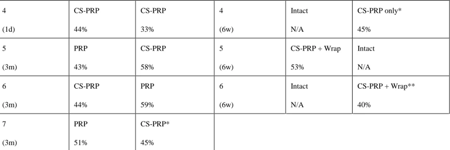
Table 6.1 Placement of meniscal defects. Values closer to 0% were near the vascularised periphery and values closer to 100% were near the avascular free border. ... 88
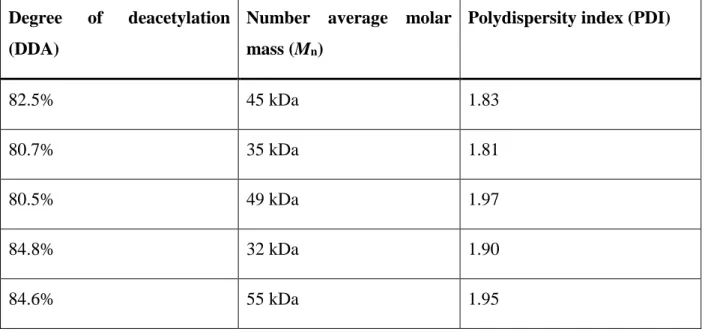
Table 7.1 Properties of the CS used in the study. ... 112
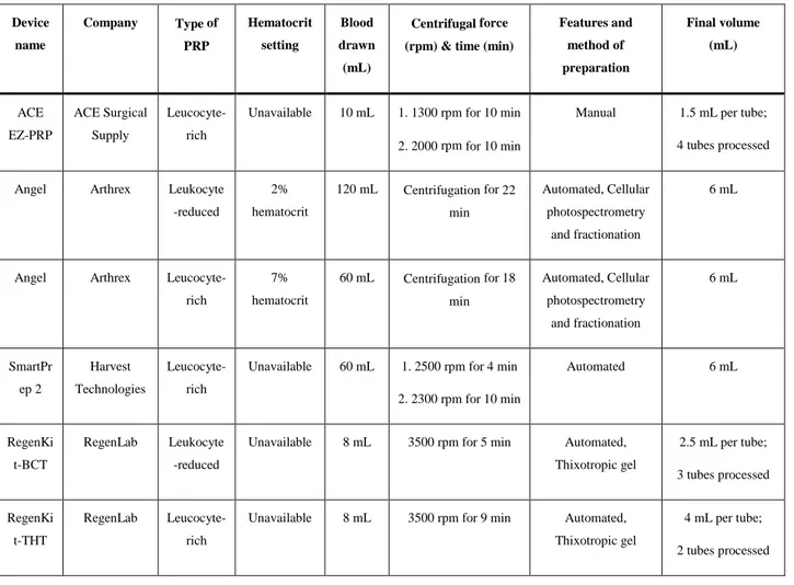
Table 7.2 Characteristics of the different PRP preparations used for the study. ... 113
Table 7.3 Complete blood count (CBC) analysis in whole blood and different PRP preparations. ... 117
Table 7.4 Pearson correlation coefficients r and corresponding p values between the performance characteristics of the CS-PRP formulations and the properties of the CS and PRP preparations used to prepare the formulations. * % clot mass lost was 0 for all CS-PRP formulations assessed; N/A = Non -applicable. ... 125
Table 7.5 Pearson correlation coefficients r and corresponding p values between the performance characteristics of the recalcified PRP controls and the properties of the PRP preparations. ... 125
Table 8.1 Definition of parameters and their values for the freeze-drying cycles used in phase I of this study ... 140
Table 8.2 Tg', Tc, and Teu of the formulation components (22, 176, 177). ... 141
Table 8.3 Detailed parameters for the freeze-drying cycles used in phase I of the study. ... 141
Table 10.1 Level I and Level II clinical studies of meniscus repair. ... 194
Table 10.2 Clinical studies of meniscus repair augmentation by trephination, rasping and abrasion. ... 200
Table 10.3 Clinical studies of meniscus repair augmentation by fibrin/blood clots. ... 205
Table 10.4 Clinical studies of meniscus repair augmentation by platelet-rich plasma. ... 209
LIST OF FIGURES
Figure 3.1 Gross anatomy of the knee joint. The menisci are two pads of fibrocartilage situated within the knee joint between the tibia and femur. Ligaments stabilize menisci within the joint. An anterior view (A) superior view (B), and the ligaments of the knee joint (28). ... 10 Figure 3.2 A frontal section of the vascularity of meniscus. Radial vessels from the perimeniscal capillary plexus (PCP) are penetrating the peripheral border of the medial meniscus via 3 zones: I. Red-red (R-R) zone is fully vascularized, II. White-red (R-W), and III. White-white (W-W) is within the avascular area of the meniscus. F: femur; T: tibia (27). ... 11 Figure 3.3 Schematic pattern of orientation of collagen fibers of the meniscus which can be categorized into 3 layers: I. Superficial network, II. Lamellar layer, and III. Circumferential fibers (27). ... 12 Figure 3.4 Schematic diagram of cell types in the meniscus. Cells of the superficial zone are fusiform; the cells in the outer periphery are mainly elongated fibroblasts with the process; moving toward the inner portion of the meniscus, the cells rounded and chondrocyte-like (40). ... 14 Figure 3.5 Drawing of common types of menisci tears (52). ... 17 Figure 3.6 Chemical structure of chitin (A) and chitosan (B) (20). ... 31 Figure 3.7 Simplified flowchart of preparation of chitosan from chitin (20, 96). ... 36 Figure 5.1 Treatment algorithm of meniscus lesions (Adapted from Mordecai et al, 2014). ... 55 Figure 6.1 A) Schematic representation of the bilateral surgical model. 10-mm long incisions were created bilaterally in the anterior portion of the medial menisci in 7 sheep (black in A). Freeze-dried chitosan formulations were solubilized in autologous PRP and 0.5 mL of chitosan-PRP was injected into the meniscal tear through two trephination channels created with 18-gauge needles (in green in A). The tears were sutured in a horizontal mattress pattern (in red in A). B) Schematic representation of the surgically induced defect model. C) Picture of a meniscus treated with CS-PRP. D) Seven ewes (2-6 years old) were included in the study and treated with either chitosan-PRP only (n = 2 knees at 1 day, n = 4 knees at 3 weeks & 3 months) or PRP only (n = 2 knees at 3 weeks & 3 months). E&F). A rhodamine-chitosan tracer was added
to the freeze-dried formulations to allow detection of the implants with epifluorescent microscopy (E&F). The chitosan-PRP implants were partly resident in the tears and trephination channels at 1-day post-surgery (E&F). Safranin O/fast green stained sections showed that chitosan-PRP implants induced cell recruitment from the vascularized periphery of the menisci towards the trephination channels (G&H). The rectangles in panel E&G indicate the regions where the higher magnification images F&H were acquired. ... 89 Figure 6.2 A) Schematic representation of the unilateral surgical model. A bone block with medial collateral ligament attached was detached to increase access to the meniscus. A 10-mm longitudinal tear with a horizontal component was created towards the anterior portion of the medial meniscus (in black in A). Two 20-gauge needles were used to create trephination channels from the capsular border of the meniscus to the tear (in green in A). 0.5 mL chitosan-PRP implant was injected into the tear through the trephination channels and the tear was stabilized with three sutures tightened in a vertical pattern (in red in A). A piece of collagen membrane (12.5 mm X 25 mm) was wrapped around the meniscus (in blue in A) and sutured first at the capsular border, and then with two vertical sutures placed through the meniscal tissue. 0.5 mL chitosan-PRP implant was then injected under the membrane. The contralateral knee was left intact. B) Schematic representation of the surgically induced defect model. C) Picture of a meniscus treated with wrapping + CS-PRP. D) Six ewes (2-6 years old) were included in the study and treated with either chitosan-PRP only (n = 2 knees), chitosan-PRP + wrap (n = 2 knees) or wrap only (n = 2 knees). The contralateral knees were left intact (n = 6 knees). ... 90 Figure 6.3 The tears were macroscopically visible at the time of necropsy 3 weeks, 6 weeks and 3 months after surgery and the edges of the tears were usually well apposed (A to G). A reddish repair tissue and signs of neo-vascularization were visible in two chitosan-PRP treated tears at 6 weeks (E) and at 3 months (B) post-surgery (white arrowheads). Sutures were apparent in all treatment groups. Aside from the surgically-induced tears, no other sign of macroscopic meniscal degeneration was observed. ... 91 Figure 6.4 Histological sections of the repaired tissue stained with safranin O/fast green were used to evaluate tissue repair in the bilateral model. A highly cellular repair tissue was seen in one chitosan-PRP treated tear at 3 weeks post-surgery (A&B). Partial integration between the
repair tissue and the original meniscal tissue was achieved in this treated tear (B). Complete healing with a highly vascularized repair tissues and seamless repair tissue integration were seen in one chitosan-PRP treated tear at 3 months (E&F). There was no repair tissue synthesis in the PRP controls at 3 weeks or at 3 months (C&D and G&H), and in the other CS-PRP treated tears. The surgical approach induced some fibroplasia in the outer portion of the menisci at 3 weeks and 3 months (A, C, E, & G). Rectangles in panels A, C, E, & G indicate regions where the higher magnification images B, D, F, & H were taken. Histological sections were scored based Zhang et al for overall meniscal tissue quality (i, ranging from 0 for the best to 26 for the worst quality) and repair tissue quality (j, ranging from 0 for the best to 7 for the worst quality) and were consistent with the histological observations41. ... 92 Figure 6.5 Histological sections of the repaired tissue stained with safranin O/fast green were used to evaluate tissue repair in the unilateral model. One tear treated with chitosan-PRP only showed complete repair (A-C), while one tear treated with chitosan-PRP with wrapping was partially healed (D-F). There was no repair tissue in the group treated with wrapping only (G-I). In the two cases where repair was observed, the repair tissue was highly cellular, well integrated to the adjacent meniscal tissue, but structurally different than the contralateral intact menisci (j-L). Significant cell recruitment into the outer portion of all treated menisci was observed compared to contralateral intact menisci. Suture tracks were frequently observed in menisci treated with the wrapping technique, along with sparse foreign body giant cells (FBGCs) in the outer vascularized area (D-I). Rectangles in A, D, G & J demonstrate regions where the higher magnification images B, C, E, F, H, I, K & L were acquired. Histological sections were scored based on Zhang et al for overall meniscal tissue quality (m, ranging from 0 for the best to 26 for the worst quality) and repair tissue quality (n, ranging from 0 for the best to 7 for the worst quality) and were consistent with the histological observations. N/A: Non-applicable. ... 93 Figure 6.6 Hematoxylin and eosin stained sections of synovial membrane (A to C). There was a mild to moderate transient synovitis in most treated knees. Changes included intimal hyperplasia, inflammatory cell infiltration (B), some sub-intimal fibrosis, and an increase in vascularization (C). Histological sections were scored as in Little et al (D to F, ranging from 0 to 12 for severe abnormalities) and scores reflected those observations. ... 94
Figure 6.7 There were mild to moderate changes to the articular surfaces as shown by safranin O/fast green stained sections of osteochondral cores collected from the medial femoral condyles (A to C) and from the medial tibial plateau (not shown). Changes included depletion of glycosaminoglycan (B&C) and some structural abnormalities (C). Histological sections were scored according to Mankin (D to F, ranging from 0 to 14 for severe abnormalities) and scores reflected those observations. There was no effect of treatment on the histological scores. ... 95 Figure 6.8 Electromechanical properties of the tibial plateau and the distal femurs were mapped across the entire articular surfaces using the hand-held Arthro-BST device (A&B). Panels a and b are representative examples of mapping of distal femurs (A) and tibial plateau (B) with corresponding QP. A high QP (shown in red) indicates weak electromechanical properties and poor load-bearing capacity and a low QP (in blue) shows strong electromechanical properties and high load-bearing capacity. Average QP values for medial femoral condyles and medial tibial plateau are shown in panels C to E and showed that articular surfaces displayed good load-bearing properties. There was no effect of treatment on QP values. ... 96 Figure 7.1 Study design. Five different CSs (with Mn ranging from 32 to 55 kDa and DDA ranging from 80.5 to 84.8%) were used to prepare freeze-dried formulations that also contained trehalose as lyoprotectant and calcium chloride as clot activator. Freeze-dried cakes were solubilized with 7 different PRP preparations (Angel with 2% hematocrit and Angel with 7% hematocrit are pictured here). Performance characteristics of the solubilized CS-PRP mixtures were assessed in vitro. ... 114 Figure 7.2 Complete blood count (CBC) analysis of whole blood and resultant PRP from each system is shown in panels a (platelet concentration), b (leukocyte concentration) & c (erythrocyte concentration). Macroscopic appearance of different PRP preparations is shown in panel d. Erythrocyte concentration (panel c) influenced the colour of PRP preparations (panel d). ... 117 Figure 7.3 pH (panel a) and osmolality (panel b) of formulations post-solubilization with PRPs isolated with the different devices. pH of CS-PRP formulations was lower than recalcified PRP controls (a). Osmolality of CS-PRP formulations was higher than recalcified PRP controls (b). Data are presented as box plots where median box indicates the 25th and 75th
percentile; Whisker extends to the most extreme data point within 1.5 times the interquartile range of data. n=5 samples for each type of CS-PRP formulation and n=1 for each recalcified PRP control. ... 119 Figure 7.4 Runniness of the CS-PRP and PRP formulations was assessed on an inclined plate. Panel a shows runniness of CS-PRP and a recalcified PRP control prepared with CS 84.6% DDA Mn 55 kDa and ACE EZ-PRP as an example. CS-PRP formulations were paste-like and
less runny than the recalcified PRP controls panel b). Note that in this assay water runs off the plate and has a runniness exceeding 310 mm. Data are presented as box plots where median box indicates the 25th and 75th percentile; Whisker extends to the most extreme data point within 1.5 times the interquartile range of data. n=5 samples for each type of CS-PRP formulation and n=1 for each recalcified PRP control. ... 120 Figure 7.5 A thrombelastograph (panel a) was used to assess clotting properties of formulations in
vitro. Panel b shows TEG traces obtained for formulations prepared with CS 84.6% DDA Mn
55 kDa and RegenKit-BCT or RegenKit-THT as an example. Clot reaction time (R) is the time in minutes from initiation of the tracing to the point where branches have diverged by 2 mm. Maximal amplitude (MA) is the maximal distance in mm between the two diverging branches and corresponds to clot strength. All CS-PRP formulations clotted and had average clot reaction times between 2 and 9 minutes (panel c). Clot reaction times of the recalcified leukocyte-rich PRP controls were greater, between 32 and 57 minutes (panel c). CS-PRP formulations had average clot maximal amplitude above 42 mm (d). Recalcification of the leukocyte-reduced PRP controls with 42.2 mM was insufficient to induce clotting in this dynamic system (c & d). Recalcified SmartPrep 2 PRP control had barely started to clot when the assay was terminated so that its clot reaction time was high (57 minutes) (c) and its clot maximal amplitude was low (11 mm) (d). Data are presented as box plots where median box indicates the 25th and 75th percentile; Whisker extends to the most extreme data point within 1.5 times the interquartile range of data. n=5 samples for each type of CS-PRP formulation and n=1 for each recalcified PRP control. ... 121 Figure 7.6 Clot retraction was assessed by gravimetric measurement. Panel a show a CS-PRP hybrid clot and a recalcified PRP control prepared with CS 84.8% DDA Mn 32 kDa and
prepared with CS 82.5% DDA Mn 45 kDa and Arthrex ACP as an example. All CS-PRP
hybrid clots remained voluminous after clotting for 1 h at 37°C and did not express any serum (panel c). Recalcification with 42.2 mM CaCl2 was sufficient to induce coagulation of all PRP
controls in this static assay. Recalcified PRP controls expressed a lot of serum upon clotting and lost 43% to 82% of their original mass upon clotting (panel c). n=5 samples for each type of CS-PRP formulation and n=1 for each recalcified PRP control. ... 123 Figure 7.7 Clot homogeneity was assessed with Fast Green and Iron Hematoxylin staining of paraffin sections. Panels a & b show a CS-PRP hybrid clot prepared with CS 84.8% DDA Mn
32 kDa and Angel with 7% hematocrit as an example and panels c & d show the recalcified PRP control. Panels e & f show a CS-PRP hybrid clot prepared with CS 84.8% DDA Mn 32
kDa and Angel with 2% hematocrit as an example and panels g & h show the recalcified PRP control. Dispersion of CS within the hybrid clots was usually homogenous (b & f). Erythrocytes were more abundant in clots prepared with Angel with 7% hematocrit compared to Angel with 2% hematocrit (compare panel d to h). Outlines in panels a, c, e & g show where higher magnification images b, d, f & h were acquired. ... 124
LIST OF SYMBOLS AND ABBREVIATIONS
APM Arthroscopic Partial Peniscectomy
α Alpha
ACL Anterior Cruciate Ligament
AAOS American Association of Orthopaedic Surgeons AC Articuler Cartilage
β Beta
b-FGF Basic-Fibroblast Growth Factor BM Bone Marrow
BMAC Bone MarrowAspirate Concenearte BMS Bone Marrow Stimulation
CS Chitosan
CBC Complete Blood Count CMI Collagen Meniscus Implants CM Chondrogenic Medium CaCl2 Calcium Chloride cm Centimeter
CD Cluster of Differentiation DP Deproteinization
DM Demineralization DC Decolorization
DDA Degree of Deacetylation D Dimensional
FU Follow-Up F Femur FD Freeze-Dried
FBGC Foreign Body Giant Cell
GF Growth Factor
GP Glycerol Phosphate
GPSIII Gravitational platelet sepration system III GAG Glycosaminoglycan
H2O Water
HA Hyaluronic Acid HCl Hydrochloric Acid
HGF Hepatocyte Growth Factor H&E Hematoxylin and Eosin
IGF Vascular Endothelial Growth Factor IL Interleukin
KOOS Knee Injury and Osteoarthritis Score
IKDC International Knee Documentation Committee L-PRF Leukocyte-Rich Platelet-Rich Fibrin
MRI Magnetic Resonance Imaging MRA Magnetic Resonance Arthrography
MOON Multicenter Orthopaedic Outcomes Network MFC Medial Femoral Condyle
MTP Medial Tibial Plateau
mL Millilitre
MW Molecular Weight
Mn Number Average Molar Mass
MA Maximal Amplitude
MSC Mesenchimal Stem Cells MCL Medial Collateral Ligament MLS Meniscus-Like strucutre MA Maximal Amplitude MPa Megapascal mTorr Millitorr μ Micro μL Microlitre
NMR Nuclear Magnetic Resonnace
N/S Not-Specified
N/A Non-Applicable
NH3 Amino
NBF Neutral Buffered Formalin NaOH Sodium Hydroxide
OA Ostheoarthtics
PCNA Proliferating Cell Nuclear Antigen PDI Polydispersity Index
PCL Posterior Cruciate Ligament PCP Perimeneiscal Capaliary Plexux PU Polyurethane
PCL Polycaprolacone
PLGA Polylactic-co-Glycolic Acid PLLA Poly-L-lactic Acid
PGA Polyglycolic Acid
PLGA Polylactic-co-Glycolic Acid PM Partial Meniscectomy PRP Platelet-Rich Plasma PPP Platelet-Poor Plasma
PDGF-AB Platelet Derived Growth Fcator-AB P-PRF Leukocyte-Poor Platelet-Rich Fibrin PG Proteoglycan
PS Polysaccharide
P-PRP Leukocyte-Poor Platelet-Rich Plasma QofL Quality of Life
QP Quantitative Parameter RBC Red Blood Cell
R-R Red-Red
R-PRP Leukocyte-Rich Platelet-Rich Plasma rhFVIIa Recombinant Human Factor -rhFVIIa RA Rheumatoid Arthritis
ROM Range of Motion Saf-O Safranin-O
SC Stem Cell
SEM Scan Electron Microscpy
TGF-B Transforming Growth Factor-Beta Tp Product Temperature
Tg Glass Transition Temperature Tg Eutectic Temperature
TM Total Meniscectomy TE Tissue Engineering TEG Thromboelastography
T Tibia
TNF- Tumor Necrosis Factor
UK-ACL University of Kentucky Anterior Cruciate Ligament Test
V/v Volume/volume
VEGF Vascular Endothelial Growth Factor WOMET Western Ontario Meniscal Evaluation Tool WORMS Whole Organ MRI Score
W-W White-White
W-R White-Red
LIST OF APPENDICES
CHAPTER 1
INTRODUCTION
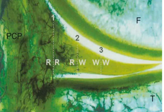
More than 1.7 billion people in the world are suffering from musculoskeletal conditions which cost an estimated $213 billion either directly or indirectly. Orthopedic disorders involving tissues such as cartilage, bone, intraarticular ligaments, and meniscus have significant socioeconomic impact (1, 2). Osteoarthritis (OA) is a slowly progressive joint disorder and the primary cause of musculoskeletal disability worldwide. Osteoarthritis is a multifactorial disease affecting the whole joint rather than only articular cartilage (AC). The meniscus is a fibrocartilaginous tissue located between the femur and tibia. The menisci are the crucial structures of knee joint acting as shock absorbent and protecting the articular cartilage. Inner zones of menisci lack blood supply and healing capacity, therefore, the potential for regeneration is very poor (3).
Meniscal tears are among the most common pathologies of the knee joint. Based on past reports, the incidence of meniscus tears are estimated to be as high as 70 per 100,000 knee injuries with even higher rate in the elderly, men, medial meniscus, and active populations (4). There is evidence suggesting that asymptomatic meniscal lesions are common incidental findings on knee magnetic resonance imaging (MRI) of at least one-third of the knees of middle or elderly individuals. Such tears limit the range of motion causing clinical symptoms such as sharp pain, swelling, effusion, locking, and catching in the knee (2, 5, 6). Arthroscopic partial meniscectomyis the most common orthopedic surgical procedure that is performed for small radial and complex meniscus tears (7-9). Although partial meniscectomy relieves pain, it is associated with a high risk of developing knee OA. Loss of meniscal function due to trauma, injury, or partial meniscectomy may lead to increased biomechanical stress and relative overloading on the joint which further progress to OA and pain over time. Knee OA is clinically associated with meniscal tear and degeneration (2, 10).
Surgical repair of meniscus tears can be performed by using suture-based devices. Complex or degenerative tears located in the avascular segments of the meniscus affect the fibrocartilage structures and surrounding tendons (4, 11). Complex tears which develop mostly in middle or older-aged patients involve a progressive horizontal cleavage of the meniscus usually in the multiplane direction without history of significant acute trauma. Patient- and clinical-reported outcomes of meniscus repair and meniscectomy have demonstrated significant failures involving re-tear or reoperation at long-term follow-up (9). Therefore, selection of appropriate management strategy depends on multiple factors for patients and needs carefull consideration (12).
Recent technologies are attempting to repair the tissue while paying more attention to preserve the structural integrity of the knee joint to lower the risk of OA. Therefore, the combination of sutures and biological augmentation strategies such as meniscus wrapping with extracellular matrix materials, trephination, synovial and meniscus rasping, fibrin/blood clot placement, and platelet-rich plasma (PRP) injections have been introduced to improve healing rate of the meniscus repair (4, 7, 13). For a tissue-engineered meniscus, selection of an optimal cell type, appropriate growth factors, and the type of scaffold are crucial. Tissue engineering approaches are still at the early development stage (14, 15). Safety and efficacy of ortho biological products need to be assessed more thoroughly in prospective and randomized clinical studies (16). The general aim of meniscus repair would be to restore the tissue structure and biomechanical function to minimize pain and OA in the long-term.
Platelet-rich plasma is an autologous biological agent with a raised platelet concentration above the physiological level and has been used predominantly in the musculoskeletal field for sports injuries. Activation of the platelets leads to the local liberation of several growth factors and cytokines from granules which play a pivotal role in the coordination of inflammation, cell proliferation, differentiation, angiogenesis, and tissue remodeling. Also, PRP contains the full complement of clotting factors which contribute to hemostasis and aggregation (17, 18). These anabolic platelet-derived growth factors have been shown to affect meniscal cell function positively and induce soft-tissue repair augmentation. Different commercial PRP preparation systems and medical devices are available, which generate PRPs that contain varying concentrations of platelets and blood components. The presence or absence of leukocytes and the structure of fibrin determine the different families of PRP (16). Potential anabolic or catabolic effecst of leukocytes and platelets in PRP is an intensive ongoing debate. Upon delivery of platelet-rich plasma to the desired site, PRP is activated which causes the fast release of growth factors from platelets. The short half-life of growth factors in PRP, the significant dilution of PRP by residual arthroscopic fluid at the time of injection, and variability in the content pose methodological challenges to investigators. To overcome theese limitations, clot activators and modified natural biopolymers can be added to PRP which resul in the formation of a dense matrix and prolongs the release of growth factors (GF). Further randomized controlled clinical trial studies with a large number of patients and a long-term follow-up remains a necessity (16, 19).
Chitosan (CS), a biocompatible and biodegradable cationic polysaccharide family derived from the alkaline deacetylation of chitin, has been used extensively for soft and hard tissue engineering (20). Chitosan accelerates healing of the wound and has some similarity to hyaluronan which is found in extracellular matrices of cartilage (23). Chitin is degraded by lysozymes and produce oligomers of N-acetylglucosamine. Theses oligomers act as templates for hyaluronan synthesis to promote cell adhesion and proliferation which lead to massive cell movement and tissue re-organization. Chitosan shows immunopotentiating activity by macrophages stimulation. This naturally-derived polysaccharide is well-tolerated at the articular synovial level and favors repair process in meniscus tissue through promotion of angiogenesis (21). Freeze-drying (FD) is a method for preservation of pharmaceutical products and drugs. FD is a three-step process that can provide increased long-term stability and minimize degradation of chitosan-based scaffold for manufacturing (22).
In the context of cartilage repair, our lab has shown that chitosan-glycerol phosphate solutions can be mixed with whole blood and implanted over cartilage defects to augment repair by increasing cell recruitment, transient vascularization, as well as a polarization of the macrophage phenotype towards the alternatively-activated pro-wound healing lineage, and stimulated secretion of anabolic wound repair factors, all of which are also expected to be beneficial in the context of meniscus repair (23, 24). We have developed freeze-dried formulations of chitosan that can be solubilized in PRP to form injectable CS-PRP implants for orthopedic tissue repair. Previous studies showed that CS-PRP implants resist platelet-mediated retraction post-clotting, release increased amounts of platelet-derived growth factors, and have prolonged residency in vivo compared to PRP alone. CS-PRP implants have also shown potential to improve repair of rotator cuff and cartilage in small and large animal models. Hence, the starting hypothesis for this project was that the biological effects of CS-PRP hybrid implants would also be beneficial for meniscus repair (19, 25).
CHAPTER 2
OBJECTIVES AND HYPOTHESIS
2.1 General objective and hypothesis
The overall objective of this thesis was to make an outstanding contribution to improve current surgical treatment repair strategies for meniscus tears. In this context, the research presented here is to enhance current surgical treatment of meniscus tears by assessing the effect of freeze-dried chitosan-PRP implants in ovine models.
Chitosan-blood hybrids have previously been shown to improve pre-clinical cartilage repair outcomes in small and large animal models through mechanisms involving increased cell recruitment, angiogenesis, cell migration, repair tissue synthesis, and tissue remodeling/integration. Since current surgical techniques for meniscus repair also rely on cell recruitment from adjacent tissues through trephination and rasping, chitosan-PRP hybrids are expected to enhance the success of meniscus repair through similar mechanisms of cell recruitment and angiogenesis. Here, platelet-rich plasma rather than whole blood will be mixed with the polymer. PRP is obtained through centrifugation of anti-coagulated blood to produce an increase in platelet concentration over baseline. Hybrid clots composed of chitosan and PRP are expected to have significant bioactivity through the release of platelet-derived growth factors in order to improve meniscus repair.
In the first study, feasibility of using CS-PRP implants to improve meniscus repair in ovine models was investigated. In the second study, compatibility of freeze-dried chitosan formulations with PRPs prepared with different commercial automated systems was assessed. Finally, in the third study, freeze-dried cycle of chitosan formulations was optimized and reduced from 3 days to 1 day and performance characteristics of the freeze-dried formulastions were evaluated.
2.2 Study 1: Freeze-dried chitosan-PRP injectable surgical implants
for meniscus repair: pilot feasibility studies in ovine models
2.2.1 Objective for Study 1
The overall purpose of this study was to assess whether ovine meniscus repair would be improved by application of freeze-dried chitosan PRP to the tears over PRP alone or/and a meniscus wrapping technique.
The first pilot study used a bilateral longitudinal surgical laceration model where tears were treated with suturing along with the CS-PRP implant or with PRP alone for 3 weeks or 3 months. The second pilot study used a unilateral complex laceration model where tears were treated with suturing along with the CS-PRP implant, or the tears were treated with a wrapping technique using a Chondro-Gide collagen membrane, or the tears were treated with both CS-PRP and the wrapping technique for 6 weeks.
The purpose of conducting sheep studies of meniscus repair is to approximate as closely as possible the clinical pattern and pathology of meniscus tears and repair. In this context, it is worth noting that the surgical model was significantly improved from the first to the second study.
2.2.2 Hypotheses for Study 1 Our original hypotheses were that:
First pilot study: Freeze-dried chitosan formulations can be reconstituted in autologous PRP and injected into meniscus defects, where they coagulate and form stable hybrid chitosan-PRP implants. Repair outcomes would be improved by using CS-PRP implants injected in the tear site through trephination channels, due to long-term residency over PRP alone. Suturing and application of CS-PRP via trephination was expected to augment repair of meniscus tears.
Second pilot study: Repair outcomes would be improved by using CS-PRP implants in conjunction with the wrapping technique over CS-PRP implants injected in the tear site alone or wrapping alone, due to increased implant retention and resulting bioactivity. Suturing, wrapping by membrane matrix, and application of CS-PRP via trephination was expected to augment repair of meniscus tears.
2.2.3 Methods for Study 1
Lyophilized formulations of chitosan were solubilized in autologous PRP and applied to surgically induced meniscus lacerations. In the first study, bilateral tears in 7 sheep were treated by suturing, trephination and injecting either CS-PRP or PRP into the tears. In the second study, unilateral tears in 6 sheep were treated by suturing, trephination and injecting CS-PRP in tears, wrapping the meniscus with a collagen membrane and injecting CS-PRP in tears and under the wrap or wrapping only. CS-PRP implants residency, recruitment of host cells, repair tissue integration, and vascularization were investigated by macroscopic, microscopic and histological procedures. Chondroprotective effects of the meniscus on the adjacent joints including articular cartilage of tibial plateau and the distal femurs were assessed by electromechanical mapping using the hand-held Arthro-BST device. Quality of adjacent articular cartilage and synovium was investigated by histology.
2.3 Study 2: Multiple platelet-rich plasma preparations can
solubilize freeze-dried chitosan formulations to form injectable
implants for orthopedic indications
2.3.1 Objective for Study 2
The purpose of this study was to determine whether the in vitro performance of the formulations depends on the type of PRP preparation used to solubilize chitosan. Our specific objectives were: I. Assess compatibility of this freeze-dried technology with the various types of PRP preparations that can be isolated with commercially available systems and II. Define a range of chitosan degree of deacetylation (DDA) and number average molecular weight (Mn) that would yield freeze-dried
formulations with acceptable performance characteristics. 2.3.2 Hypotheses for Study 2
Our starting hypothesis was that although the different PRP preparation systems would yield PRPs with varying properties, all PRP preparations would be compatible with our freeze-dried chitosan technology.