OATAO is an open access repository that collects the work of Toulouse
researchers and makes it freely available over the web where possible
Any correspondence concerning this service should be sent
to the repository administrator:
[email protected]
This is an author’s version published in:
http://oatao.univ-toulouse.fr/26797
To cite this version:
Quintero Rincón, Antonio and D’Giano, Carlos and Batatia,
Hadj Mu-suppression detection in motor imagery
electroencephalographic signals using the generalized
extreme value distribution. (2020) In: 2020 International Joint
Conference on Neural Networks (IJCNN), 19 July 2020 - 24
July 2020 (Glasgow, United Kingdom).
Official URL:
https://doi.org/10.1109/IJCNN48605.2020.9206862
Mu-suppression detection in motor imagery
electroencephalographic signals using the
generalized extreme value distribution
Antonio Quintero-Rinc´on, Carlos D’Giano
Centro Integral de Epilepsia y Telemetr´ıa
Fundaci´on Lucha contra las Enfermedades Neurol´ogicas Infantiles (FLENI)
Buenos Aires, Argentina
[email protected]
Hadj Batatia
IRIT - INPT
University of Toulouse
Toulouse, France
Abstract—This paper deals with the detection of
mu-suppression from electroencephalographic (EEG) signals in
brain-computer interface (BCI). For this purpose, an efficient
algorithm is proposed based on a statistical model and a linear
classifier. Precisely, the generalized extreme value distribution
(GEV) is proposed to represent the power spectrum density
of the EEG signal in the central motor cortex. The associated
three parameters are estimated using the maximum likelihood
method. Based on these parameters, a simple and efficient
linear classifier was designed to classify three types of events:
imagery, movement, and resting. Preliminary results show that
the proposed statistical model can be used in order to detect
precisely the mu-suppression and distinguish different EEG
events, with very good classification accuracy.
Index Terms—Motor imagery, Mu-suppression, Generalized
extreme value, Electroencephalography, Brain-computer
inter-face
I. I
NTRODUCTIONElectroencephalograms (EEG) are a non-invasive
longstan-ding medical modality that measures the brain’s activity by
recording the electromagnetic field at the scalp. Since its
creation, EEG has played a fundamental role in understanding
several major neurological disorders, by analyzing their
ma-nifestation into brain rhythms. For example, the study of
de-ceases such as depression, age-related cognitive deterioration,
epilepsy, anxiety disorders and subnormal brain development
in children have benefited from this technology. The typical
brain rhythms are distinguished by their different frequency
ranges, called delta (δ) within the range 0.5 to 4Hz, theta (θ)
within the range 4 to 7.5Hz, alpha (α) within the range 8
to 13Hz, beta (β) within the range 14 to 30Hz, and gamma
(γ) within the range 30 to 64Hz. In this study, we focus
on the brain rhythm called mu (µ) within the range 7.5 to
11.5Hz. Mu-waves are considered to emerge naturally and
may convey information about what the functioning of brain
hierarchies [1]. According to [2], there exist three historical
theoretical hypotheses to explaining the mu-brain rhythm: i)
the neuronal hyperexcitability related to the rolandic cortex;
ii) the superficial cortical inhibition explaining its suppression
with motor activity; and iii) the somatosensory cortical idling,
related to the afference-dependent phenomenon. This study
considers the second hypothesis, as the mu-rhythm relates
strongly to the sensorimotor cortex and associated areas, in
particular, the changes in the bilateral brain activities subject
to physical and imaginary movements [3]. Based on the same
consideration, this rhythm has been studied in brain-computer
interface (BCI) [4], [5]. The underlying idea of BCI is to
supply communication and control of devices through the
monitoring of brain activity, by using EEG channels.
The generalized extreme value (GEV) distribution is a
family that includes continuous probability distributions
ob-tained as the limit of maxima of a sequence of independent and
identically distributed random variables [6]. This distribution
has been used in [7] to detect interictal spikes in epileptic EEG
signals, using time-frequency properties. The underlying idea
was to identify strong outliers using GEV to model normalized
EEG data. In [8], showed that both EEG and MEG signals
can be correctly modeled using GEV distribution. Luca et
al. [9] used an unsupervised method to detect hyper-motor
epileptic seizures, where GEV was applied for extracting
maxima in EEG signals, using multivariate kernel density. In
[10], GEV was used to assess characteristics of Alzheimer’s
disease using EEG signals, where the variance of the power
of each frequency were used to derive an index of neuronal
abnormality. For applications in other biomedical signals see
[11].
The purpose of this paper is to present a novel and rapid
algorithm for detecting mu-suppression in EEG signals by
using the generalized extreme value distribution. The
under-lying idea is to estimate the maximum and minimum values
of the signal in the central motor cortex using statistical
modeling, for motor imagery events, corresponding to
mu-suppression. To the best of our knowledge, this statistical
model has not been investigated yet for detecting the
mu-suppression in EEG signals, despite the extensive study of
this phenomenon [12]–[14]. Several other methods have been
proposed in the literature to estimate mu-suppression in motor
imagery, see [15], [16] for a comprehensive state-of-the-art.
Table I summarizes the most common methods, such as
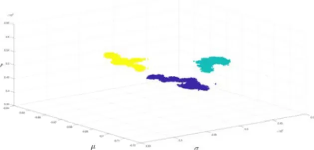
Fig. 6. Scatter plot example with randomized data. It is clearly possible to use a linear classifier as imagery events (grey), the movement events (yellow), and the resting events (blue) are well separated in the GEV parameter space.
classifier derived from the linear discriminant analysis (LDA)
was designed to distinguish between event types using these
parameters. The performance of the proposed method was
evaluated on a real dataset from 52 subjects achieving 100%
accuracy. In addition to its performance, an advantage of this
method is its low computational cost compared to existing
methods. These good results have the potential to shed new
light on mu-suppression detection in motor imagery EEG
signals in the central motor cortex.
The noise and artifacts were not taken into consideration in
this work, which constitutes its main limitation. Future work
will focus on the study of the noise and artifacts in order to
make the method applicable in real-time. A large scale testing
campaign will also be undertaken with other databases and
other possible channel locations with the idea of using the
least amount of channels possible.
V. A
CKNOWLEDGMENTSThe authors would like to thank Joaquin Ems, Lourdes
Hirschson, and Catalina Carenzo for useful comments on
an earlier version of the manuscript. We are grateful to the
professor emeritus Jaime A. Pineda from the University of
California, San Diego, La Jolla, CA, USA, for all the useful
discussions during the process of this study.
R
EFERENCES[1] K. Friston, “Waves of prediction,” PLOS Biology, vol. 10, no. 17, p. e3000426, 2019.
[2] D. L. Schomer and F. L. da Silva, Niedermeyer’s Electroencephalo-graphy: Basic Principles, Clinical Applications, and Related Fields. Lippincott Williams and Wilkins, 2010.
[3] S. Sanei, Adaptive Processing of Brain Signals. Wiley-Blackwell, 2013. [4] S. Arroyo, R. P. Lesser, B. Gordon, S. Uematsu, D. Jackson, and R. Webber, “Functional significance of the mu rhythm of human cortex: an electrophysiologic study with subdural electrodes,” Electroencepha-lography and Clinical Neurophysiology, vol. 3, no. 87, pp. 76–87, 1993. [5] G. Pfurtscheller and C. Neuper, “Event-related synchronization of mu rhythm in the EEG over the cortical hand area in man,” Neuroscience Letters, vol. 1, no. 174, pp. 93–96, 1994.
[6] E. Castillo, A. S. Hadi, N. Balakrishnan, and J. M. Sarabia, Extreme Value and Related Models with Applications in Engineering and Science. Wiley-Interscience, 2004.
[7] N. Gaspard, R. Alkawadri, P. Farooque, I. I. Goncharova, and H. P. Za-veri, “Automatic detection of prominent interictal spikes in intracranial EEG: Validation of an algorithm and relationsip to the seizure onset zone,” Clinical Neurophysiology, vol. 6, no. 125, pp. 1095–1103, 2014.
[8] N. Nazmia, S. A. Mazlana, H. Zamzuria, M. Azizi, and A. Rahman, “Fitting distribution for electromyography and electroencephalography signals based on goodness-of-fit tests,” IEEE International Symposium on Robotics and Intelligent Sensors, no. 76, pp. 468–473, 2015. [9] S. Luca, P. Karsmakers, K. Cuppens, T. Croonenborghs, A. V. de Vel,
B. Ceulemans, L. Lagaee, and B. V. Sabine Van Huffel a, b, “Detecting rare events using extreme value statistics applied to epileptic convulsions in children,” Artificial Intelligence in Medicine, no. 60, pp. 89–96, 2014. [10] T. Ueda, T. Musha, and T. Yagi, “Research of the characteristics of alzheimer’s disease using EEG,” Annual International Conference of the IEEE EMBS, no. 2009, pp. 4998–5001, 2009.
[11] S. J. Roberts, “Extreme value statistics for novelty detection in biome-dical data processing,” IEE Proceedings - Science, Measurement and Technology, no. 147, pp. 363–367, 2000.
[12] J. Pineda, “The functional significance of mu rhythms: Translating ”seeing” and ”hearing” into ”doing”,” Brain Research Reviews, vol. 68, no. 57, pp. 57–68, 2005.
[13] H. M. Hobson and D. V. M. Bishop, “The interpretation of mu suppression as an index of mirror neuron activity: past, present and future,” Royal Society Open Science, vol. 3, no. 4, p. 160662, 2017. [14] Y. Nishimura1, Y. Ikeda1, A. Suematsu1, and S. Higuchi, “Effect of
visual orientation on mu suppression in children: a comparative EEG study with adults,” Journal of Physiological Anthropology, vol. 16, no. 37, pp. 1–9, 2018.
[15] M. Hamedi, S.-H. Salleh, and A. M. Noor, “Electroencephalographic motor imagery brain connectivity analysis for BCI: A review,” Neural Computation, no. 6, pp. 999–1041, 2016.
[16] K. Xu, Y.-Y. Huang, and J.-R. Duann, “The sensitivity of single-trial mu-suppression detection for motor imagery performance as compared to motor execution and motor observation performance,” Frontiers in Human Neuroscience, vol. 13, no. 302, pp. 1–12, 2019.
[17] C. Guger, G. Edlinger, W. Harkam, I. Niedermayer, and G. Pfurtscheller, “How many people are able to operate an EEG-based brain-computer interface (BCI)?” IEEE Transactions on Neural Systems and Rehabili-tation Engineering, vol. 2, no. 11, pp. 145–147, 2003.
[18] M. Schroder, T. N. Lal, T. Hinterberger, M. Bogdan, N. Hill, N. Bir-baumer, W. Rosenstiel, and B. Scholkopf, “Robust EEG channel selec-tion across subjects for brain-computer interfaces,” EURASIP Journal on Applied Signal Processing, no. 19, pp. 3103–3112, 2005.
[19] M. Naeem, C. Brunner, R. Leeb, B. Graimann, and G. Pfurtscheller, “Seperability of four-class motor imagery data using independent com-ponents analysis,” Journal of Neural Engineering, no. 3, pp. 208–216, 2006.
[20] B. Blankertz, F. Losch, M. Krauledat, G. Dornhege, G. Curio, and K.-R. Muller, “The Berlin brain-computer interface: Accurate performance from first-session in BCI-naive subjects,” IEEE Transactions on Biome-dical Engineering, vol. 10, no. 55, pp. 2452–2462, 2008.
[21] B. Wan, Z. Zhou, L. Xu, D. Ming, H. Qi, and L. Cheng, “Mu rhythm desynchronization detection based on empirical mode decomposition,” 31st Annual International Conference of the IEEE EMBS, pp. 2232– 2235, 2009.
[22] M. Arvaneh, C. Guan, K. K. Ang, and C. Quek, “Optimizing the channel selection and classification accuracy in EEG-based BCI,” IEEE Transactions on Biomedical Engineering, vol. 6, no. 58, pp. 1865–1873, 2011.
[23] H. Cho, M. Ahn, S. Ahn, M. Kwon, and S. C. Jun, “EEG datasets for motor imagery brain computer interface,” Gigascience, vol. 7, no. 6, pp. 1–8, 2017.
[24] R. B. D’Agostino and M. A. Stephens, Goodness-of-Fit Techniques. Dekker, 1986.
[25] S. Kotz and S. Nadarajah, Extreme Value Distributions: Theory and Applications. ICP, 2001.
[26] A. Quintero-Rinc´on, M. Pereyra, C. D’Giano, M. Risk, and H. Batatia, “Fast statistical model-based classification of epileptic EEG signals,” Biocybernetics and Biomedical Engineering, vol. 38, no. 4, pp. 877– 889, 2018.
[27] A. Quintero-Rinc´on, C. D’Giano, and M. Risk, “Epileptic seizure prediction using Pearson’s product-moment correlation coefficient of a linear classifier from generalized Gaussian modeling,” Neurolog´ıa Argentina, vol. 10, no. 4, pp. 201–217, 2018.
[28] A. Quintero-Rinc´on, M. Flugelman, J. Prendes, and C. D’Giano, “Study on epileptic seizure detection in EEG signals using largest Lyapunov exponents and logistic regression,” Revista Argentina de Bioingenier´ıa, Bioengineering Argentinian Society, vol. 23, no. 2, pp. 17–24, 2019.