UNIVERSITÉ DU QUÉBEC À MONTRÉAL
NEW TOOLS FOR STUDYING CDXNEURAL FUNCTIONS
THE SIS
PRESENTED
AS PART
OF A MASTER'S IN BIOCHEMlSTRY
BY
DANIEL AGUlRRE MARTINEZ
Avertissement
La diffusion de ce mémoire se fait dans le respect des droits de son auteur, qui a signé le formulaire Autorisation de reproduire et de diffuser un travail de recherche de cycles supérieurs (SDU-522 - Rév.0?-2011). Cette autorisation stipule que «conformément à l'article 11 du Règlement no 8 des études de cycles supérieurs, [l'auteur] concède
à
l'Université du Québecà
Montréal une licence non exclusive d'utilisation et de publication de la totalité ou d'une partie importante de [son] travail de recherche pour des fins pédagogiques et non commerciales. Plus précisément, [l'auteur] autorise l'Université du Québec à Montréalà
reproduire, diffuser, prêter, distribuer ou vendre des copies de [son] travail de rechercheà
des fins non commerciales sur quelque support que ce soit, y compris l'Internet. Cette licence et cette autorisation n'entraînent pas une renonciation de [la] part [de l'auteur]à
[ses] droits moraux nià
[ses] droits de propriété intellectuelle. Sauf entente contraire, [l'auteur] conserve la liberté de diffuser et de commercialiser ou non ce travail dont [il] possède un exemplaire.»UNIVERSITÉ DU QUÉBEC À MONTRÉAL
NOUVEAUX OUTILS POUR L'ETUDE DES FONCTIONS NEURALES DES GENESCDX
MÉMOIRE
PRÉSENTÉ
COMME EXIGENCE PARTIELLE
DE LA MAÎTRISE EN BIOCHIMIE
PAR
DANIEL AGUIRRE MARTINEZ
ACKNOWLEDGEMENTS
I would like to address my sincerest thanks to the following people who have helped me throughout the last two years of my master' s pro gram:
Nicolas Pilon, my superviser and one of my professors at the university, for giving me the opportunity to learn in his lab. Despite ali the piles of books and articles keeping him busy in his office he is always available to talk to.
Ouliana Souchkova for imparting on me valuable technical and scientific knowledge. Especially at the beginning when she had to do ali that plus deal with my broken ankle and limited mobility.
Ali the members of the lab who have helped me in sorne way or another
ACKNOWLEDGEMENTS ... i
FIGURE LIST ... vii TABLE LIST ... ix
ABBREVIATIO , SIG A D ACRO YM LLST ... xi
RÉSUMÉ ... xiii ABSTRACT ... xv INTRODUCTION ... 1 CHAPTER I 3 BACKGROUND ... 3 1.1 Yertebrate embryogenesis ... 3 1.2 Molecular mechanisms ... 8
1.2.1 Signais ofthe posterior neuroectoderm ... lü 1 .3 Gene regulation and NC development ... .; ... 18 1.3.1 Enhancers ... 18
1.3.2 Cdx2NSE ... 22
1.4 Diseases ofNCC origin and neural tube defects ... 24
1.4.1 Neurocristopathies ... 24
1.4.2 Neural tube defects ... 26
1.4.3 Tools for studying neurocristopathies and NTDs ... 27
CHAPTER JI 29 HYPOTHESES A D OBJECTIYES ... 29
IV
2.2 Objectives ... 29
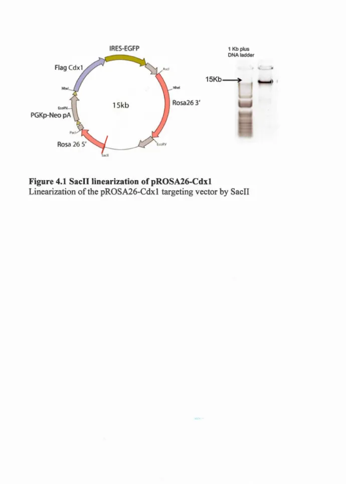
CHAPTER III 31 MATERlALS AND METHODS ... 31 3.1 Construction of pROSA26-Cdx 1, ES targeting and verification ... 31 3.1. t Vector construction ... 31 3.1.2 Targeting ES ce lis ... 31 3.1.3 Southern Blotting ... 32
3.2 Testing the neural specifie regulation of Cdx2NSE ... 33
3.2.1 Plasn1ids ... 33
3.2.2 Luciferase assay conditions ... 33
3.2.3 Luciferase assay test and statistical analysis ... 35 CHAPTER!V 37 TOWARD THE GENERATlON OF MICE CARRYING THE CRE-INDUCIBLE CDXJ TRANSGENE ... 37 4.1 Introduction ... 37 4.2 Results ... 37 4.2.1 Construction of the targeting vector ... 37 4.2.2 Targeting the ROSA26 locus ... 38 CHAPTER V 43 TtSTfi-.JG THE NEURAL SPECIFIC REGULATiON OF CDX2NSE ... 43
5.1 Introduction ... 43
5.2 Results ... 44
5.2.1 Potential binding activators ... 44
5.2.2 NSE and potential TFBS sequence homology ... 46 5.2.3 Directional preference and synergy ... 49
5.2.4 Testing the reverse NSE ... 51 CHAPTER VI 53 DISCUSSION ... 53 6.1 ln pursuit of a conditional Cdxl overexpression mouse mode! ... 53 6.2 Insights into Cdx2NSE neural regulation and potential application ... 56
APPENDlX A ... 63
EVOLUTIONARY CONSERVATION OF CDX2NSE POTENTIAL TFBS ... 63
APPENDIX B ... 65
REGULATION OF PAX3 NCEJ BY CDX2 PROTErNS ... 65
APPENDTX C ... 67
CDX2NSE AND FGF SIGNALLING ... 67
Figure Page
1.1 Steps of neurulation ... 7
1.2 NCC migration throughout four different levels ... 7
1.3 Neural crest gene regulatory network ... 9
1.4 AP axis patterning ... 11
1.5 The canonicat Wnt signaling pathway .............................. 11
1.6 The FGF signalling pathway ... 13
1.7 The spatial and temporal differences of murine Cclx gene expression ... 17
1.8 Types of cis-regulatory elements ... 19
1.9 Techniques for identifying and testing enhancers ... 21 1.10 Identification and testing the Cdx2 neural specifie enhancer (NSE) ... 23
1.11 The enhancer is not entirely neural specifie ... 23
4.1 Sacll linearization of pROSA26-Cdx 1 ... 39
VIII
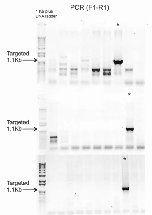
4.3 PCR screen of targeted ES clones ... 41
5.1 Potential Lefl1/Tcfand STEM family binding sites ... .45
5.2 Vertebrate homology ofthe neural specifie enhancer ... 48
5.3 Cdx2NSE synergy and directional preference ... 50
Table Page
1.1 Diversity ofNeural Crest Cel! derivatives ... 8
3.1 Pritners for PCR screen ... 32
3.2 P-value guide ... 35
AP BMP Cd x CNS DLHP DY EMT EGFP FGF GRN TRES LEFl MEF MHP NC Anterior-Posterior
Borre morphogenie protein
Caudal-related homeobox
Central nervous system
Dorsal-lateral hinge points
Dorsal-Ventral
Epithelial-mesenchymal transition
Enhanced green fluorescence protein
Fibroblast growth factor
Gene regulatory network
Internai ribosome entry site
Lymphoid enhancer-binding factor 1
Mouse Embryo Fibroblast
Median hinge points
XII NCC NCE NP NPB NSE NT NTD PNS SOX2 TCF TF TFBS Wnt Neural crest cel! Neural crest enhancer Neural Plate
Neural Plate Border Neural Specifie Enhancer
Neural Tube
Neural Tube Defects Peripheral nervous system
SRY (sex determining region Y)-box 2
T-cell specifie transcription factor Transcription factor
Transcription factor binding site Wingless-related integration site
Le système nerveux est un composant fondamental chez les vertébrés qui implique un réseau complexe de gènes régulateurs (GRN). Sa mise en place commence avec l'induction du neuroectoderme par l'activité des signaux Wnt, FGF et BMP le long de l'axe antéro-postérieur nouvellement formé. Plusieurs signaux transcriptionnels et
morphogén iques coopèrent pour indu ire l'in vagi nation du neuroectoderme
ventralement afin de former le tube neural et la crête neurale qui donneront naissance, respectivement, au système nerveux central et au système nerveux périphérique. Le neuroectoderme postérieur possède son unique lot de molécules de signalisation et toute perturbation dans les évènements précoce de son développement entraîne un groupe de maladies et malformations congénitales distinct. L'utilisation de modèles transgéniques murins est une façon idéale de comprendre les activités transcriptionnelles et morphogéniques dans cette région. Nous proposons deux modèles basés sur les facteurs de transcription de la famille Cdx- essentiels pour le
développement postérieur de 1 'embryon, et des acteurs majeurs dans le
développement de la crête neurale (Sanchez-Ferras et al., 2014; Sanchez-Ferras et al., 2012). Le premier serait un modèle pour l'anomalie qui pourrait être entraînée par la surexpression conditionnelle du gène Cdxl et nous permettrait ainsi de mieux comprendre le rôle de ce gène dans les cancers dérivés des cellules de la crête neurale. A ce jour, un criblage par PCR nous a permis d'identifier des cellules souches po si ti v es pour 1' intégration d'une cassette Cdx 1 mais une confirmation par southern blot est nécessaire. Concernant la deuxième souris transgénique, elle
représente un modèle unique pour étudier les évènements précoces dans le
neuroectoderme postérieur. En effet, elle est basée sur 1' uti 1 isation de 1 'enhancer (NSE) de Cdx2 qui permet de générer une lignée transgénique avec une induction spécifique au tube neural via la technique Cre-LoxP. Cependant, Cdx2NSE s'est
révélée n'être que partiellement active dans le mésoderme. Cette activité
mésodermale étant mineure, il serait possible de retirer les éléments non-neuraux de NSE afin d'obtenir une activité entièrement neurale. Pour déterminer la nature de l'activité des différents éléments deNSE, des essais luciférase ont été effectués par co-transfection du facteur pro-neural Sox2 et d'une protéine de fusion Lefl-~caténine agissant comme effecteur de la voie Wnt, ces deux derniers étant importants pour le maintien de l'identité neuroectodermale postérieure. Les résultats ont montré que NSE est régulé par Wnt et Sox2 de manière synergistique, mais les éléments no n-neuraux restent à identifier.
Mots-clés: GRN, neuroectoderme, postérieur, développement, Cdx, régulation, outils
->
The nervous system is a fundamental feature of vertebrates whose formation involves a complex gene regulatory network (GRN). The process can be said to commence with the induction of the neuroectoderm by Wnt, FGF and BMP activity along the
newly formed anterior-posterior axis. Severa! transcriptional and morphogenie signais
in concerted action internalize the neuroectoderm ventrally where it becomes the neural tube and neural crest, the precursors of the central and peripheral nervous system, respectively. The posterior end of the neuroectoderm has its own unique set
of signalling molecules and disruptions in the early events in this region engender a distinct set of congenital diseases and malformations. One way to understand the
transcriptional and morphogenie activity in this posterior region is through the use of
mo use mode! s. Described are two proposed mo use systems based on the genes of the Cdx family of transcription factors- important for posterior embryonic development, and key players in neural crest development (Sanchez-Ferras et al., 2014; Sanch
ez-Ferras et al., 2012). The first would serve as a mode! for the potential pathogenesis
brought on by Cdxl conditional overexpression, in particular its potential role in the oncogenesis of neural crest cel! derived cancers. So far, positive embryonic stem cel! clones with the Cdxl overexpression cassette have been identified via a preliminary
PCR screen but require confirmation by southern blot. The second mouse mode! would take advantage of a modified neural specifie enhancer of Cdx2 to generate a truly neural specifie Cre driver line for the posterior neuroectoderm, a unique and
useful mode! for studying the early events in the posterior neuroectoderm. Tnitially
considered as entirely neural specifie (Wang and Shashikant, 2007), a Cre mouse line
driven by the Cdx2NSE later revealed partial activity in the mesoderm as weil (Coutaud and Pilon, 2013). Since gene expression was minor in the mesoderm, it
seems conceivable that the mesoderm expression can be removed, resulting in true
neural specifie expression. To do this the non-neural specifie elements in Cdx2NSE sequence have to be identified and either removed or made non-functional. To this end, luciferase assays were carried out via co-transfecting neural Sox2 and Wnt
effectors Lefl-~catenin. Both are important for maintaining posterior neuroectoderm
identity. Results show the NSE is regulated by the synergy of Wnt activity and Sox2.
However, the non-neural elements have yet to be identified.
A hallmark of vertebrate embryogenesis is the development of the nervous system. The process begins with the induction of the neuroectoderm across the midline. The new structure is not only physically and behaviourally unique from the surrounding epidennal ectoderm but has itself two distinct domains: the neural plate border (NPB), and the neural plate (NP). The NPBs, located on the lateral edges of the neuroectoderm, contain the precursors of the neural crest cells (NCC). The neural plate, located between the NPBs, encompasses a relatively large piece of neuroepithelium that becomes the nascent central nervous system via the formation of the neural tube (NT). The neuroectoderm eventually invaginates ventrally to form the NT and NCC population. The NCCs are a multipotent migratory group of cells that can become peripheral neurons, glia, pigment cells, cartilage cells and others (Garnett et al., 20 12; Perris, 1997; Stuh 1 mi lier and Garcia-Castro, 20 12).
The transition from ectoderm to neural plate to multipotent neural crest cells requires a finely tuned neural crest-gene regulatory network (NC-GRN) whose interactions are constantly being updated, as they are made known (Amore et al., 2003; Hinman et al., 2003; Oliveri et al., 2003; Van Otterloo et al., 2013). Work in revising and updating this regulatory gene network is valuable not just to improve our understanding of neurogenesis in general but how changes to this network can affect development. One way to shed sorne light on this gene network is through transgenic mouse models. Mouse systems can provide in vivo evidence on NC-GRN processes both spatially and temporally. Two proposed mouse models to be described in this memoire have their origins in the caudal end of development, as they are based on the genes encoding the caudally-restricted Cdx family of transcription factors. Cdx transcription
2
factors are known for their impo1tance in posterior embryonic development, but have just recently been implicated in the initial steps of the caudal NC-GRN, acting as mediators, inducers and co-operators of known players of the NC regulatory circuit (Sanchez-Ferras et al., 2014; Sanchez-Ferras et al., 2012).
The first mouse mode! would elucidate the effects of conditionally overexpressing Cdxl. Overexpression targeting NCC development is believed to result in the pathogenesis of NCC derived cancers, such as neuroblastoma. So far, potentially positive embryonic stem cell clones possessing the transgene construct have been identified via a preliminary PCR screen but verification by southern blot has of yet been inconclusive. Once veritied, microinjection and rearing of transgenic mice capable of inducible Cdxl overexpression could proceed. The second mode! is envisioned to serve as a Cre driver line for Cre-LoxP systems aiming to study the early events of the posterior neuroectoderm. Developed in the lab by Coutaud and Pilon (2013), Cdx2NSE-Cre was designed to provide Cre expression solely in the posterior neuroectoderm. Initially characterized as a neural specifie enhancer (NSE) by Wang and Shashikant (2007) it was later revealed to be partially active in no n-neural mesodermal tissue (Coutaud and Pilon, 20 13). To address this problem, it was imperative that the non-neural characteristics of the enhancer be identitied and removed or made non-functionai thereby preserving a true NSE capabie of being employed as a Cre driver line for true neural specifie induction in Cre-LoxP systems. To test the absence or presence of a neural identity, luciferase assays were carried out on a Cdx2NSE-Luc reporter, co-transfecting neural Sox2 and Wnt signalling effectors Lefl-~catenin. Wnt is important for determining posterior structures and regulating Cdx genes (Pi lon et al., 2006; Prinos et al., 2001 ), and Sox2 is a neural marker of the neuroectoderm (Graham et al., 2003; Uchikawa et al., 20 Il). So far, results show the NSE is regulated in synergy by combined Wnt activity and Sox2. However, we have not been able to identify the specifie areas of the NSE architecture responsible for the non-neural activity.
BACKGROUND
1.1 Vertebrate embryogenesis
Following fettilization and cleavage, the third and fourth major events in vertebrate
development are gastrulation and neurulation. Prior to gastrulation, the developing embryo is composed of a spherical layer of cells. Gastrulation transforms the single
layer into three distinct germ layers. Known as the ectoderm, mesoderrn, and
endoderm, each of the three germ layers esta bi ishes the framework of ali future · tissues. Moreover, gastrulation begins to shape the anterior-posterior (AP) axis and dorsal midline (Tam and Behringer, 1997). These axes are defined by the formation
of the mesodermal derived notochord (Purves and Wi Il iams, 2001 ).
Neurulation (Figure l.la) begins with the induction of the neural plate along the AP
axis, followed by specification of the neural plate borders (NPB). Housed in the NPB
are the precursors of the neural crest cells (NCC). The NPB separates the non-neural
ectoderm (NNE) from the neuroectoderm. Next, the neuroectoderm begins to bend inward creating neural folds at the lateral edges. Bending continues inward as the
neural folds elevate; such is the degree of the bending that the resulting U-shape is called the neural groove. As the groove lowers the neural folds rise, meet, fuse and close the loop. The closed loop, nowa cylinder of neuroepithelial cells known as the
neural tube (NT), is freed from the overlying ectoderm. The NT is completed first in the middle of the AP axis, next extending cranially and caudally; in birds and
4
mammals the NT fonns first in cranial then truncal levels (Figure 1.1 b) (Colas and
Schoenwolf, 2001; Duband, 201 0).
At the same time the NT fonns, NCCs are freed from what used to be the NPB and they begin to migrate. NCCs exhibit an epithelial to mesenchymal transition (EMT) allowing them to delaminate from the neuroepithelium, and migrate on pathways that
will specify their lineage. Delamination and migration occurs in a rostral to caudal
wave, coinciding with NT formation, with the wave of migration ultimately fi !ling up four levels: cranial, cardiac/vagal, truncal and sacral (Figure 1.2) (Huang and Sa int-Jeannet, 2004; Mason, 2007). In the trunk, NCCs migrate around the NT and through,
and sometimes between, somites * and in the head through rhombomeres'i" (Ghysen,
2003; Go mez et al., 2008; Guthrie and Lumsden, 1991 ). Ventral regions are filled-up first before more dorsal regions (Weston and Butler, 1966) and depending on where
they end up, NCCs can give rise-to a diverse set of derivatives of mesenchymal, neuronal, secretory or pigmented identity (Table 1.1) (Simoes-Costa and Bronner,
20 15; Smith and Schoenwolf, 1997).
There is sti Il some controversy as to whether NCCs are fate-restricted prior to migration, during migration or if they maintain multipotency (Jessen and Mirsky,
2005; Krispin et al., 2010; Mayor and Theveneau, 2013; McKinney et al., 2013). In vitro experiments have demonstrated the multipotent capacity of NCCs to become
multiple derivatives such as neurons, osteoblasts and melanocytes (Dupin et al., 20 1 0;
Dupin and Sommer, 20 12). Moreover, recent advances in in vivo cell-label ing and time-lapse imaging show that pre-migratory NCCs from any location along the dorsal
• Segments of paraxial mesoderm th at will form the skeletal muscles, vertebraes and ribs
NT may contribute to multiple NC targets, and that microenvironment eues along the migration pathway may be responsible for specifying NCC fate (McKinney et al.,
2013). There is also recent evidence suggesting that most migratory NCCs are
multipotent and only a few in the population are fate-restricted (Baggiolini et al.,
A
(
1
)
(•) 17 cUys(2)
{b) 19 clays (cl 20 daya Ne ur.l l old(3)
(d) 22 d. y a N e ur al plate Neu r al Primit i ve struk Cu t ~ of a m nion toldB
Ch ick aan i a l region ( Midbra l n ) Y o l k sac:•
.__ NolochO< d 2 4 h ( 3 s) 28 h ( 5 s) Ch ick trunca l region (Bracll lal)) 4 2 h (13 s ) 48 h (17 s) 52 h ( 20 s ) 54h (23 s) 68h (30 s ) 80 h (38 s)Figure 1.1 Steps of neurulation.
(A) Dorsal and transverse view of human neurulation. Neurulation entails at !east 3
spatially and temporally distinct stages: (1) formation and shaping of the neural plate (NP); (2) bending of the NP into the neural groove; (3) closure of the neural groove
into the neural tube (NT) and subsequent neural crest cell migration. Image retrieved
from Marieb and Hoehn (2007).
(B) Chick cranial and truncal temporal differences in NT closure. Birds and mammals exhibit NT closure temporal differences depending on the region along the midline. Yellow: presumptive NCCs; Green: delaminating NCCs; Blue: migrating NCCs.
Image retrieved from Du band (20 1 0)
CRANIAL
,-:;:::r-~---~==~cranlofaclal cartilage and bona
connective tissue cranial ganglia
r--~~-lpigment cells enterlc ganglla smooth muscle
TRUNK pigment cells dorsal root ganglia sympathetic ganglla adrenal medula
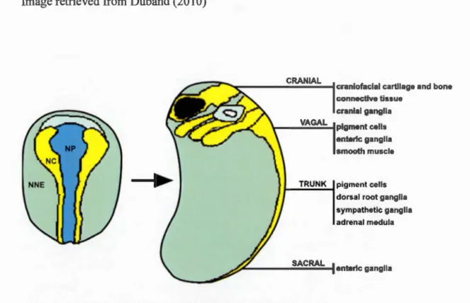
Figure 1.2 NCC migration throughout four different levels.
Migration is the first step in NCCs adopting different lineages. In a process known as epithelial to mesenchymal transition (EMT), presumptive NCCs exit the
neuroepithelium and take on different migration pathways each one leading to a
change in gene expression and thereby a change in cell type (McKinney et al., 2013). Grey: NNE non-neural ectoderm; Yellow: NC neural crest; Blue: NP neural plate.
8
T bi 11 D' a e
.
1vers1ry o 't fN eu ra IC res tCIId. e enva 1ves f .Neural crest cell derivatives
Mesenchymal Neuronal Cells Secretory Cells Pigmented Cells ce Us
Chondroblasts Sensory neurons Chromaffin cells Melanocytes Osteoblasts Cholinergie
Calcitonin-neurons producing cells Fibroblasts Adrenergic Parafollicular cells
neurons Odontoblasts Satellite cells
Cardiac Schwann cells
mesenchyme
Myoblasts Glial cells Adipocytes
Adapted from Simoes-Costa and Bronner (20 15). 1.2 Molecular mechanisms
From the transformation of ectoderm to neuroectodenn, to the changing expression profiles of migrating NCCs, there are a multitude of morphogenie signais, eues and gradients, both spatial and temporal that are responsible for these very physical and behavioural changes. This complex network is built on effective communication from
one cell to another, relaying severa) specifying signais at once. This complex network
is known as the neural crest gene regulatory network (NC-GRN) (Meulemans and Bronner-Fraser, 2002; Oliveri et al., 2003). Neurulation and NCC migration/specification rely on the NC-GRN (Figure 1.3). The process begins with the induction of the neural plate via the Wnt, FGF and BMP signalling pathways
(Huang and Saint-Jeannet, 2004). It is the combination ofthese three signais and their
antagonists that create the gradients responsible for cell specification and
morphogenesis (Niehrs, 2004). The specifies as to how these three signalling pathways work together in a concerted manner to induce the NPB is not fully understood and is active! y being researched (Garnett et al., 20 12; Monsoro-Burq et aL, 2005).
Signaling module Neural pla border mo te du le est Neural cr specificat module ion -Neural crest migration module Neural crest diversification circuits
WNTs
BMPs
Notch
FGFs
+
..
Zic
Ms x
Gbx2
Pax3/7 Tfap2 Dlx5/6
,,
FoxD3 Pax3/7 Tfap2 Sox10 Pax3/7 Neurons and glia•
..
Snai1/2 Twist Sox5 Sox9 Ets1 Mye
Id Sox10 Myb
1
EMT
t.
Sox5 FoxD3 Ebf1 Tfap2 RxrG Id Snai1/2 Chondroblasts and osteoblasts Mye Myb Melanocytes
Figure 1.3 Neural crest gene regulatory network.
-Through different molecular signais, both temporal and spatial dependent, the NC-GRN is responsible for the process ofNC development, from the induction of the NP to the diversification ofNC targets. Image retrieved from Simoes-Costa and Branner (2015).
10
1.2.1 Signais of the posterior neuroectoderm
The posterior neuroectoderm receives a different concentration, combination and
timing of signais than the anterior level. The outcome is a later onset of the NT and
NCCs in the posterior region as weil as different NCC lineages. In summary, anterior
structures are patterned first wh ile posterior on es occur later (Durs ton, 20 15).
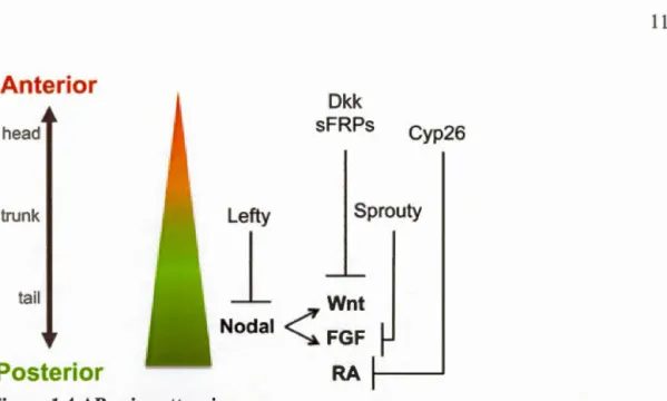
The patterning of the anterior-posterior (AP) axis is regulated in part by Wnt and
FGF, as weil as Nodal and Retinoic acid morphogens and their antagonists (McGrew
et al., 1997). Higher Wnt and FGF concentrations specify posterior fates, and their
antagonism anterior fates (Figure 1.4) (Kiecker and Niehrs, 2001; Kudoh et al.,
2002). Meanwhile, BMP and its antagonists· pattern the dorsal-ventral (DY) axis
(Marchant et al., 1998; Patten and Placzek, 2002; Smith and Harland, 1992). Higher
BMP concentrations specify ventral fates and lower levels dorsal fates (Endo et al.,
2002; Little and Mullins, 2006). Again, ali three - Wnt, FGF and
BMP-morphogens are needed to induce the formation of the NPB (home of the future
NCCs) but Wnt and FGF signais are mostlyt posteriorizing; BMP mostlyt
ventralizing (Hendrickx et al., 2009; Tuazon and Mullins, 2015).
• such as Noggin (Nog), Follistatin (Flst), and Chordin (Chd)
t Wnt and FGF signaling are crucial for AP patteming but they can be involved in DY patterning as they can affect regulation of BMP activity
~ BMP is crucial for DY patterning but isoform BMP4 has posteriorizing effects. ln ail, there is a trend where Wnt and FGF : posteriorizing and BMP : ventralizing, but overlap may occur
Ante ri or
head trunk ta ilPosterior
Dkk sFRPs Cyp26 Lefty Sprouty1
Wnt
Nodal< FGF RA Figure 1.4 AP axis patterning.Higher concentrations of Wnt FGF Nodal and RA activity specify posterior fates;
antagonists to these posteriorizing signais specify anterior fates. Image retrieved from
Tuazon and Mullins (20 15)
A OFF B ON LRP516
•
Frizzled ~tenltl) TrCP, p-catenin )Figure 1.5 The canonical Wnt signalling pathway.
( lkatemn )
( P..catenln )
( ~-catenin )
Wnt
raspanslve-(A) With no Wnt ligand present, the ~-catenin destmction complex is active and thus
no ~-catenin is freely available to mediate Wnt responsive gene transcription. (B) Upon introduction of a Wnt ligand to the Frizzled receptor, the Dishevelled proteins
are activated and impede the destruction of ~-catenin. This causes cytoplasmic levels
of ~-catenin to rise to where they are then able to enter the nucleus and associate with
Lefl/Tcf family of DNA-binding proteins and activate target gene transcription.
12
1.2.1.1 Wnt and FGF signalling
The canonicat Wnt signalling pathway begins with the binding of Wnt ligands to
Frizzled receptors, which in turn stops the ~-catenin destruction complex. An
increasing concentration of ~-catenin in the cytoplasm favours its translocation into the nucleus where it can then associate with transcription factors of the Lefl/Tcf family and activate target gene expression (Figure 1.5) (Angers and Moon, 2009;
MacDonald et al., 2009; Petersen and Reddien, 2009). This modulation of gene
expression by the Wnt pathway is implicated in most stages of embryogenesis
throughout various phyla including deuterostomes, protostomes, and pre-bilaterians
(Petersen and Red dien, 2009; Stuhlmiller and Garcia-Castro, 20 12). In vertebrates, the Wnt pathway is responsible for cell proliferation, cell fate, and body AP axis patterning (Hi kas a and Sokol, 20 13). Particular to AP neural patterning is how W nt specifies caudal CNS cell fa tes (Tuazon and Mullins, 20 15). lnversely, anterior levels require antagonists of Wnt activity (Houart et al., 2002). For the most part, posterior markers are tied to an increase in Wnt activity whereas a decrease in Wnt activity increases an teri or markers (Kudoh et al., 2002; McGrew et al., 1997; Petersen and
Reddien, 2009).
Like the Wnt pathway, the FGF pathway is important in establishing the vertebrate AP axis and specifying posterior cell fates (Figure 1.4; 1.6) (Tuazon and Mullins,
20 15). Suppressing FGF blacks posterior tissue development; overexpressing FGF
blacks anterior tissue development (Draper et al., 2003; Isaacs et al., 1994;
Stuhlmiller and Garcia-Castro, 2012). As such, bath Wnt and FGF signais inhibit
anterior gene expression (Kudoh et al., 2002). Concerning their origin, Wnt signais
come from the non-neural ectoderm and the paraxial mesoderm; FGF only cames
from the paraxial mesoderm (Huang and Saint-Jeannet, 2004). NPB induction may
Extracellular Plasma membrane ~ PIP42 'PIP3 I
!
P3 • DAG Cytoplum /!
l
f 1;) ----~~ PD ~ cal· K!
\
Cytoskeleton Targ ts Figure 1.6 The FGF signalling pathway.FGF binding to FGF receptors leads to the recruitment of Grb2 and Ras among other effectors to activate the Erkl/MAP kinase pathway which in turn phosphorylates a diverse set of transcription factors initia ting their regulatory activity implicated in ce li growth, migration, and morphogenesis. In AP patterning, antagonists of the FGF pathway, such as Sprouty, work to inhibit posteriorizing fates, thus prompting anterior ones. Image retrieved from Mason (2007).
14
the NPB or indirect) y by activating the Wnt pathway (Garnett et al., 20 12). Severa)
studies by Hong et al. (2008) have suggested that Wnt originating from the paraxial
mesoderm is activated by FGF, indicating that FGF activates mesodermal Wnt which
in turn helps induce NCC formation.
Together, through overlap, direct or indirect methods, Wnt, FGF and BMP signalling
induce the NC precursors at the NPB. The first phase involves Wnt and FGF to
activate the expression of border specifiers su ch as Msxl 12 Pax3/7 and Zic. The
second phase involves Wnt, BMP as weil as BMP-antagonizing Notch signalling to
further specify the NCC, by stimulating the expression of Snai/2, FoxD3, and
Sox9/10 (NC-GRN Figure 1.3) (Huang and Saint-Jeannet, 2004; Tuazon and Mullins,
20 15).
1.2.1.2 Sox2
Important for the development of the neural primordia as well are the Sox-B l
transcription factors: Sox 1, Sox2 and Sox3 (Uchikawa et al., 2011 ). Sox2 expression
in particular is the most extensive, with full expression across the neural primordia
(Okuda et al., 2010). In fact, Sox2 is the most definitive marker of the early neural
plate (Papanayotou et al., 2008; Pevny and Ni colis, 201 0; Rex et al., 1997). From
embryo to adult, Sox2 plays a ro!e in 1naintaining neural progenitor populations
(Brazel et al., 2005; Ellis et al., 2004). Constitutively ex pressing Sox2 suppresses
neuronal differentiation; inhibiting Sox2 results in early neuronal differentiation
(Graham et al., 2003). Suppressing neuronal differentiation in the early
neuroectoderm is essential for maintaining the neural plate identity (Kishi et al.,
2000). In the posterior levels, during neural plate development, Wnt and FGF signais
1.2.1.3 Cd'C genes
Caudal Cdx genes are also implicated in the development of the posterior NP and in
AP patterning (Marom et al., 1997), as weil as mediating the closure of the NT
(Savary et al., 20 Il a) and players of the trun cal NC-GRN (Sanchez-Ferras et al.,
20 14; Sanchez-Ferras et al., 20 12). In addition, they play a significant role in gut
development (Beek and Stringer, 201 0; Silberg et al., 2000).
Cdx genes are related to the caudal Drosophila gene (cad) (Barad et al., 1988;
Mlodzik et al., 1985). There are three Cdx genes: Cdxl, Cdx2, and Cdx4. Du ring
embryogenesis, the three are expressed in the caudal regions, occupying ali tissues
surrounding the primitive streak (Houle et al., 2003a). Their timing and location around this area varies; the Cdxl expression domain has the most rostral reach,
followed by Cdx2 (Figure 1.7). Cdx2 is the earliest expressed, E3.5 at the
trophoectoderm. Cdx2-null mice die at this stage because they fail to implant
(Strumpf et al., 2005). In embryonic tissues, Cdxl/4 are expressed around E7.5 before Cdx2 (E8.5). Expression of ali three attenuates as ti me goes on and recedes caudally until Cdx4 expression is gone at E10.5 and Cdxl/2 expression remain localized in the
gut where they continue throughout !ife (Lohnes, 2003; Silberg et al., 2000). In AP
patterning, Cdx l/2 are functionally similar despite sequence variability (Savary et al.,
2009b; van den Akker et al., 2002). Knockout studies show that Cdxl is important for
AP patterning (Subramanian et al., 1995); Cdx2 is important for AP patterning and
gut development (Beek and Stringer, 201 0; Chawengsaksophak et al., 2004) and
Cdx4 has subtle importance in AP patterning (van Nes et al., 2006).
The three Cdx proteins regulate Hox gene expression, both directly (Beek et al., 1995;
Marom et al., 1997; Subramanian et al., 1995) and indirectly (Savary et al., 2009a).
Tn vertebrates, Hox genes control axial regionalization, as weil as the subdivision of
the nascent vertebrae (Iimura et al., 2009). Studies suggest that Cd'C genes regulate
16
al., 2002; Houle et al., 2003b; Keenan et al., 2006; Lohnes, 2003; Pilon et al., 2006;
Pilon et al., 2007; Shimizu et al., 2005). Wnt in particular seems to be important in the process, with combinations of Wnt and FGF (Keenan et al., 2006) and Wnt and RA (Pilon et al., 2007) demonstrating the signiticant contribution of Wnt signais in inducing Cdx expression. Moreover, Cdx genes have been found to mediate Wnt
signal ling in specifying posterior morphogenesis in vertebrates (Sanchez-Ferras et al.,
2012; Shimizu et al., 2005; Zhao et al., 2014). Cdxl can regulate its own promoter,
and significantly more so with Lefl (a Wnt effector), suggesting the existence of
Wnt-Cdx joint regulatory complex (Beland et al., 2004). Cdx4 can also be regulated
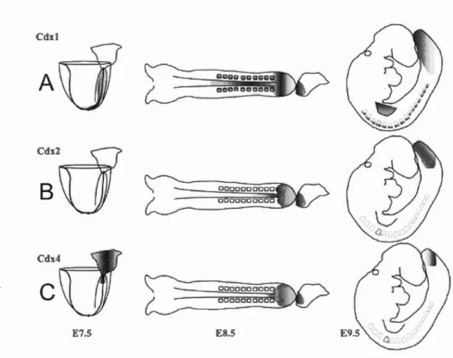
Cdxl
A
c
--E7.5 8.5
Figure 1.7 The spatial and temporal differences of rn urine Cdx gene expression.
(A) Cclxi expression begins at E7.5 at the ectoderm and mesoderm of the primitive streak. At E8.5, expression extends to the neural plate, paraxial mesoderm, hindgut and tailbud; At E9.5, expression regresses caudally, and lingers in somites and presumptive dermamyotome; at El2.5, expression is limited to the gut where it persists (Houle et al., 2003a; Meyer and Gruss, 1993; Silberg et al., 2000). (B) Cdx2
embryonic expression begins at E8.5 in the neural plate, neural tube, part of the notochord, the hindgut and ali tissues of the tailbud; at E9.5, expression regresses caudally in the NT, NP and notochord, hindgut and ali tissues of the tailbud more toward the tait bud; expression is limited to the gut at El2.5 where it persists (Beek et al., 1995; Silberg et al., 2000). (C) Cdx4 expression begins at E7.5 near the posterior end of the primitive streak; at E8.5-9.5, expression regresses caudally in the mesoderm and hindgut endoderm; at E 1 0.5, expression ends (Gamer and Wright,
18
1.3 Gene regulation and NC development
NC development relies on a feed-forward system of regulatory events known as the gene regulatory network (Figure 1.3) (Meulemans and Bronner-Fraser, 2004). Vital to any GRN is proper gene regulation by cis-regulatory regions. These regions are so important that it has been suggested that mutations in pre-vertebrate cis-regulatory regions were critical for NC evolution, and by extension the evolution of the NC-GRN (Jandziket al., 2015; Van Otterloo et al., 2013).
1.3.1 Enhancers
Enhancers are small cis-regulatory elements of around 200-500bp that can be up to 1.5 Mb downstream, upstream or even intronic of their target gene; (Figure 1.8) (Epstein, 2009; Rada-Iglesias et al., 201 3). Their function is to enhance target gene expression by activating the promoter of the target gene (Pennacchio et al., 2013). Transcription factors bind to specifie regulatory motifs on the enhancer and mediate activation of the target promoter. In nature, an ac ti vated enhancer e 1 icits a conformation of the chromatin structure so that the enhancer can loop near the target promo ter and mediate its activation (Kranz et al., 20 Il).
During development enhancers play a role in cellular growth, differentiation and migration by reiaying the activator signais from growth factors and transcription factors (Howard and Davidson, 2004; Kranz et al., 20 1 1 ). In the context of the NC-GRN, for example, enhancers of NPB specifïers, such as Msx, Pax3/7, Zic 1, Dlx3/5 would be at the receiving end of effectors of Wnt, FGF, BMP and otch signalling. Then, these NPB specifier transcription factors, along with other signais, would be at the delivery end of the enhancers of NC specifiers, such as Zic3, Sox9, Foxd3,
TSS
B
R
pp
lE
Figure 1.8 Types of cis-regulatory elements.
From left to right, BE: boundary element; E: enhancer; R: repressor (the counterpart
of the enhancer-instead of promoting the activation of a target promoter they
suppress activation) PP: proximal promoter; CP: core promoter; TSS: transcription
start site; JE: intronic enhancer. E1/2 represent exons. Image retrieved from Epstein
(2009)
Also of note regarding the current NC-GRN, as it relates to enhancer activity, is the
absence of known overlying epigenetic regulation needed for enhancer activity to occur in the first place. In fact, several epigenetic modifiers regulating NC
development have been identified, and constitute a fairly recent area of active
research (Hu et al., 20 14; Liu and Xiao, 2011 ).
1.3 .1.1 Approaches for identifying and tes ting enhancers
According to Simoes-Costa and Branner (2013), there are three main methods used in
identifying enhancers (Figure 1.9). Ail three require: the construction of the putative
enhancer sequence with a minimal promoter to drive the expression of a reporter, and
the tes ting of the putative enhancer via activation through ce li transfections, or in vivo
20
The first method requires inserting severa! non-coding sequences, one by one, into a reporter construct in the hopes of identifying an enhancer. This method has the potential advantage that random screening might find activity the other more focused methods do not detect. However this method is out-dated, costly, more uncertain, and laborious.
The second method screens only non-coding sequences with high vertebrate
homology. White homology does not necessarily equate to an enhancer, it is an important indicator as highly conserved regions have been found to be enhancer enriched (Bejerano et al., 2004; Pennacchio et al., 2006). Therefore, this method has a significantly better chance of identifying an enhancer than the previous method. In addition, a lot of sequencing data from numerous vertebrate species is freely and readily available.
Lastly, the third method screens areas where ChiP-seq has identified areas of histone modification and therefore possible sites with enhancer activity (Rada-Iglesias et al., 20 12). This method relies on the temporal qualities of histone modification in a specifie cell type, and can therefore target a specifie moment and axial leve! in potential enhancer activity. If the Chip-seq datais not currently available, this would add additional time and cost if it needs to be obtained.
APPROACHESFORENHANCER IDENTIFICATION
A. Screening of non-coding regions
'\ \
.\
'
' \ \ \\
'
., ' \.
.
\
\
~
\
,
i
...
1 / / , / ! 1 .j. .. .j B. Evolutionary conservationC. Profillng of histone modifications
TEST ENHANCER FOR ACTIVITY
Cell transfection
Stable transgenesls
Figure 1.9 Techniques for identifying and testing enhancers.
First step: build a putative enhancer reporter construct, with the enhancer obtained
from (A) Using a blind screen of non-coding areas; (B) Using a genome browser to
identify conserved regions; (C) Using Chip-seq data to uncover areas of histone modification Second step: testing the enhancer by cel! transfections, transient and/or stable transgenic models. Image retrieved from Simoes-Costa and Branner (2013),
- - - · - - - -- - - -- - - -- - - -- - -
-22
1 .3 .1.2 Characterization of binding motifs
While the previous methods can identify the existence of an enhancer, the location of
activity, and even test severa! potential TF activators, the characterization of binding motifs requires different steps. Bioinformatic tools can be used to identify potential
binding motifs but characterization is not addressed until the binding of transcription
factor to DNA is demonstrated. Electrophoretic mobility shift assays (EMSA) are often used in promoter/enhancer studies to reveal the binding of transcription factors (Pares-Matos, 2013). A DNA pull-down assay followed by Mass-Spectrometry can also be used to identify DNA-binding TFs (Drewett et al., 2001; Hubner et al., 2015).
1.3.2 Cdx2NSE
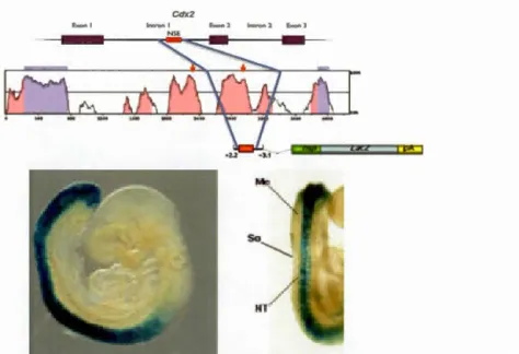
The enhancer identification method, using evolutionary conserved regions, described
above and in Figure 1.9b, was used in the discovery of a neural specifie enhancer (NSE) of Cdx2. Stable transgenesis showed reporter (LacZ) activity in the neural tube
(Figure 1.1 0) (Wang and Shashikant, 2007).
1.3.2.1 Cre-LoxP application of Cdx2NSE
As weil as providing evidence of regulatory interactions, enhancers can be used in a variety of appiications, inciuàing fate mapping, time-iapse imaging and targei.ed ioss of- and gain-of-function assays (Simoes-Costa and Bronner, 2013). One use of the
Cdx2NSE is to exploit its neural specifie activity and caudal localization in the
developing vertebrate. Cre-LoxP mouse systems in particular, are a great tool and way of exp loi ting the temporal and spatial qualities of an enhancer. In this system, the enhancer drives expression of Cre-recombinase in a transgenic line of mice. This enzyme can recognize and excise DNA flanked by loxP sites. Conditional knockouts are generated from the successful cross of a tissue specifie Cre-expressing line and a
Cdx2
Figure 1.10 Identification and testing the Cdx2 neural specifie enhancer (NSE).
Cdx2NSE was identified by its high vertebrate homology, and tested by a transgenic
line canying the presumptive enhancer driving the activation of a LacZ reporter.
Expression seems confined to the neural tube. Images retrieved from Wang and
Shashikant (2007)
Sox2 YFP Sox2YFP
B B' B" r,·
c
C' C" f', 1·'11 '.
!
.,, D D' D" ,. 1 E E' E"Figure 1.11 The enhancer is not entirely neural specifie.
An e8.5 embryo obtained from a cross between Cdx2NSE-Cre driver line and
R26R-YFP had Cre and therefore YFP expressed mostly in the neuroectoderm with sorne
present in mesodermal cells. Compare YFP to neural marker Sox2. Image retrieved
24
Here, the gene of interest is removed in specifie Cre expressing tissues. Another use
of the system is to have loxP sites impeding the expression of a gene of interest. In
this example, the successful recombination leads to the tissue-specifie expression of
the target gene.
Seeing as how no such Cre-driver line targeting the posterior neuroectoderm existed, Coutaud and Pilon (2013) decided to generate such a line using the Cdx2NSE. However, once the Cre-driver line was created and tested it was revealed that the
enhancer had minor activity in the mesoderm as weil and was therefore not entirely
neural specifie (Figure 1.11).
1.4 Diseases ofNCC origin and neural tube defects
A better understanding of the NC-GRN and associated epigenetic mechanisms is important because disruptions in these processes are believed to be the source of severa! human congenital birth defects and cancers. Relatedly, mechanisms affecting
proper neurulation can lead to severa( neural tube defects (NTDs ). The result of these
regulatory disturbances vary, but may drive changes affecting cell-fate/programming decisions, differentiation timing and/or migration patterns.
1.4.1 Neurocristopathies
Neurocristopathies are NC-derived developmental anomalies comprising over 700*
known syndromes and defects (Trainor, 201 0). Sorne of the best-known birth defects due to NC malformation result in craniofacial defects, heart defects and agang1ionosis of the colon. Many of these defects affect severa( known NC-GRN factors, such as SoxlO, Pax3, Snail2 in Hirschsprung's disease and Waardenburg syndrome (Kim et
al., 20 Il; Para tore et al., 2002), or Sox9 and Twist in CHARGE syndrome (Hu et al.,
2014).
1.4.1.1 Neural crest cell-derived cancers
NCCs possess many of the same attributes as metastatic cells, such as migration and
invasion. The assumption then is that cells of NC origin restatt many of the same regulatory mechanisms needed for embryonic development (Simoes-Costa and Bronner, 2013). Two of the most well-known cancers of NC lineage are
neuroblastoma and melanoma.
Neuroblastoma arises from cel! types of the sympatico-adrenal NCC lineage and is
the most common sol id childhood tumour accounting for 7-l 0% of paediatric cases
(Brodeur, 2003; Jiang et al., 2011; Schulte et al., 2013). The change from NCC to
mature sympathetic ganglia, involves an epigenetic switch needed to silence two genes that promote cel! cycle progression and cel! death aversion- respectively,
Mycn and Arid3b- the same switch left on is believed to lead to the oncogenesis of
neuroblastoma (Kobayashi et al., 2013). Mycn in particular has been shown to be
expressed in early NCC during ventral migration and in NCC undergoing neuronal
differentiation (Wakamatsu et al., 1997).
Studies have pointed out that the progression of metastatic melanoma may involve
severa! genes of the NC specification module (of the NC-GRN) such as Twist, SoxJO,
Slug and FoxD3 (Shakhova et al., 2012; Shirley et al., 20 12). Slug is needed by NCC
for EMT (Peinado et al., 2004), and Sox 10 for earl y melanoblast formation and
postnatal maintenance (Harris et al., 2013; Potte rf et al., 200 1 ). Twist 1 promotes
invasion of melanoma cells by mediating the regulation of a matrix metalloproteinase
(MMP), an enzyme needed to breakdown the extracellular matrix (Weiss et al.,
20 12). Twist in particular is overexpressed in many sol id tu mours ofNCC origin, not just melanoma, but also glioma and neuroblastoma, highlighting its imp01tance in the
26
1.4.2 Neural tube defects
Neural tube defects are congenital deformities brought about by an inadequate closure
of the neural tube. Prevalence of NTDs is from 0.75 to 1.12 per thousand, depending
on ethnie origin (Feuchtbaum et al., 1999). NTDs are complex in that a diet rich in
folie acid may weil be an impotiant factor in prevention, as public health data seems
to indicate (Czeizel et al., 201 1; Erickson, 2002; Pitkin, 2007). Interestingly, there is
also evidence indicating that the NCC may also be protected by folate
supplementation, suggesting the neuroepithelium may be sensitive to folate-related
pathways (Antony and Hansen, 2000; Li et al., 20 Il). Other possible NTD risk
factors may include environmental exposure to teratogenic agents such as heavy
metals, organic solvents and agricultural chemicals (Sever, 1995). Depending on
where the anomaly occurs the result is usually ::,pina bifida or anencephaly, although
there are severa! others. Improper closure near the caudal neuropore leads to spina
b{fida and anencephaly in the rostral neuropore. The degree of affliction varies, as
does neurological dysfunction. In spina bifida cognition, behavior, vertebral column
and organ systems can be affected depending on severity (Fletcher and Brei, 201 0).
Anencephaly is usually more severe, because the neural folds of the brain remain
open (Co pp and Greene, 20 13) leaving a large portion of the brain undeveloped.
Survival rate is very low, i.e. 40% after 24 hours (Baird and Sadovnick, 1984). Proper
closure of the cranial and spinal neural tube depends a lot on the bending of the
neuroepithelium at median (MHP) and dorsal-lateral hinge points (DLHPs). MHP
and DLHPs are both features of cranial and intermediate spinal neurulation. However,
MHP is a feature of upper spinal neurulation and DLHP of the lower spi ne (Copp et
al., 2003; Yamaguchi and Miura, 2013; Ybot-Gonzalez et al., 2007). BMP antagonist
Noggin is needed for cranial as weil as lower and intermediate spinal DLHPs
(Stottmann et al., 2006). Conversely, the upper spine MHP relies on inhibition of
Noggin by Sonic-hedgehog, hence higher BMP levels (Ybot-Gonzalez et al., 2002;
1.4.3 Tools for studying neurocristopathies and NTDs
Many of the molecular mechanisms so far described have been clarified through the
use of mutant mo use models (Britsch et al., 2001; Dixon et al., 2006; Shakhova et al., 20 12; Southard-Sm ith et al., 1 998; Weiss et al., 1997). Dom (Dom ina nt megacolon) mice are mutant (chromosome 15) models for Hirschsprung's disease that arose spontaneously, i.e. were not purposefully engineered, and they were instrumental in
demonstrating the importance of Sox 1 0 for development of the peripheral/enteric
nervous system (Puliti et al., 1995; Southard-Smith et al., 1998). Splotch mice also arose spontaneously (Russel, 194 7), with a mutation in chromosome 1 (Epstein et al.,
199la) and are often used as models for NTDs (Moase and Trasler, 1992). In homozygous mice, the mutation is embryonic lethal by E14 (Li et al., 1999),
exhibiting NT and heart defects (Auerbach, 1954; Conway et al., 1997). The
heterozygous Splotch line carries one nul! Pax3 allele, and is viable having minor
pigmentation abnormalities. ln the early 90's when the Pax3 deletion was found to be the cause of the mutation, it quickly indicated the importance of Pax3 for normal
neural development (Epstein et al., 1991 b; Goulding et al., 1993).
Conditional knockouts (KO) have also been used, many exploiting the NC-specific
Wntl-Cre transgenic line (Danielian et al., 1998). Wnt1 is active in the embryonic NT
and migrating NCC, and is important for the formation of the midbrain-hindbrain
boundary (Echelard et al., 1994; Lewis et al., 2013). Using this NCINT specifie Cre
-driver line, Dudas et al. (2004) knocked-out a BMP receptor (Aik2) which then produced craniofacial defects including cleft palate and mandible malformation and
suggested the importance of Alk2 in regulating the formation of specifie cranial
features. Akiyama et al. (2004) employed it to generate conditional Sox9 KO embryos to examine cardiac NCC and the essential role of Sox9 in heart EMT. Degenhardt et al. (201 0) used it to rescue !ost Pax3 expression specifically in the NC, and demonstrated the redundancy of Pax3 "neural crest enhancers". Other Cre-driver
2
8
1 ines used include Nestin-Cre for the neuroectoderm (Dubois et al., 2006; Tronche et
al., 1999) and Pax3pro-Cre for the dorsal NT and NC (Li et al., 2000).
Overexpression knock-in mutants are also instrumental models for disease. Gene
amplification in particular is known to be the direct cause of many known human
diseases and cancers (Santarius et al., 201 0; Shastry, 1995). For example, the classic
mainstay mouse model for neuroblastoma is the TH-MYCN, wherein Mycn
expression is driven by the TH rat tyrosine hydroxylase promoter (Weiss et al., 1997).
As previously discussed, Mycn is normally expressed in migrating NCC switching to
neuronal lineage (Wakamatsu et al., 1997). At the time, MYCN was known to be a
proto-oncogene of human neuroblastoma but had not yet been tested in an animal
model. Weiss et al. (1997) provided direct in vivo evidence that Mycn amplification
can contribute to the oncogenesis of neuroblastoma. The TH-MYCN transgene is
integrated in chromosome 18 in a distal region, and the effect it has in disturbing this
region is unknown. Due to this uncertainty, Althoff et al. (20 15) created another
Mycn-driven neuroblastoma mouse model targeting the ROSA26 locus (chromosome 6). This locus has the advantage that it is ubiquitously expressed, disruptions in the
locus have no physical effect in mice, and can be targeted with high efficiency
(Soriano, 1999; Zambrowicz et al., 1997). As of 2010, there were already over 130
knock-in iines targeting the ROSA26 locus (Casoia, 20 i 0).
Ali these mutant models have their own limitations, and advantages, and yet they
share the fact that the disruptions they create help fuel our understanding of the
HYPOTHESES AND OBJECTIVES
2.1 Hypotheses
Conditional overexpression of Cdxl plays an oncogenic role in the development of
neural crest cell derived cancers in envisioned mouse model.
The intronic neural specifie enhancer (NSE) of Cdx2 contains identifiable non-neura
l-specifie activator binding sites.
2.2 Objectives
Genera te a mo use mode! carrying the Cre-inducible Cdx 1 overexpression trans gene. The steps required to genera te the mode!: Construction of the targeting vector,
transfection of the vector into ES cells, selection of the colonies, verification of
positive clones, microinjection and rearing.
lnvestigate regulation of Cdx2NSE, and identify activator-binding sites with no n-neural specifie characteristics. Testing the regulation would give us a better understanding as to how the NSE architecture could be modified to drive truly neural
specifie expression in a new Cre-driver line. Luciferase assays using neural Sox2
MATERJALS AND METHODS
3.1 Construction of pROSA26-Cdx 1, ES targeting and verification
3. 1. 1 Vector construction
To make the pROSA26-Cdx 1 targeting vector, a PGKneoPA-Flag-Cdx
1-IRES-EGFP-BGH cassette was inserted into the PacT and Ascl sites of the pROSA26-PA
targeting vector (Srinivas et al., 2001). pROSA26-PA was a gift from Frank
Costantini (Addgene plasmid # 2 L271). lt is a vector used to target the ubiquitous
ROSA26 locus, and contains the 3' and 5' ROSA26 arms necessary for homologous
recombination (see Figure 4.2) as weil as a diphtheria toxin gene (PGK-DTA) for
negative selection in ES cells. The final construct was digestion and sequencing
verified. ln total, the final construct contains a loxP-flanked PGKneoPA cassette
capable of kanamycin resistance expression in E.coli and eukaryotic promoter PGK
for expression of neomycin resistance in the mammalian ES cells. Next the N
-terminal FLAG-tag is upstream of a full Cdxl ORF (807-bp) as previously described
(Beland et al., 2004), followed by an IRES-EGFP reporter sequence.
3.1.2 Targeting ES cells
RI ES cells were cultured on mouse embryo fibroblast (MEF) feeder cells that were
mitotically inactivated by 1 Oflg/ml mitomycin C treatment. The cul turing of the ES
cells on MEF feeder cells was done on gelatin-coated plates to improve conditions.
Also, the ES medium was supplemented with leukemia inhibitory factor (LIF) to
32
linearized by Sacll and (12.5 ~tg DNA) electroporated (250Y, 500 ~F, in a 4mm cuvette) into the R 1 ES cells. Selection of stable integrants was done with 200~g/ml
0418 for one week. Genomic DNA was extracted and isolated. Screening of successful homologous recombination was carried out first by PCR using an external
forward ROSA26 primer and an internai reverse primer to create a 1.1 kb fragment (Table 3.1 ). Three clones were identified. Confirmation by Southern blot, albeit inconclusive, was followed soon after.
Table 3.1 Primers for PCR screen.
PRIMERNAME 1 LENGTH 1 SEQUENCE
ROSA26TARG-F1 1 23 IAAGAAGAGGCTGTGCTTTGGGGC
ROSA26TARG-RJ 1 20 JAGGGCGGCTTGGTGCGTTTG
3.1.3 Southern Blotting
Around 20 ~g of genomic DNA was digested with EcoRI and Kpnl and separated on
a 0.8% agarose gel. The gel was then depurinated, denatured and neutralized. Afterwards, the DNA was transferred over night onto a Hybond membrane (Amersham) via a simplified downward capillary system, i.e. without added transfer buffer.
Once transferred, the membrane was rinsed with 2x SSC, dried, UV cross-iinked and
pre-hybridized. The pre-hybridization buffer solution contained salmon sperm ssDNA to block non-specifie sites. After pre-hybridization, the membrane was
hybridized with probe ssDNA.
Two DNA probes were prepared to target the 5' or 3' ROSA26 ends (Figure 4.2), at 25ng in 45~1 of TE buffer. The probes were [a.-32P] dCTP-labelled using the Rediprime Il labelling kit (Amersham), and purified by sepharose columns. The final
probe concentration was approximately around 2.8ng/mL of hybridization buffer, as per manufacturer's instructions. Hybridization occurred over night at 65°C. The
membrane was la ter washed at least 2 times (2x SSC
+
0.1% SDS) before placing inan exposure cassette for l-7 days.
3.2 Testing the neural specifie regulation of Cdx2NSE
3.2.1 Plasmids
The full Cdx2NSEforward-LUC and Cdx2NSEreverse-LUC reporter constructs were created in our lab (Coutaud, 20 13) and were readily available. They contain an 852
-bp Cdx2NSE sequence upstream of an Hsp68 minimal promoter in a luciferase gene expression vector. Four Cdx2NSEreverse fragments were created by PCR with the
f01·ward primers containing a Hindlii site and reverse primers containing a Kpnl eut
sites (Table 3.3). PCR products were amplified with Taq polymerase (Feldan) and
cloned into a pGEMT-easy vector (Promega) and verified by sequencing. The Hindlll and Kpnl restriction sites were then used to clone into an Hsp68-LUC reporter vector.
The resulting four fragment NSE reporter constructs can be seen in Figure 5.4, and include the NSE sequences containing putative Lef1 /Tcf sites : 4-3-2-1 (676-bp);
3-2-1 (607-bp); 2-1 (437-bp); and 5 (193-bp). The construct with LEF/TCF site « 1 »
(219-bp) was made by digesting Cdx2NSEreverse-LUC with BamHT to remove a 666bp fragment containing potential Lefl /Tcf binding sites 5-4-3-2. PGL3-0T served
as positive control in transfections; it contains 3 copies of wildtype Tcf-4 binding sites and was a gift from Bert Yogelstein (Addgene plasmid # 16558) (Shih et al.,
2000). Negative control was an empty vector containing just the promoter and
luciferase gene. Serving as the activators for the transfections were Sox2, and Lef
l-~catenin.
3.2.2 Luciferase assay conditions
P 19 cells were cultured at 37°C (5% C02) in a-MEM supplemented with 7.5% hea
t-inactivated bovine calf serum and 2.5% heat-inactivated fetal bovine serum (Wisent).
Approximately 2 hours prior to transfection, the P19 cells were plated in 24 weil
34
Co-transfection was carried out to test the effect of Sox2 and Lefl-~caten in (individually and together) on the full Cdx2NSE forward and reverse reporter
sequences. Cdx2NSEforward-Luc and Cdx2NSEreverse-Luc reporter DNA was maintained at 1 Oûng/well, as was the negative and positive control DNA. On the
other hand, Sox2 and Lefl-~catenin activator DNA ranged from Ong, 25ng, SOng or
1 Oûng/well. The total amou nt of DNA per weil was kept at 300ng. Tf needed, an
empty IRES-GFP expression vector was used to complete this amount. GeneJu ice
was the transfection reagent (Novagen) and the ratio of reagent to DNA was 3~tl to
1 ~Lg DNA, as recommended, i.e. each weil had 0.9~tl of transfection reagent and
300ng of total DNA. This set of transfections was performed at !east three times in
triplicate.
Assay conditions were similar for the tests with the truncated Cdx2NSEreverse DNA
reporter sequences. 1-!owever, this set underwent two key differences in conditions:
the cell density used and activator DNA concentration. Cell density was kept at
1.5x 10⁴ cells/well and activa tor DNA (Sox2 and/or Lefl-~catenin) was kept at Ong or
1 Oûng/well. This set oftransfections was performed at !east six times in triplicate.
Ali transfections were incubated in the a-MEM FBS+CBS media for about 48 hours
at 37°C before performing post-transfection tests and analyses. After 48 hours,
transfection efficiency was visually assessed by m1croscopy, 1.e. identifying GFP
expressiOn. Next, transfection analysis (gene expression) was quantified by
luminometry. The steps for this included: media removal, rinsing wells with
phosphate buffered saline 1 X, and lysing the cells. In a tube, 20~tl of the ce li lysate
was added to 1 00~1 of luciferase assay buffer. Then, 50~1 of luciferin was delivered
before placing tubes in the luminometer,
Luciferase activity was expressed as fold activation relative to the appropriate
reporter vector atone. Each independent experiment was carried out in triplicate, at
3.2.3 Luciferase assay test and statistical analysis
A Il the results of the luciferase assays are expressed as means+S.D., and the differences in luciferase activity are expressed as fold activation. The differences in
fold activation were examined by student's t-test for two-group comparisons. One-way ANOVA was performed to identify differences in multiple group comparisons Graphs and ali statistical analyses were done using GraphPad Prism version 6 software, and p-value ranges, wording and asterisk representation follow the Graph Pad statistics gu ide (Table 3 .2).
T a bi e 3 2 P -va ue gUI "d e
P VALUE DESCRIPTION REPRESENTATION
< 0.0001 Extremely significant
****
0.0001 to 0.001 Extremely significant***
0.001 to 0.01 Very significant **
0.0 l to 0.05 Significant
*
36
T ba i e . 3 3