Université de Montréal
Comparison between two different antibiotic regimens for
the placement of dental implants:
A phase-I randomized clinical trial
Par Issam Kersheh
Département de santé buccale Faculté de médecine dentaire
Université de Montréal
Mémoire présenté à la Faculté des études supérieures et postdoctorales en vue de l’obtention du grade de
Maîtrise ès Sciences (M.Sc.) en sciences buccodentaires
Juillet 2017
Université de Montréal
Faculty of Graduate and Postdoctoral Studies
This thesis entitled:
Comparison between two different antibiotic regimens for
the placement of dental implants:
A phase-I randomized clinical trial
By Issam Kersheh
Was evaluated by a jury composed of the following members:
Dr. Elham Emami, President of the jury Dr. Robert Durand, Thesis supervisor Dr. Nathalie Rei, Thesis co-supervisor
Résumé
Introduction : Afin de minimiser la morbidité postopératoire après une chirurgie implantaire,
plusieurs régimes d’antibiotiques péri-opératoires ont été suggérés, mais leurs effets sur le remodelage osseux péri-implantaire n’a pas été clairement établi. De plus, l’utilisation répandue des antibiotiques en médecine dentaire et en médecine est remise en question étant donné l’émergence récente des résistances bactériennes aux antibiotiques.
Objectifs : L’objectif primaire de cette étude pilote était de produire des données
préliminaires et d’évaluer si des doses postopératoires d’antibiotiques après la pose d’implant prises sur sept jours influenceraient les niveaux osseux péri-implantaires après 4 mois chez les patients en santé subissant la pose simple d’un implant de type « platform-switching ». Les objectifs secondaires étaient d’évaluer la sévérité de la douleur, la morbidité postopératoire, et le taux de survie après un an.
Méthodes : Trente-huit participants ont été recrutés dans un essai clinique parallèle randomisé
à double insu. Les participants du groupe intervention ont reçu 2 g d’amoxicilline une heure avant la chirurgie implantaire, et un régime postopératoire de 500 mg d’amoxicilline d’une durée de sept jours. Les participants du groupe contrôle ont pris seulement une dose de 2 g d’amoxicilline une heure avant la chirurgie et un placébo postopératoire. Les changements du niveau osseux péri-implantaire mésial et distal (résultat primaire) ont été mesurés à la pose de l’implant et quatre mois plus tard à l’aide de radiographies rétroalvéolaires standardisées. La sévérité de la douleur et les morbidités postopératoires (résultats secondaires) ont été évaluées à l’aide d’examens cliniques et de questionnaires auto-administrés. Le taux de survie implantaire a été évalué un an plus tard. Des analyses bivariées et descriptives ont été utilisées
pour analyser les données. Une valeur de P ≤ 0.05 a été considérée statistiquement significative.
Résultats : Trente-sept participants ont complété l’étude (âge moyen : 57,4 ± 11,3 ans). Les
changements moyens du niveau osseux péri-implantaire combiné pour le groupe intervention et le groupe contrôle étaient respectivement de -0.29±0.36mm et de -0.11±0.35mm. Les différences entre les groupes pour le changement moyen du niveau osseux péri-implantaire combiné et la sévérité de la douleur n’étaient pas statistiquement significatives (P> 0.05). Les interférences avec les activités quotidiennes étaient parfois significativement plus importantes pour le groupe contrôle comparativement au groupe intervention (p< 0.05), dépendamment du critère évalué et du nombre de jours écoulés depuis la chirurgie. Le taux de survie implantaire était de 100 % dans les deux groupes après un an.
Conclusions : Les résultats de cette étude pilote suggèrent qu’un régime postopératoire
d’antibiotiques chez les patients en santé subissant la pose simple d’implant de type « platform-switching » n’est pas nécessaire. Des investigations additionnelles sont nécessaires afin de confirmer les résultats de cette étude pilote.
Abstract
Introduction: In order to minimize postoperative morbidity and failure of dental implant
therapy, several antibiotic regimens have been proposed in the literature. However, the extensive use of antibiotics in health care has been debated due to adverse effects and bacterial resistance. Furthermore, the impact of postoperative antibiotics on peri-implant bone level is still not clear.
Objectives: The primary objective of this pilot study was to produce preliminary data and to
assess whether giving postoperative antibiotics after implant placement over seven days would influence peri-implant crestal bone levels after four months in healthy patients undergoing platform-switched implant placement. The secondary objectives were to evaluate postoperative pain severity, surgery-associated morbidities, and one-year implant survival rate.
Methods: Thirty-eight individuals were enrolled in a double-masked two-arm randomized
clinical trial. Participants in the intervention group received 2 g of amoxicillin one hour before implant placement followed by a seven-day post-operative course of 500 mg of amoxicillin. Participants in the control group took only 2 g of amoxicillin before surgery and an identical placebo postoperatively. The changes in mesial and distal crestal bone level (primary outcome) were measured at baseline and four-month follow-up using standardized periapical radiographs. Pain severity and surgery-associated morbidities (secondary outcomes) were evaluated by clinical examinations and self-administered questionnaires. Implant survival rate was assessed at the one-year follow-up. Descriptive and bivariate analyses were used to analyze the data. A P value ≤ 0.05 was considered statistically significant.
Results: Thirty-seven participants completed the study (mean age: 57.4 ± 11.3 years). The
mean combined peri-implant crestal bone level change for the intervention and control group was -0.29±0.36mm and -0.11±0.35mm, respectively (n=37 participants). The differences between groups for mean combined crestal bone level change and pain severity were not statistically significant (P> 0.05). Interferences with daily activities were sometimes significantly more important for the control group compared to the intervention group (P< 0.05), depending on the criteria and on the number of days elapsed since the surgery. The implant survival rate was 100% in both groups after one year.
Conclusions: Results from this study suggest that an additional postoperative intake of
antibiotics in healthy patients undergoing straightforward platform-switched implant placement might not be necessary. Further investigations are needed to confirm these pilot study findings.
TABLE OF CONTENTS
Résumé ... iii
Abstract ... v
TABLE OF CONTENTS ... vii
LIST OF TABLES ... x
LIST OF FIGURES ... xi
LIST OF SYMBOLS AND ABBREVIATIONS ... xii
DEDICATION ... xiii ACKNOWLEDGMENTS ... xiv CHAPTER I ... 1 LITERATURE REVIEW ... 1 1.1 INTRODUCTION ... 1 1.2 OSSEOINTEGRATION IN IMPLANTOLOGY ... 3
1.3 EVALUATION OF DENTAL IMPLANT OUTCOMES ... 6
1.3.1 Criteria to determine implant success ... 6
1.3.2 Bone remodeling around dental implants ... 11
1.4 PAIN EXPERIENCE IN IMPLANTOLOGY ... 14
1.5 THE ROLE OF ANTIBIOTICS IN DENTISTRY ... 17
1.5.1 Risks associated with antibiotics overuse ... 17
1.5.2 Effects of antibiotics on dental implant outcomes ... 18
CHAPTER II ... 27 METHODOLOGY ... 27 2.1 OBJECTIVES, HYPOTHESIS... 27 2.1.1 Objectives ... 27 2.1.2 Hypothesis... 27 2.2 RESEARCH METHODOLOGY... 28
2.2.1 Study design, participants, eligibility and intervention ... 28
2.3 DATA COLLECTION AND EXPERIMENTAL PROCEDURES ... 30
2.3.1 Medical and sociodemographic questionnaires ... 30
2.3.2 Clinical procedures ... 30 2.3.3 Radiographical methodology ... 34 2.4 STATISTICAL ANALYSIS ... 37 2.5 ETHICAL CONSIDERATIONS ... 37 2.6 STUDY RELEVANCE ... 38 CHAPTER III ... 39 RESULTS ... 39 3.1 RECRUITMENT OF PARTICIPANTS ... 39 3.2 CALIBRATION RESULTS ... 40
3.3 SOCIODEMOGRAPHICS AND MEDICAL BACKGROUND ... 40
3.4 SURGICAL PARAMETERS AND IMPLANT CHARACTERISTICS ... 42
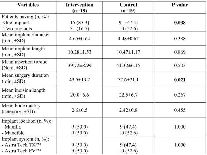
3.5 PERI-IMPLANT CRESTAL BONE CHANGE ... 43
3.6 PAIN EXPERIENCE ... 45
3.7 INTERFERENCE WITH DAILY ACTIVITIES ... 48
3.8 POSTOPERATIVE MORBIDITIES ... 53
3.9 ORAL HYGIENE AROUND IMPLANTS ... 57
3.10 EFFECTS OF SURGERY DURATION AND NUMBER OF IMPLANTS ON CRESTAL BONE CHANGE ... 58
CHAPTER IV ... 59
DISCUSSION ... 59
4.1 CLINICAL OUTCOMES ... 59
4.2 PATIENT-BASED OUTCOMES ... 62
4.3 STUDY STRENGTH AND LIMITATIONS ... 64
4.4 CLINICAL RELEVANCE AND PRACTICAL IMPLICATIONS ... 66
4.5 FUTURE RESEARCH ... 66
CHAPTER V ... 68
CONCLUSION ... 68
BIBLIOGRAPHY ... 69
APPENDIX I. CONSENT FORM ... i
APPENDIX II. TIMELINE OF RESEARCH APPOINTMENTS ... ix
APPENDIX III. MEDICAL QUESTIONNAIRE ... x
APPENDIX IV. SOCIODEMOGRAPHIC QUESTIONNAIRE ... xii
APPENDIX V. POSTOPERATIVE INSTRUCTIONS ... xv
APPENDIX VI. PAIN AND INTERFERENCE WITH DAILY ACTIVITIES QUESTIONNAIRE ... xvii APPENDIX VII. CLINICAL AND RADIOGRAPHICAL DATA COLLECTION FORMS ... xlix APPENDIX VIII. INFORMATION TO RECRUIT PARTICIPANTS ... lviii
LIST OF TABLES
Table 1: Health Scale for Dental Implants ...10
Table 2: Inclusion and exclusion criteria ...28
Table 3: Participants’ sociodemographics and medical characteristics ...41
Table 4: Surgical parameters and implant characteristics ...42
Table 5: Postoperative morbidities after one week ...54
Table 6: Postoperative morbidities after three weeks ...54
Table 7: Postoperative morbidities after sixteen weeks ...55
Table 8: Postoperative morbidities after one year ...56
LIST OF FIGURES
Figure 1: Individualized silicon bite block and x-ray positioning technique ...32
Figure 2: Radiographic evaluation of crestal bone change: a) Baseline periapical radiograph; b) month periapical radiograph; c) Superimposed radiographs with baseline and four-month measurements ...36
Figure 3: Study flowchart ...39
Figure 4: Mean crestal bone changes after four months: a) At the mesial aspect; b) At the distal aspect ...43
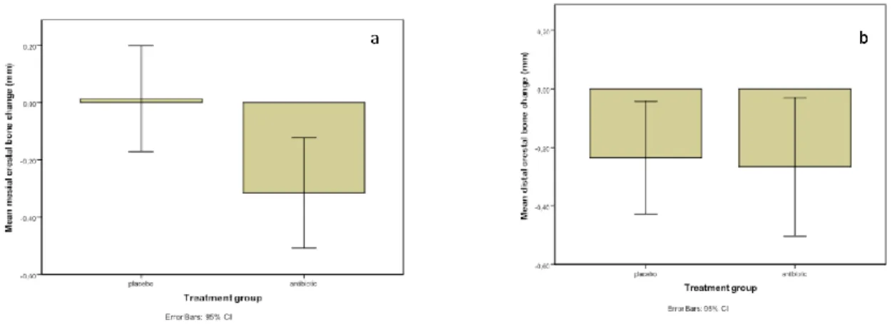
Figure 5: Mean combined crestal bone changes after four months ...44
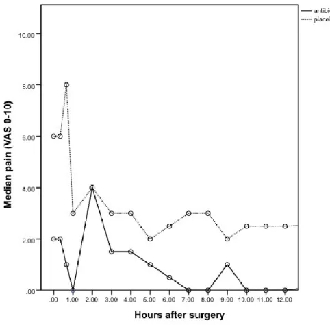
Figure 6: Pain severity for the first twelve hours after surgery ...46
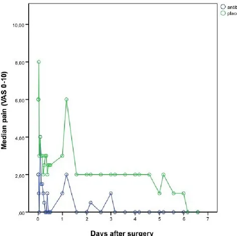
Figure 7: Pain severity for the first seven days after surgery ...47
Figure 8: Interference with chewing ...49
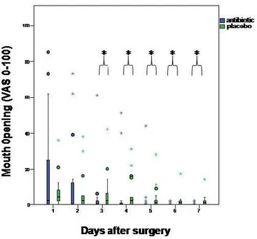
Figure 9: Interference with mouth opening ...49
Figure 10: Interference with speech...50
Figure 11: Interference with sleep ...50
Figure 12: Interference with work or school ...51
Figure 13: Interference with social life ...51
Figure 14: Interference with recreational activities ...52
LIST OF SYMBOLS AND ABBREVIATIONS
Vs Versus
SLA® Sand-blasted, large-grit, acid-etched
I kB Inhibitor of nuclear factor kappa-B % Percentage
mm Millimeter
ICOI International Congress of Oral Implantologists CBCT Cone Beam Computed Tomography
DAS Dental Anxiety Scale VAS Visual analog scale
EMA Ecological Momentary Assessment PC Personal computer
E. coli Escherichia coli C. difficile Clostridium difficile g Gram
mg Milligram
NNT Number Needed to Treat
AMSTAR A Measurement Tool to Assess Systematic Reviews
mPI Modified Plaque Index
CRCHUM Centre de recherche du Centre hospitalier de l’Université de Montréal
μg Microgram
DEDICATION
To my father’s spirit To my beloved family and friends
To all who have supported me through hard times
Thank you Chaza, Majd, Joyce, Marie Rose, Salem, Munif, Faheem, Aminah for being a source of love, support and encouragement.
ACKNOWLEDGMENTS
I am deeply grateful to my director, Dr. Robert Durand, who has taught me on all aspects of dentistry and who has supported me when I was in need.
I would like to thank Dr. Elham Emami and my co-director, Dr. Nathalie Rei, for their expertise in clinical research. I am also grateful to Dr. Thierry Cresson who standardized the radiographic images and Dr. Matthieu Schmittbuhl for his expertise in oral radiology.
I would like to thank Dr. René Voyer and Dr. Pierre Boudrias. They were amazing professors and good supporters throughout this research project.
I am also thankful to Mr. Pierre Rompré for his expertise with statistical analysis.
I would like to thank all the implantology clinic staff members at the Faculty of Dentistry at the Université de Montréal, especially Marie-Josée and Kim, who helped me to collect clinical data and to recruit participants.
I am thankful to my wife Chaza, my dear friend Dr. Munif Rabadi, and my brother Salem. Thank you for giving me tremendous support and help during my studies and thesis writing period.
This study would not have been possible without the financial support from the Ernest Charron and Vice Dean of Research funds at the Université de Montréal Faculty of Dentistry.
CHAPTER I
LITERATURE REVIEW
1.1 INTRODUCTION
No one can deny the importance of antibiotics in medicine and dentistry. However, misuse or overuse of antimicrobial medications may have detrimental effects on one’s health. Indeed, widespread usage of antibiotics increases the risk of developing antibiotic-resistant bacteria strains (1, 2), specifically, the community-acquired species, which has been observed over the past two decades (3, 4).
The population’s demand for dental implants has been increasing due to their high survival rate and their significant improvements of a patient’s quality of life. On the other hand, failures have been reported and bacterial infections are thought to play an important role (5). Clinical studies have shown conflicting results regarding the effect of perioperative use of antibiotics on implant survival rate, while their consumption may cause adverse consequences (6, 7). However, other studies have proven that the benefits outweigh the risks of secondary effects (8). Different prophylactic regimens can be found in the literature in order to increase the survival rate of dental implants by reducing the risk of infection (8, 9). Two recent meta-analyses of randomized clinical trials comparing patients with implants who received antibiotics pre- and/or post-operatively to those who did not take any antibiotics, have shown that in the latter group, there were statistically significant higher implant failures (10, 11). The authors of the latest Cochrane review concluded that preoperative antibiotics given one hour before implant placement surgery significantly reduced implant failure rates (9). However, the
authors could not assess whether it was beneficial to give postoperative antibiotics in addition to a preoperative intake or as a sole antibiotic regimen. Consequently, in order to prevent the overuse of antibiotics and the potential emergence of drug resistant bacteria, it would be advisable to find an optimal protocol including minimal antibiotic exposure while maintaining an acceptable implant survival rate. In addition, investigators have found that patients who were taking antibiotics postoperatively have shown less peri-implant crestal bone loss after six months of implant placement compared to individuals who did not receive any post-operative antibiotics (12). However, there is very little data available on the influence of antibiotics on the crestal bone level change.
Subjective outcomes after implant placement are poorly documented in the literature. Indeed, patients’ pain and discomfort have not been taken into account in most of the dental implant clinical trials and little is known about their prevalence and intensity after surgery, more specifically with regards to different antibiotic regimens (11). Moreover, very few implantology studies have compared the effects of different antibiotic regimens on the implant survival rate including subject-based, clinical, and radiographic outcomes simultaneously. Therefore, the goals of this two-arm double-masked randomized phase-I clinical trial was to evaluate the influence of postoperative antibiotics on peri-implant crestal bone remodeling after four months, postoperative pain and morbidity, and one-year implant survival rate in healthy patients undergoing straightforward platform-switched implant placement.
1.2 OSSEOINTEGRATION IN IMPLANTOLOGY
Osseointegration was first described by Swedish scientist Per-Ingvar Branemark and his coworkers as a direct, structural and functional connection between living bone and the surface of a load-carrying implant (13). The first implant patient was treated in 1965 by Dr. Branemark (14). The initial implant surface was polished and the implant was cylindrical and screw-shaped. In the 1980s, efforts were made by several implant companies to enhance surface energy and accelerate osseointegration in order to increase implant survival rate and improve patient care. Nowadays, most implants remain screw-shaped or tapered, and their surfaces are micro-textured and/or nano-textured to enhance osseointegration. The success of an implant’s osseointegration depends on the following factors: biocompatibility of the implant material, macroscopic and microscopic nature of the implant surface, status of the implant bed (non infected) and bone quality, surgical technique, quality of infection control during surgery, condition of the patient’s immune system and subsequent prosthetic design.
Histological studies have shown that the osseointegration process is more complex than initially demonstrated, although similar to direct fracture healing (15). The osseointegration process starts with early events beginning within two hours of implant placement. The threads of self-tapping screw-shaped implants usually provide initial mechanical stability. A blood clot is formed around the implant, serving as a matrix for neoangiogenesis, extracellular matrix deposition, and bone forming cells (16, 17). Bone remodeling occurs within one week of implant placement. It starts with contact osteogenesis, where osteogenic cells migrate directly onto the implant surface and generate the bone matrix (16). This phenomenon has only been observed on textured implant surfaces. Fourteen days later, the woven bone
formation is more pronounced. Most of this bone formation starts at a distance from the implant, from the borders of the drill hole, and is thus called “distant osteogenesis”, where osteoblasts migrate to the surface of the implant cavity, differentiating and stimulating new bone formation. Osteoclasts play a role in bone resorption, especially in the zones where there is implant pressure in the osteotomy site. After two to three weeks, the implant’s stability is at its lowest because of the smaller percentage of mature mineralized bone matrix in contact with the implant as a result of the remodeling process. Four weeks after implant placement, the newly formed bone extends and covers most of the implant walls. At six to twelve weeks of healing, mineralized bone fills all the remaining space between the implant and the native bone. At that point, secondary implant stability is at its highest, increasing only slightly thereafter. Its strength depends largely on new bone formation at the bone-to-implant interface (18).
Recent human and animal studies have shown that roughened sandblasted and acid-etched implants showed a better bone-to-implant contact compared to implants with a polished surface. This observation was noted as early as one week after implant placement. A limited number of human studies have shown healing with screw-type sandblasted acid-etched dental implants (SLA®, Straumann AG, Basel, Switzerland) (19-22). The findings from these studies indicated an average bone-to-implant contact of 22% of the total implant surface in SLA® and SLActive® (Straumann AG, Basel, Switzerland) implants at the end of the first week, which consisted mainly of native bone. This number went up to 28% at the end of the second week, and new bone covered 12.2% and 14.8% of the surface in SLA® and SLActive® implants respectively. At four weeks, the old bone covered 28.3% and 13.9% of SLA® and SLActive®
implant surfaces respectively, and the new bone covered 32.4% and 48.3% of SLA® and SLActive® implant surfaces. At six weeks, the percentage of the old bone being in contact with the titanium implants decreased to between 8 and 13.6%, and the new bone covered 61.5% of the surface for both types of implants.
The molecular mechanisms involved in the osseointegration process were not well understood until recently. Major signalling pathways such as IkB kinase/nuclear factor KappaB, start early during osseointegration and subsequently decrease over time (23). Those pathways provide areas of interest that might be modified to enhance osseointegration. Other molecular mechanisms also playing an important role are inflammation, angiogenesis, neurogenesis and skeletogenesis. A human study using whole-genome transcriptional analysis described the principal molecular mechanisms during the first two weeks of the osseointegration process (20, 21).
It was shown that immuno-inflammatory genes were expressed early during the osseointegration process and down-regulated over time. However, their role is not fully understood. For example, enhanced macrophage cytokine expression suggests that modified implant surfaces may accelerate osseointegration as macrophage cytokines are well known to play a part in the inflammatory process after injury, eventually leading the healing process. Further investigation is needed to fully understand their exact role as well as the integration mechanism in dental implants.
During the osteogenesis phase, osteoblast differentiation, ossification and biomineral formation was observed. In human patients, most of these events occur in the first two weeks of osseointegration. Of interest is the period between day four and seven when the embryonic skeletogenesis-associated genes are differentially regulated. The importance of angiogenesis is another area where osseointegration can be targeted for modification since blood vessels provide a network and direct access to the bone-implant interface for growth factors and bone forming cells such as osteoblasts. A very prominent over-expression of genes associated with neurogenesis was observed during the early stages of osseointegration, though their role is unclear. Neuropeptide Y is one molecule that was shown to modulate osteoblast function.
The capacity of micro-textured implant surfaces to enhance and accelerate gene expression of bone matrix molecules and surface hydrophilicity increases osteogenesis and angiogenesis. This finding provides new avenues in implant surface modification and development. Newer implant designs do indeed incorporate such surface modifications in order to enhance the implant survival rate and accelerate the osseointegration process. Nevertheless, little has been done in clinical research regarding potential biochemical or pharmacological methods to accelerate the osseointegration process.
1.3 EVALUATION OF DENTAL IMPLANT OUTCOMES
1.3.1 Criteria to determine implant success
The success of a dental implant may be subjective and depends on both the surgeon’s and the patient’s perceptions. Clinical, radiographic and patient-based outcomes have been used since
the 1980s. On the other hand, dental technologies and dental implant designs have significantly changed since then, and implant success criteria have evolved over the years. More importantly, implant success is not synonymous with implant survival. Implant success is characterized by specific preselected criteria that are met, while implant survival simply means that the implant remains in situ or in function without any other criterion being considered. It must be measured once the implant’s osseointegration is completed and the implant is restored, which usually takes between two and six months. The ten-year cumulative implant survival rate has recently been reported in a systematic review to be around 95% (24). The success rate of implants may greatly vary depending on the outcomes measured. Several success criteria have been proposed by different groups of experts. The original criteria for implant success were described by Albrektsson and colleagues (25) and included the following:
1) The individual, unattached implant should be immobile when tested clinically; 2) No radiographical evidence of peri-implant radiolucency;
3) Less than 0.2mm annually of vertical bone loss after first year of service;
4) Absence of persistent pain, infection, neuropathies, paresthesia or violation of mandibular canal.
5) Based on these criteria, a success rate of 85% at the end of a five-year observation period and 80% at the end of a ten-year period were minimum levels for success at that time.
It is important to note that these success rate thresholds were measured with the original Branemark® polished-surface implants. It was determined that the mean crestal bone loss for
Branemark® osseointegrated implants was 1.5 mm for the first year followed by a mean crestal bone loss of 0.1 mm/year (26). Thus, a mean bone loss threshold of 0.2 mm per year after the first year in function was accepted as a criterion for success. Three years later, an additional criterion for success was added to take into account the implant restoration aesthetic appearance:
6) The implant design does not preclude placement of a prosthesis or crown with an aesthetic appearance that is satisfactory to the patient and dentist (27).
A consensus report was published later on using the same success criteria but removing the expected success rate and radiographical peri-implant radiolucency (28). The authors emphasized the importance of using standardized radiographs to measure crestal bone loss with predetermined reference points and angulations.
With the advent of implant surface texturing methods, Buser and colleagues studied soft and hard tissue integration around Straumann® rough-surface implants after one year using several clinical parameters to determine implant success: plaque index, sulcus bleeding index, probing depth, distance between implant shoulder and mucosal margin, attachment level, width of keratinized mucosa, and mobility (29). Standardized radiographs were also taken to measure the distance between the implant shoulder and the first visible bone contact. The authors used the following success criteria:
1) Absence of persistent subjective complaints, such as pain, foreign body sensation and/or dysesthesia;
2) Absence of a recurrent peri-implant infection with suppuration; 3) Absence of mobility;
4) Absence of a continuous radiolucency around the implant; 5) Possibility for restoration.
Furthermore, the authors classified implant failures as early or late failures. Early failures occur during the first five months following implant placement. Overheating of the bone during drilling procedures, lack of primary stability of the implant, masticatory loading forces, and/or bacterial contamination during surgery may contribute to implant failures. Late failures occur during the maintenance phase after successful osseointegration. The clinical signs and symptoms were pain, bleeding on probing, peri-implant suppuration, and increased probing depth. Upon analyzing the data of 100 consecutively placed implants by one surgeon, 98 implants were considered to be successful, while osseointegration was not achieved in one implant, and a peri-implant infection developed in another one, for a success rate of 98% after one year (29).
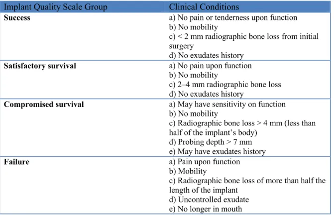
In an effort to develop a more comprehensive classification system for implant success, The International Congress of Oral Implantologists (ICOI) held a Consensus Conference in 2007 to update implant success criteria and health status based on an Implant Quality Health Scale using four categories based on the original James-Misch Health Scale (30, 31). Implant success, survival and failure were defined based on the following clinical and radiological parameters such as pain, mobility, crestal bone loss, probing depths, and peri-implant disease as presented in Table 1 (32).
Table 1: Health Scale for Dental Implants
Implant Quality Scale Group Clinical Conditions
Success a) No pain or tenderness upon function b) No mobility
c) < 2 mm radiographic bone loss from initial surgery
d) No exudates history
Satisfactory survival a) No pain upon function b) No mobility
c) 2–4 mm radiographic bone loss d) No exudates history
Compromised survival a) May have sensitivity on function b) No mobility
c) Radiographic bone loss > 4 mm (less than half of the implant’s body)
d) Probing depth > 7 mm e) May have exudates history
Failure a) Pain upon function
b) Mobility
c) Radiographic bone loss of more than half the length of the implant
d) Uncontrolled exudate e) No longer in mouth
Although the Albrektsson (25) and Buser (29) implant success criteria remain the most commonly used, a lack of international consensus still reigns. This is illustrated by the fact that several authors have used their own criteria for implant success in recent years (33-35). During the 8th European Workshop in Periodontology held in 2012, several working groups
were organized to assess the quality of reporting of clinical research in implant dentistry (36). The consensus report identified three main outcome domains that should be included in future implantology studies: patient-reported outcome measures, peri-implant tissue health, and performance of implant supported restorations. More specifically, health-related quality of life, satisfaction, marginal bone level, tissue inflammation, probing depth, longevity and functionality of the implant-supported restoration as well as technical complications were among the outcomes that should be collected in prospective implantology clinical trials.
1.3.2 Bone remodeling around dental implants
Crestal bone loss around implants is a key parameter affecting implant success (37). The marginal bone around the implant crestal region is a major indicator of implant health and its preservation will affect the long term and predictable success of an implant (38). The level of the crestal bone may be measured from the crestal position of the implant at the initial implant surgery. Initial peri-implant bone remodeling occurs as soon as the implant is connected with a healing or prosthetic abutment as a result of establishing a peri-implant attachment called “biologic width” and may take a few months (39). The key factors that may affect initial peri-implant bone remodeling include peri-implant surface texturization and peri-implant platform design (40).
The platform-switching concept was first described in 2006 (41). In this concept, implants with a wider diameter are used with prosthetic abutments of a smaller diameter in order to move the microgap between the prosthetic abutment and the implant platform further away from the crestal bone. Long-term radiographic observations on the use of platform-switched implants have shown less than expected peri-implant crestal bone loss compared to implants restored with prosthetic components of matching diameters. These findings were supported by a systematic review that reported a mean difference of -0.37 mm in peri-implant bone level changes (95% CI: -0.55 to -0.20; P <0.0001) in favour of platform-switched implants (42). Subgroup analyses showed that an implant-abutment diameter difference > or= 0.4 was associated with a reduction in peri-implant bone loss. In recent years, most implant companies have modified their implant design to include the concept of platform-switching to minimize initial peri-implant bone remodeling.
Implant surface texturization has optimized the host-to-implant tissue response by increasing surface area and roughness. In fact, implant surface modifications have led to high implant survival and better predictability, even for more challenging conditions such as immediate implant placement (43) and immediate loading (44-46). In 2009, the proceedings of a consensus meeting of the European Federation of Periodontology regarding the evidence on the effect of the different commercially-available implant surface modifications on marginal bone loss were published (47). The authors concluded that implants with moderately rough surfaces obtained the highest percentage of bone-to-implant contact, that newer generation of surfaces from four of the major world implant companies enhanced bone integration compared to their predecessors, while they could not find any clinically significant evidence that the platform-switched implant design was superior. However, a recent systematic review and meta-analysis has found that the severity of peri-implant bone loss after at least five years in function was significantly less around minimally rough implants compared to moderately rough and rough implants (48). These conflicting results underline the necessity of conducting further well-controlled clinical studies including confounding factors. In fact, time of measurement (38), implant platform location in relation to the crestal bone, soft tissue thickness (49), oral hygiene (50), and smoking habits (51, 52) have been shown to influence bone level changes after the initial healing phase.
The most common method to assess crestal bone remodeling is by radiographic evaluation. There are three types of radiograph that can be used to evaluate peri-implant bone remodeling around an implant: periapical radiographs, orthopantomographs, and cone beam computerized tomography (CBCT). The first two methods are easily accessible and can be performed
quickly and at a low cost, while the latter one is expensive and will expose the patient to a significantly higher radiation dose. Orthopantomographs are mainly inconvenient because they magnify and distort the images, which affects their sharpness compared to periapical radiographs. Investigators have compared periapical radiographs and CBCT to evaluate peri-implant bone levels around peri-implants and have found significant disparities between the two methods (53). A mean difference of 0.47 mm (range: -0.47 to 3.13) was found, indicating that CBCT images underrated the bone level systematically. Hence, intra-oral radiographs should be considered as the standard to monitor the peri-implant bone remodeling over time. Vandeweghe et al. reported that bone remodeling did not undergo significant changes after 15 weeks (54). Other researchers have demonstrated that the median crestal bone loss between the time of implant placement and three months postoperatively was 1 mm (55). When the median crestal bone loss was calculated between three and six months, it was close to zero. A recent systematic review reported a mean marginal bone loss of 1.3 mm over a mean duration of 13.4 years after implant placement (24). Although this value seems insignificant, one must bear in mind that statistical analysis is often patient-based. This might hide the outliers that lost a significant amount of bone, often being represented by a small number of implants. For example, it was found in a long-term study of Branemark implants that the mean bone loss was 0.8 mm after five years, and insignificant changes were reported thereafter (56). However, the prevalence of implants losing more than 3 mm of bone in that study was 5.6% after one year, 10.8% after five years, and 15.2%, 17.2% and 23.5% after 10, 15 and 20 years respectively. Consequently, it is recommended to measure crestal bone changes at both patient- and implant-level in patients with multiple implants to better visualize extreme values and trends over time (57).
Although several risk factors have been identified with peri-implant bone loss, some consider peri-implantitis as the main risk factor while others see crestal bone loss as an unavoidable physiological process following implant placement and loading. While new developments in implant surfaces and design have help minimize peri-implant bone remodeling, very little research has focused on ways to minimize or even prevent peri-implant bone loss using pharmacological or other non-traditional therapies.
1.4 PAIN EXPERIENCE IN IMPLANTOLOGY
Dental implant placement procedures involve surgical trauma to both the soft tissues and the alveolar bone that will result in an acute inflammatory reaction. Swelling is a classic manifestation occurring after surgery and it may trigger pain, loss of function or neural damage (58). Pain intensity has been showed to be low to moderate after dental implant placement and although it decreases over time, it may last up to a week postoperatively (59, 60). The fear of pain might keep a significant portion of patients from seeking implant placement to replace their missing teeth (61). Pain perception after implant placement is influenced by various factors such as previous experiences, stress, the clinical situation, the complexity of surgery, the number of implants, the surgeon’s experience, the sex of the patient, the pain experienced earlier and anxiety (62-64).
One of the earliest studies focusing on pain and anxiety related to implant placement was published in 2003 (62). Sixty patients were recruited from a specialist’s private practice to participate in the study. Dental anxiety scale (DAS) (65), state anxiety on a visual analog scale
(VAS) and the patient’s evaluation of pain on a VAS were collected immediately before and after surgery, and four weeks post-operatively. Significant correlations were found between the subjects’ anxiety and pain at different occasions. The best predictor of pain evaluation immediately after surgery and four weeks postoperatively was state anxiety. Another study evaluated pain and anxiety in 18 subjects undergoing implant surgery using several patient-based outcomes (66). The authors found that for the first three days, 27% of patients experienced “lots” or “quite a bit” of interference with chewing, which decreased to 11% by the sixth postoperative day. Swelling was the most frequent symptom reported during the first three days after surgery, dropping from 72% on the first postoperative day to 39% by the sixth day. The mean VAS score for average pain was the highest on the first postoperative day (24/100) and it decreased gradually to 9/100 on the sixth day, 0 representing no pain and 100 being the most intense pain imaginable. Most patients reported some limitations in their daily activities during the first postoperative day with a mean VAS score of 25/100, and this score decreased by half on the second day and gradually decreased further thereafter.
A more recent study including 89 participants investigated pain and anxiety before implant surgery (T0), immediately after surgery (T1), one day (T2), and one week postoperatively (T3) (59). Participants were instructed that VAS scores of 1 to 3 were indicative of mild pain, 4 to 6 moderate pain, and 7 to 10 severe pain, 0 representing “no pain” and 10 “the most intense pain imaginable”. The pain score increased from 1.03±0.83 at T1 to 4.13±1.37 at T2, then dropped to 0.98±0.94 at T3. The pain score at T2 was the highest. At T1, 31.4% reported having no pain, while at T2, 33.7% reported mild pain, 62.9% reported moderate pain, and 3.3% reported severe pain. At T3, 39.3% reported having no pain and 60.6% reported mild pain. There was a
significant correlation between the pain score at T2 and the state of anxiety at T1 and T0, and between the pain score at T2 and the state of anxiety at T1 and T2. The authors concluded that anxiety affected pain intensity after implant surgery, corroborating the findings of Eli and colleagues.
Investigators have recently used cellphone-based real-time pain and swelling questionnaires after dental implant surgery (67). In an attempt to boost the validity level of the assessments and increase the number of time point assessments after implant surgery, the authors used a cellular phone-based Ecological Momentary Assessment (EMA) system regulated by a host PC server that automatically sent e-mails to cell phones every two hours on the first day of surgery, and every 24 hours onward until the seventh postoperative day. The outcomes measured were the patient’s preoperative anxiety level, postoperative pain and subjective swelling sensation. Subjective intensity of pain and swelling were measured by an 11-grade and a 4-grade rating-scale questionnaire respectively. For the pain level assessment, a score of 0 represented “painless” and a maximum score of 10 indicated “intolerable pain”. Regarding swelling sensation assessment, a score of 0 represented “no swelling”, a score of 1 meant “swelling of a limited area”, a score of 2, “swelling of an extended area”, and a score of 3, “swelling extended to an extra-oral region”. The data from 25 participants was analyzed. Mean postoperative pain and swelling peaked at six and 36 hours after surgery with a mean rating of 0.87 (0-11) and 1.32 (0-3) respectively. Six risk factors were significantly associated with postoperative pain: presence of diabetes and/or hypertension, duration of surgery, intake of premedication, bone quality, preoperative anxiety and total swelling sensation, while three
were significantly associated with a swelling sensation: accumulated postoperative pain, absence of diabetes and/or hypertension, and bone quality.
Several factors have been shown to influence pain experienced after implant surgery but only a few studies have simultaneously evaluated these potential factors. Although the intensity of pain experienced by individuals undergoing implant surgery has been described as low to moderate, it represents one of the most common barriers keeping patients from seeking dental implant therapy. Controlled clinical trials including the above mentioned factors may shed some light on the extent of the influence operator-based, patient-based and surgery-based factors have on a patient’s perceived pain after implant placement.
1.5 THE ROLE OF ANTIBIOTICS IN DENTISTRY
1.5.1 Risks associated with antibiotics overuse
Extensive antibiotic exposure in health care is a well-known risk factor for antibiotic resistance (68). Multidrug-resistant bacteria such as E. coli strains have been associated with the overprescription of antibiotics in medicine and dentistry (69). Antibiotics are occasionally associated with many clinical complications ranging from a simple rash to life-threatening superimposed infections. One of the most common and dangerous complications is the C. difficile infection. In 2015 alone, about 500,000 Americans suffered from this infection and one out of three patients with this infection was 65 years or older (70). Therefore, proper diagnosis and adequate use in clinical practice are important strategies that will reduce antibiotic exposure (71). In a recent study comparing the side effects of amoxicillin/
clavulanate potassium and clindamycin for the treatment of odontogenic infections, at least 50% of patients who used either antibiotic had at least one side effect ranging from minor complaints such as nausea, abdominal discomfort and diarrhea to elevated liver enzymes, a reaction associated with liver damage (72). The routine use of antibiotics to prevent postoperative complications in oral surgery, especially after dental implant placement, has been subject of much debate in recent years.
1.5.2 Effects of antibiotics on dental implant outcomes
One of the first antibiotic protocols described relating to dental implant surgery was Branemark’s protocol (26). One hour before the surgical placement of one or multiple implants, 2 g of phenoxymethylpenicillin was given orally along with a 2% chlorhexidine rinsing solution, and 20 to 25 mg of diazepam per os to reduce patient anxiety. Phenoxymethylpenicillin was also given for the first ten postoperative days (2g bid). This protocol was aimed at preventing postoperative infections and increasing chances of osseointegration. However, a recent survey of oral and maxillofacial surgeons in the U.S. has shown different prescription patterns being used for prevention of implant complications (73), underlining a lack of standardization among dental practices. There was no consensus among surgeons regarding perioperative antibiotic regimens for implant placement and their effectiveness at decreasing the implant failure rate. In addition, most of the antibiotic regimens used did not comply with current scientific evidence.
Finland is one of the few countries in the world that has a Dental Implant Registry. A retrospective study was done using non-public information obtained with permission from this registry (74). This study examined a total of 110,543 dental implant placement procedures and
1,038 dental implant removal operations performed between April 1994 and April 2012. The aim of the study was to review the type of antibiotic regimens used for dental implant placement and to analyze the association between antibiotic usage and early implant removal. A total of 61 different antibiotics or combinations of antibiotics were prescribed peri-operatively. Antibiotics were prescribed in 1,640 of the 2,521 (65.1%) implant placement operations. Early implant failure generally occurred within six weeks of implant placement. Its prevalence was 12.7% in those cases where antibiotics were prescribed, and 15% when no antibiotics were prescribed. These differences were not statistically significant. Therefore, the authors concluded that the use of prophylactic antibiotics had little effect on implant complications related to the initial surgery and the success rate.
One of the first major prospective clinical studies investigating the effects of the perioperative use of antibiotics on dental implants success rate was published in 1997 (75). The study included data from 2,973 implants, but the type, timing (pre- vs post-op), and duration of the antibiotic treatment were left to the discretion of the surgeon. Follow-up of patients was done for up to three years after the prosthesis was loaded. Higher survival rates were found in patients who had received preoperative antibiotic coverage.
Ten years later, a well-controlled randomized clinical trial investigated the effects of the antibiotics on subjective signs and symptoms after implant placement (76). The study included two groups of 40 patients treated consecutively for a total of 128 implants. In the first group, a pre- and post-operative antibiotic regimen was given. More specifically, participants took 1 g of amoxicillin one hour before the surgery and 2 g during two days. The participants of the
second group were not given any antibiotic coverage. Bacterial samples were taken from the peri-oral skin and a VAS questionnaire was used to evaluate symptoms of infection and inflammation by both the patient and the surgeon. No significant differences were found in the clinical and microbiological parameters between the two groups, although the patient’s perception of postoperative discomfort experienced was significantly milder in the antibiotics group.
A multicenter, placebo-controlled, randomized clinical trial included 105 patients recruited from 12 private practices in Spain (77). The goals of the study were to compare the safety and efficacy of 2 g of amoxicillin given one hour prior to dental implant placement with identical placebo tablets given when placing single implants in types II and III bone density. The study participants were divided into two groups. Fifty-two individuals received 2 g of amoxicillin one hour preoperatively, and 53 participants were given identical placebo tablets. Two participants in each group experienced an implant/crown failure, and six participants in each group suffered from a postoperative infection. No adverse events were reported and no statistically significant differences were observed for any of the outcome measures.
Another study had similar results while comparing (78) participants who were given 2 g of amoxicillin one hour preoperatively with participants who were given identical placebo tablets for dental implant placement. The antibiotic group and the placebo group included 254 and 255 participants, respectively. The investigators evaluated the implant and prosthesis failure rates, as well as the presence of adverse events and postoperative complications. Three participants were excluded because they did not complete the study. Four participants in the
group taking antibiotics experienced prosthesis failures versus ten in the group taking a placebo. No adverse events were reported and no significant differences were observed for any of the outcome measures.
Another study including 100 patients undergoing implant placement surgery compared the effects of four different antibiotic regimens on dental implants failure rate (7). Participants in the first antibiotic regimen group were given 2 g of amoxicillin one hour preoperatively and no antibiotics given postoperatively. In the second group, the antibiotic regimen was 2 g of amoxicillin to take one hour preoperatively and 1 g twice a day for seven days following surgery. For the third regimen, participants took 1 g twice a day after surgery for seven days. In the fourth group, no antibiotic regimen was prescribed. Each group included twenty-five participants. They were examined for internal and external oedema, internal and external erythema, pain heat and exudates, as well as implant failure. Two participants in the no antibiotic group had single implant failures versus none in any of the three antibiotic groups. However, this difference was not statistically significant. No infections or side effects were reported.
A double-blind randomized controlled trial evaluated the effects of antibiotic coverage on subjective outcomes such as postoperative morbidity, pain and interference with daily activities, and implant survival rate (79). The 27 participants in the first group were given 3 g of amoxicillin one hour preoperatively and the 28 individuals in the second group were given identical placebo tablets. The surgeons were residents in a specialty program. There was no implant loss in any of the participants of the antibiotic group but five participants from the
placebo group each lost their implant (P= 0.0515). No participant in the antibiotic group presented clear signs of infection versus two participants in the placebo group. Post-operative pain and interference with daily activities were significantly lower in the antibiotic group versus the placebo group at seven days. The authors concluded that the prophylactic use of antibiotics may be beneficial for implant osseointegration and to reduce postoperative pain, especially in cases with longer surgery.
The most recent Cochrane review evaluating the effects of various prophylactic antibiotic regimens vs no antibiotics on implant survival rates in patients undergoing dental implant placement was published in 2013 (9). The authors searched several databases up to June 17th,
2013 including the Cochrane Oral Health Group’s Trials Register, the Cochrane Central Register of Controlled Trials (CENTRAL), MEDLINE and EMBASE. The studies included in the review were randomized clinical trials with a follow-up of at least three months. Outcome measures included prosthesis failures, implant failures, postoperative infections and adverse events (gastrointestinal, hypersensitivity, etc.). The data of 1,162 participants from the studies were included in the statistical analyses. The meta-analyses of the six trials showed a statistically significant higher number of participants experiencing implant failures in the groups not receiving antibiotics (RR: 0.33; 95% CI: 0.16 - 0.67, P = 0.002). The number needed to treat (NNT) for one additional beneficial outcome to prevent one person from having an implant failure was 25 (95% CI: 14 - 100), based on an implant failure rate of 6% in participants not receiving antibiotics. There was a borderline statistical significance for prosthesis failures (RR: 0.44; 95% CI: 0.19 - 1.00), with no statistically significant differences for infections (RR: 0.69; 95% CI: 0.36 - 1.35), or adverse events (RR 1; 95% CI: 0.06 -
15.85). The authors concluded that the perioperative use of antibiotics for implant placement is beneficial to improve the implant survival rate, and that taking 2 g or 3g of amoxicillin one hour prior to implant surgery might be recommended. However, the effects of postoperative use of antibiotics could not be determined in this study.
Several randomized clinical trials were published after the latest Cochrane review. The first study was aimed at evaluating the clinical benefits of preventing early dental implant failure by adding a postoperative antibiotic regimen after giving a single dose before dental implant placement (80). Eighty participants were recruited and randomly divided into two groups. The first group received 1 g of amoxicillin before the procedure, and the second group received the same preoperative dose as well as a three-day antibiotic course. Follow-ups were scheduled at three days, seven days and twelve weeks. Pain, swelling, wound dehiscence, and pus formation were assessed. There were no implant failures in either group. The author concluded that a single preoperative antibiotic dose was sufficient to prevent implant failure.
A multicenter randomized controlled clinical trial investigated the effects four different antibiotic regimens on patient-oriented outcomes and postoperative complications after implant placement (81). In this study, 329 healthy adults who had a single tooth edentulous area with sufficient bone width and height for the placement of a dental implant without simultaneous bone grafting were recruited in seven centers located in Singapore, China, Australia, Spain, Taiwan and Iceland. Heavy smokers were excluded from the study. The surgeons and examiners were not made aware of the antibiotic regimen selected, although this was not the case for all participants. Participants were randomly assigned to one of four
treatment groups. The first group took 2 g of amoxicillin one hour before surgery. The second group took 2 g of amoxicillin immediately after surgery. The third group took 2 g of amoxicillin one hour before surgery and 500 mg twice daily on the second and third days after surgery. The fourth group took 2 g of a placebo one hour before surgery. The primary outcomes included patient-reported outcomes such as VAS score on pain, swelling, bruising and bleeding. The secondary outcome variables included clinical recordings of flap closure, pain, swelling, suppuration, and implant stability. Postsurgical complications were assessed at the first, second, fourth, and eighth week postoperatively. There were no significant differences in subject profile among the four treatment groups in terms of age, gender, smoking status, sites of implantation profile, and implant dimensions. No statistically significant differences were found among the four groups regarding pain, swelling, bruising, bleeding, suppuration and implant stability. There was a statistically significant difference between the placebo group and the other three groups when it came to flap closure, where 5% of the participants did not achieve complete flap closure at week four compared to 0% for the other groups. However, at the other time points, there were no significant differences between the groups. The authors concluded that none of the three prophylactic regimens excelled at preventing post-surgical complications or improving patient-reported outcomes.
Another recent multicenter randomized clinical trial evaluated the perioperative use of antibiotics for dental implant placement by comparing the differences between a single dose of amoxicillin given preoperatively and the same dose of amoxicillin given for two additional days postoperatively in patients undergoing conventional dental implants (82). Two dental surgeons in two private practices recruited 360 participants and randomly divided them into
two groups of 180 individuals. Participants in group A received only one dose of amoxicillin preoperatively (2 g) while participants in group B received 2 g of amoxicillin before surgery as well as 1 g of amoxicillin in the evening and 1 g twice a day for two additional days after surgery. Both groups were followed during six months to evaluate the implant failure rate as well as side effects of the antibioprophylaxis. The data from 14 patients in group A and three patients in group B were not available. Two patients in group B experienced a prosthetic failure, losing four implants, while no prosthetic failures were reported in group A. Six patients in group A and four patients in Group B experienced early postoperative complications. However, there were no statistically important differences found between the two groups. As observed by others (80, 81), postoperative antibiotic use was not beneficial to reduce the implant failure rate.
As part of the fourth European Association for Osseointegration Consensus Conference, a complex systematic review was done of the best available scientific evidence regarding the effects on implant survival of the perioperative use of antibiotics during dental implant placement (11). The literature search yielded 846 articles, including ten primary studies and seven systematic reviews that were analyzed as part of the systematic review – two were considered to have a moderate risk of bias and five had a high risk of bias according to the AMSTAR criteria. The two systematic reviews with a moderate risk of bias showed divergent numbers needed to treat (NNT) to prevent one patient from having an implant failure. Four of the primary placebo-controlled studies were included in the meta-analysis. They had a low or moderate risk of bias and the heterogeneity between the studies was low. It was found that preoperative antibiotic use significantly reduced the risk of implant loss by 2% (P = 0.02). The
NNT was 50 to prevent one patient from losing an implant. More importantly, none of the primary studies showed that perioperative antibiotics used on their own had any statistically significant benefit. A sub-analysis suggested that in uncomplicated implant placements in healthy patients, using antibiotics was not beneficial to prevent implant loss. On the other hand, the authors concluded that in more complex cases or for medically compromised patients, using antibiotics during dental implant placement could prove to be significantly beneficial.
To date, there have been no randomized clinical trials using a double masked study design including a placebo control given postoperatively to evaluate the potential effects of a postoperative antibiotic regimen in addition to one preoperative dose of antibiotics on simultaneous crestal bone remodeling, postoperative morbidity, implant success rate, clinical outcomes and patient-based outcomes such as pain and interference with daily activities.
CHAPTER II
METHODOLOGY
2.1 OBJECTIVES, HYPOTHESIS
2.1.1 Objectives
The primary objectives of this phase-I clinical trial were:
1- To produce preliminary data and to assess whether giving additional antibiotics after implant placement over seven days would influence peri-implant crestal bone levels after four months in healthy patients undergoing platform-switched implant placement.
The secondary objectives were:
2- To evaluate postoperative pain severity, surgery-associated morbidities, and one-year implant survival rate.
2.1.2 Hypothesis
We tested the following null hypotheses:
1- There is no difference in peri-implant crestal bone loss between healthy patients who received a pre- and postoperative antibiotic regimen compared to those who received only a preoperative dose of antibiotics during surgery for straightforward platform-switched implant placement.
2- There are no differences in pain severity, surgery-associated morbidities, and one-year implant survival rate between healthy patients who received a pre- and postoperative antibiotic regimen compared to those who received only a preoperative dose of antibiotics during surgery for straightforward platform-switched implant placement.
2.2 RESEARCH METHODOLOGY
2.2.1 Study design, participants, eligibility and intervention
The study used double-masked two-arm randomized controlled clinical trial.
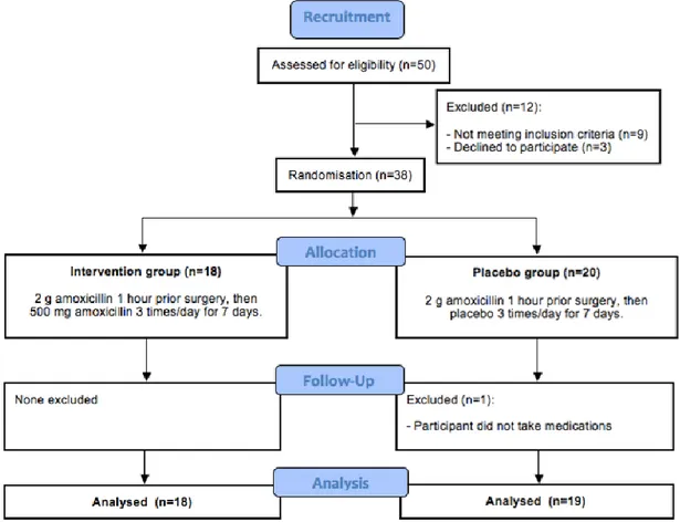
Fifty patients from the Dental Clinic at the Faculty of Dentistry of the Université de Montréal were invited to participate in this study. The eligibility criteria are presented in Table 2.
Table 2: Inclusion and exclusion criteria Inclusion criteria:
- Periodontally healthy remaining dentition or presenting with mild gingivitis with adequate oral hygiene.
- Presence of a partially edentulous alveolar ridge that will be restored with no more than two adjacent implants.
- To have one or two implants restored with a crown or fixed bridge. - Absence of any active infection in site.
- Presence of enough bone and soft tissue for the implant to be placed without any bone grafting procedure in a one-stage approach (with healing abutment).
- Implants 8 mm long or longer using the Dentsply AstraTech Implant System™ (Osseospeed TX or EV™).
- Subjects able and willing to provide written informed consent and comply with study procedures.
Exclusion criteria:
- Individuals taking regular analgesics or antidepressants.
- Allergies to amoxicillin, cephalosporin, and non-steroidal anti-inflammatory analgesics.
- Smoking ten cigarettes/cigars or more per day. - Drug abuse.
- Completely edentulous individuals. - Pregnant and nursing women.
- Individuals who have an active peptic ulcer or are susceptible to peptic ulcers.
- Any systemic or local immunodeficiency.
(ex.: Coumadin).
- Presence of uncontrolled periodontitis or poor oral hygiene. - Presence of any acute oral infection.
- Presence of uncontrolled diabetes or other systemic diseases.
- Individuals who have received previous radiation therapy in the head and neck area.
- Individuals who receive intravenous bisphosphonates.
- Individuals who have been taking oral bisphosphonates for more than three years.
- Individuals with long-term intake of corticosteroids.
- Individuals who need routine prophylactic antibiotics prior to dental surgery.
- Individuals who have taken antibiotics three months prior to surgery.
Eligible participants were randomized in two groups using block randomization. Individuals in the intervention group received 2 g of amoxicillin one hour prior to surgery and 500 mg three times a day for seven days. Those in the control group received only 2 g of amoxicillin one hour prior to surgery and an identical placebo three times a day for seven days.
Prior to data collection, the ethics committee for health research at the Université de Montréal (Comité d'éthique de la recherché en santé: 13-094-CERES-D) approved this study, which was registered at www.clinicaltrials.gov (NCT01851681). All study procedures were undertaken with the understanding and written consent of each participant and in accordance with ethical principles including the World Medical Association Declaration of Helsinki (Appendix I). The participants were informed of the sequence, duration and number of appointments they would need to attend in order to remain in the study (Appendix II).
2.2.2 Outcomes measures
The primary outcome was the changes in crestal bone level measured by periapical radiographs using a standardized technique at baseline and at four-month follow-up. The secondary outcomes were pain severity, surgery-associated morbidities (interference with
daily activities, swelling, suppuration, ecchymosis, dehiscence, infection, neuropathy, paresthesia, mobility and radiolucency) evaluated by clinical examination and self-administered questionnaires, and one-year implant survival rate. The explanatory variables included the participants’ sociodemographics and medical background such as mean age, sex, language, ethnic background, civil status, living status, education, yearly household income, smoking status, and diabetes. They also included surgical parameters such as the number of implants per patient, mean surgery duration, mean incision length, mean bone quality, implant location, as well as implant characteristics such as implant diameter, implant length, insertion torque and implant system.
2.3 DATA COLLECTION AND EXPERIMENTAL
PROCEDURES
2.3.1 Medical and sociodemographic questionnaires
A research assistant approached and recruited patients who were willing to restore a partially edentulous area with a fixed implant prosthesis at the implantology clinic of the Faculty of Dentistry at the Université de Montréal. Medical history, smoking habits, and sociodemographic data were obtained through self-administered questionnaires (Appendices III and IV).
2.3.2 Clinical procedures
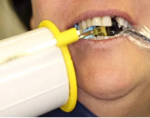
All participants were instructed to rinse with chlorhexidine gluconate 0.12% for one minute, and were given 600 mg of ibuprofen and 2 g of amoxicillin one hour prior to surgery under the supervision of a research assistant. Standard measures of asepsis included the use of sterile
drapes around the patient’s head and over the supine body of the patient as well as sterile scrubs and gloves for the surgeon. Screw-type, two-piece dental implants with a moderately rough surface (Osseospeed™ TX, Osseospeed™ TX Profile or Astra EV™, Dentsply Implants, Mölndal, Sweden) were placed in a one stage procedure without simultaneous bone grafting, in accordance with the manufacturer’s recommendations, by two board-certified specialists who had a minimum of 10 years of experience in surgical implantology. Mucoperiosteal flaps were raised to access the underlying alveolar bone for all implant surgeries. The healing abutment was inserted at the time of implant placement and soft tissues were sutured with interrupted sutures (4-0 silk, Perma Sharp®, Hu-Friedy Mfg Co., Chicago, IL, U.S.A.). A standardized radiograph was taken immediately after dental implant placement. The research assistant placed the x-ray cone perpendicular to the crestal bone to assess the baseline crestal bone level on the mesial and distal aspects of each implant using a bite registration material (Blu-Mousse®, Parkell Inc., Edgewood, NY, U.S.A.) adapted to a paralleling device (XCP film holding system, Dentsply Rinn, Elgin, IL, U.S.A.) for each participant (Figure 1). The customized bite registrations with each participant’s study identification number were kept in a locked cabinet in a cool room for the subsequent four-month follow-up period. Surgical parameters such as the length of the incision, implant system, implant dimensions, insertion torque, bone quality (83), and the duration of the surgery were recorded by the surgeon. Participants were asked to refrain from performing mechanical plaque control in the surgical area and were advised to remain on a soft diet during the first postoperative week.