nih.no
Hody S.
1,2, Cumming K.T.
2, Wernbom M.
3, Bjørnsen T.
4, Paulsen G.
2,5and Raastad T.
21University of Liege, Belgium; 2Norwegian School of Sport Sciences, Norway; 3University of Gothenburg, Sweden; 4University of Agder,5The Norwegian Olympic and Paralympic
Committee and Confederation of Sport, Norway
Effect of a failure- versus a submaximal low-load blood flow
restriction training protocol on Heat-Shock Protein responses
References:
(1) Wernbom et al. (2008). Scand J Med Sci Sports. Aug;18(4):401-16. (2) Loenneke et al. (2014). Scand J Med Sci Sports. 2014 Dec;24(6):e415-422.
(3) Cumming et al. (2014). Acta Physiol (Oxf). Aug;211(4):634-46. (4) Sieljacks et al. (2016). Eur J Appl Physiol. Mar;116(3):513-25.
Correspondance: stephanieh@nih.no
Introduction
Blood flow restricted exercise (BFRE) with low loads has gained interest during the last years because it induces muscle hypertrophy to a similar extent as conventional heavy load strength training1. Although it
is highly debated2, BFRE has recently been suggested
to elicit considerable stress to exercising muscles and in some cases, muscle damage3,4. The degree of
stress caused by BFRE has, however, been poorly investigated at the cellular level.
The heat shock proteins (HSP), such as αB-crystallin, are typical intracellular markers of cellular stress and known to translocate and accumulate in the affected areas within the cell.
AIM: To compare the acute and long-term effects of a failure (FA) vs submaximal (SU) BFRE protocols on αB-crystallin response in exercising muscles.
Methods
Sixteen untrained men (18-45 yrs) completed 14 BFRE sessions divided into 2 blocks of 7 sessions in 5 days, interspersed by 10 days of rest (Fig.1). Legs were randomly assigned to either FA (4 sets to voluntary failure) or SU protocol (30-, 15-, 15-, 15 reps) using unilateral knee extensions at 20% of 1RM with 30s rest between sets. BFRE was conducted with partial blood flow restriction (100 mmHg) induced by a 15 cm wide pressure cuff.
Biopsies from the m.vastus lateralis were collected before (Pre), 2h after the first session (+2h), during the rest period (+11d) and 10 days post intervention (+29d).
The HSP response investigated was changes in αB-crystallin staining intensity on muscle cross sections analyzed by immunofluorescence. The staining intensity was measured using ImageJ (with a mean ± SD of 209 ± 107 fibers/time point).
Results
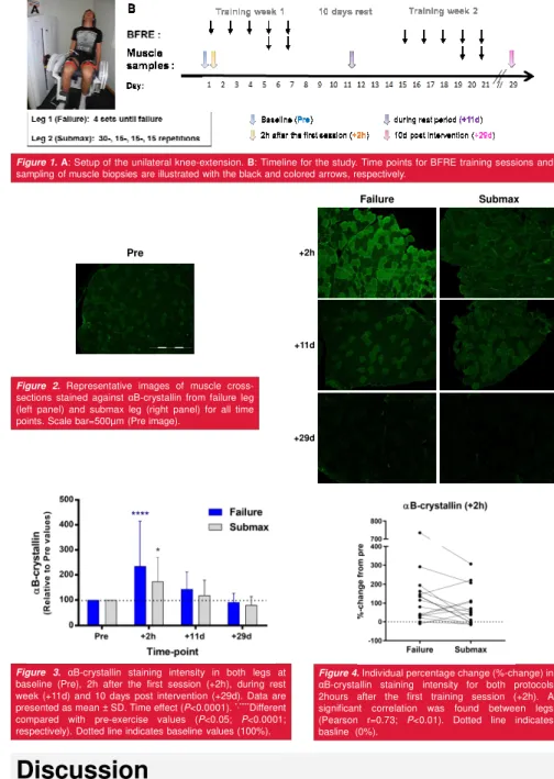
Relative to pre-exercise (100%), a significant increase in αB-crystallin staining intensity (reflecting cytoskeletal bound proteins) was observed 2h after the first session of both BFRE protocols (FA: 234.7 ± 179.6%, P<0.0001 and SU: 173.7 ± 95.7%, P<0.05; respectively) (Fig. 2-3). There was no significant difference between protocols at any time point, but the acute response of αB-crystallin tended to be larger in FA legs than SU legs. The αB-crystallin staining intensity gradually decreased to baseline values during the rest period (FA: 142.6 ± 68.8% and SU: 118.2 ± 60.13%) and 10 days post intervention (FA: 91.10 ± 35.83% and SU: 80.70 ± 33.28%). Note that a large intersubject variability was observed, especially in the acute response (Fig. 4).
Discussion
The increase in αB-crystallin staining intensity indicates that both FA and SU BFRE are able to induce cell stress and possible damage to cytoskeletal structures. The translocation of HSP to myofibrillar structures after low-load BFRE is probably related to ischemia rather than mechanical stressors. It should be noted that some subjects were not able to complete all the repetitions during SU. Consequently, our SU protocol was close to failure in the first training sessions. In addition, since fiber-type specific adaptations after BFRE have already been observed3, one main perspective
is to investigate αB-crystallin in type 1 and type 2 fibers. In parallel to the immunohistological analyses, immunoblots and ELISA for HSP are carried out and should improve the understanding of acute and chronic HSP response after FA and SU training.
Conclusions
(1) The results in this study suggests that cytoskeletal proteins are stressed after the first session of both low-load FA and SU BFRE protocols. (2) No accumulation of this small HSP in cytoskeletal structures seems to occur after a period of BFRE training.
Figure 1.A: Setup of the unilateral knee-extension. B: Timeline for the study. Time points for BFRE training sessions and sampling of muscle biopsies are illustrated with the black and colored arrows, respectively.
Figure 2.Representative images of muscle cross-sections stained against αB-crystallin from failure leg (left panel) and submax leg (right panel) for all time points. Scale bar=500µm (Pre image).
Pre
Failure Submax
+2h
+11d
+29d
Figure 4.Individual percentage change (%-change) in αB-crystallin staining intensity for both protocols 2hours after the first training session (+2h). A significant correlation was found between legs (Pearson r=0.73; P<0.01). Dotted line indicates basline (0%).
Figure 3. αB-crystallin staining intensity in both legs at baseline (Pre), 2h after the first session (+2h), during rest week (+11d) and 10 days post intervention (+29d). Data are presented as mean ± SD. Time effect (P<0.0001).*,****Different
compared with pre-exercise values (P<0.05; P<0.0001; respectively). Dotted line indicates baseline values (100%).