Remerciements
La thèse est une grande aventure faite de rencontres, d’opportunités et de nouveautés. C’est un tout nouveau monde qui s’ouvre à soi, inconnu, inconfortable mais exaltant. Au cours de mon parcours de thèse parfois tumultueux, j’ai eu la chance de faire de belles rencontres scientifiques et humaines. Je souhaiterais remercier toutes ces personnes et espère n’oublier personne.
J’ai accompli ce travail de thèse au Laboratoire de Pharmacologie et Biologie pour le Développement (Pharma-DEV) ainsi qu’au Laboratoire de Chimie de Coordination (LCC). Je remercie le Pr. Nicolas Fabre et le Dr. Azzedine Bousseksou pour leur accueil au sein de ces deux laboratoires.
Je souhaiterais adresser mes remerciements aux Dr. Marius Réglier et Dr. Jérôme Santolini qui ont référé ce travail avec beaucoup d’attention et d’intérêt. Je tiens également à remercier les différents membres du jury pour leur présence ainsi que pour les discussions scientifiques qui ont eu lieu au cours de ma soutenance et qui m’ont beaucoup apporté.
Pendant ces trois années de thèse, j’ai eu la chance d’être encadrée par des personnes exceptionnelles, scientifiquement et humainement. Je tiens à remercier très chaleureusement Fabrice Collin, Christelle Hureau et Peter Faller qui m’ont apporté leur confiance, leur soutien et m’ont appris tellement… Je n’aurais pas pu être mieux encadrée qu’avec vous ! Merci aussi de m’avoir donné l’opportunité de faire des activités « extra-labos » comme les enseignements, la vulgarisation scientifique, le club jeunes SCF, toutes ces conférences auxquelles j’ai pu assister et présenter, et les sessions à l’ESRF. Ce n’est pas donné à tous les doctorants d’avoir autant d’opportunités et de liberté, et je vous remercie de m’avoir donné cette chance.
Fabrice, tout d’abord merci de m’avoir donné ma chance en me choisissant pour ce projet de thèse. Merci pour ta disponibilité, tes précieux conseils et pour ton optimisme contagieux. Tu as toujours su partager ta passion pour la chimie mais aussi ta bonne humeur, même quand j’étais au fond du trou, notamment quand le peptide oxydé n’en faisait qu’à sa tête. Je te remercie également pour ta patience et ta pédagogie. Corriger mon premier article en « franglais » tout en faisant attention de ne pas me vexer n’a pas dû être une mince affaire !! Merci pour les longues discussions scientifiques, pour le temps que tu as passé avec moi quand les manips ne fonctionnaient pas (masse ou autre). J’ai passé des supers moments avec toi en conf’ et à l’ESRF en mode 007 !
Christelle, je souhaiterais te remercier pour (i) la générosité et la passion avec laquelle tu partages ton savoir scientifique, (ii) ta disponibilité pour discuter de chimie ou de choses plus personnelles et ce, même dans les moments où tu as déjà accordé 110% de ton temps à l’ERC et 90% à l’équipe, (iii) avoir été une super coloc de bureau prête à dégainer du chocolat à tout moment et enfin (iv) ne pas m’avoir viré quand j’ai cassé ta voiture ! Je te remercie de m’avoir fait me sentir comme ton égale lors de nos longues conversations scientifiques pendant lesquelles j’ai pu développer mon sens critique et proposer toutes les idées (quelquefois farfelues) qui me venaient à l’esprit, sans peur du ridicule. Merci également de montrer à toutes les femmes de l’équipe qu’on peut être une super chercheuse et une super maman de 4 bambins en même temps !
Peter, tout d’abord je te remercie de m’avoir accueillie dans ton équipe. Merci pour tes précieux conseils, pour les discussions scientifiques qui m’ont beaucoup apporté mais aussi pour ton enthousiasme permanent. Un grand merci aussi pour la confiance que tu m’as accordée et pour ta disponibilité, même après ton départ pour Strasbourg. Ta passion pour les sciences est vraiment contagieuse, alors propage la autant que tu peux !
Cette thèse n’aurait pu se faire sans l’aide précieuse de :
- Emilien Jamin pour la HRMS, je te remercie pour tes précieux conseils et pour le temps que tu m’as accordé. Et désolée pour la poisse que j’emmenais (souvent) avec moi à l’orbitrap !
- Christian Bijani pour les spectres RMN, merci de ne pas t’être lassé de voir des RMN avec des pics super larges tout le temps !! Et merci d’avoir pris le temps de regarder la partie RMN du manuscrit.
- Lionel Rechignat pour la RPE, merci pour les conseils et pour la conversation sur la théorie de la RPE (toujours aussi obscure pour moi !!).
- Vanessa Soldan pour le TEM, je tiens à te remercier pour les précieux conseils et le temps que tu as passé avec moi à sonder l’échantillon à la recherche de fibres (ou d’absence de fibres !).
- Toute l’équipe FAME de l’ESRF. Merci à Isabelle Kieffer pour toutes les explications sur le XAS et pour la visite guidée de la nouvelle ligne. Merci à Denis Testemale pour sa disponibilité et sa patience quand la ligne plantait en notre présence (juré c’est pas de notre faute !) et pour les précieux conseils prodigués en somnambule à toute heure de la nuit !!
- Stéphanie Sayen et Emmanuel Guillon lors des sessions à l’ESRF. Merci pour l’aide précieuse apportée, pour toutes les réponses à mes questions floues sur le XANES. Merci aussi d’avoir partagé les sessions blind-test et les bonbons avec moi !
- Petit Poney qui a subi nos expériences plus ou moins douteuses avec l’azote liquide. Merci pour les photos souvenirs !
Coté Pharma-DEV, je remercie Françoise Nepveu, Karine Reybier, Paul-Louis Fabre, Mohamed Haddad, Marieke Vansteelandt, Geneviève Bourdy ainsi que tous les autres membres des équipes Redstress et PEPS, avec qui j’ai eu la chance de travailler, de partager un repas ou bien un café. Merci à Pierre pour sa gentillesse et sa bonne humeur quotidienne, ainsi que pour l’aide précieuse et les bons conseils qu’il m’a apporté. Merci également à Franck qui a toujours le sourire et qui m’a beaucoup aidé pour les questions administratives. Franck, ton punch est à tomber par terre ! Il me faut la recette ! Merci à tous les stagiaires, doctorants et post-doc de Pharma-DEV qui m’ont permis de travailler dans une super ambiance : Ennaji, Nambinina, Rémi, Thi Thu, Luyen, Solomiia, Filip, Marion, Lucie, Mireia, Laure-Estelle (LEC), Cynthia. Malgré le froid glacial l’hiver, j’ai adoré partager les repas du midi avec vous dans le « couloir-cantine ». Et j’aurais bien besoin de me remettre aux mots croisés du midi !! Un énormissime merci à LEC et Cynthia pour l’accueil très chaleureux que vous m’avez fait à mon arrivée au laboratoire, je me suis sentie bien tout de suite et c’est grâce à vous. Merci pour vos encouragements au quotidien, pour les discussions scientifiques (ou pas !) et pour tous les bons moments passés ensemble. Vous avez été mes modèles de réussite ! Coté LCC, je remercie toute l’équipe F qui m’a chaleureusement accueillie et m’a fait mourir de faim jusqu’à 13h30 pendant mes deux premières années de thèse ! Merci à Manu pour sa bonne humeur et ses super blagues, mais aussi pour les discussions autour des sciences, de l’administration (et des mouches !), de la musique et de l’œnologie ! Merci à Béatrice qui a toujours le sourire, pour sa gentillesse et ses conseils. Laurent, je te remercie de ne pas m’avoir détesté d’office quand j’ai cassé ta voiture ! Je suis très contente d’avoir pu te connaître ces derniers mois et je te remercie pour l’aide et le soutien que tu m’as apporté. Viviane, merci pour ton rire contagieux, ta bonne humeur quotidienne et ta gentillesse ! Je tiens également à remercier tous les étudiants / postdoc actuels ou anciens de l’équipe : Olivia, Hélène, Adam, Olena, Carine, Melisa, Daniel (bon aprèm les garces !), Mireia (mais c’est vrai ça ?!), Megan (so cute !), Gabriel (Mr Marcel), Rufus (c’est qui Clémence ?!), Sara (à ne pas confondre avec Sara !), Marie, Alex (petit puits), Omar (Ahomalll), Elena (a fare l’amore, encore merci pour la chanson !), Valentina (la mammmmmmmmma) et Amandine (ACDC <3). Merci pour la super ambiance que vous apportez dans l’équipe, pour les discussions scientifiques qui permettent de faire avancer nos manips qui rament, mais aussi un grand merci pour les nombreux moments passés ensemble à l’extérieur du labo, les soirées mojitos / danses tahitiennes à La Plage… Le labo a été un super endroit pour se faire des amis !
Mes remerciements s’adressent ensuite à toute l’équipe pédagogique Chimie pharmaceutique aux cotés desquels j’ai eu le plaisir de découvrir l’enseignement pendant mes deux années de DCE : Geneviève Baziard, Salomé El Hage, Barbora Lajoie, Jean-Luc Stigliani, Fatima El Garah,
Christelle Recoche-Gueriot et Laurent Amielet. J’ai beaucoup appris auprès de vous et je tiens à vous remercier pour votre accueil et votre bienveillance.
Je tiens à remercier tout le bureau jeune de la SCF Midi-Py avec qui j’ai eu la chance d’animer des ateliers scientifiques et de monter des projets sympas : Claudia, Cécile, Alix, Morgane, Jérémy, Stéphane, et tous les étudiants de l’équipe F. Les sessions Chimie & Terroir vont me manquer, mais pas les ventes des kits chimistes ! Merci aussi à Lydie Valade et plus largement à Chimie et Société de m’avoir fait partager leur passion pour la vulgarisation scientifique.
Je remercie les organisatrices des fameux et très sélects « Zumbapéro » qui m’ont permis de garder la forme pendant ces années de dur labeur ! Julie, merci d’avoir toujours la pêche et d’avoir été là pour les sessions papotage nécessaires à la survie de la thèse ! Amandine, je ne sais même pas quoi dire… Merci d’avoir été là pour moi pendant les bons mais aussi les mauvais moments, de m’avoir fait rire à en pleurer, d’avoir fait mon copilote pendant les missions… et tant d’autres choses ! Bref, merci d’être toi !! Merci aussi à Laurent de nous avoir supporté toutes les deux, je sais qu’on peut être intenables parfois !!
Léa, mon petit, merci d’être là pour moi, avec ta pêche d’enfer et ton hyperactivité !! Merci aussi à ma Flow qui est dans un pays lointain (une fois !). Votre amitié m’a beaucoup aidé à traverser cette thèse.
Merci à mes deux petites angevines Noémie et Marine que j’ai le plaisir de revoir quand je remonte dans le grand nord ! Merci d’être toujours là pour moi après toutes ces années…
Pierre, cette thèse est aussi la tienne. Je n’aurais jamais pu aller au bout sans toi. Merci d’avoir été là pendant toutes ces années, de m’avoir soutenu, nourri (j’ai fait des envieux au labo avec mes gamelles du midi !!), remis sur les rails quand je perdais toute motivation. Tu as été d’une patience à toute épreuve et un vrai roc pour moi.
Enfin, un grand merci à toute ma famille qui m’a toujours soutenu pendant mes longues années d’études et m’a encouragé dans les moments de doute. Je sais que je ne suis pas toujours facile à suivre quand je parle de mon travail, c’est comme si je vous parlais chinois, mais vous êtes toujours intéressés et ça me fait très plaisir. Bref, tout ça pour vous dire : 我愛你 (et cette fois-ci c’est vraiment du chinois, pas du langage de chimiste !).
List of abbreviations
1
List of abbreviations
AAD: Alzheimer’s Disease AFM: Atomic Force Microscopy
AICD: Amino-Terminal APP Intracellular Domain
Ala: Alanine
APP: Amyloid Precursor Protein Arg: Arginine
Asc: Ascorbate Asn; Asparagine Asp: Aspartate
Aβ: Amyloid-β peptide
AβDPs: Aβ-degrading proteases Aβox: Oxidized Amyloid-β Peptide C
CCA: Coumarin-3-Carboxylic Acid CID: Collision Induced Dissociation CNS: Central Nervous System CTF: Carboxyterminal Fragments Cu(I): Cuprous ion
Cu(II): Cupric ion Cu: Copper D
Da: Dalton E
EDTA: Ethylenediaminetetraacetic acid ENDOR: Electron Nuclear Double Resonance EPR: Electron Paramagnetic Resonance ESI: Electrospray ionization
F
FDA: Food and Drug Administration FDG: Fluoro-deoxy-D-glucose Fe : Iron
FID: Free Induction Decay
FPLC: Fast Protein Liquid Chromatography G G: Gauss Gln: Glutamine Glu: Glutamate Gly: Glycine GSH: Glutathione GS-SG: Glutathione disulfide H H2O2: Hydrogen peroxide H2SO4: Sulfuric acid His: Histidine HO•: Hydroxyl radical
HPLC: High Performance Liquid Chromatography
HRMS: High-Resolution Mass Spectrometry HYSCORE: Hyperfine Sublevel Correlation I
IBS: “In-between” state L
LC: Liquid Chromatography Leu: Leucine
List of abbreviations
2
M
m/z: Mass to charge ratio
MCO: Metal-catalyzed oxidation Met: Methionine
MRI: Magnetic Resonance Imaging MS/MS: Tandem Mass Spectrometry MS: Mass Spectrometry
N
NaOH: Sodium Hydroxide NMDA: N-Methyl-D-Aspartate NMR: Nuclear Magnetic Resonance O
O2: Dioxygen
O2•-: Superoxide anion
P
PD: Parkinson’s Disease PDA: Photodiode array
PET: Positron Emission Tomography Phe: Phenylalanine
PSEN1: Presenilin 1 PSEN2: Presenilin 2 R
ROS: Reactive Oxygen Species RS: Resting State
S
sAPP: Secreted Amyloid Precursor Protein Ser: Serine
SOD: Superoxide Dismutase
T
TEM: Transmission Electron Microscopy ThT: Thioflavin T
TIC: Total ion current TMS: Tetramethylsilane Tyr: Tyrosine
U
UV-Vis: Ultraviolet-Visible Spectroscopy V
Val : Valine X
XANES: X-Ray Absorption Near Edge Structure
XAS: X-Ray Absorption Spectroscopy Z
Zn: Zinc Greek letters
ε : Molar Attenuation Coefficient λ : Wavelength
Numbers
7-OH-CCA: 7-Hydroxycoumarin-3-carboxylic acid
Table of contents
3
Table of Contents
LIST OF ABBREVIATIONS ... 1
GENERAL INTRODUCTION ... 7
CHAPTER I: CONTEXT OF THE PROJECT ... 10
I.A. ALZHEIMER’S DISEASE ... 10
I.A.1. Prevalence ... 10
I.A.2. Clinical signs ... 11
I.A.3. Histopathological signs ... 11
a. Brain size [6] ... 11
b. Amyloid plaques ... 12
c. Neurofibrillary tangles... 12
I.A.4. Risk factors ... 12
a. Age ... 12 b. Gender ... 13 c. Genetic mutations ... 13 I.A.5. Diagnosis ... 13 a. Clinical diagnosis ... 13 b. Neuroimaging [18]... 14
I.A.6. Current treatments ... 14
I.B. AΒ AND THE AMYLOID PLAQUES FORMATION ... 15
I.B.1. Aβ: Structure and formation ... 15
I.B.2. APP and Aβ mutations ... 16
I.B.3. Amyloid cascade hypothesis ... 17
I.B.4. Aggregation ... 18
I.C. AΒ, METAL IONS AND REACTIVE OXYGEN SPECIES ... 20
I.C.1. Coordination of Aβ with metal ions ... 20
a. Zn(II) coordination to the Aβ peptide... 20
b. Cu(II) coordination to the Aβ peptide ... 21
c. Cu(I) coordination to the Aβ peptide ... 22
I.C.2. Reactive Oxygen Species ... 23
a. ROS and oxidative stress ... 23
b. Metal-catalyzed ROS production... 25
c. Coordination of copper with Aβ during redox cycling ... 26
I.C.3. Metal-catalyzed oxidation of Aβ ... 27
a. Histidines ... 28
Table of contents 4 c. Tyrosine ... 30 d. Phenylalanines ... 31 e. Methionine ... 31 f. Other cleavages ... 32 REFERENCES ... 33
CHAPTER II: METHODOLOGIES ... 40
II.A. PREPARATION OF THE AΒ PEPTIDE ... 40
II.A.1. Solubilisation and monomerization ... 40
II.A.2. Dosage [1] ... 40
II.A.3. Oxidation and purification of Aβ ... 41
II.A.4. Preparation for aggregation ... 42
II.B. MASS SPECTROMETRY ... 42
II.B.1. General principles ... 42
II.B.2. Electrospray Ionization (ESI) ... 43
II.B.3. Ion trap ... 44
a. Principle ... 44
b. Orbitrap [7] ... 46
c. Tandem Mass Spectrometry (MS/MS) applied to proteomic analysis ... 46
II.B.4. Analysis of Aβ by MS and MS/MS ... 47
a. MS/MS ... 47
b. High-Resolution Mass Spectrometry (HRMS) ... 49
II.C. UV-VISIBLE SPECTROSCOPY ... 50
II.C.1. General principles ... 50
II.C.2. Ascorbate consumption ... 52
II.D. FLUORESCENCE SPECTROSCOPY ... 53
II.D.1. General principles [10] ... 53
II.D.2. Fluorescence of 7-hydroxycoumarin-3-carboxylic acid ... 55
II.D.3. Thioflavin T fluorescence ... 56
II.E. PROTON NUCLEAR MAGNETIC RESONANCE ... 58
II.E.1. General principles ... 58
II.E.2. Application to Aβ chemical structure study ... 61
a. Samples preparation ... 61
b. NMR conditions ... 61
II.F. ELECTRON PARAMAGNETIC RESONANCE ... 61
II.F.1. General principles ... 61
II.F.1. Application to Cu(II) coordination study ... 64
II.G. X-RAY ABSORPTION NEAR EDGE STRUCTURE (XANES) ... 66
II.G.1. X-Ray absorption: general principles [19] ... 66
Table of contents
5
a. Cu K-edge XANES conditions ... 68
b. Zn K-edge XANES conditions ... 69
REFERENCES ... 70
CHAPTER III: OXIDATION OF THE AΒ PEPTIDE ... 71
III.A. CHARACTERIZATION OF THE OXIDATION SITES ... 71
III.A.1. Experimental section ... 72
III.A.2. Results ... 72
a. Detection of the oxidized tryptic peptides by LC-HRMS ... 72
b. Characterization of the oxidation sites by LC-MS/MS ... 76
III.B. KINETICS OF AΒ40 OXIDATION ... 80
III.B.1. Experimental section ... 81
III.B.2. Results ... 81
III.C. NMR STUDY OF AΒOX ... 83
III.C.1. Experimental section ... 84
III.C.2. Results ... 84
III.C.3. Summary ... 86
III.D. CONCLUSION ... 87
REFERENCES ... 89
CHAPTER IV: CONSEQUENCES OF AΒ OXIDATION ... 91
IV.A. CU COORDINATION AND ROS PRODUCTION WITH AΒOX ... 91
IV.A.1. Article ... 91
IV.A.2. French summary ... 101
IV.B. ZN COORDINATION WITH AΒOX ... 105
IV.B.1. Experimental section... 105
IV.B.2. Results ... 105
IV.C. AGGREGATION OF AΒOX ... 108
IV.C.1. Experimental section ... 108
IV.C.2. Results ... 109
a. Aggregation of Aβ and Aβox ... 109
b. Morphology of Aβ aggregates ... 110
IV.C.3. Outlook ... 111
IV.D. CONCLUSION ... 112
REFERENCES ... 113
SUPPORTING INFORMATION ... 115
CHAPTER V: CHARACTERIZATION OF A REDOX COMPETENT CU BINDING MODE IN THE « IN-BETWEEN » STATE………. .127
Table of contents
6
V.B. FRENCH SUMMARY... 139
V.C. SUPPORTING INFORMATION ... 143
REFERENCES ... 157
CHAPTER VI: PRO VERSUS ANTIOXIDANT PROPERTIES OF ASCORBATE ... 158
VI.A. COMMUNICATION... 158
VI.B. FRENCH SUMMARY... 164
VI.C. SUPPORTING INFORMATION ... 168
REFERENCES ... 177 GENERAL CONCLUSION ... 178 ANNEX I ... 181 ANNEX II ... 182 ANNEX III ... 185 RESUME ... 188
General introduction
7
General introduction
Le cerveau humain est un organe très surprenant. Alors qu’il ne pèse que 2 % du poids total du corps et qu’il est constitué majoritairement d’eau (75%), il assure les fonctions cognitives et motrices du corps et traite les informations provenant de la vue, de l’ouïe, de l’odorat, du toucher et du goût. Il est l’une des parties du corps dont l’activité métabolique est la plus intense, et utilise au repos, à lui tout seul, 20% de l’oxygène consommé par l’organisme entier. Il est constitué de 100 milliards de neurones qui communiquent entre eux grâce aux neurotransmetteurs, messagers chimiques qui traversent les synapses. Le cerveau étant un organe extrêmement complexe et multitâche, les origines biologiques et chimiques de ses nombreuses fonctions ne sont pas encore toutes parfaitement connues.
De nombreuses pathologies sont liées à un dysfonctionnement métabolique du cerveau, mais pour la plupart d’entre elles, leur étiologie est inconnue. C’est le cas des maladies neurodégénératives, ou démences, qui touchent actuellement plus de 45 millions de personnes à travers le monde. En raison de l’augmentation de l’espérance de vie liée aux avancées de la médecine, le nombre de personnes atteintes de démence ne fait que s’accroître. Parmi les différentes maladies neurodégénératives, la maladie d’Alzheimer, découverte il y a plus d’un siècle, est la plus répandue. Bien que la cause de son développement soit encore inconnue à l’heure actuelle, deux types de lésions cérébrales sont observés chez les patients : (i) les enchevêtrements neurofibrillaires, ayant pour origine l’hyperphosphorylation de la protéine Tau et (ii) la formation de plaques amyloïdes extracellulaires. Les recherches scientifiques sont donc principalement dirigées vers l’étude de ces deux caractéristiques de la maladie, que ce soit d’un point de vue mécanistique, pour comprendre l’étiologie de la maladie, ou d’un point de vue thérapeutique, pour tenter de trouver un médicament efficace.
Dans ce cadre global, le travail présenté ici s’est focalisé sur la problématique des plaques amyloïdes – appelées aussi plaques séniles – et plus précisément sur son composant principal, le peptide amyloïde-bêta (Aβ). Aβ est un peptide composé de 40 à 42 acides aminés, naturellement présent sous forme monomérique dans le cerveau. Dans le cas de la maladie d’Alzheimer, il est retrouvé sous forme agrégée dans les plaques amyloïdes. Ces dernières peuvent être formées dans l’espace inter-synaptique et empêcher le bon fonctionnement des neurones en obstruant le passage des neurotransmetteurs entre les synapses de deux cellules
General introduction
8
nerveuses. Un lien entre maladie d’Alzheimer et stress oxydant a également été démontré. En plus de sa capacité d’agrégation dans des conditions spécifiques à la maladie d’Alzheimer, Aβ est également à l’origine de la production d’espèces réactives de l’oxygène (ROS) car il est capable de chélater des ions métalliques ayant des propriétés oxydo-réductrices, ions cuivre ou fer par exemple. En présence d’un agent réducteur tel que l’ascorbate, naturellement présent dans le cerveau à des concentrations pouvant être localement importantes, le complexe Cu-Aβ formé peut catalyser la production de l’anion superoxyde (O2•–), du peroxyde d’hydrogène
(H2O2) et du radical hydroxyle (HO•) à partir du dioxygène. Ces ROS, et plus spécifiquement
le radical hydroxyle, sont des espèces oxydantes réactives qui peuvent endommager les biomolécules environnantes (lipides, protéines, ADN). Lors de la production de ROS, le peptide Aβ, lié au cuivre, subit également des attaques oxydantes. Dans ce contexte général, le projet de thèse a consisté à caractériser l’oxydation du peptide Aβ lors de la production de ROS, catalysée par le cuivre, et en l’étude des conséquences des dommages d’oxydation subis par Aβ sur la coordination d’ions métalliques, la production de ROS et l’agrégation du peptide oxydé. Le premier chapitre situe le contexte du projet de thèse. La maladie d’Alzheimer y est décrite avec les différentes étapes connues de son développement, ainsi que quelques approches diagnostiques et thérapeutiques. Les lésions cérébrales associées à la maladie sont présentées et une attention particulière est portée aux plaques amyloïdes et à la caractérisation du peptide Aβ qui fait l’objet de notre étude. Les différents événements impliquant le peptide Aβ y sont décrits : la formation des plaques amyloïdes, la production d’espèces réactives de l’oxygène (ROS) en présence d’ions métalliques pouvant entrainer l’oxydation des biomolécules et du peptide Aβ. Enfin, les sites d’oxydation du peptide Aβ décrits dans la littérature sont présentés.
Le second chapitre expose la méthodologie du projet de thèse. La préparation d’échantillons, étape essentielle pour l’étude des peptides, y est présentée. Différentes techniques spectroscopies ont été utilisées : la Spectrométrie de Masse (MS), les spectroscopies UV-Visible et de fluorescence, la Résonance Magnétique Nucléaire du proton (1H RMN) ainsi
que la Résonance Paramagnétique Electronique (RPE) et l’absorption des rayons X (XANES). Les principes généraux de chaque technique sont rappelés, et les conditions d’utilisation (matériel et méthodes) pour notre sujet d’étude sont présentées.
L’oxydation du peptide Aβ40 catalysée par le cuivre est étudiée dans le chapitre III par
General introduction
9
d’oxydation de chacun d’eux est présentée. Une étude du peptide oxydé est également réalisée par 1H RMN.
Le chapitre IV se concentre sur l’étude des conséquences de l’oxydation de Aβ. Le mode de coordination du peptide oxydé (Aβox) avec les ions métalliques Cu(I) et Cu(II), catalyseurs de la production de ROS, est étudié par XANES et RPE respectivement, et l’effet de l’oxydation de Aβ sur la production de ROS est étudié par spectroscopie de fluorescence. La coordination du peptide Aβox avec l’ion Zn(II) est également étudiée par XANES et les conséquences de l’oxydation de Aβ sur le phénomène d’agrégation sont investiguées par spectroscopie de fluorescence et par microscopie électronique à transmission (TEM).
Dans le chapitre V, la production de ROS catalysée par le cuivre est étudiée par spectroscopie de fluorescence, avec une série de peptides Aβ ayant subi des modifications (troncation, mutation d'un acide aminé, blocage de l'amine N-terminale), afin de déterminer quels acides aminés sont liés au cuivre pendant la production de ROS.
Enfin, le chapitre VI se focalise sur l'ascorbate, molécule connue pour ses propriétés antioxydantes, mais qui participe également à la production de ROS catalysée par un métal. Afin d'évaluer ses effets pro- et anti-oxydants dans le cadre de la maladie d'Alzheimer, les oxydations des molécules environnantes et du peptide Aβ lors de la production de ROS catalysée par le cuivre sont évaluées par spectroscopie de fluorescence et MS respectivement, pour différentes concentrations d'ascorbate.
Chapter I: Context of the project
10
Chapter I: Context of the project
I.A.
Alzheimer’s Disease
In 1907, Aloïs Alzheimer related in the article “Über eine eigenartige Erkankung der Hirnrinde” (“On an unusual Illness of the Cerebral Cortex”) the uncommon case of a 51-year-old patient who was suffering from memory loss, disorientation, hallucinations and cognitive impairment. After the death of the patient, the post-mortem examination showed an atrophic brain with “striking changes of the neurofibrils” and “minute military foci” caused by the “deposition of a special substance in the cortex”.[1] One century later, this “unusual illness”
named Alzheimer’s Disease (AD) has become the most widespread neurodegenerative disease whose etiology is still unknown.[2]
I.A.1. Prevalence
According to the World Alzheimer Report,[3] 46.8 million people were suffering from
dementia worldwide in 2015 and this number is expected to almost double every 20 years. Approximately 5% - 8% of individuals over age 65, 15% - 20% of individuals over age 75, and 25% - 50% of individuals over age 85 are affected by dementia.[4] Figure I.A-1 shows the
repartition of the 46.8 million people living with dementia. In Europe, 10.5 million people are estimated to suffer from a neurodegenerative disease.
Alzheimer’s disease is the most common form of dementia, accounting for 50% - 75% of all dementias.[4]
Chapter I: Context of the project
11 I.A.2. Clinical signs
AD is characterized by a progressive deterioration of cognitive functions and progresses in three stages: mild, moderate and severe.[5]
At the early-stage of AD, the patient can have memory lapse, difficulties to complete familiar tasks and be confused with time or place. New problems with words in speaking or writing can arise, and the possible changes in mood can be misinterpreted as depression.
At the middle-stage, the symptoms become stronger, the patient can get frustrated or angry and the personality and behavior are impacted. The memory troubles worsen: the patient can have forgotten events or parts of his personal history.
At the severe stage, the patient requires around-the-clock assistance. He loses the ability to carry on a conversation and to control movement and can be bedbound. Individual is then vulnerable to infections (especially pneumonia) and the cause of death is usually external to the disease.
I.A.3. Histopathological signs
a. Brain size [6]
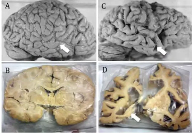
In AD, the volume of the brain is significantly reduced compared to normal brain (Figure I.A-2). This atrophy results from the degeneration of synapses and the death of neurons. Hippocampus, the brain region playing a role in memory and spatial orientation, is particularly affected. The reduction of brain size and the progression of AD are related.
Figure I.A-2: Neuroanatomical comparison of normal brain (A, B) and AD brain (C, D). Prominent atrophy in C compared with A (arrows). B, D: Coronal plane of A and C respectively. Arrow on D shows
Chapter I: Context of the project
12
b. Amyloid plaques
The first hallmarks of AD described by Aloïs Alzheimer in 1907 [1] as “minute military
foci” caused by the “deposition of a special substance in the cortex” are amyloid plaques (Figure I.A-3a). They are composed of deposits of a peptide named Amyloid-β peptide (Aβ) in aggregated forms [8] mostly fibrils. Those plaques, also named senile plaques, are found in the
extracellular media of AD brain and more especially located in the hippocampus region. Aβ is mainly a 40 to 42-amino acid residues peptide originating from the cleavage of Amyloid Precursor Protein (APP) by two enzymes: β- and γ-secretases (see Section I.B for more details).
c. Neurofibrillary tangles
The other hallmarks of the disease are intracellular neurofibrillary tangles (Figure I.A-3b). Those tangles are also observed in Parkinson’s disease (PD)[9] and are composed of
hyper-phosphorylated Tau proteins.[10] This microtubule-associated protein interacts with
tubulin to stabilize microtubules. In AD and PD, the abnormal phosphorylation of Tau induces accumulation as paired helical filaments that aggregate inside neurons in neurofibrillary tangles, making unstable the microtubules. Since microtubules are essential to preserve the structure of the neuron, the neuron loses its functionality.
Figure I.A-3: Neuropathological lesions revealed by immunohistochemistry. (a) Senile plaques observed by immunohistochemistry with antibodies against Aβ. (b) Neurofibrillary tangles observed by immunohistochemistry with antibodies against phosphorylated Tau (Pictures from reference [11])
I.A.4. Risk factors
a. Age
The age is the higher risk factor: the more aged the individuals, the higher the risk to develop AD. Indeed, around 5% of 65 years old individuals suffer from dementia and the risk
Chapter I: Context of the project
13
of developing the disease reaches 50% for individuals beyond age 85.[4] As the life expectancy
increases with the medicine advances, more and more people are likely to develop AD.
b. Gender
Women are known to have a higher life expectancy than men, thus being more susceptible to suffer from AD. Furthermore, studies suggest that the decrease in estrogen levels due to menopause could increase the risk of having AD. Indeed, clinical trials have shown that women who had been treated with hormone therapy have a lower risk of AD.[12]
c. Genetic mutations
There are two major forms of AD: the sporadic or late-onset form that is the most common, and the familial or early-onset form, representing less than 5 % of the cases. [13]
Individuals living with Down’s syndrome (also called trisomy 21) have an increased risk of early-onset AD. Indeed, they carry an extra copy of chromosome 21 in which is located the gene that is responsible for the APP formation.[14]
Mutations in genes coding for APP, Presenilin 1 and Presenilin 2 (parts of the γ-secretase which is responsible for cleavage of APP in Aβ) and ApoE (involved in Aβ clearance) genes increase the risk of developing AD as these proteins are involved in Aβ regulation in the brain.[13, 15]
I.A.5. Diagnosis
The diagnosis of AD is realized at 70% accuracy with clinical examination and neuropsychological assessment of the patient combined with brain imaging techniques.[7]
However, the definitive diagnosis of AD requires histopathologic confirmation and is made post-mortem, based on the observation of specific pathological lesions: intracellular neurofibrillary tangles and senile plaques.[11]
a. Clinical diagnosis
In 1984, the National Institute of Neurological and Communicative Disorders and Stroke–Alzheimer’s Disease and Related Disorders Association (NINCDS-ADRDA) has proposed diagnostic criteria for dementia and for AD.[16] Medical history, clinical examination,
Chapter I: Context of the project
14
neuropsychological testing and laboratory assessment are the recommended standard methods of examination. Criteria are made for several stages: probable AD dementia, possible AD dementia and probable or possible AD dementia with evidence of the AD pathophysiological process. The diagnostic criteria have been revised in 2011[17] and are still used as no specific
marker for AD is known. However, they are realized in combination with brain imaging techniques.
b. Neuroimaging [18]
Structural magnetic resonance imaging (MRI) and positron emission tomography (PET) are two brain imaging techniques clinically used for the detection of abnormalities in the brain.[19] Structural MRI is commonly used to visualize brain atrophy caused by neuronal and
dendritic losses. However, the cerebral atrophy is not specific to AD and can be the result of another pathology. Structural MRI has thus limitations in AD recognition as it cannot detect the histopathological hallmarks of AD (amyloid plaques and neurofibrillary tangles).
Fluoro-deoxy-D-glucose (FDG) PET is used as an indicator of brain metabolism by measuring the synaptic activity. Nevertheless, as metabolism can be disrupted for different reasons, FDG is not a specific marker for AD either.
Specific biomarkers of AD hallmarks are now under focus. Recently, the European Medicines Agency granted marketing authorization for the Florbetapir F18 (18F-AV-45) (also
called Amyvid), a PET marker with a good affinity for Aβ plaques (Kd = 3.7 nM[20]). This active
substance can be used to detect amyloid plaques on living patient. [20-21] This is a great advance
for AD diagnosis.
I.A.6. Current treatments
There is currently no cure for AD, only symptomatic treatments. The U.S Food and Drug Administration (FDA) has approved four medications that are marketed in France and classified in two groups: acetylcholinesterase inhibitors (Donepezil, Rivastigmine and Galantamine) and N-Methyl-D-Aspartate (NMDA) receptor antagonists (Memantine).
Apart from amyloid plaques and neurofibrillary tangles, AD is characterized by a deficit of acetylcholine, a neurotransmitter that diffuses signal across the synapse, between two neurons. Acetylcholinesterase inhibitors avoid acetylcholine degradation by inhibiting
Chapter I: Context of the project
15
acetylcholinesterase, an enzyme that catalyzes the breakdown of acetylcholine. The level of acetylcholine thus remains stable, allowing neurotransmission.
An over-concentration of glutamate is also observed in synaptic clefts of AD patients. This molecule is an excitatory neurotransmitter that plays a role in neural activation but it can also lead to neuronal death if physiologically over-concentrated. NMDA receptor antagonists block the glutamate receptors to avoid the loss of neurons.
These symptomatic treatments only slow down the cognitive deterioration and have no effect on patients in the severe stage of AD. Researchers particularly focus on amyloid plaques and neurofibrillary tangles to find new therapeutic pathways that could prevent or stop the progression of the disease.[22]
I.B.
Aβ and the amyloid plaques formation
I.B.1. Aβ: Structure and formation
Aβ is a 38 to 43 amino acid residue peptide (Figure I.B-1) derived from the enzymatic cleavage of APP. Depending on the exact location of the cleavage on the C-terminal part,
several lengths can be formed, from Aβ1-38 to Aβ1-43. However, the most abundant species
produced in the brain are Aβ1-40 and to a lesser extent Aβ1-42. Aβ is amphiphilic: the N-terminal
moiety is hydrophilic while the C-terminal one is hydrophobic.
Figure I.B-1: Amino acid sequence of Aβ1-43 (1-letter code)
APP is a type-1 trans-membrane protein expressed in various tissues of the organism, especially in the central nervous system (CNS).[23] Its major neuronal isoform encompasses 695
amino acid residues. [24] Although its physiological function is still unclear, APP would play an
important role in brain development, memory and synaptic plasticity.[24]
Two different pathways of APP metabolism can occur, as shown in Figure I.B-2. In the non-amyloidogenic pathway, the predominant one, APP is first cleaved by α-secretase and then by γ-secretase to form truncated Aβ17-40/42 (P3) peptides or by β-secretase leading to the
formation of the truncated Aβ1-16. In the amyloidogenic pathway that occurs to a minor extent,
Chapter I: Context of the project
16
peptides (mainly Aβ1-40/42). Both pathways also lead first to the formation of amino terminal
fragments (secreted APP (sAPP) α or β) and carboxyterminal fragments (CTF83 or CTF99) and then to the formation of the amino-terminal APP intracellular domain (AICD).[25]
Figure I.B-2: A schematic view of APP proteolytic cleavage. In the non-amyloidogenic pathway, APP is first cleaved by α-secretase and then by γ-secretase to form truncated Aβ17-40/42 peptides or by β-secretase
leading to the formation of the truncated Aβ1-16. In the amyloidogenic pathway, APP is cleaved
consecutively by the β- and γ-secretases leading to the formation of full-length Aβ1-40/42 peptides.
Thus, Aβpeptides are the product of a minor pathway of APP metabolism,[26] released
in the extracellular space of healthy brain during neuronal activity, without leading necessarily to Alzheimer’s pathology. Aβ is subject to a proteolytic degradation by Aβ-degrading proteases (AβDPs), which regulates Aβ levels in the brain.[27] Its functions in the brain are still unknown,
although Aβ could play a role in the synaptic plasticity and the memory.[28]
I.B.2. APP and Aβ mutations
Familial AD are caused by mutations on a gene of APP, Presenilin 1 (PSEN1) or Presenilin 2 (PSEN2). PSEN1 and PSEN2 are two subunits of γ-secretase. The mutations on both PSEN1 and PSEN2 lead to a higher Aβ production, PSEN1 mutations specifically conducting to an increased Aβ1-42 formation.[13]
Chapter I: Context of the project
17
For APP, 65 mutations are indexed in the Alzheimer Disease & Frontotemporal Dementia Mutation Database, with only 15 being non-pathogenic.[29] The mutations are divided
in three categories: mutations at the β-secretase cleavage site, at the γ-secretase cleavage site and in the mild-domain amyloid-β region.[30] The mutations at the γ-secretase cleavage site can
alter the cleavage position and lead to an increase of the Aβ1-42/Aβ1-40 ratio. The mutations at
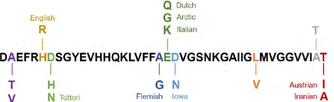
the β-secretase cleavage site increase the rate of APP proteolysis by the β-secretase. The mutations in the mild-domain of Aβ region in APP alters Aβ assembly by increasing the propensity of Aβ to form oligomers and fibrils.[31] As APP mutations can occur in the Aβ
domain, APP proteolysis by both β- and γ-secretases leads to the formation of a mutated peptide. The possible mutations are shown in Figure I.B-3.
Figure I.B-3: Familial AD mutations on Aβ1-43. The amino acid residues mutated and the names of the
mutations are colored. (1-letter code). [13]
I.B.3. Amyloid cascade hypothesis
AD is a multifactorial disease and the multiple mechanisms related to the disease are unclear. However, since Aβ has been found in healthy brain on soluble form but on aggregated form in AD patient’s brain,[8] a hypothesis has been proposed to explain the formation of senile
plaques composed of aggregated Aβ. The amyloid cascade hypothesis (Figure I.B-4) formulated in the early 1990s [32-35] has become the dominant model for AD pathogenesis,[36]
although still controversial.[37-38]
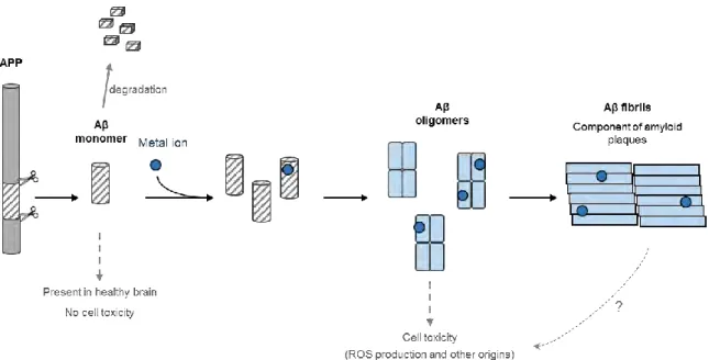
It is proposed that an abnormal extracellular increase of Aβ levels in brain could lead to its aggregation in β-sheet rich structures.[39] The aggregation starts with the formation of
oligomers species that are reorganized into protofibrils and fibrils, which are found in amyloid plaques, a hallmark of AD. In particular, oligomers accumulated in AD patient brains [40] are
proposed to be the more toxic species for cells [41-42] as they can permeabilize cellular
membranes, thus initiating a series of events leading to cell dysfunction and death.[43] According
to this hypothesis, the others events such as the intracellular formation of neurofibrillary tangles and the disruption of synaptic functions would ensue from this early and key event.
Chapter I: Context of the project
18
Figure I.B-4: Schematic representation of the amyloid cascade hypothesis with the intervention of metal ions.
Metal ions such as zinc, iron and copper ions have been found in amyloid plaques.[44] In
addition, Cu and Zn are exchanged within the synaptic cleft of some neurons. They are supposed to play an important role in the aggregation according to the amyloid cascade hypothesis.[45] Actually, metal ions can bind Aβ and thus modulate the aggregation process.
They act either on the kinetics or on the thermodynamics by impacting the morphology of the formed aggregates.[46] Furthermore, amyloid aggregates with entrapped redox-active metal ions
such as copper ions are considered as more toxic since they can produce Reactive Oxygen Species (ROS), deleterious for the biomolecules.[47]
I.B.4. Aggregation
Aβ is a natively unfolded peptide with no defined 3D structure. As this peptide is highly flexible and unstructured, it can easily undergo aggregation to amorphous or ordered structures (Figure I.B-5, green and blue pathways respectively), the β-sheet rich structures being thermodynamically the most stable.[48-49]
The fibrils formed during the aggregation process are organized in stacked parallel or anti-parallel β-sheets structures. In Aβ40 fibrils, the 12-24 and 30-40 residues would be
responsible for β-sheet formation.[50] The aggregation process of Aβ in β-sheet structures is
dynamic and complex, consisting in multiple self-assembly steps. Two different steps are observed over time: nucleation and elongation.
Chapter I: Context of the project
19
Figure I.B-5: Schematic representation of amyloid aggregation (top section) and AFM images (2 × 2 µM) of the Aβ peptide at the oligomeric and fibrillary stages superimposed with the typical sigmoid curve of
fibril formation (bottom section). Picture from reference [46].
During nucleation, the unstructured monomers in solution cluster in small aggregates called oligomers that further form nuclei (red pathway in Figure I.B-5). Nucleation is the slower and limiting step of aggregation since association of monomers that occurs during nucleation is not thermodynamically favorable.[46] The nucleation phase is then followed by a rapid elongation
phase in which protofibrils and finally fibrils are formed from nuclei (orange pathway in Figure I.B-5).
Several techniques have been developed to monitor Aβ aggregation.[46, 51] Among them,
fluorescence of Thioflavin T (ThT) dye is widely used. ThT interacts with β-sheet and upon interaction undergoes a strong fluorescence enhancement.[52] This allows to monitor the
aggregation kinetics. Atomic force microscopy (AFM) and transmission electron microscopy (TEM) give information about the size and shape of aggregates (see AFM images in Figure I.B-5).
Chapter I: Context of the project
20
I.C.
Aβ, metal ions and Reactive Oxygen Species
I.C.1. Coordination of Aβ with metal ions
Metal ions such as zinc, iron and copper are present in the brain. They are necessary and required to regulate the neuronal activity in the synapses and involved in biological functions of metallo-proteins. In several diseases such as AD, the metal ion homeostasis is disrupted and the concentration is very far from the physiological one, with Cu and Zn levels that can reach up to three times the control levels.[53] Moreover, high content of these metal ions is found in
amyloid plaques extracted from AD brains. [44] In addition, such ions can bind to Aβ under
physiological concentrations. Thus, knowing the coordination mode of these metal ions with Aβ is a pre-requisite to understand their role in AD.
a. Zn(II) coordination to the Aβ peptide
Zn ion exists only as Zn(II) and its coordination to Aβ is still not well-established.[54-55]
Although it is consensual that a complex 1:1 is formed,[55] the nature of the amino acid residues
involved in the coordination sphere is still under debate. A novel binding model has been recently proposed, based on Nuclear Magnetic Resonance (NMR) and X-ray Absorption Spectroscopy (XAS) studies of Zn coordination with mutated and N-terminal acetylated peptides (Figure I.C-1).[56] In this model, Zn(II) would be bound by imidazole rings of His6 and
either His13 or His14 residues, the carboxylate group of Glu11 and the carboxylate group of Asp1, Glu3 or Asp7.
Chapter I: Context of the project
21
b. Cu(II) coordination to the Aβ peptide
The Cu(II) coordination to Aβ has been widely studied for years and is challenging as several species are formed depending on the pH. Numerous studies have been realized in the past decade and the results have been recently reviewed,[54, 57-59] leading to a consensual model
with different Cu(II) binding modes depending on the pH. The four binding modes observed for pH values higher than 6.5 are shown in Figure I.C-2.
For component I, it is now established that Cu(II) is bound to the NH2 terminus, the
adjacent CO function from Asp1-Ala2 and to imidazole rings of His6 and either His13 or His14.[60-64]
Figure I.C-2: Schematic representation of equatorial Cu(II)-Aβ binding sites depending on the pH. The pKa of the different components are indicated in the pH scale (Picture from reference [65]).
For component II, two distinct models have been proposed. In the first one, Cu(II) is bound via the carbonyl function from Ala2-Glu3 and the imidazole rings of the three His. [62, 64]
In the second one, Cu(II) is bound to the N-terminal amine of Asp1, the amidyl function of Asp1-Ala2, the carboxylate group of Ala2 and the imidazole ring of one His. [60-61, 65] The first
model does not explain the effect of pH on the coordination as all the residues involved in Cu(II) coordination that can undergo deprotonation are already deprotonated. The second model explains the change of Cu(II) binding mode that occurs around pH 7.8 with the deprotonation of the Asp1-Ala2 amide function, leading to its coordination. Furthermore, Electron Nuclear Double Resonance (ENDOR), Hyperfine Sublevel Correlation (HYSCORE) and NMR studies highlight the involvement of both the NH2 terminus of Asp1 and the deprotonated Asp1-Ala2
amide bond, favoring the second model.[57] Thus, the second proposed model (illustrated in
Figure I.C-2) is the most accepted model and it will be used as the component II model thereafter.
The other two components (called III and IV) are formed at higher pH with the deprotonation of the Ala2-Glu3 and Glu3-Phe4 amide functions, respectively.[65] Cu(II) is
Chapter I: Context of the project
22
residue in component III and via the NH2 terminus and the three amidyl functions between
Asp1 and Phe4 in component IV.
A carboxylate group has also been proposed to be involved in apical position for several components, coming from Asp1[60-62] or from Glu3, Asp7 and Glu11 carboxylates in
equilibrium with Asp1 for component I.[61]
c. Cu(I) coordination to the Aβ peptide
Copper is a redox-active ion which is present physiologically in two redox states: Cu(I) and Cu(II). Cu(I) coordination with Aβ has been investigated more recently than Cu(II) coordination and the involvement of histidine residues is now consensual. Several binding models are suggested, two of them being most populated (Figure I.C-3).
Figure I.C-3: Schematic view of the two proposed models for Cu(I) coordination in the Aβ peptide. Adapted from reference [66].
Model A proposes a linear binding of histidines with a dynamic exchange between His6, His13 and His14. Model B involves an equilibrium between the His dyad and the His triad. NMR studies have shown the implication of the three histidines in the Cu(I) coordination with a dynamic exchange, in line with the two proposed models.[66] However, XAS studies [66-67] and
a comparison of synthetized Cu(I) complexes HisHis dipeptides and Cu(I) complexes with truncated Aβ6-14 and Aβ10-14 peptides [68-69] highlight a linear binding mode with 2 histidines,
corroborating the model A.
In addition, according to a tandem mass spectrometry (MS/MS) study on Cu(I)-Aβ structure, the two histidines mostly involved in Cu(I) coordination would be His13 and His14
[70]. Thus, evidences suggest that Aβ is bound to Cu(I) by histidine residues in a linear fashion
Chapter I: Context of the project
23
His14 dyad. This is in line with affinity studies realized on three Cu(I) complexes with one His-Ala mutation on Aβ peptide (named H6A, H13A and H14A) [71-73] that point out to a slightly
lower affinity than for the native peptide, H6A having a stronger affinity than the other two mutants. These results indicate that Aβ only needs two histidines for binding Cu(I), His13-His14 dyad being the major form.
I.C.2. Reactive Oxygen Species
a. ROS and oxidative stress
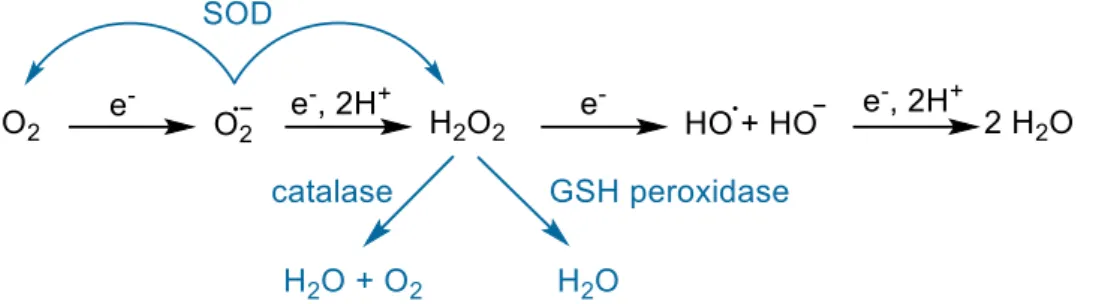
Reactive oxygen species (ROS) are radicals and molecules deriving from the incomplete reduction of dioxygen. They are produced in small quantity during the in vivo metabolism of oxygen, through four successive 1-electron reductions of O2 leading to H2O formation (Figure
I.C-4). They are necessary to maintain the homeostasis in cells and play an important role in signaling [74] but are also reactive oxidants, able to damage biomolecules. In cells, endogenous
enzymes are in charge of the antioxidant defense to prevent the ROS mediated damages.[47]
Figure I.C-4: Schematic view of the ROS production during oxygen reduction (black pathway) and the enzymes involved in ROS detoxification (blue pathways)
The superoxide (O2•) anion, the first ROS produced by the one-electron reduction of
dioxygen, is capable of inactivating few enzymes,[47] but has a poor reactivity with most of the
bio-inorganic substrates due to low rate constant (usually below 102 L mol-1 s-1).[75-76] To
remove a potential excess of O2•, endogenous enzymes called superoxide dismutases (SOD)
are present in cells and can catalyze the superoxide dismutation (Figure I.C-5, reaction (1)) with a diffusion rate close to the limit (k around 109 L mol-1 s-1).[76-77]
Chapter I: Context of the project
24
Figure I.C-5: (1) Dismutation of superoxide into dioxygen and hydrogen peroxide catalyzed in living systems by SOD. (2) Dismutation of hydrogen peroxide into dioxygen and water catalyzed in living systems by catalase. (3) Hydrogen peroxide reduction catalyzed by the glutathione peroxidase (GSH).
GS-SH: Glutathione disulfide.
Hydrogen peroxide (H2O2) is the product of the one-electron reduction of superoxide. It
can oxidize proteins with thiol groups and is deleterious in the presence of redox-active metal ions such as iron and copper as it can produce the hydroxyl radical during the Fenton reaction (Figure I.C-6, reaction (1)) or during the Haber-Weiss reaction with superoxide (Figure I.C-6, reaction (2)). H2O2 is regulated in vivo by two enzymes. The catalase catalyzes the dismutation
of H2O2 into two molecules of water and one dioxygen (Figure I.C-5, reaction (2)) whereas the
glutathione peroxidase (GSH) catalyzes its reduction in water (Figure I.C-5, reaction (3)).[78]
The hydroxyl radical (HO•) is the result of the third one-electron reduction of oxygen.
It can also be produced in the presence of metal ions from H2O2 or H2O2 and O2• by the Fenton
reaction or the Haber-Weiss reaction respectively (Figure I.C-6). HO• has a very short half-life
(10-9 s) compared with O2• (10-6 s) and is thus the more reactive and deleterious ROS,[74] being
able to oxidize the biomolecules such as proteins, lipids, DNA[79] because of its very high redox
potential (E°’=2.34 V [80]).
Figure I.C-6: (1) Fenton reaction (2) Haber-Weiss reaction catalyzed by iron ions.
To control the quantity of pro-oxidants (ROS) and prevent the damages on the biomolecules, the body has protecting mechanisms including enzymatic and chemical antioxidants. However, in some diseases such as AD [81], an imbalance may occur between
pro-oxidants and antipro-oxidants, due to a higher ROS production or a reduced activity of the enzymes responsible for the ROS degradation, leading to oxidative damages on biomolecules.[82]
Chapter I: Context of the project
25
b. Metal-catalyzed ROS production
Redox active metal ions such as copper and iron are involved in the ROS production via Fenton and Haber-Weiss reactions (Figure I.C-6). In the presence of a reducing agent, they can have a catalytic activity.[83] In AD, Cu and Fe can be coordinated to Aβ and the resulting
complex could be directly involved in the ROS production. ROS production has been mostly studied with Cu-Aβ, as Fe-Aβ has a lower redox activity.[84] Iron is found in the amyloid plaques
predominantly in a colloidal form (originating from ferritin), however histochemical studies indicate that it could also be bound to Aβ.[85] The coordination mode of Fe(II) with Aβ has been
characterized [86] and Fe(III) does not form a stable complex with Aβ because it finally converts
into Fe(III)(HO)3 and precipitates. Thus, the physiological stable formation of Fe(III)-Aβ is
unlikely. However, ROS production by Fe-Aβ still might be relevant as Fe(II)-Aβ is stable and the Fe(III) complex formed during ROS production might not have time to precipitate. As the involvement of iron bound to Aβ in ROS production is still unclear, we focus here only on Cu-Aβ.
In the case of copper, the pro-oxidant role of the Cu-Aβ system is not clearly established as the complex is more active in ROS production than several biological relevant Cu-peptides or Cu-proteins [87] but less efficient than loosely-bound copper.[84, 87-91] However, in vitro studies
have shown that Cu-Aβ is able to catalyze the formation of H2O2 and HO•in the presence of O2
and a reducing agent such as ascorbate (Figure I.C-7).[84, 87-88, 92] Moreover, although it was
generally proposed that H2O2 production by Cu-Aβ occurs via a two-electron process, a recent
study has highlighted the formation of superoxide as an intermediate in the production of H2O2
by Cu-Aβ and O2.[93]
Figure I.C-7: Mechanism of ROS production from a reductant and dioxygen catalyzed by the Cu-Aβ complex. The ROS produced are the superoxide anion (O2•), hydrogen peroxide (H2O2) and the hydroxyl
Chapter I: Context of the project
26
c. Coordination of copper with Aβ during redox cycling
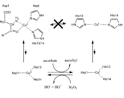
Copper is redox-active and cycles between the +I and +II oxidative states when bound to Aβ. An electrochemistry study has shown that a preorganization mechanism was needed to allow the electron transfer for the oxidation of Cu(I) or the reduction of Cu(II) since the Cu(II) and Cu(I) coordination spheres are very different (Figure I.C-8, resting states).[94]
Figure I.C-8: Top: Resting states that are the most populated states of Cu(II)-Aβ (left) and Cu(I)-Aβ (right). The redox reaction between these states is sluggish due to a high reorganization energy. Bottom: putative mechanism of HO• production from H2O2 and ascorbate, through the efficient redox reaction of
the in-between state. Picture from reference [95].
The energy required for the rearrangement between the Cu(I) and Cu(II) geometries (linear and square-planar respectively) being very high, the electron transfer would rather proceed via a low-populated redox-active state in which Cu(I) and Cu(II) binding modes are highly similar, thus inducing a low reorganization energy. This transient state, called “in-between” state, is in equilibrium with the resting states (Figure I.C-8, bottom section). It has been studied by calculations [96] and characterized with MS/MS by identifying the sites of
oxidative damage on the peptide.[95] By comparing the non-specific oxidations detected on Aβ28
after the radiation-induced ROS production with the copper-mediated oxidations of Aβ28, Asp1,
His 13 and His14 have been found to be the metal-specific targeted amino acid residues. Furthermore, kinetic studies of the copper-mediated Aβ28 oxidation have shown that Asp1
would be the first amino acid residues damaged. Thus, in this study, the proposed ligands for both Cu(II) and Cu(I) coordination in the in-between state are Asp1, His 13 and His14. As they
Chapter I: Context of the project
27
have been found to be the main targets for HO•, they are supposed to be the amino acid residues
the closest from copper during the metal-catalyzed ROS production. I.C.3. Metal-catalyzed oxidation of Aβ
During the metal-catalyzed ROS production, the Aβ peptide undergoes oxidative damages. This is in line with the detection of oxidized Aβ in amyloid plaques in vivo.[97] Studies
on single amino acid residue oxidations could allow a prediction on the residues targeted during the metal-catalyzed oxidation (MCO) of Aβ.[98-100] The physiological main targets for HO• are
the sulfur-containing amino acids (methionine, cysteine), the basic amino acids (arginine, histidine, lysine) and the aromatic amino acids (phenylalanine, tyrosine, tryptophan).[101]
Table I.C-1 provides the main oxidation products of these amino acid residues. Oxidation of Aβ28 by HO• produced by γ-radiolysis has shown that His and Phe residues are
mainly targeted,[95] in line with the oxidations reported previously for free amino acid residues.
However, in the case of MCO of Aβ, the ROS are produced at the metal center. Thus, the oxidations are site-specific and can differ from the amino acid oxidations usually detected without metal.
Table I.C-1: Main oxidation products of the principal amino acid residues undergoing HO• attack.[101]
Amino acid residue 3-letter abbreviation Products of oxidation by HO•
Cysteine Cys Cysteic acid
Cystine Methionine Met Methionine sulfoxide
Methionine sulfone Arginine Arg 5-hydroxy-2-amino valeric acid
Histidine His 2-oxohistidine
Lysine Lys 3,4 or 5-hydroxylysine Phenylalanine Phe 2-hydroxyphenylalanine
Tryptophan Trp N'-Formylkynurenine Kynurenine
Tyrosine Tyr Dihydroxyphenylalanine (DOPA) Dityrosine
Chapter I: Context of the project
28
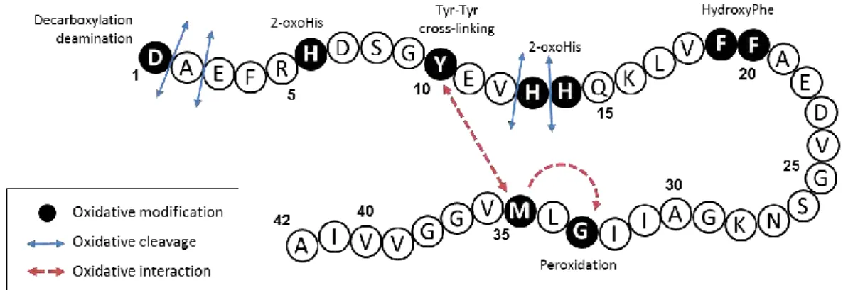
Several studies have reported the damages undergone by the Aβ peptide during the copper-mediated oxidation. The amino acid residues damaged are summarized in Figure I.C-9 and further described in the following paragraphs.
Figure I.C-9: Schematic view of the different oxidative modifications (black circle), cleavages (blue arrows) and interactions (red arrows) undergone by the Aβ42 peptide during the copper-mediated
oxidation (from reference [87]).
a. Histidines
The Aβ peptide contains 3 histidines located at the positions 6, 13 and 14. They are involved in both Cu(II) and Cu(I) coordination by their imidazole ring (see section I.C.1). Thus, they are targeted during MCO. Histidines have been found oxidized into 2-oxohistidines (Figure I.C-10) during MCO of Aβ bound to copper in the presence of ascorbate [88, 95, 102-103] or
hydrogen peroxide.[104]
Figure I.C-10: Structural formula of histidine (left) and 2-oxo-histidine (right).
The mechanism of oxidation is shown in Figure I.C-11. His13 and His14 have been found to be more sensitive to oxidation, His6 being not detected on its oxidized form [95, 103-104]
or affected after longer oxidation time.[102] This is in line with the predominant binding mode
Chapter I: Context of the project
29
Figure I.C-11: 2-oxo-histidine formation from the oxidative attack of hydroxyl radical at the C-2 position of the imidazole ring of Histidine [105]
b. Aspartate
The Aβ peptide has 3 aspartates residues at positions 1, 7 and 23. In the literature, only Asp1 has been found to be oxidized. Actually, as Asp1 is involved in the coordination of Cu(II),[57-58] it is a preferential target for the hydroxyl radical produced at the metal center.
Different damages have been detected during MCO of Asp1 both in the presence of ascorbate [95, 106] and of hydrogen peroxide [104]. Figure I.C-12 shows an oxidative mechanism
leading to the formation of either pyruvate (blue pathway), isocyanate (red pathway) or 2-hydroxyaspartate (green pathway) function through the formation of an alkoxyl radical.
Figure I.C-12: Mechanism of aspartate oxidation with three different pathways starting from alkoxyl radical and leading to the formation of 2-hydroxyaspartate (green), isocyanate (red) and pyruvate (blue).
Chapter I: Context of the project
30
The oxidative decarboxylation and deamination of Asp1 leads to the formation of a pyruvate function (Figure I.C-12, blue pathway).[95, 104, 106] Asp1 is also subject to a backbone
cleavage on the α-position of the peptide, leading to an isocyanate function (Figure I.C-12, red pathway).[95, 106] Another oxidation of Asp1 into 2-hydroxyaspartate corresponding to the
formal addition of an oxygen atom has also been described (Figure I.C-12, green pathway).[95]
c. Tyrosine
Although the amino acid residues involved in copper coordination are more vulnerable to oxidation, non-coordinating amino acid residues can also be oxidized. It is the case for Tyr10 which is sensitive to oxidation and is responsible for the Aβ peptide cross-linking by dityrosine formation (Figure I.C-13).[98] This latter, induced by Cu(II), has been detected for Aβ in the
presence of H2O2.[107] MCO of Tyr10 into dityrosine was found to have an impact on
aggregation as Aβ cross-linking was correlated with the formation of covalent oligomers. [108-109]
Furthermore, a study has proposed that Tyr10 acts as a gate that promotes the electron transfer from Met35 to Cu(II) for its reduction in Cu(I).[110]
Chapter I: Context of the project
31
d. Phenylalanines
Three phenylalanines are present in the Aβ sequence at positions 4, 19 and 20. None of them are involved in the Cu(II) or Cu(I) coordination, nevertheless Phe19 and Phe20 have been found oxidized during MCO of Aβ in the presence of Cu(II) and ascorbate.[95] Phe19 and Phe20
has been detected with the formal addition of an oxygen atom, likely oxidized into hydroxyphenylalanine (Figure I.C-14).[98] This oxidation seems to occur after the oxidation of
Asp1 which is involved in Cu binding.[95]
Figure I.C-14: Structural formula of phenylalanine and the three hydroxyphenylalanines.
e. Methionine
Methionine is an amino acid residue very sensitive to oxidation. In vivo, the enzyme methionine sulfoxide reductase is responsible for the reduction of the methionine sulfoxide (Figure I.C-15), a main oxidized form of the methionine.[112] Methionine can also be converted
into sulfuranyl / hydroxysulfuranyl radical cation by a one-electron oxidation.[113]
Chapter I: Context of the project
32
Reviews have reported about oxidation of the methionine of the Aβ peptide located at position 35 and its role in toxicity and oxidative stress.[114-115] Although methionine is very
sensitive to oxidation, its conversion into methionine sulfoxide occurs only after the oxidation of His13 and His14 during the in vitro MCO of Aβ in the presence of Cu(II)/ascorbate.[102] This
highlights the site-specificity of the amino acid residue oxidation catalyzed by the bound copper.
Met35 has also been found to promote Tyr10 oxidation [116] and to interact with Gly33,
inducing its peroxidation by promoting the formation of a carbon-centered radical, leading to a hydroperoxide.[90, 117]
f. Other cleavages
Other oxidative cleavages have been reported for Aβ bound to Cu(II) in the presence of H2O2 such as the cleavage of the peptide bond of Asp1/Ala2, Ala2/Glu3, Val12/His13 or
Chapter I: Context of the project
33
References
[1] R. A. Stelzmann, H. Norman Schnitzlein and F. Reed Murtagh, Clinical Anatomy 1995, 8, 429-431.
[2] M. Goedert and M. G. Spillantini, Science 2006, 314, 777-781.
[3] M. Prince, A. Wimo, M. Guerchet, G. Ali, Y. Wu and M. Prina, Alzheimer's Disease International, London 2015.
[4] B. Duthey, Priority Medicines for Europe and the World. " A public Health Approach to Innovation" 2004, 1-74.
[5] Alzheimer's & dementia: the journal of the Alzheimer's Association 2015, 11, 332. [6] M. P. Mattson, Nature 2004, 430, 631-639.
[7] L. O. Soto-Rojas, F. de la Cruz-López, M. A. O. Torres, A. Viramontes-Pintos, M. del Carmen Cárdenas-Aguayo, M. A. Meraz-Ríos, C. Salinas-Lara, B. Florán-Garduño and J. Luna-Muñoz, Neuroinflammation and Alteration of the Blood-Brain Barrier in Alzheimers Disease, in Alzheimer's Disease - Challenges for the Future, I. Zerr, InTech, 2015.
[8] G. G. Glenner and C. W. Wong, Biochemical and Biophysical Research Communications 1984, 120, 885-890.
[9] M. Goedert, Science 2015, 349, 1255555.
[10] I. Grundke-Iqbal, K. Iqbal, Y. C. Tung, M. Quinlan, H. M. Wisniewski and L. I. Binder, Proceedings of the National Academy of Sciences of the United States of America 1986, 83, 4913-4917.
[11] L. Minati, T. Edginton, M. G. Bruzzone and G. Giaccone, American journal of Alzheimer's disease and other dementias 2009, 24, 95-121.
[12] S. C. Janicki and N. Schupf, Current neurology and neuroscience reports 2010, 10, 359-366.
[13] R. C. Barber, Scientifica (Cairo) 2012, 2012, 246210.
[14] F. K. Wiseman, T. Al-Janabi, J. Hardy, A. Karmiloff-Smith, D. Nizetic, V. L. Tybulewicz, E. M. Fisher and A. Strydom, Nature Reviews Neuroscience 2015.
[15] J. Nasica-Labouze, P. H. Nguyen, F. Sterpone, O. Berthoumieu, N.-V. Buchete, S. Coté, A. De Simone, A. J. Doig, P. Faller and A. Garcia, Chemical Reviews 2015, 115, 3518-3563. [16] G. McKhann, D. Drachman, M. Folstein, R. Katzman, D. Price and E. M. Stadlan, Neurology 1984, 34, 939-944.


![Figure I.C-11: 2-oxo-histidine formation from the oxidative attack of hydroxyl radical at the C-2 position of the imidazole ring of Histidine [105]](https://thumb-eu.123doks.com/thumbv2/123doknet/2086480.7260/40.892.129.784.104.241/figure-histidine-formation-oxidative-hydroxyl-position-imidazole-histidine.webp)
![Figure I.C-13: Tyrosine cross-linking mechanism leading to the formation of dityrosine [111]](https://thumb-eu.123doks.com/thumbv2/123doknet/2086480.7260/41.892.124.772.653.1004/figure-tyrosine-cross-linking-mechanism-leading-formation-dityrosine.webp)