8355
Identification and phytochemical screening of
Endophytic fungi from stems of Phragmanthera
capitata (Sprengel) S. Balle (Loranthaceae)
Ladoh-Yemeda CF*1, Nyegue MA2, Ngene JP3, Benelesse GE1, Lenta B4, Wansi5 JD, Mpondo Mpondo E3, Dibong SD1,3
(1) Laboratory of Biology and Physiology of Plant Organisms. University of Douala, P.O. BOX 24157 Douala, Cameroon.
(2) Laboratory of Microbiology. University of Yaoundé I, P.O. BOX 812 Yaoundé, Cameroon.
(3) Department of Pharmaceutical sciences, Faculty of Medicine and Pharmaceutical University of Douala, P.O. BOX 2701 Douala, Cameroon.
(4) Laboratory of Natural Product Chemistry and Organic Synthesis. Department of Chemistry, Higher Teachers’ Training College, University of Yaoundé I, P.O. BOX 47 Yaoundé, Cameroon.
(5) Laboratory of Chemistry/Biology. University of Douala, P.O. BOX 24157 Douala, Cameroon. *Corresponding author: [email protected]
Original submitted in on 27th March 2015. Published online at www.m.elewa.org on 30th June 2015
http://dx.doi.org/10.4314/jab.v90i1.7
ABSTRACT
Objective: The purpose of this study was to identify some endophytic fungi, which were associated with the stems of Phragmanthera capitata (Loranthaceae), and to determine the phytochemical composition of their extracts.
Methodology and results: The isolation of endophytic fungi was made on PDA medium (Potato dextrose agar) and the identification was based on macroscopic and microscopic observations of the different strains isolated and using identification keys. The qualitative phytochemical analysis of acetate ethyl extracts of the endophytes was carried out using standard procedures. Eleven fungi species belonging to 4 genera were isolated: Aspergillus (06 species), Penicillium (03), Trichoderma (01) and Fusarium (01). The phytochemical analysis revealed the presence of flavonoids, anthroquinones, tannins, phenols, steroids, coumarins and terpenoids and absence of alkaloids and saponins in all the extracts.
Conclusion and application of results: The study shows that endophytic fungi of P. capitata could be a potential source of new bioactive compounds, which can be used in the fields of health and agriculture.
RÉSUMÉ
Objectif: Le but de cette étude a été d’identifier quelques champignons endophytes qui sont associés aux tiges de Phragmanthera capitata (Loranthaceae) et de déterminer la composition phytochimique de leurs extraits.
Méthodologie et résultats: L’isolement des champignons endophytes s’est fait sur milieu de culture PDA (Potato dextrose agar) et l’identification sur la base des observations macroscopique et microscopique des différents isolats et des clés d’identification. L’analyse phytochimique qualitative des extraits à l’acétate d’éthyl des endophytes a été
Journal of Applied Biosciences 90:8355– 8360
8356
réalisée suivant les procédures standards. Onze espèces fongiques appartenant à 4 genres ont été isolés : Aspergillus (06 espèces), Penicillium (03), Trichoderma. (01) et Fusarium. (01). L’analyse phytochimique a révélé la présence des flavonoïdes, anthroquinones, tanins, phénols, stéroïdes, coumarines et terpenoïdes et l’absence des alcaloïdes et saponines dans tous les extraits.
Conclusion et application des résultats: L’étude montre que les champignons endophytes de P. capitata seraient une source potentielle de nouveaux composés bioactifs applicables dans les domaines de la santé et de l’agriculture. INTRODUCTION
Phragmanthera capitata is a hemiparasite epiphytic plant belonging to the family of Loranthaceae. It appears as the most abundant and most widely distributed in the Littoral Region (Cameroon) (Dibong et al., 2009). Different host plants of Loranthaceae species include Spondias mangifera, Garcinia kola, Manniophyton fulvum, Persea americana, Psidium guajava, Citrus spp. and Theobroma cacao (Dibong et al., 2011). Loranthaceae plays an important and complex role in biological systems where they interact with ants, birds and mammals (Watson, 2001; Ladoh et al., 2013.). The plants during their evolution, have developed symbiotic interactions with microorganisms. These interactions play a critical role in the success of the conquest or adaptation to habitat by these plants (Cao et al., 2009). Recent works have revealed the presence of endophytes in symbiotic interactions with Loranthaceae (De Abreu et al., 2010; Guimarães et al., 2013; Chao-Dong et al., 2014, Sadananda et al., 2014). Endophytes are microorganisms (bacteria or fungi) that live intra- and/or intercellular in the internal tissues of plants without causing apparent symptoms (Pimentel et
al., 2011). Endophytic fungi are the most frequently isolated microorganisms as endophytes (Strobel et al., 2004). These microorganisms live in complex interaction with plants. Their role is to improve the ecological and physiological performance of their hosts (improvement of nutritional status, disease resistance, pest and physical stress). In return, they receive from their host plant nutrition and protection (Saikkonen et al., 1998). Dreyfuss and Chapela (1994) and Hawksworth and Rossman (1997) estimated about 1.5 million species of endophytic fungi on plants and only a small fraction of about 70,000 to 100,000 or 5% was identified. The exploration of medicinal plants of endophytic habitats could be a favourable way to the discovery of new species of endophytes and bioactive secondary metabolites synthesized by them. The extracts of P. capitata have numerous biological activity such as antibacterial, antioxidant, analgesic and anti-pyretic (Ladoh et al., 2014; Takem et al., 2014).The objective of this study was to identify some endophytic fungi of P. capitata and to determine the chemical composition of their extracts.
MATERIALS AND METHODS
Plant material: The stems of Phragmanthera capitata harvested in Theobroma cacao were collected in June 2014 at the orchard of Chiefdom 1st degree Ndogbong (Douala, Cameroon). The plant was botanical identified by the Laboratory of Biology and Plant Physiology of Organisms (University of Douala). The collected samples were placed in sterile bags and stored at 4°C until isolation procedure was started.
Isolation and identification of endophytic fungi: The stems of Phragmanthera capitata were surface sterilized using the methods of Pimentel et al. (2006) and Khan et al. (2010). The stems were cleaned under running tap water for ten minutes to remove impurities and surface debris. After air-drying, the cleaned stems were cut into small pieces using sterile blade and then surface sterilized in a sterile hood by immersion in 70°ethanol for 1 min, 2.4% sodium hypochlorite solution for 4 min, 70° ethanol for 30 s and then
washed three times with sterile distilled water for 1 min. The surface-sterilized samples were dried on sterile blotting paper, then cut into pieces (1 mm2) and aseptically placed in petri dish containing sterile potato dextrose agar (PDA medium) supplemented with chloramphenicol (250 mg/l) to inhibit bacterial growth. All plates were incubated at room temperature until mycelium grew out hyphal tips that were cut and transferred to PDA. All the isolated endophytic fungi were stored on PDA tube at 4°C. The identification procedure was based on microscopic and macroscopic observations of colony and hyphal morphology of the fungal cultures, characteristics of the spores according to the standard taxonomic key (Navi et al., 1999; Chabasse et al., 2002 ; Dufresne, 2014).
Fermentation and extraction of metabolites: Each isolated endophytic fungus strain was introduced into a test tube containing sterile distilled water supplemented
8357 with tween 80 to dissociated spores. Each inoculum was introduced in flasks containing sterilized solid medium using rice as the base (100 g rice, 100 ml distilled water, sterilized for 1 h in an autoclave). The flasks were incubated at room temperature for 3 weeks. After incubation, fungal metabolites were extracted with ethyl acetate as organic solvent for 48 h, then filtrate and concentrated by rotary evaporation to obtain crude extract.
Phytochemical Screening of endophytic fungi extracts: Preliminary phytochemical analysis of the crude extracts of fungi was carried out for the presence of the following metabolites: alkaloids, flavonoids, anthraquinones, tannins, phenols, steroids, coumarins, saponins and terpenoids using standard procedures as described by Sofowora (1993) and Harborne (1998)
RESULTS
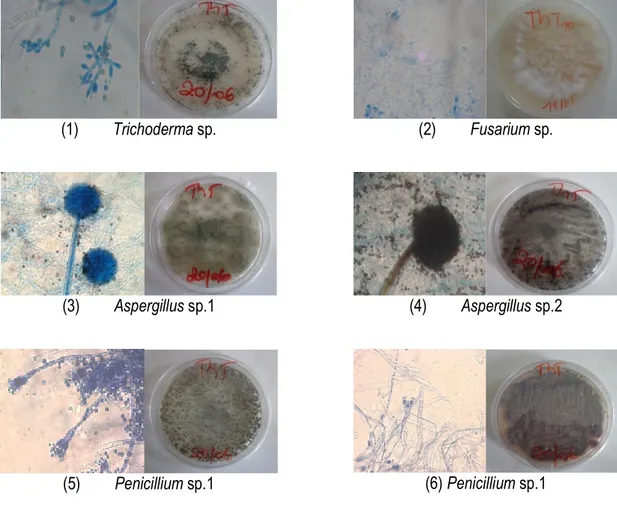
Identification of isolated endophytic fungi: The identification of fungal strains isolated from stems of Phragmanthera capitata collected on Theobroma cacao was done under the basis of macroscopic and
microscopic observations of cultural characters (Fig. 1). A total of 11 species belonging to four genera were isolated and identified: Aspergillus (06 species), Penicillium (03), Trichoderma (01) and Fusarium (01).
(1) Trichoderma sp. (2) Fusarium sp.
(3) Aspergillus sp.1 (4) Aspergillus sp.2
(5) Penicillium sp.1 (6) Penicillium sp.1
Figure 1: Microscopic and macroscopic characters cropping of some isolated endophytic fungi. Screening phytochemical extracts of endophytic
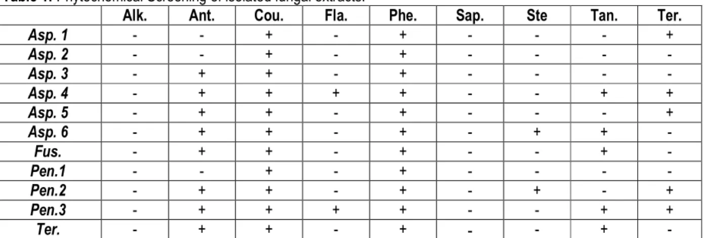
fungi: Preliminary phytochemical screening of fungal
ethyl acetate extracts showed the presence of flavonoids, anthraquinones, tannins, phenols, steroids,
8358 coumarins and terpenoids and the absence of alkaloids and saponins. The result of qualitative analysis of the
phytochemicals were summarised in table 1.
Table 1: Phytochemical Screening of isolated fungal extracts.
Alk. Ant. Cou. Fla. Phe. Sap. Ste Tan. Ter.
Asp. 1 - - + - + - - - + Asp. 2 - - + - + - - - - Asp. 3 - + + - + - - - - Asp. 4 - + + + + - - + + Asp. 5 - + + - + - - - + Asp. 6 - + + - + - + + - Fus. - + + - + - - + - Pen.1 - - + - + - - - - Pen.2 - + + - + - + - + Pen.3 - + + + + - - + + Ter. - + + - + - - + -
(+): Presence, (-): Absence, Asp. : Aspergillus, Pen.: Penicillium, Fus.: Fusarium; Tri.: Trichoderma, Alk. : Alkaloïds, Ant. : Anthraquinone, Cou. : Coumarins, Phe. : Phenols, Tan. : Tannins, Fla. : Flavonoïds, Sap. : Saponins, Ter. : Terpenoïds.
DISCUSSION
In traditional medicine, plants are assumed to have some healing power that may be due to unknown bioactive compounds (Strobel and Daisy, 2003). Phragmanthera capitata was selected for the isolation of endophytic fungi based on medicinal importance. Eleven species of endophytic fungi from stems of Phragmanthera capitata were isolated and identified, belonging to 4 genera (Apergillus, Penicillium, Trichoderma and Fusarium). Sadananda et al., (2013) also reported the presence of endophytes in different parts of Viscum album (Viscaceae). Similarly, another study conducted in Brazil on another Loranthaceae also revealed the presence of Aspergillus sp. and Trichoderma sp. as endophytic fungi from Cladocolea micrantha (Guimarães et al., 2013). These endophytes were also isolated from several host plants (Giordano et al., 2009; Suresh et al., 2013; Mbouobda et al., 2014.). Generally many species of the genus Aspergillus such as A. fumigatus, and A. niger in addition to species of Penicillium and Fusarium are adapted to different plant tissues (Ilyas et al., 2009). A recent study showed that endophytes are not host specific (Jalgaonwala et al., 2011). They colonize multiple host species of the same plant family within the same habitat, and their distribution can be similar in closely related plant species (Huang et al., 2008). The
present study is one of the first report that several fungal endophytes are associated with P. capitata. The preliminary phytochemical screening of all the isolates showed the presence of secondary metabolite secretions such as flavonoïds, anthraquinones, tannins, phenols, steroids, coumarins and terpenoids. The chemical composition of endophytic fungi varies depending on the species; however, there was lack of alkaloids and saponins in all fungal extracts. The ability of an endophyte to produce some metabolites but not others has been described by Selim et al. (2012) where different endophytes in a plant may produce different secondary metabolites hence play different functions in the plant and that the total number of metabolites in a plant extract may be a contribution of all the endophytes that live in the plant. The production and quality of bioactive compounds from endophytic fungi depends on natural conditions of the association and the nature of the synthetic medium used (Shinyar et al., 2008).Strategies can be developed to use these fungi for exploitation of bioactive compounds. In addition, the use of endophytes as potential factories for the production of secondary metabolites might revolutionize agricultural, pharmaceutical and biotechnological research in the near future (Suresh et al., 2013).
8359 CONCLUSION
The present study reveals that Phragmanthera capitata live with endophytic fungi which synthesize secondary metabolites. For our knowledge, this is the first report that fungal endophytes associated with P. capitata that
could be a potential source of bioactive compounds to explore for the development of new drugs for human diseases and phytopathogens.
REFERENCES BIBLIOGRAPHIQUES
Cao R, Liu X, Gao K, Mendgen K, Kang Z, Gao J, Dai Y, Wang X, 2009. Mycoparasitism of endophytic fungi isolated from reed on soil borne phytopathogenic fungi and production of cell wall-degrading enzymes in vitro. Current Microbiology, 59: 584-592
Chabasse D, Bouchara JP, Gentile L, Brun S, Cimon B, Penn P, 2002. Les moisissures d’intérêt médical. Laboratoire de parasitologie Mycologie CHU, Angers. 159 p
Chao-Dong Qian, Yu-Hang Fu, Fu-Sheng Jiang, Zheng-Hong Xu, Dong-Qing Cheng, Bin Ding, Cheng-Xian Gao, Zhi-Shan Ding, 2014. Lasiodiplodia sp. ME4-2, an endophytic fungus from the floral parts of Viscum coloratum, produces indole-3-carboxylic acid and other aromatic metabolites. BMC Microbiology, 14: 297.
De Abreu LM, Almeida AR, Salgado M, Pfenning LH, 2010. Fungal endophytes associated with the mistletoe Phoradendron perrottettii and its host tree Tapirira guianensis. Mycol Progress, 9: 559-566.
Dibong SD, Mony R, Ladoh CF, Boussim IJ, Amougou A, 2011. Parasitism evolution of Loranthaceae in the Ndogbong chiefdom’s orchard (Douala, Cameroon). Int. J. Plt. An. Env. Sci., 1 (3) 2231-4490.
Dibong SD, Engone Obiang NL, Din N, Priso RJ, Taffouo VD, Fankem H, Salle G, Amougou A, 2009. Niveau d’infestation des arbres fruitiers des groupements végétaux par Phragmanthera capitata (Sprengel) S. Balle (Loranthaceae) dans la région littorale du Cameroun. Int. J. Biol. Chem. Sci., 3 (2): 347-354.
Din N, Saenger P, Priso JR, Dibong SD, Amougou A, 2008. Logging activities in mangrove forests: A case study of Douala Cameroon. African Journal of Environmental Science and Technology, 2 (2): 22-30.
Dreyfuss MM, Chapela IH, 1994. Potential of fungi in the discovery of novel, low-molecular weight
pharmaceuticals. In: The discovery of natural products with therapeutic potential (Ed.): V.P. Gullo. Butter-Worth-Heinemann, London, United Kingdom. 49-80.
Dufresne Philippe, St-Germain Guy, 2014. Identification des champignons d’importance médicale. Laboratoire de santé publique. Québec. 56 p. Gallo MB, Guimaraes DO, Momesso LS, Pupo MT,
2008. In: Saikia R, Bezbaruah, RL, Bora, TC. (eds). Natural products from endophytic fungi. Microbial Biotechnology. New India Publishing Agency, New Delhi, India; p. 139
Giordano L, Gonthier P, Varese GC, Miserere L, Nicolotti G, 2009. Mycobiota inhabiting sapwood of healthy and declining Scots pine (Pinus sylvestris L.) trees in the Alps. Fungal Diversity, 38: 69-83.
Govindappa M, Channabasava R, Sowmya DV, Meenakshi J, Shreevidya MR, Lavanya A, Gustavo Santoyo, Sadananda TS, 2011. Phytochemical screening, antimicrobial and in vitro anti-inflammatory activity of endophytic extracts from Loranthus sp. Pharmacognosy Journal, 3: 82-90.
Guimarães AC, Siani AC, Bezerra JL, Lima de Souza AQ, Sarquis MI, 2013. Endophytic Mycobiota Characterization of the Amazonian Mistletoe Cladocolea micrantha Hosted in Cashew Tree. American Journal of Plant Sciences, 4: 917-921.
Harbone JB, 1998. Phytochemical methods. A guide to modern techniques of plants analysis. Third edition. Chapman and Hall. London. 150 p. Hawksworth DL, Rossman AY, 1997. Where are all the
undescribed fungi? Phytopathology, 87: 888-891.
Huang WY, Cai YZ, Hyde KD, Corke H, Sun M, 2008. Biodiversity of endophytic fungi associated with 29 traditional Chinese medicinal plants. Fungal Div. 33: 61-75.
Ilyas M, Kanti A, Jamal Y, Hertina, Agusta A, 2009. Biodiversity of endophytic fungi associated
8360 with Uncaria gambier Roxb. (Rubiaceae) from west Sumatra. Biodiversitas, 10: 23-28. Jalgaonwala RE, Mohite BVM, Mahajan RT, 2011. A
review: natural products from plant associated endophytic fungi. J. Microbiol. Biotechnol, 1: 21-32.
Khan R, Shahzad S, Choudhary MI, Khan SA, Ahmad A, 2010. Communities of endophytic fungi in medicinal plant Withania somnifera. Pakistan Journal of Botany, 42: 1281-1287.
Khan R, Shahzad S, Iqbal choudhary M, Khan SA, Ahmed A, 2007. Biodiversity of the endophytic fungi isolated from Calotropis procera (Ait.) R. Br. Pakistan Journal of Botany, 39: 2233-2239. Ladoh Yemeda CF, Mony R, Tchatat M, Dibong SD, 2013. Contribution des fourmis à la lutte biologique contre les Loranthaceae. Int. J. Biol. Chem. Sci, 7(3): 924-937.
Ladoh Yemeda CF, Dibong SD, Nyegue MA, Djembissi Talla RP, Lenta Ndjakou B, Mpondo Mpondo E, Yinyang J, Wansi JD, 2014. Activité antioxydante des extraits méthanoliques de Phragmanthera capitata (Loranthaceae) sur Citrus sinensis. Journal of Applied Biosciences, 84: 7636-7643.
Mbouobda HD, Fotso B, Muyang RF, Chiatoh TA, Omokolo Ndoumou D, 2014. Enzymes and qualitative phytochemical screening of endophytic fungi isolated from Lantana camara Linn. Leaves. Journal of Applied Biology and Biotechnology, 2 (06):1-6.
Navi SS, Bandyopadhyay R, Hall AJ, Bramel-Cox, 1999. A pictorial guide for the identification of mold fungi on sorghum grain. International Crops Research Institute for the Semi-Arid Tropics, 118 pp.
Pimentel IC, Glienke-Blanco C, Gabardo J, Stuart RM, Azevedo JL. 2006. Identification and colonization of endophytic fungi from soybean (Glycine max (L.) Merril) under different environmental conditions. Brazilian archives of biology and technology, 49: 705-711.
Pimentel MR, Molina G, Dionisio AP, Marostica Junior MR, Pastore GM, 2011. The use of endophytes to obtain bioactive compounds and their application in biotransformation process. Biotechnol. Res Int., pp 150.
Sadananda TS, Govindappa M, Ramachandra YL, 2013. Antibacterial activity of Viscum album endophytic fungal lectin. International Journal
of Biological & Pharmaceutical Research, 4(12): 1033-1042.
Saikkonen K, Faeth SH, Helander M and Sullivan TJ, 1998. Fungal endophytes: A continuum of interactions with host plants. Annual Review of Ecology and Systematics, 29: 319-343. Selim K, El-Beih A, Abdel-Rahman T, El-Diwany A,
2012. Biology of endophytic fungi. Current Research in Environmental & Applied Mycology, 2(1): 31-82.
Sofowora A. 1993. Medicinal plants and Traditional Medicine in Africa. Spectrum Books, badan. Strobel G, Daisy B, Castillo U, Harper J. 2004. Natural
products from endophytic microorganisms. Journal of Natural Products, 67: 257-268. Strobel GA and Daisy B. 2003. Bioprospecting
formicrobial endophytes and their natural products. Microbiol. Mol. Biol. Rev., 67: 491-502.
Suresh Bhargav Desaraju, Vijaya Tartte, Venkateswarulu Nagam, Chandramouli Kalla, Krishnakanth Sarma, Chamarthi Nagaraju. 2013. Endophytic fungal diversity from endemic plants of Tirumala hills in Eastern ghats and their anti-candidal activity. World Journal of Pharmaceutical Research, 3 (1): 834-847.
Takem LP, Abe NP and Ogbonna OJ. 2014. Anti-Pyretic and Analgesic Potentials of Aqueous Extract of Phragmanthera capitata S. Balle in Albino Rats. American Journal of Pharmacy and Pharmaceutical Sciences, 1(2):37-43. Watson DM, 2001. Mistletoe-A keystone resource in
forests and woodlands worldwide. An. Rev. Eco. Sys,. 32: 219-249.