HAL Id: hal-02696333
https://hal.inrae.fr/hal-02696333
Submitted on 1 Jun 2020
HAL is a multi-disciplinary open access
archive for the deposit and dissemination of
sci-entific research documents, whether they are
pub-lished or not. The documents may come from
teaching and research institutions in France or
abroad, or from public or private research centers.
L’archive ouverte pluridisciplinaire HAL, est
destinée au dépôt et à la diffusion de documents
scientifiques de niveau recherche, publiés ou non,
émanant des établissements d’enseignement et de
recherche français ou étrangers, des laboratoires
publics ou privés.
Stress-induced enhancement of colitis in rats: CRF and
arginine vasopressin are not involved
M. Gué, C. Bonbonne, J. Fioramonti, C. del Rio-Lachèze, Christine Coméra,
Lionel Bueno
To cite this version:
M. Gué, C. Bonbonne, J. Fioramonti, C. del Rio-Lachèze, Christine Coméra, et al.. Stress-induced
enhancement of colitis in rats: CRF and arginine vasopressin are not involved. AJP - Gastrointestinal
and Liver Physiology, American Physiological Society, 1997, 272 (35), pp.G84-G91. �hal-02696333�
Stress-induced
enhancement
of colitis in rats:
CRF and arginine
vasopressin
are not involved
MICHkLE GUIk,l CATHY BONBONNE,2 JEAN FIORAMONTI,2 JEAN MORl$2
CHANTAL DEL RIO-LACHhZE,2 CHRISTINE COMlt?RA,2 AND LIONEL BUlkNO
Vnstitut de Recherches Jouveinal, 94265 Fresnes; and 2Department of Pharmacology,
Institut National de la Recherche Agronomique, 31931 Toulouse Ckdex, France
Gu6, Mich&le, Cathy Bonbonne, Jean Fioramonti, Jean MO&, Chantal Del Rio-Lach&ze, Christine Corn&a, and Lionel Bukno. Stress-induced enhancement of colitis in rats: CRF and arginine vasopressin are not involved. Am. J. Physiol. 272 (Gastrointest. Liver PhysioZ. 35): G84-G91,
1997.-Because exacerbation of colitis seems to be associated with stress, we proposed evaluating the influence of stress and the involvement of corticotropin-releasing factor (CRF) and arginine vasopressin (AVP) on experimental colitis in rats. Partial restraint stress was applied during 4 consecutive days, before or after intracolonic 2,4,6-trinitrobenzenesul- fonic acid (TNB) instillation (15 mg) in rats. Finally, two groups of rats were centrally injected with a-helical CRF-(9- 41) (5 ug) or AVP antagonist (5 ug) before each session of stress. Stress was applied before or right after TNB enhanced colitis, with an increase in macroscopic and histological scores and myeloperoxidase activity. a-Helical CRF-(g-41) or AVP antagonist had no effect on TNB-induced colitis but enhanced the effects of stress on colitis. These results show that stress may exacerbate experimental colitis in rats and that CRF and AVP are not responsible for this effect.
2,4,6-trinitrobenzenesulfonic acid; inflammatory bowel dis- ease; experimental colitis
ULCERATIVE COLITIS and Crohn’s disease are inflamma- tory bowel diseases (IBD) that exhibit an unpredictable
clinical course usually characterized by successive exac-
erbations and remissions of variable intensity and
duration. In patients with IBD, emotional stress is
clinically associated with exacerbation of IBD (6). In
rats, 4 h of cold-restraint stress increases prostaglan-
din E2 (PGE2) and leukotriene Cd (LTCJ synthesis in
the proximal colon (28). McHugh et al. (18) observed
that stress reactivated experimental colitis when it was
applied several weeks after its induction in rats. The
inflammatory reaction in IBD is characterized by promi-
nent colonic mucosal polymorphonuclear leukocyte in-
filtration and the presence of LTBd (17). Activated
neutrophils (9) and macrophages (29) are major compo-
nents of active lesions in both ulcerative colitis and
Crohn’s disease (17). In rats, intrarectal instillation of
2,4,6-trinitrobenzenesulfonic acid (TNB) causes a pro-
found colonic inflammation with histological changes
and mediator release mimicking that found in human
IBD (19).
Exposure to adverse stimuli or stressors modulate
various aspects of immune function. For instance,
prolonged restraint stress resulted in a decreased
incidence and severity of experimental autoimmune
encephalomyelitis when applied before but not after its
induction (15). Integrative physiological models ini-
tially proposed that stress-induced immune suppres-
sion was mediated by activation of the pituitary-
adrenal axis with the release of the glucocorticoids (27).
Now there is no doubt that a counterregulatory feed-
back loop exists between the immune system and
central nervous system (CNS), in which immune or
proinflammatory mediators stimulate corticotropin-
releasing factor (CRF) activation of the hypothalamic-
pituitary-adrenal (HPA) axis (23). The resultant in-
crease in plasma glucocorticoids serves to restrain and
limit the intensity of the inflammatory-immune re-
sponse. However, stress-induced gastrointestinal distur-
bances are mediated through a mechanism involving
the central release of CRF but are not linked to the
stimulation of the HPA axis (8).
In the present study, we have examined the ability of
partial restraint stress (PRS) applied during 4 consecu-
tive days to modify the course of colitis induced by TNB
in terms of macroscopic and histological scores and
granulocyte recruitment. Finally, because CRF acts in
synergy with arginine vasopressin (AW) to regulate
pituitary adrenocorticotropic hormone (ACTH) secre-
tion and ultimately the activity of the pituitary-adrenal
axis (5, 31), we have also attempted to determine the
roles of CRF and AVP in the modulation of colitis
severity by stress.
METHODS
AnimaZs. Twenty groups of eight male Wistar rats (Centre d’I?levage R. Janvier, Le Genest Saint Isle, France) weighing 225-300 g were used for these experiments. Animals were housed individually in polypropylene cages (37.5 x 17 x 15 cm) and kept in a temperature-controlled room (21 2 l°C, 50 ? 5% rh) on a 12:12-h light-dark cycle (lights on at 800 A.M.). The rats were fed standard laboratory diet and tap water ad libitum. All experimental procedures described in this report were performed in accordance with the guidelines of the local ethics committee for in vivo animal studies (Agreement 94.203 A).
Under general anesthesia with ketamine (Imalgene 1000, Rhone Merieux, Lyon, France; 100 mg/kg ip), six groups of eight rats were fitted with a small polyethylene catheter (0.3 mm ID, 0.7 mm OD) inserted into a lateral ventricle of the brain with the following coordinates from bregma: anteropos- terior -1.3 mm, lateral 1.8 mm, and ventral 3.5 mm. Two screws were implanted in the bone surface, and dental cement secured the catheter.
Induction of coZitis. Rats (n = Qroup) were randomized into treated groups. After an overnight fast and under general anesthesia (ketamine, 100 mg/kg ip), colitis was induced by intracolonic administration of 0.15 ml of 50% ethanol (vol/vol) containing 15 mg of TNB as previously described (19). A rubber catheter (2 mm OD) was inserted rectally into the colon so that the tip was 8 cm proximal to the anus, approximately at the splenic flexure. The instillation proce- G84 0193-1857/97 $5.00 Copyright 0 1997 the American Physiological Society
CRF, AVP, AND STRESS-ENHANCED COLITIS G85
dure required 30 s to complete. After instillation of the TNB-ethanol solution, the cannula was left in place for a few seconds and then gently removed.
Stress procedure. PRS, a relatively mild nonulcerogenic model of restraint (31), was used in all stress sessions. Animals were lightly anesthetized with ethyl ether, and the foreshoulders, upper forelimbs, and thoracic trunk were wrapped in a confining harness of paper tape to restrict but not to prevent body movements; the animals were then placed in their home cages for 2 h. The rats recovered from ethyl ether within 2-3 min, immediately moved about in their cages, ate, and drank but had restricted mobility of their forelimbs, which prevented them from grooming the face, upper head, and neck. Control sham-stressed animals were anesthetized but were not wrapped. After recovering from ethyl ether anesthesia, control rats diligently groomed the face, head, and abdomen. PRS or sham-stress was applied for 4 consecutive days to mimic a repetitive situation of stress.
Two stress protocols were used as shown in Fig. 1. In protocoZ A, two groups of eight rats were subjected to chronic PRS before either saline (noninflamed group) or TNB instilla- tion at 15 mg. Groups of rats were killed on day 4 after TNB for assessment of colitis. In protocoZ & chronic PRS was applied from days 1 to 4 after instillation of saline or TNB (15 mg). Groups of rats were killed on day 4 after TNB instilla- tion. For each protocol, a group of eight rats was subjected to sham stress and received saline or TNB (15 mg) as in the corresponding protocol. PRS was always performed between 1O:OO and 12:OO A.M., and assessment of damage was always performed between 2:00 and 4:00 P.M. Rats were weighed before intracolonic instillation and the day of colitis assess- ment.
Effect of CRF antagonist and AVP antagonist. In a last series of experiments, rats were injected intracerebroventricu- larly with saline alone (10 ul) or containing 5 ug of either a-helical CRF-(g-41) or [deamino-Pen1,Va14,D-Arg8]vasopres- sin, a synthetic analogue ofAVP with antagonist properties, 5 min before each PRS or sham-stress session. The animals were daily submitted to PRS or sham stress during 4 consecu- tive days and were instilled with TNB (15 mg) as in protocoZ A. The rats were killed on day 4 after induction of colitis. Doses of CRF antagonist and AVP antagonist were chosen in accordance with previous studies (5,21).
Assessment of colonic injury and inflammation. The sever- ity of the colitis was assessed in three ways: macroscopic
Protocol A:
Protocol B:
-4 -3 -2 -1 0 2 3 4
Days of experiments
Fig. 1. Schematic representation of stress procedures (protocok A and B). TNB, 2,4,6-trinitrobenzenesulfonic acid; n , partial restraint stress (PRS); 0, damage assessment.
Table 1. Criteria for macroscopic scoring of colonic
ulceration and inflammation
Score 0 1 2 3 4 5-10 Appearance Normal appearance
Focal hyperemia, no ulcers
Ulceration with inflammation at 1 site lkvo or more sites of ulceration and inflammation Major sites of damage extending >l cm along length
of colon
When an area of damage extended ~2 cm along length of colon, score was increased by 1 for each additional cm of involvement
Modified from Ref. 22.
scoring, histological evaluation, and quantification of granulo- cyte infiltration through measurement of myeloperoxidase (MPO) activity. MPO is an enzyme found in cells of myeloid origin, especially neutrophils, and has been used as a quanti- tative marker of granulocyte infiltration into gastrointestinal tissues (19).
Rats of the randomized treated groups were weighed and killed by cervical dislocation, and the 10 cm of distal colon were removed. The colon was opened by a longitudinal incision, rinsed with saline, and pinned out on a wax block. The macroscopic scoring of colonic damage was performed using the criteria outlined in Table 1 (modified from Ref. 22), which take into consideration the area of damage involve- ment and the presence or absence of ulcers. The presence or absence of adhesions between the colon and other organs was also noted. Scoring of damage and excision of tissue samples were performed by an observer unaware of the treatment group (C. Bonbonne and M. Gue).
The distal portion of the colon (6-7 cm proximal to anus) was excised in two pieces. One piece was immersed in neutral buffered formaldehyde solution and was then processed by routine techniques before embedding in paraffin. Thin sec- tions (5 urn) were mounted on glass slides and stained with hematoxylin and eosin to reveal structural features. Histologi- cal assessment was performed on coded slides to prevent observer (J. More) bias by the criteria outlined in Table 2 (modified from Ref. 7). The other piece of tissue sample was frozen on dry ice and stored at -8O’C for subsequent measure- ment of MPO activity no more than 10 days later. MPO activity was measured by the modified technique of Bradley et al. (3). The MPO assays were performed in a blinded fashion on coded tubes. Protein concentration was measured by the modified method of Lowry et al. (16) using the Bio-Rad DC test, and results ‘were expressed as units of MPO assay per gram of tissue protein.
StatisticaZ anaZysis. Data are expressed as means 5 SE. Comparisons among groups of data were made by one-way analysis of variance followed by a Newman-Keuls post hoc test. Differences in the incidence of adhesions between groups were compared with a median test and x2. P 5 0.05 was considered significant.
MateriaZs. TNB was obtained from Fluka Chemie (Buchs, Switzerland). a-Helical CRF-(9-41) and [deamino-Pen1,Va14, D-Arg8]vasopressin were obtained from Sigma Chemical (St. Louis, MO). Bio-Rad DC test was obtained from Bio-Rad (Ivry sur Seine, France).
RESULTS
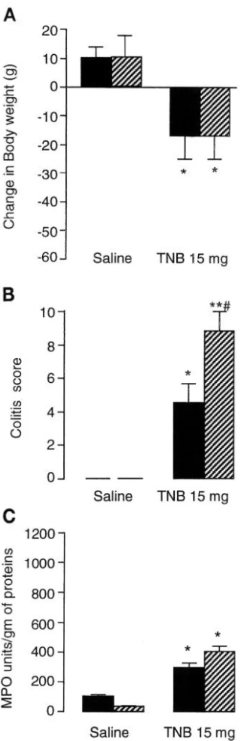
Induction of colitis in prestressed rats (protocol A).
Figure 2 shows the severity of colitis expressed as mean
G86 CRF, AVP, AND STRESS-ENHANCED COLITIS
Table 2. Criteria for histological scoring of colonic mucosa
Severity of Changes
Variables 0 1 2 3
Ulceration No ulcer Erosion or single ulceration not exceeding lamina mus- cularis mucosae
Mucous cell depletion Preserved mucous Mild depletion in a few cells cell
Mucosal atrophy Normal thickness Mild atrophy (> 10%) Edema (submucosa) Normal thickness Mild edema (submucosal
expansion > 10%) Inflammatory cell No inflammatory Mild inflammatory cell infil-
infiltration cell infiltration tration; few scattered cells Vascular dilatation Normal blood Mild dilatation of single
vessels blood vessels
Multifocal ulcerations not exceeding the submucosa Moderate depletion (~30% of
cells)
Moderate atrophy (1040%) Moderate edema (submuco- sal expansion, lo-100%) Moderate: distributed but
not dense inflammatory cell infiltration
Moderate: dilatation of sev- eral blood vessels
Ulcerations exceeding the submucosa
Severe depletion or complete disappearance of mucosa Severe atrophy (X0%) Severe edema (submucosal
expansion > 100%) Dense: inflammatory cell
infiltration
Severe: dilatation of several blood vessels
Modified from Ref. 7.
scores, and MPO activity in colonic tissue in non-
stressed rats and after chronic PRS applied during 4 consecutive days. Chronic PRS applied in healthy rats had no effect on the mean weight gain or colitic aspect,
since no change was detected macroscopically or histo-
Four days after TNB administration, rats exhibited a
logically, even though chronic PRS reduced by 66% the
drastic colitis characterized by severe hyperemia and
ulceration extending along the distal colon up to 2 cm.
The colitic damage scores reached a value of 5.6 2 0.8.
MPO activity compared with control (sham-stressed)
Adhesions between the affected portion of the colon and
other organs, usually the small intestine, were ob-
animals (35 2 3 vs. 104 * 8 U/g protein).
served in 39% of TNB-treated rats (Table 3). When they
existed, adhesions were invariably located very close to
a site of ulceration. Histological observations showed
mucosal extended ulceration and moderate-to-severe
mucin cell depletion in addition to mucosal atrophy and
edema (lo-30%), dilated vessels, and inflammatory
cell infiltration into the submucosa (Fig. 3, Table 3).
MPO activity was increased by approximately three
times for TNB-treated rats over that of saline controls
(293 IL 31 vs. 104 2 8 U/g protein). Colitis observed in
rats treated with TNB was paralleled by changes in
body weight. Rats in the TNB-treated group lost an
average of 21 g over a 4-day period.
With the chronic PRS applied during 4 consecutive
days before induction of colitis, administration of TNB
resulted in a significant increase of severity of colitis
over that of nonstressed rats, assessed both macroscopi-
tally, with the areas of ulceration extended up to 5-6
cm instead of 2 cm, and by the colonic damage score
(9.6 5 0.5 vs. 5.6 2 0.8); furthermore, the incidence of
adhesions was 84%. Histological examination of tissues
provided results complementary to the macroscopic
data (Fig. 3, Table 3). This consisted of mucosal ulcer-
ations that did not exceed the superior third of the
lamina propria. The edema was moderate, and the
mucosal atrophy was ~50%. The mucous cell depletion
was severe as was the vascular dilatation (Fig. 3). In all
rats, there was marked inflammation in the colon as
shown by the significant difference between sham-
stressed and prestressed rats submitted to colitis (Fig.
2, Table 3). In addition, the colonic tissue of these rats
exhibits a significant increase by 118% in MPO activity
compared with the TNB-treated group. Chronic PRS
Effect of chronic PRS applied after induction of colitis
applied after induction of colitis slightly reduced the
fprotocoZ B). Applied during 4 consecutive days after
induction of colitis, PRS increased the colitic damage
change in body weight, even though there was no
score in rats treated with TNB (8.8 2 1.2 vs. 4.6 2 1.4 in
nonstressed rats), which paralleled an increase in the
incidence of adhesions (66 vs. 36%). Furthermore, PRS
significant difference compared with the sham-stress
applied after TNB instillation increased MPO activity
by 38% (403 group (Fig. 2).
* 35 vs. 293 2 31 U/g protein; Fig. 4).
Histologically, there was mucosal ulceration with se-
vere mucous cell depletion, mucosal atrophy was ~70%,
and the edema with 100% of submucosal expansion
(Table 3). Ho wever, chronic PRS had no significant
effect on the changes in body weight that accompanied
the development of colitis (Fig. 4).
Central injection of a-helical CRF-(g-41) and AVP
antagonist. Intracerebroventricular injection of either
a-helical CRF-(9-41) or Aw antagonist performed
before each session of PRS had no effect per se on MPO
activity in saline-treated rats.
In rats injected with a-helical CRF-(9-41) or AVP
antagonist before each session of sham stress, the
severity of colitis was not significantly different com-
pared with the group treated intracerebroventricularly with saline, as assessed by the colitic damage score, the
histological score, the weight body change, and the
MPO activity (Fig. 5, Table 4).
In rats injected with a-helical CRF-(9-41) (5 ug
intracerebroventricularly) before each session of PRS,
the colitis was much more severe when measured 4
days after TNB (15 mg) instillation (Fig. 5). Indeed,
MPO activity significantly (P < 0.05) increased com-
pared with the PRS group (1,429 * 347 vs. 702 2 31 U/g
protein), as did the macroscopic score (9.8 2 1.1 vs.
5.9 2 0.8). At the same time, rats lost an average of 45 5 6 vs. 8 2 3 g in PRS (Fig. 5). In one of eight
CRF, AVP, AND STRESS-ENHANCED COLITIS G87
animals, cicatricial tissue was observed, whereas seven of eight rats presented ulcerations exceeding the submu- cosa, severe edema and mucosal atrophy, inflammatory cell infiltration in the submucosa, and severe dilatation of blood vessels (Fig. 6). Similarly, AW antagonist (5 pg/kg) centrally injected before the stress session signifi- cantly increased the MPO activity and the loss of weight in rats treated with TNB and increased the
-60
J
B
10 8C
1200 .$ 1000 5 ?i 800&
600T3
400.e
s
g
200=
0
Saline TNB 15 mg -- Saline TNB 15 mg Saline TNB 15 mgFig. 2. Body weight changes (‘A), colitic score (B ), and myeloperoxidase (MPO) activity CC) in sham-stressed (solid bars) and PRS (hatched bars) rats 4 days after saline or TNB (15 mg) instillation. PRS was applied before induction of colitis after protocol A. *P -C 0.05, sig- nificantly different from corresponding saline values. **P < 0.01, significantly different from corresponding saline values. #P c.O.05,
significantly different from corresponding sham-stress values. n = 8 rats/group.
Table 3. Histologically assessed damage and incidence
of adhesions induced by TNB: effects of PRS before
(protocol A) or after (protocol B) induction of colitis
with TNB
Treatment
Protocol A
Days Histological Incidence of After TM3 Damage Score Adhesions, % Sham stress + TNB PRS + TNB Protocol B 4 1.58 k 0.65 39 4 2.69 L! 0.47* a4* TNB + Sham stress 4 1.49 k 0.52 36 TNB + PRS 4 2.53 ? 0.51* 66* Histological damage scores are given as means 2 SE. TNB, 2,4,6-trinitrobenzenesulfonic acid; PRS, partial restraint stress. *P < 0.05, significantly different from corresponding TNB + sham-stress values (Newman-Keuls test).
severity of colitis assessed macroscopically (Table 4) and histologically (Fig. 6, Table 4).
DISCUSSION
Our findings show that PRS applied during 4 consecu- tive days considered as a chronic stress increases the severity of experimental colitis in rats. These changes were expressed by an increased colitic damage score, an aggravation of microscopic aspect, and increased granu- locyte recruitment assessed by MPO activity. Second, our present study shows that central injection of either CRF antagonist or AVP antagonist before PRS en- hanced the colitis already increased by stress. Our results reinforce the hypothesis that stress may be involved in exacerbation of colitis, at least in the experimental conditions described herein.
Intrarectal administration of TNB in ethanol results in acute inflammation, with ulcers, that evolves into chronic inflammation of the distal colon in rats. When TNB binds to tissue proteins, it elicits cell-mediated immune responses and induces an inflammation of the gut comparable to human Crohn’s disease. In particu- lar, the histologically observed infiltration of lympho- cytes and histiocytes is similar to that described for the human disease (19). Various inflammatory mediators such as PGE2, thromboxane As, prostacyclin, LTBd, LTC*, platelet-acivating factor (PAF), and interleukin (IL) may be involved in TNB colitis as in human IBD (25). Among various inflammatory mediators in TNB- induced colitis, IL-1 is considered to be the most significant indicator of mucosal inflammation because its level correlates with MPO activity (20).
Peripherally generated inflammatory mediators and immune cytokines derived from various inflammatory and immune cells activate the HPA axis at some or all of its levels, which include hypothalamic CRF neurons, pituitary corticotrophs, and the adrenal cortex (23). Included among these substances are mast cell-derived PAF, lymphocyte-derived y-interferon, IL-2, IL-6;mac- rophage-derived IL-l, tumor necrosis factor (TNF), and other interleukins. In addition, the CNS contains neu- ronal pathways and receptors for cytokines such as IL-l, especially in the hypothalamus, an area of the brain that is important in the mediation of acute-phase
G88 CRF, AVP, AND STRESS-ENHANCED COLITIS
Fig. 3. Light micrographs of colon corre- sponding to protocol A. Appearance of normal colon in control (A) and stressed rats (B): observe mild erosion of surface epithelium. Inflammation and injury of colonic mucosa 4 days after intracolonic instillation ofTNB (15 mg): in nonstressed rat (Cl, note infiltration of mucosa and submucosa by inflammatory cells; in stressed rat CD), mucosa is shrunken and partially necrotic.
response (4). It is not known whether peripheral IL-l
interacts with CNS IL-l, these two systems are indepen-
dent, or peripheral IL-1 crosses the blood-brain barrier
to exert its effect. Whatever the mechanism of action,
IL-1 stimulates the HPA axis and then the release of
glucocorticoids, which are anti-inflammatory and block
the production and action of several lymphokines, such
as IL-2 and y-interferon, as well as IL-1 production by
macrophages. In fact, there exists a feedback glucocorti-
coid-associated immunoregulatory system that may
exert a continuous surveillance of the immunological
state. However, when overstimulation of the pituitary-
adrenal axis by cytokines produced by immune-
inflammatory cells occurs, it leads to pathological states.
The principal effecters of the stress response include
the CRF and locus ceruleus-norepinephrine systems in
the CNS. CRF is widely distributed in many brain
regions, including the paraventricular nucleus of the
hypothalamus, brain stem, limbic system, and cortex.
CRF was initially isolated as the principal hypotha-
lamic stimulus to the pituitary-adrenal axis (30). CRF
is involved in behavioral and physiological responses to
stress. These responses include HPA axis activation,
sympathetic nervous system (SNS) activation, an-
orexia, and changes in motor activity (30).
Increased levels of CRF have been implicated in the
stress-induced suppression of immune function (10). In
CRF, AW, AND STRESS-ENHANCED COLITIS G89
during 4 consecutive days induces a decrease of MPO
activity in noninflamed rats. Because MPO level is
related to neutrophil infiltration (3), this result is in
accordance with the knowledge of stress-suppressive
A
Sham stress PRS
action on the immune system (12), even though studies
in adren alectomized rats have shown that this re-
sponse is independent of glucocorticoids (22). Al .though
the ess ential pathophysiology of stress-i nduced immu-
nosuppression -- has not been determined, there is consid-
erable evidence demonstrating that CRF is an essential
agent in stress-induced impairment of immune func-
g
--
27
2 -40 c .- % -50 2 3 -60 -70 -80 I3 1800 1600 c 1400 .- al 25 & 1200 -5 g) 1000 \ CA - .- c 800 i z 600 400 200 0 tion (13).Our study shows the well-recognized stress-induced
suppression of the immune system in intact rats but
UJ s -30 * 6 -40 Saline TNB 15 mg
#
P
Saline TNB 15 mgC
1200- ii .- al lOOO- I3 k 800- # 5 ** E 600- m 2 .- 400- c G 200- L 2 Sham stress PRSFig. 5. Effects of o-helical corticotropin-releasing factor (CRF)-( 9- 41) [5 ug intracerebroventricularly (icv); open bars] and arginine vasopressin (AW) antagonist (5 ug icv; hatched bars) injected before each session of sham stress or PRS on body weight (A) and MPO activity (B) 4 days after rectal instillation of TNB (15 mg). Solid bars, saline (10 ul icv). Rats were submitted to same stress procedure as in protocol A. *P < 0.05, significantly different from corresponding saline values. #P < 0.05, significantly different from corresponding sham stress values. n = 8 rats/group.
also provides evidence that chronic PRS has a proinflam-
matory effect in inflamed rats. Indeed, the MPO activ-
ity and the colitic damage score were higher in animals
stressed before TNB instillation and, to a lesser extent,
in rats stressed after induction of colitis. Cytokines
appear to play a role in the response to stress and may mediate the release of hormones from the HPAaxis. For instance, IL-6, which is not only produced by the cells in
immune tissues but also by the cells in neuroendocrine
Saline TNB 15 mg
Fig. 4. Effects of PRS applied from &zys 1 to 4 after induction of colitis on body weight changes (A), MPO activity (C), and colitic score (B) in sham-stressed (solid bars) and PRS (hatched bars) rats 4 days after saline or TNB (15 mg) instillation. These results correspond to protocol B. *P < 0.05, significantly different from corresponding saline values. **P < 0.01, significantly different from corresponding saline values. ##P < 0.05, significantly different from corresponding sham-stress values. n = 8 rats/group.
G90 CRF, AVP, AND
Table 4. Colitis score and histologically assessed
damage induced by TNB instillation: effects of
a-helical CRF-(g-14) and anti-AVP injected
before each session of sham stress or PRS
STRESS-ENHANCED COLITIS
Treatment Colitis Score Histological Score
Saline icv + sham stress + TNB 6.1 k 1.2 1.56 2 0.53 Saline icv + PRS + TNB 10.3 2 1.1+ 2.612 0.42” CRF-(g-14) + sham stress + TNB 5.9kO.9 1.42 ? 0.41 CRF-(g-14) + PRS + TNB 14.3 k 1.6*t 2.98 2 0.39y Anti-AVP + sham stress + TNB 6.2kO.8 1.52 2 0.52 Anti-AVP + PRS + TNB 15.12 1.4*t 2.8950.36V
Values are means k SE. icv, Intracerebroventricular; CRF-(g-141, d-helical corticotropin-releasing factor (5 pg icv); anti-AVP, arginine vasopressin antagonist (5 pg icv). *P < 0.05, significantly different from corresponding sham-stress values (Newman-Keuls test). +P < 0.05, significantly different from PRS + TNB values (Newman-Keuls test).
and endocrine tissues, such as the hypothalamus (241,
anterior pituitary (261, and adrenal cortex (111, is
increased after tissue injury, infection, and inflamma-
tion in which there is activation of the HPA axis and
SNS. However, in rats exposed to a mild psychological
stressor, blood IL-6 increased through a mechanism
independent of endotoxemia, tissue injury, or inflamma-
tion (141. Recently, Zhou et al. (32) reported that
physical or psychological stressors elevated plasma
levels of IL-6 in a manner resembling that of corticoste-
rone. Consequently, we can hypothesize that PRS stimu-
lates the release of IL-6, which in turn stimulates and
sensitizes the tissues that respond by an increase of
colitis after TNB instillation. Of course, this hypothesis
needs to be confirmed in further experiments.
When animals received central administration of
either CRF antagonist [o-helical CRF-(g-4111 or AVP
antagonist before each PRS session applied before
induction of colitis, these treatments enhanced the
level of inflammation already increased by stress.
Indeed, the level of MPO was much higher than that
observed in rats intracerebroventricularly injected with
vehicle. The central injection of either o-helical CRF-
(g-41) or AVP antagonist blocked the stress-induced
stimulation of HPA axis and therefore the subsequent
glucocorticoid release. Thus the colonic inflammation
related to TNB was exacerbated, as shown by increased
MPO activity. These results provide evidence that,
during a stress situation, CRF and AVP exert protective
effects against inflammation and avoid overinflamma-
tion.
With regard to body weight during colitis, we ob-
served that central administration of o-helical CRF-(9-
411 or AVP antagonist before a stress session increased
the loss in body weight, suggesting that CRF and AVP
prevented the worsening of inflammation-induced
weight loss. These observations reinforce the hypoth-
esis that, during stress, CRF and AVP act to minimize
the influence of general colitis, at least through the
stimulation of HPA axis. Indeed, the physiologically
relevant ACTH secretagogues are CRF and AVP (31).
AVP alone has a weak stimulatory effect on ACTH
secretion but strongly potentiates the ACTH-releasing
capacity of CRF (13) and seems to modify the inhibitory
effect of glucocorticoids on pituitary corticotroph cells (2).
Stress-induced alterations of gastrointestinal func-
tions such as intestinal (311 and colonic (8) motility are
directly linked to the central release of either CRF or
Fig. 6. Histology of TNB colitis tissue obtained from o-helical CRF-(g-41) and AVP antagonist-pretreated rats before each session of stress. A: colonic mucosa 4 days after induction of colitis in e-helical CRF-(9-4l&pretreated rat. B: colonic mucosa 4 days after induction of colitis in AVP antagonist-pretreated rat. Note in both cases invasion of whole colonic wall by inflammatory cells and necrosis of mucosa.
CRF, AW, AND STRESS-ENHANCED COLITIS G91
AW (16) but are not linked to the stimulation of the 13.
HPA axis (8, 23). Consequently, we could not reject the
hypothesis that CRF and AW may act on other path-
~~ *
ways than the HPA axis to decrease the severity of
colitis during stress. 15.
In conclusion, this study provides evidence that
stress may enhance experimental colitis; however, the
mediators and the mechanisms involved in such a re-
16 ’ sponse need to be determined.
17. The authors thank L. Ressayre for skillful technical assistance.
This work was presented in part at the 95th Annual Meeting of the American Gastroenterological Association, May 14-17, 1995, San
Diego, CA. 18.
Address for reprint requests: M. Gue, Dept. of Pharmacology, Institut National de la Recherche Agronomique, 180 Chemin de Tournefeuille, BP3,31931 Toulouse Cedex, France. 19. Received 10 April 1996; accepted in final form 30 August 1996.
REFERENCES 20. 1. 2. 3. 4. 5. 6. 7. 8. 9. 10. 11. 12.
Antoni, F. A. Hypothalamic control of adreno-corticotropin secretion: advances since the discovery of 41-residue corticotro- pin releasing factor. EndocrinoZ. Reu. 7: 351-381,1986.
Bilezikjian, L. M., A. L. Blount, and W. Vale. The cellular actions of vasopressin on corticotrophs of the anterior pituitary: resistance to glucocorticoid action. MoZ. EndocrinoZ. 1: 451-458,1987.
Bradley, P. P, D. A. Priebat, R. D. Christensen, and G. Rothstein. Measurement of cutaneous inflammation: estima- tion of neutrophil content with an enzyme marker. J. Inuest. Dermatol. 78: 206-209,1982.
Breder, C. D., C. A. Dinarello, and C. B. Saper. Interleukin-1 immunoreactive innervation of the human hypothalamus. Sci- ence Wash. DC 240: 321-324,1988.
B&no, L., M. Gue, and C. Del Rio. CNS vasopressin mediates emotional stress and CRH-induced colonic motor alterations in rats. Am. J. Physiol. 262 (Gastrointest. Liver Physiol. 25): G427-G431,1992.
Collins, S. M. Is the irritable gut an inflamed gut? Stand. J. Gastroenterol. 27, Suppl. 192: 102-105,1992.
Fabia, R., A. Ar’rajab, M. L. Johansson, R. Willen, R. Anderson, G. Molin, and S. Bengmark. The effect of exog- enous administration of LactobaciZZus reuteri R2LC and oat fiber on acetic acid-induced colitis in the rat. Stand. J. GastroenteroZ. 28: 155-162,1993.
Gue, M., J. L. Junien, and L. B&no. Mental stress in rats enhances colonic motility through the central release of corticotro- pin-releasing factor. GastroenteroZogy 100: 964-970, 1991. Hallgren, R., J. F. Colombel, R. Dahl, K. Fredens, A. Kruse, N. 0. Jacobsen, P. Venge, and J. C. Rambaud. Neutrophil and eosinophil involvement of the small bowel in patients with celiac disease and Crohn’s disease: studies on the secretion rate and immunohistochemical localization of granulocyte granule constituents. Am. J. Med. 86: 56-64, 1989.
Irwin, M. Stress-induced immune suppression: role of brain corticotropin releasing hormone and autonomic nervous system mechanisms. Adv. Neuroimmunol. 4: 29-47,1994.
Judd, A. M., and R. M. MacLeod. Adenocorticotropin increases interleukin-6 release from rat adrenal zona glomerulosa cells. EndocrinoZogy 130: 1245-1254,1992.
Keller, S. E., J. M. Weiss, S. J. Schleifer, N. E. Miller, and M. Stein. Suppression of immunity by stress: effect of a graded series of stressors on lymphocyte stimulation in the rat. Science Wash. DC 213: 1397-1400,198l. 21. 22. 23. 24. 25. 26. 27. 28. 29. 30. 31. 32.
Kort, W. J. The effect of chronic stress on the immune response. Adv. Neuroimmunol. 4: l-11,1994.
LeMay, L. G., A. J. Vander, and M. J. Kluger. The effects of psychological stress on plasma interleukin-6 activity in rats. Physiol. Behav. 47: 957-961,199O.
Levine, S., R. Strebel, E. Wenk, and P. Harman. Suppression of experimental allergic encephalomyelitis by stress. Proc. Sot. Exp. Biol. Med. 109: 294-298,1962.
Lowry, 0. H., N. J. Rosebrough, A. L. Farr, and R. J. Randall. Protein measurement with the Folin phenol reagent. J. Biol. Chem. 193: 265-275,195l.
MacDermott, R. P., and W. F. Stenson. Inflammatory bowel disease. In: Immunology and Immunopathology of the Liver and GastrointestestinaZ Duct, edited by S. R. Targan and F. Shana- han. Tokyo: Igaku-Shoin, 1990, p. 459-486.
McHugh, K., H. P. Weingarten, I. Khan, R. Riddell, and S. M. Collins. Stress-induced exacerbation of experimental colitis (Abstract). GastroenteroZogy 104: A803, 1993.
Morris, G. P., I? L. Beck, M. S. Herridge, W. Depew, M. R. Szewczuk, and J. L. Wallace. Hapten-induced model of chronic inflammation and ulceration in the rat colon. GastroenteroZogy 96: 795-803,1989.
Rachmilewitz, D., P. L. Simon, L. W. Schwartz, D. E. Griswold, J. D. Fondacaro, and M. A. Wasserman. Inflammatory mediators of experimental colitis in rats. Gastroen- terology 97: 326-337, 1989.
Rivier, C., and W. Vale. Modulation of stress-induced ACTH release by corticotropin releasing factor, catecholamines and vasopressin. Nature Lond. 305: 325-327,1983.
Saperstein, A., H. Brand, T. Audhya, D. Nabriski, B. Hutchinson, S. Rosenzweig, and C. S. Hollander. Interleu- kin ll3 mediates stress-induced immunosuppression via cortico- tropin-releasing factor. EndocrinoZogy 130: 152-158,1992.
Sapolsky, R., C. Rivier, G. Yamamoto, P. Plotsky, and W. Vale. Interleukin-1 stimulates the secretion of hypothalamic corticotropin releasing factor. Science Wash. DC 238: 522-524. 1987.
Schobitz, B., D. A. M. Voorhuis, and E. R. De Kloet. Localization of interleukin 6 mRNA and interleukin 6 receptor mRNAin rat brain. Neurosci. Lett. 136: 189-192,1992.
Sharon, P., and W. F. Stenson. Enhanced synthesis of leukot- riene Bd by colonic mucosa in inflammatory bowel disease. Gastroenterology 86: 453-460,1984.
Spangelo, B. L., R. M. MacLeod, and P. C. Isakson. Produc- tion of interleukin 6 by anterior pituitary cells in vitro. Endocri- nology 126: 582-586,199O.
Stein, M., R. C. Schiavi, and M. Camerino. Influence of brain and behavior on the immune system. Science Wash. DC 191: 435-440,1976.
Stein, T.A., L. Keegan, L. J. Auguste, B. Bailey, and L. Wise. Stress induced experimental colitis. Mediators InfZammation 2: 253-256,1993.
Tanner, A. R., M. J. P. Arthur, and R. Wright. Macrophage activation, chronic inflammation and gastrointestinal disease. Gut 25: 760-783,1984.
Vale, W., J. Spiess, C. Rivier, and J. Rivier. Characterization of a 41-residue ovine hypothalamic peptide that stimulates secretion of corticotropin and beta-endorphin. Science Wash. DC 213: 1394-1397,198l.
Williams, C. L., R. G. Villar, J. M. Peterson, and T. F. Burks. Corticotropin-releasing factor directly mediates colonic response to stress. Am. J. Physiol. 253 (Gastrointest. Liver Physiol. 16): G582-G586,1987.
Zhou, D., A. W. Kusnecov, M. R. Shurin, M. dePaoli, and B. S. Rabin. Exposure to physical and psychological stressors elevates plasma interleukin 6: relationship to the activation of hypothalamic-pituitary-adrenal axis. EndocrinoZogy 133: 2523- 2530.1993.


![Fig. 5. Effects of o-helical corticotropin-releasing factor (CRF)-( 9- 41) [5 ug intracerebroventricularly (icv); open bars] and arginine vasopressin (AW) antagonist (5 ug icv; hatched bars) injected before each session of sh](https://thumb-eu.123doks.com/thumbv2/123doknet/2351525.36385/7.867.489.768.112.819/effects-corticotropin-releasing-intracerebroventricularly-arginine-vasopressin-antagonist-injected.webp)

