Science Arts & Métiers (SAM)
is an open access repository that collects the work of Arts et Métiers Institute of
Technology researchers and makes it freely available over the web where possible.
This is an author-deposited version published in: https://sam.ensam.eu
Handle ID: .http://hdl.handle.net/10985/10201
To cite this version :
Denis AUBRY, M. GUPTA, B. LADOUX, Rachele ALLENA - Mechanical link between durotaxis, cell polarity and anisotropy during cell migration - Physical Biology - Vol. 12, p.026008 - 2015
Mechanical link between durotaxis, cell polarity and anisotropy
during cell migration
D Aubry1
, M Gupta2,3
, B Ladoux2,3
and R Allena4
1 Ecole Centrale Paris, Laboratoire MSSMat UMR CNRS 8579, Grande Voie des Vignes, 92295 Châtenay-Malabry, France 2 Mechanobiology Institute, Cell adhesion and Mechanics Laboratory, T-Lab, #09-01 5 A, Engineering Drive 1, Singapore 11741 3 Institut Jacques Monod, Cell adhesion and Mechanics Laboratory UMR CNRS 7592, 10 rue Hélène Brion, 75205 Paris Cedex 13, France 4 Arts et Metiers ParisTech, LBM, 151 Bd de l'hôpital, 75013 Paris, France
E-mail:[email protected] Keywords: durotaxis, polarity, anisotropy
Supplementary material for this article is availableonline
Abstract
Cell migration, a fundamental mechanobiological process, is highly sensitive to the biochemical and
mechanical properties of the environment. Efficient cell migration is ensured by the intrinsic polarity
of the cell, which triggers a transition from an isotropic to an anisotropic configuration of the
acto-mysion
filaments responsible for the protrusion–contraction movement of the cell. Additionally,
polarity may be highly influenced by the substrate rigidity, which results in a phenomenon called
durotaxis. In the present work, we propose a two-dimensional
finite element model able to capture
three main features of cell migration: durotaxis, cell polarity and anisotropy. The cell is modelled as a
continuum able to develop cyclic active strains regulated by the polymerization and depolymerization
of the acto-myosin
filaments and synchronized with the adhesion forces between the cell and the
substrate underneath. A generalized Maxwell model is used to describe the viscoelastic behaviour of
the cell constituted by a solid anisotropic branch with active strains (i.e. the acto-myosin
filaments)
and a
fluid viscoelastic branch (i.e. the cytoplasm). Several types of substrate have been tested which
are homogeneously soft or stiff or include both regions. The numerical results have been qualitatively
compared with experimental observations showing a good agreement and have allowed us to
find the
mechanical link between durotaxis, cell polarity and anisotropy.
1. Introduction
Cell migration plays a fundamental role during several biological phenomena and it is sensitive to both the biochemical and mechanical properties of the envir-onment. Actually, cells probe the stiffness of the extracellular matrix (ECM) by adhering to the sur-roundingfibres and pulling on them. Thus, ECM may be critical for many cellular functions such as adhesion [1], migration [2], differentiation [3] and polarization [4]. Among the potential candidates responsible for the cell mechanosensitivity, focal adhesion (FA) seems to be the most plausible due to the existing correlation between their surfaces and the exerted force inducing an elastic strain [5–8]. It has been shown that the ion calcium channels may be involved in the building up of the cellular tension in response to a mechanical signal [2, 9, 10]. Additionally, recent observations
[11, 12] as well as a mechanical model [13] have
suggested that the acto-myosin complexes can act as global sensors of rigidity [14]. Huge efforts are continuing to be made to understand such responses and uncover the molecular details of these biomecha-nical pathways [15]. However, the coupling between local and global scales of the mechanical signalling modules described above is far from being understood [16]. The difficulty lies in the complexity of the mechanosensitive feedback responses that occur at various time and length scales. The most challenging problem is to identify how all these signals are integrated in order to induce a global response at the cell scale.
From a physical point of view, it is still unclear whether the mechanosensitivity is regulated by the stresses generated by the cell or by strains undergone by the ECM. Nevertheless, it is evident that
determining the mechanical principles at the basis of the forces’ transmission between the cell and the ECM would allow to us explain the behavioural divergences during cellular activities within environments with different stiffness.
1.1. Cell polarity and durotaxis
Cell polarity or polarization is the ability of a cell to create and maintain an asymmetric distribution of intrinsic subdomains with distinct chemical, physical and mechanical properties. Cell polarity is relevant during migration and induces a transition from a symmetric and isotropic configuration (i.e. acto-myosinfilaments radially oriented) to an asymmetric and anisotropic configuration (i.e. acto-myosin fila-ments oriented in the direction of migration).
Mechanical properties of the ECM and more spe-cifically its rigidity may induce cell polarization [4,17– 19]. This results in a phenomenon called durotaxis which consists of the orientation of the acto-myosin filaments along the stiffness gradient of the ECM or along the stressfields generated by neighbour cells in order to reduce the elastic energy [4,20,21]. In fact, the acto-myosinfilaments tend to adapt to the ECM rigidity and to develop higher traction forces on stiffer substrates [12,22]. Such adaptation, coupled with the FA sensitivity, might explain how the ECM stiffness triggers cell polarization and migration.
During the last few years, several analytical and numerical works have been proposed in the literature to investigate durotaxis during cell migration [16,23].
Moreo et al [24] proposed an extension of the Hill’s model for skeletal muscle behaviour to investi-gate cell mechanosensing, migration and prolifera-tion. Their results allow us to predict the cell response on elastic substrates and under different loading con-ditions. Dokukina and Gracheva [25] developed a 2D discrete model of a viscoelasticfibroblast cell using a Delaunay triangulation. At each node the balance of the forces is calculated as the contribution of the fric-tional force between the cell and the substrate, the pas-sive viscoelastic force and the active force. The authors have evaluated the cell behaviour over a substrate with a rigidity step and their results are in agreement with specific experimental observations. In fact, they found that the cell (i) preferentially moves on stiffer substrate and (ii) turns away from the soft substrate when it approaches it as reported by [2].
In Harland et al [26] the cell is a collection of stress fibres undergoing contraction and the birth/death
process. The formation of new fibres, whose rate
depends on the substrate stiffness, is stochastic and centred at the cell centre of mass. The model shows that cells for which the adhesions slide more slowly and stressfibres form readily on stiff substrates also exhibit durotaxis. Stefanoni et al [27] employed Lan-gevin equations to take into account the local mechan-ical properties of the substrate underneath and analyze
two distinct configurations for an isotropic and a biphasic substrate. Trichet et al [18] introduced a phe-nomenological model based on active gel theory showing that cells preferentially migrate over stiff sub-strates andfind an optimal range of rigidity leading to efficient migration. Finally, in Allena and Aubry [28] a 2D mechanical model is proposed to simulate cell migration over an heterogeneous substrate. The cell is able to adopt two different strategies (i.e. ‘run-and-tumble’ and ‘look-and-run’ strategies) to avoid the soft regions inhibiting the adhesion.
1.2. Objective of the present work
In the present paper, we propose a 2Dfinite element work to simulate single cell migration over substrate with different rigidities. To do so, the model is based on the following assumptions:
(1)To take into account the durotaxis phenomenon, the underneath substrate is represented as a square which may be homogenously soft or stiff or include both types of regions. Additionally, a viscous force which inhibits the cell progression is associated to the soft domains;
(2)As in previous works [28,29], the cell is modelled as a continuum, with initial circular shape. It is able to develop radial and cyclic active strains of protrusion and contraction, which are assumed to be regulated by the polymerization and depolymer-ization processes of the actin filaments, respec-tively. Such strains are synchronized with the adhesion forces between the cell and the substrate and exerted over the frontal and rear adhesion surfaces in the direction of migration;
(3)The direction of migration is triggered by an external attractive source [28];
(4)A generalized viscoelastic Maxwell model has been used to describe the mechanical behaviour of the cell and it includes a viscoelastic (i.e. the cyto-plasm) and an anisotropic elastic (i.e. the actin filaments) branch.
The main objective of the work is to highlight the mechanical link between durotaxis and cell aniso-tropy and polarity and the influence of one of these aspect over the other two. The paper is organized as follows. First, the geometrical description of both the cell and the substrate is proposed. Second, the mechanical framework, the constitutive model and the active strain implementation are described. Finally, the results are presented. The cell efficiency has been evaluated in terms of covered distance, migration speed and mechanical stresses and the out-comes have been qualitatively compared to experi-mental observations.
2. Material and methods
2.1. Stiff and soft substrateThe underneath substrate is represented by a square. Both stiff (Ωstiff)and soft (Ωsoft)regions are described
by two characteristic functions and two configurations have been tested. First, both the stiff (hstiff,r) and the soft (hsoft,r) regions are considered as rectangles separated by a sharp frontier. Therefore, the domains are defined as = < = > h x x h x x 1 if 0 otherwise 1 if 0 otherwise (1) r t r stiff, 0 sof , 0 ⎧ ⎨ ⎩ ⎧ ⎨ ⎩
where x is the horizontal coordinate of any particle and x0is a constant value.
Second, a mapped substrate constituted byfive cir-cular stiff regions surrounded by a soft domain has been obtained and the associated characteristic func-tions read
∑
= − + − − = −(
)
(
)
h x x y y r h h 1 if 0 otherwise 1 (2) c i i c c stiff, stiff, 2 stiff, 2 st2 soft, stiff, ⎪ ⎪ ⎧ ⎨ ⎩where y,xstiff,i, ystiff,i and rstiff are respectively the
vertical coordinate of any particle and the spatial coordinates of the centres and the radius of the circular regions.
According to the substrate stiffness, a viscous force
fsubstrateapplies and reads
μ Ω Ω = − f v on 0 on (3) substrate substrate soft stiff ⎧ ⎨ ⎩
where μsubstrate and v are the substrate friction coefficient and the cell velocity, respectively.
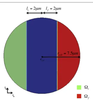
2.2. Cell geometry
We consider a cell with initial shape approximated by a two-dimensional (2D) circle Ωcell of radius rcell
(figure1). The cell is equipped with frontal (Ωf)and
rear (Ω )r adhesion regions, which allow the adhesion
between the cell and the substrate underneath [28,30] (figure 1(b), section A.1) and are developed in the direction of migration d. Both regions are described through two characteristic functions as follows
= − > = − < −
(
)
(
)
p p c d p p c d h l h l ( ) 1 , 0 otherwise ( ) 1 , 0 otherwise (4) f f r r cell cell ⎪ ⎪ ⎪ ⎪ ⎧ ⎨ ⎩ ⎧ ⎨ ⎩with d= cos ( )iθ x + sin ( )iθ y the direction of
migration as function of the angle θ, lf and lr the distances ofccellfrom the boundaries of Ωf and Ωr
respectively (figure1), p= x−u the initial position
of any particle, where x and u are respectively the actual position and the displacement.
2.3. Mechanics and constitutive behaviour of the cell Letρ be the cell density, a the acceleration, σ the Cauchy stress,F the deformation gradient and J its determinant, then conservation of momentum with respect to the initial configuration in the coordinates systemp is given by
σ
ρ =a Div
(
J F−)
+f +f (5)p T adh substrate
where fadh indicates the viscous adhesion forces
between the cell and the substrate (section2.3). Here, all the body forces but the inertial effects are neglected. In fact, it has been shown that they may play a significant role during the rapid protrusion phase
[31,32]. Additionally, from a numerical point of view,
taking into account small accelerations improves the convergence performances. The cell is constituted of two main phases: a solid (i.e. the actinfilaments) and a fluid (i.e. the cytoplasm) phase. Then, the actin filaments are considered rather elastic, while the cytoplasm shows a viscoelastic behaviour. Addition-ally, we assume that the polymerization of the actin filaments, which occurs at the frontal edge of the cell [33], is responsible of the cell protrusion, whereas their depolymerization generates the contractile stress at the rear of the cell [34]. Nevertheless, the polymer-ization of the actinfilaments only occurs when the cell is able to adhere to the underneath substrate, whereas in the absence of adhesion the cell pulses in place [28]. In the former case, the cell acquires an elongated shape in the direction of migration d,which becomes the principal axis of anisotropy. In the latter case instead, the cell radially expands and contracts in an isotro-pic way.
Figure 1. Geometry of the cell with the frontal (red) and the rear (green) adhesion regions.
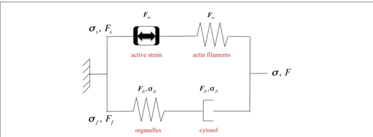
As in previous works [28,30], we use a generalized Maxwell model to describe the global mechanical behaviour of the cell (figure2). Consequently, the total Cauchy’s stressσis equal to
σ=σs+σf (6)
withσsand σfthe solid and thefluid Cauchy’s stresses,
respectively. The transformation gradientF is the same in the solid and thefluid branch, so that we can write
= + = =
F D up I Fs Ff (7)
with = ∑ = ∂ ⊗
∂ D up m3 1 pu i ,m
m u the displacement and I
the identity matrix [35,36].
In the solid phase (i.e. the actin filaments),
σse=σsawith se and sa standing for solid elastic and
solid active respectively. Thus, we have
σ = F S F J 1 (8) se se se se se T
where Jseis the determinant of solid elastic deforma-tion tensorFse,Sseis the second Piola–Kirchoff solid elastic stress tensor calculated in the global system of coordinates, which is computed as an anisotropic hyperelastic Saint-Venant material as follows
=
(
)
Sse RCloc R E R RT se T (9) where R is the classical rotation matrix leading to
=
ploc Rp, with ploc the initial position in the local orthonormal system of coordinates
(
iθ,iα)
. For thesake of clarity, we provide here the expression of the inverse of the local elastic tensorC−
loc1as
∑
= ⊗ ⊗ ⊗ + × ⊗ ⊗ ⊗ θ α θ α − = =(
) (
)
(
) (
)
C A i i i i B i i i i (10) m n mn m m n n mn m n m n loc1 , ,with Amn and Bmn two symmetric matrices whose
components are ν ν = = − = − = = = = = θθ θ θα θα θ αθ αθ α αα α θθ αα θα αθ θα A E A E A E A E B B B B G 1 1 0 (11) with νθαand ναθthe Poisson ratios andGθαthe shear
modulus of the cell in the local coordinates system. The Young moduliEθandE are deα fined as follows
Ω Ω = = α θ α θ E E E E on 0.1 on (12) soft stiff
The Green–Lagrange solid elastic strain tensorEse
is expressed as =
(
−)
E 1 F F I 2 (13) se seT se whereFseis given by = − Fse F Fs sa1 (14)with Fs and Fsa being respectively the total solid and
the solid active deformation tensors.Fseis triggered by the interaction between the cell and the underneath substrate, whereasFsadescribes the cyclic and active pulsatile movement of the cell and is defined in the next section. Here, we have chosen the active strain approach since it appears to be more robust from a mathematical point of view than the active stress one [37]. Additionally, its physiological relevance has already been shown in several biological context
[28,38–41].
In thefluid phase (i.e. the cytoplasm), the defor-mation gradientFfis also multiplicatively decomposed as
=
Ff F Ffe fv (15)
Figure 2. Scheme of the generalized Maxwell model used to describe the viscoelastic behaviour of the cell. The solid branch is constituted by the active strains and the anisotropic elastic actinfilaments. The fluid branch, the cytoplasm, is constituted by the elastic organelles embedded in the viscous cytosol.
where fe and fv stand for fluid elastic and fluid viscoelastic respectively.
The Cauchy’s stress σfreads
σf =2μDfv (16)
with μ the viscosity of the cytoplasm and Df v, the
eulerian strain rate computed from the strain gradient velocity as follows = − + − D F F F F 2 fv fv fv fvT fv (17) T 1
2.4. Active strains and intra-synchronization To describe the oscillating movement of the cell, some assumptions have been made.
(1)The cell is able to develop radial active strains of protrusion and contraction;
(2)As soon as the cell is able to adhere to the underneath substrate, a lamellipodium is formed at the leading edge and in the direction of migra-tion d.
Therefore, the solid active deformation tensorFsa reads π π π π = ⊗ > ⊗ < F i i i i e t T t T e t T t T sin 2 if sin 2 0 2 sin 2 if sin 2 0 (18) sa a r r a r r 0 0 ⎜ ⎟ ⎜ ⎟ ⎜ ⎟ ⎜ ⎟ ⎧ ⎨ ⎪⎪ ⎩ ⎪ ⎪ ⎛ ⎝ ⎞⎠ ⎛⎝ ⎞⎠ ⎛ ⎝ ⎞⎠ ⎛⎝ ⎞⎠
whereea0is the amplitude of the active strain, t is time, T is the migration period, ⊗indicates the tensorial
product andiris defined as
φ φ
= +
ir cos ( )ix sin ( )iy (19)
where φ =arctg
( )
yx .
As numerically shown [28], in order to be able to effectively migrate, the cell must adhere on the sub-strate; otherwise, it would only deform in place. Thus, an intra-synchronization is required which coordi-nates the cyclic protrusion–contraction deformations with the adhesion forcesfadh(equation (5)) generated between the cell frontal and rear adhesion surfaces and the underneath substrate in the direction of migration
d. As in previous works [28,30,42,43], such forces are assumed to be viscous and may be distinguished into a frontal (fadh,f) and a rear (fadh,r) force as follows
μ Ω μ Ω = − −∂ ∂ = − ∂ ∂ f n F v f n F v h t h t ( ) on ( ) on (20) f sa f r sa r adh, cell adh sync
adh, cell adh sync
⎜ ⎟ ⎜ ⎟ ⎛ ⎝ ⎞ ⎠ ⎛ ⎝ ⎞ ⎠
withncellthe outward normal to the cell boundary,
μadhthe friction coefficient and v the cell velocity. The characteristic function hsyncis the key ingredient of the
preceding equations since it couples the adhesion forces with the active strains, which results in the
intra-synchronization mentioned above. Thus, we observe two main phases during the migratory move-ment of the cell: (i) the protrusion and the adhesion at the rear edge, and (ii) the contraction and the adhesion at the frontal edge.
3. Results and discussion
Simulations have been run using Comsol Multiphysics 3.5 a. At the initial time point, the cell has a circular shape centred inccell(0, 0) with radius rcell equal to 7.5μm (figure1). The cell Young modulusEθis equal
to 104Pa [44], whereasEαruns from 103Pa to 104Pa
over stiff and soft substrates respectively. The Pois-son’s ratios νθαand ναθhave been set to 0.3 and the
shear modulus Gθα varies from 384 Pa to 3846 Pa
respectively over soft and stiff substrates. The cell density ρ has been set to 1000 kg m−3 [45] and the viscous friction coefficient μadhis equal 108Pa-s/m. Finally, the intensity of the active strain ea0 and the
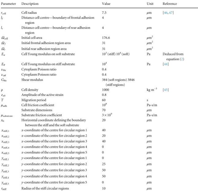
migration period T have been chosen equal to 0.2 and 60 s, respectively. The underneath substrate has a square shape with dimensions 70μm × 70 μm and is centred in (−15 μm, −15 μm). All the geometrical and mechanical parameters of the model have been reported in table1.
3.1. Soft versus stiff substrates
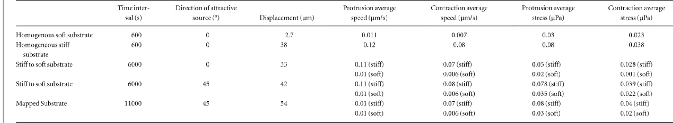
For thefirst set of simulations we want to analyze the behaviour of the cell over homogeneous soft and stiff substrates, respectively, in order to point out the main differences between the two. Thus, we have set μsubstrate= 3 × 10⁹Pa-s/m and an external attractant source is introduced atθ =0.The simulations cover a period of 600 s. The main quantitative results have been reported in table2.
For the soft substrate, the cell is not able to adhere and pulses on place by protruding and contracting radially sinceEα=E and an isotropic behaviour isθ
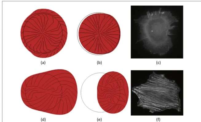
observed (equation (12)) (available atstacks.iop.org/ PB/12/026008/mmedia/movie 1). As the processes of polymerization and depolymerization of the actin fila-ments are responsible for the polarization of the cell, it is interesting to analyze the arrangement of the streamlines of the principal stresses, which provide an accurate picture of the load transfer inside the cell dur-ing migration and of the ability of the cell to maintain the necessary asymmetric distribution of thefilaments to move forward (available at stacks.iop.org/PB/12/
026008/mmedia/movie 1). In the specific case of a
homogeneous soft substrate, the streamlines are
radially oriented during both the protrusion
(figure 3(a)) and contraction (figure 3(b)) phases, which reflects the absence of polarization leading to the inefficient pulsatile movement of the cell in place. The same distribution may be observed for the actin filaments in a rat embryonic fibroblast (REF52), which scarcely migrate over a soft substrate (figure 3(c)). 5
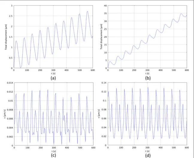
From a quantitative point of view, the average stresses developed inside the cell are equal to 0.03μPa and 0.023μPa respectively during protrusion and
contrac-tion. Thus, the cell only migrates over 2.7μm
(figure4(a)) and this is mostly due to the inertial and viscous effects rather than to the active strain–adhe-sion forces machinery. The average velocity of the cell
centre of inertia is equal to 0.011μm s−1 and
0.007μm s−1 during the protrusion and contraction phases respectively (figure4(c)).
For the stiff substrate, no additional viscous force is applied and the cell is able to develop normal adhe-sion forces. Furthermore, according to equation (12), the mechanical behaviour is anisotropic which leads to an asymmetric strain in the direction of migration. During the protrusion phase, the streamlines of the principal stresses are still radially oriented, but more elongated at the leading edge in the direction of migra-tion (θ = 0) so that the cell is able to polarize and to efficiently migrate (figure3(d), available atstacks.iop.
org/PB/12/026008/mmedia/movie 2). A similar
arrangement is found for a REF52 migrating over a
stiff substrate. In this case the actin filaments are oriented straight in the direction of migration, show-ing a very sharp polarity (figure3(f)). During the con-traction phase, the stresses are rather mixed up, but it is possible to observe a contraction at the rear edge, which allows the cell to pull its body forward (figure3(e)). Then, the cell migrates over the substrate for 38μm (figure4(b)) with an average speed of about 0.12μm s−1and 0.08μm s−1respectively during pro-trusion and contraction (figure 4(d)) and generates higher average stresses compared to the previous case (0.08μPa and 0.038 μPa during protrusion and con-traction, respectively).
3.2. From stiff to soft substrate
For the second series of simulations we have consid-ered a substrate made of both a stiff and a soft region. Three simulations have been run as described in the following.
First, we have kept attractive source at θ = 0
(available at stacks.iop.org/PB/12/026008/mmedia/ movie 3) and the boundary between the stiff and the
Table 1. Main geometrical and mechanical parameters of the model.
Parameter Description Value Unit Reference
rcell Cell radius 7.5 μm [46,47]
lf Distance cell centre—boundary of frontal adhesion
region
4 μm
lr Distance cell centre—boundary of rear adhesion
region
4 μm
Ωcell Initial cell area 176.6 μm2
Ωf Initial frontal adhesion region area 31 μm2
Ωr Initial rear adhesion region area 31 μm2 α
E Cell Young modulus on soft substrate 103(stiff) 104(soft) Pa Deduced from
equation (2)
θ
E Cell Young modulus on stiff substrate 104 Pa [44]
νθα Cytoplasm Poisson ratio 0.4
ναθ Cytoplasm Poisson ratio 0.4 θα
G Shear modulus 384 (soft regions) 3846 (stiff regions)
ρ Cell density 1000 kg m−3 [45]
ea0 Amplitude of the active strain 0.8
T Migration period 60 s
μadh Cell friction coefficient 108 Pa-s/m
Substrate dimensions 70 μm
μsubstrate Substrate friction coefficient 3 × 10⁹ Pa-s/m
x0 Horizontal coordinate defining the boundary
between the stiff and the soft substrate
20 μm
xstiff,1 x-coordinate of the centre for circular region 1 40 μm
xstiff,2 x-coordinate of the centre for circular region 2 20 μm
xstiff,3 x-coordinate of the centre for circular region 3 40 μm
xstiff,4 x-coordinate of the centre for circular region 4 0 μm
xstiff,5 x-coordinate of the centre for circular region 5 0 μm
ystiff,1 y-coordinate of the centre for circular region 1 0 μm
ystiff,2 y-coordinate of the centre for circular region 2 25 μm
ystiff,3 y-coordinate of the centre for circular region 3 50 μm
ystiff,4 y-coordinate of the centre for circular region 4 50 μm
ystiff,5 y-coordinate of the centre for circular region 5 0 μm
Table 2. Quantitative results for the numerical simulations. Time inter-val (s) Direction of attractive source (°) Displacement (μm) Protrusion average speed (μm/s) Contraction average speed (μm/s) Protrusion average stress (μPa) Contraction average stress (μPa)
Homogenous soft substrate 600 0 2.7 0.011 0.007 0.03 0.023
Homogeneous stiff substrate
600 0 38 0.12 0.08 0.08 0.038
Stiff to soft substrate 6000 0 33 0.11 (stiff) 0.07 (stiff) 0.05 (stiff) 0.028 (stiff)
0.01 (soft) 0.006 (soft) 0.02 (soft) 0.001 (soft)
Stiff to soft substrate 6000 45 42 0.11 (stiff) 0.08 (stiff) 0.078 (stiff) 0.039 (stiff)
0.01 (soft) 0.006 (soft) 0.035 (soft) 0.022 (soft)
Mapped Substrate 11000 45 54 0.01 (stiff) 0.07 (stiff) 0.08 (stiff) 0.04 (stiff)
0.01 (soft) 0.006 (soft) 0.03 (soft) 0.02 (soft)
7 Phys. Biol. 12 (2015) 026008 D Aubry et al
stiff region has been obtained via equation (1) for which x0has beenfixed equal to 20 μm. In this case,
the cell migrates over 33μm during 6000 s
(figure5(a)), but a plateau is observed as soon as the cell comes into contact with the soft substrate. In fact, the average speed of the cell centre of migration
switches from a value of around 0.08μm s−1 to
0.008μm s−1around t = 245 s (figure5(d)). In
avail-able at stacks.iop.org/PB/12/026008/mmedia/movie
3, it is possible to notice such a slowing down which also coincides with a reorganization of the principal stresses. In fact, over the stiff substrate, the streamlines of the principal stresses are radially oriented but elon-gated in the direction of migration leading to the polarization of the cell in the direction of migration, whereas over the soft substrate they are isotropically and radially arranged. Then, the cell efficiently moves over the stiff substrate since it is able to synchronize the active strains of protrusion and contraction with the adhesion forces, while it mainly pulses on place over the soft substrate.
Second, the boundary between the stiff and the soft substrates has been kept the same, but the external source has beenfixed at θ = 45° (available atstacks.iop. org/PB/12/026008/mmedia/movie 4). Here, the cell
covers a total distance of 42μm over 6000 s and once again it is possible to notice both a plateau for the total displacement (figure5(b)) and a change in the migra-tion velocity as the cell reaches the soft substrate (t = 375 s) (figure 5(e)). In fact, the average speed decreases from 0.12μm s−1 to 0.006μm s−1. One might wonder why the cell does not avoid contact with the soft substrate and look for an alternative path to reach the external source at 45° as it has been observed, for instance, in [2] where the cell moves along the boundary instead of crossing from the stiff to the soft substrate. Actually, in the present model the direction of migration isfixed and the cell does not have any notion of rotation as we proposed in [28] where it was able to detect an obstacle and change its orientation to circumvent it. Consequently, a change in direction may only occur if the balance of the momentum pro-duces a rotation. This is the case here where the rear region of the cell is still on the stiff domain where the adhesion is higher, whereas the frontal edge is already on the soft domain. Thus, as the cell comes into con-tact with the soft region, it starts to slip and rotate so that the original direction of migration towards the external signal at θ = 45° is not maintained, but it becomes almost equal to 0°.
Figure 3. (a), (b) Displacement of the front of the cell over a soft (a) and a stiff (b) substrate. (c), (d) Velocity of the cell centre of inertia over a soft (c) and a stiff (d) substrate.
Third, the attractant is still place atθ = 45°, but this time the substrate is made of circular stiff regions sur-rounded by soft regions (equation (2)), which leads to
a mapped substrate (available atstacks.iop.org/PB/12/
026008/mmedia/movie 5). The cell covers 54μm over
11 000 s and two plateaux are observed corresponding
Figure 4. Comparison between the numerical results and the experimental observations. (a), (b) Snapshot of the numerical simulation for the migration over a soft substrate at t = 15 s (protrusion phase) and t = 45 s (contraction phase) (red = cell domain, black lines = principal stresses). (c) Snapshot of the actin cytoskeleton of a rat embryonicfibroblast (REF52) migrating over a soft substrate (staining for actinfilaments). (d), (e) Snapshot of the numerical simulation for the migration over a stiff substrate at t = 15 s (protrusion phase) and t = 45 s (contraction phase) (red = cell domain, black lines = principal stresses). (f) Snapshot the actin cytoskeleton of a rat embryonicfibroblast (REF52) migrating over a stiff substrate (staining for actin filaments).
35 30 25 20 15 10 5 0 45 40 25 20 15 10 5 0 0 1000 2000 3000 4000 5000 6000 0 1000 2000 3000 4000 5000 6000 Total displacement [µm] v [µm/s] v [µm/s] v [µm/s]
Total displacement [µm] Total displacement [µm]
35 30 70 60 50 40 30 20 10 0 0 0.1 0.2 0.3 0.4 0.5 0.6 0.7 0.8 0.9 1 1.1 t [s] t [s] t [s] t [s] t [s] t [s] x104 x104 0.14 0.12 0.1 0.08 0.06 0.04 0.02 0 0.14 0.12 0.1 0.08 0.06 0.04 0.02 0 0 1000 2000 3000 4000 5000 6000 0 1000 2000 3000 4000 5000 6000 0.12 0.1 0.08 0.06 0.04 0.02 0 0 0.1 0.2 0.3 0.4 0.5 0.6 0.7 0.8 0.9 1 1.1
Figure 5. (a)–(c) Displacement of the front of the cell over a ‘stiff to soft’ substrate with direction of migration θ is equal to 0 (a) and to 45° (b) and over a mapped substrate (c). (d)–(f) Average velocity of the cell centre of inertia over a ‘stiff to soft’ substrate with direction of migrationθ is equal to 0 (d) and to 45° (e) and over a mapped substrate (f) (red dashed line: transit from stiff to soft substrate; green dashed line: transit from soft to stiff substrate).
to the passage of the cell over the soft regions (t = 120 s: 2190 s and t = 3775 s: 7500 s) (figure5(c)). Similarly, the average speed of the cell centre of inertia is highest over the stiff regions (0.1μm s−1) and lowest over the soft ones (0.007μm s−1) (figure5(f)).
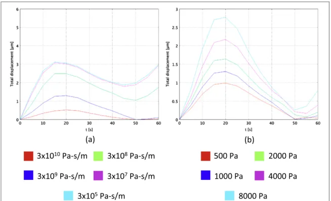
3.3. Influence of the substrate stiffness and anisotropic Young modulus
For the last series of simulations, we have considered a homogenous soft substrate with an external source placed atθ =0and we have let independently vary the
friction coefficient of the substrate μsubstrate
(equation (3)) and the Young modulus Eα
(equation (12)). Then, we have evaluated the total displacement of the frontal edge of the cell over afirst migration period (60 s) (figures 6(a) and (b)). On one hand, asμsubstratedecreases, the displacement increases since the cell is less inhibited by the additional viscous force exerted by the underneath substrate (figure 6 (a)). For a value of 3 × 10⁵Pa-s/m (light blue line in figure6(a)), the cell covers approximately 3μm during thefirst protrusion phase (from 0 to 15 s), which is close to the value found for the migration over a
homogeneous stiff region of about 4μm (see
section3.1). On the other hand, as E increases, theα
total displacement increases too (figure6(b)). In fact, the higher E ,α the less the cell shows an anisotropic
behaviour, which leads to a larger elongation in the direction of the attractant source θ =0.We found a minimal value of 1μm for Eα=500 Pa (red line in
figure 6(b)) and a maximal value of 2.7μm for
=
α
E 8000 Pa(light blue line infigure6(b)).
4. Conclusions
We have proposed a 2Dfinite element model of cell migration over flat substrates including three main aspects of the process that are (i) durotaxis, (ii) cell polarity and (iii) cell anisotropy. The cell has been modelled as a continuum and a generalized Maxwell model with anisotropic elastic branch has been employed to describe the viscoelastic behaviour of the system. The cell is able to synchronize the active strains of protrusion and contraction with the adhesion forces with the underneath substrate, which is represented as a 2D square and may include both stiff and soft regions. The latter trigger a further viscous force inhibiting the cell progression. First, we have analyzed the cell behaviour over homogenous stiff and soft regions and we have observed a clear difference in terms of efficiency. In fact, over the soft substrate the cell is not able to adhere and is almost stuck in place, whereas over the stiff region it is able to normally migrate. The numerical results have also been qualita-tively compared to specific experimental images. Second, we have tested three different configurations: (i) a stiff-to-soft region with sharp boundary and external source placed at 0°, (ii) a stiff-to-soft region with sharp boundary and attractive source placed at 45° and (iii) circular stiff regions surrounded by a soft matrix and external source placed at 45°. We have quantitatively evaluated the total cover distance, the migration velocity, and the average stress inside the cell. Finally, we have investigated the influence of both the substrate stiffness and the anisotropic Young modulus on the cell efficiency.
Figure 6. Influence of the friction coefficient of the substrate μsubstrate(a) and of the Young modulus Eα(b) on the cell total
References
[1] Giannone G, Dubin-Thaler B J, Döbereiner H-G, Kieffer N, Bresnick A R and Sheetz M P 2004 Periodic lamellipodial contractions correlate with rearward actin waves Cell116 431–43
[2] Lo C M, Wang H B, Dembo M and Wang Y L 2000 Cell movement is guided by the rigidity of the substrate Biophys. J. 79 144–52
[3] Engler A J, Sen S, Sweeney H L and Discher D E 2006 Matrix elasticity directs stem cell lineage specification Cell126 677–89 [4] Bischofs I B and Schwarz U S 2003 Cell organization in soft
media due to active mechanosensing PNAS100 9274–9 [5] Balaban N Q, Schwarz U S, Riveline D, Goichberg P, Tzur G,
Sabanay I, Mahalu D, Safran S, Bershadsky A, Addadi L and Geiger B 2001 Force and focal adhesion assembly: a close relationship studied using elastic micropatterned substrates Nat. Cell Biol.3 466–72
[6] Tan J L, Tien J, Pirone D M, Gray D S, Bhadriraju K and Chen C S 2003 Cells lying on a bed of microneedles: an approach to isolate mechanical force Proc. Natl Acad. Sci. USA 100 1484–9
[7] Nicolas A, Geiger B and Safran S A 2004 Cell
mechanosensitivity controls the anisotropy of focal adhesions PNAS101 12520–5
[8] Shemesh T, Geiger B, Bershadsky A D and Kozlov M M 2005 Focal adhesions as mechanosensors: a physical mechanism PNAS102 12383–8
[9] Glogauer M, Arora P, Yao G, Sokholov I, Ferrier J and McCulloch C A 1997 Calcium ions and tyrosine
phosphorylation interact coordinately with actin to regulate cytoprotective responses to stretching J. Cell. Sci. 110 11–21 (Pt 1)
[10] Hayakawa K, Tatsumi H and Sokabe M 2008 Actin stressfibers transmit and focus force to activate mechanosensitive channels J. Cell. Sci.121 496–503
[11] Mitrossilis D, Fouchard J, Guiroy A, Desprat N, Rodriguez N, Fabry B and Asnacios A 2009 Single-cell response to stiffness exhibits muscle-like behavior PNAS106 18243–8
[12] Lam W A, Chaudhuri O, Crow A, Webster K D, Li T-D, Kita A, Huang J and Fletcher D A 2011 Mechanics and contraction dynamics of single platelets and implications for clot stiffening Nat. Mater.10 61–6
[13] Walcott S and Sun S X 2010 A mechanical model of actin stress fiber formation and substrate elasticity sensing in adherent cells Proc. Natl Acad. Sci. USA107 7757–62
[14] Kobayashi T and Sokabe M 2010 Sensing substrate rigidity by mechanosensitive ion channels with stressfibers and focal adhesions Curr. Opin. Cell Biol.22 669–76
[15] Iskratsch T, Wolfenson H and Sheetz M P 2014 Appreciating force and shape—The rise of mechanotransduction in cell biology Nat. Rev. Mol. Cell Biol.15 825–33
[16] Ladoux B and Nicolas A 2012 Physically based principles of cell adhesion mechanosensitivity in tissues Rep. Prog. Phys.75 116601 [17] Saez A, Ghibaudo M, Buguin A, Silberzan P and Ladoux B
2007 Rigidity-driven growth and migration of epithelial cells on microstructured anisotropic substrates PNAS104 8281–6 [18] Trichet L, Digabel J L, Hawkins R J, Vedula S R K, Gupta M,
Ribrault C, Hersen P, Voituriez R and Ladoux B 2012 Evidence of a large-scale mechanosensing mechanism for cellular adaptation to substrate stiffness PNAS109 6933–8 [19] Raab M, Swift J, Dingal P C D P, Shah P, Shin J-W and
Discher D E 2012 Crawling from soft to stiff matrix polarizes the cytoskeleton and phosphoregulates myosin-II heavy chain J. Cell Biol.199 669–83
[20] Zemel A and Safran S A 2007 Active self-polarization of contractile cells in asymmetrically shaped domains Phys. Rev. E76 021905
[21] De R, Zemel A and Safran S A 2007 Dynamics of cell orientation Nat. Phys.3 655–9
[22] Mitrossilis D, Fouchard J, Pereira D, Postic F, Richert A, Saint-Jean M and Asnacios A 2010 Real-time single-cell response to stiffness PNAS107 16518–23
[23] Schwarz U S and Safran S A 2013 Physics of adherent cells Rev. Mod. Phys.85 1327–81
[24] Moreo P, García-Aznar J M and Doblaré M 2008 Modeling mechanosensing and its effect on the migration and proliferation of adherent cells Acta Biomater.4 613–21 [25] Dokukina I V and Gracheva M E 2010 A model offibroblast
motility on substrates with different rigidities Biophys. J.98 2794–803
[26] Harland B, Walcott S and Sun S X 2011 Adhesion dynamics and durotaxis in migrating cells Phys. Biol.8 015011 [27] Stefanoni F, Ventre M, Mollica F and Netti P A 2011 A
numerical model for durotaxis J. Theor. Biol.280 150–8 [28] Allena R and Aubry D 2012‘Run-and-tumble’ or
‘look-and-run’? A mechanical model to explore the behavior of a migrating amoeboid cell J. Theor. Biol.306 15–31
[29] Aubry D, Thiam H, Piel M and Allena R 2015 A computational mechanics approach to assess the link between cell
morphology and forces during confined migration Biomech. Model. Mechanobiol.4 143–57
[30] Allena R 2013 Cell migration with multiple pseudopodia: temporal and spatial sensing models Bull. Math. Biol.75 288–316
[31] Gracheva M E and Othmer H G 2004 A continuum model of motility in ameboid cells Bull. Math. Biol.66 167–93 [32] Felder S and Elson E L 1990 Mechanics offibroblast
locomotion: quantitative analysis of forces and motions at the leading lamellas offibroblasts J. Cell Biol.111 2513–26 [33] Schaub S, Bohnet S, Laurent V M, Meister J-J and
Verkhovsky A B 2007 Comparative maps of motion and assembly offilamentous actin and myosin II in migrating cells Mol. Biol. Cell18 3723–32
[34] Mogilner A 2009 Mathematics of cell motility: have we got its number? J. Math. Biol.58 105–34
[35] Holzapfel G A 2000 Nonlinear Solid Mechanics: A Continuum Approach for Engineering (Chichester: Wiley)
[36] Taber L A 2004 Nonlinear Theory of Elasticity: Applications in Biomechanics (Singapore: World Scientific)
[37] Ambrosi D and Pezzuto S 2011 Active stress versus active strain in mechanobiology: constitutive issues J. Elast.107 199–212
[38] Taber L A and Perucchio R 2000 Modeling heart development J. Elast.61 165–97
[39] Cherubini C, Filippi S, Nardinocchi P and Teresi L 2008 An electromechanical model of cardiac tissue: constitutive issues and electrophysiological effects Prog. Biophys. Mol. Biol.97 562–73
[40] Muñoz J J, Barrett K and Miodownik M 2007 A deformation gradient decomposition method for the analysis of the mechanics of morphogenesis J. Biomech.40 1372–80 [41] Conte V, Muñoz J J and Miodownik M 2008 A 3Dfinite
element model of ventral furrow invagination in the Drosophila melanogaster embryo J. Mech. Behav. Biomed. Mater.1 188–98
[42] Phillipson M, Heit B, Colarusso P, Liu L, Ballantyne C M and Kubes P 2006 Intraluminal crawling of neutrophils to emigration sites: a molecularly distinct process from adhesion in the recruitment cascade J. Exp. Med.203 2569–75 [43] Sakamoto Y, Prudhomme S and Zaman M H 2011 Viscoelastic
gel-strip model for the simulation of migrating cells Ann. Biomed. Eng.39 2735–49
[44] Laurent V M, Kasas S, Yersin A, Schäffer T E, Catsicas S, Dietler G, Verkhovsky A B and Meister J-J 2005 Gradient of rigidity in the lamellipodia of migrating cells revealed by atomic force microscopy Biophys. J.89 667–75
[45] Fukui Y, Uyeda T Q P, Kitayama C and Inoué S 2000 How well can an amoeba climb? PNAS97 10020–5
[46] Ronot X, Doisy A and Tracqui P 2000 Quantitative study of dynamic behavior of cell monolayers during in vitro wound healing by opticalflow analysis Cytometry41 19–30 [47] Ngalim S H, Magenau A, Zhu Y, Tønnesen L, Fairjones Z,
Gooding J J, Böcking T and Gaus K 2013 Creating adhesive and soluble gradients for imaging cell migration withfluorescence microscopy J. Vis. Exp.74 50310