Journal of Fundamental and Applied Sciences is licensed under aCreative Commons Attribution-NonCommercial 4.0 International License.Libraries Resource Directory. We are listed underResearch Associationscategory.
COMPARISON OF PROTEIN EXTRACTION METHODS FOR THE LEAVES OF FICUS DELTOIDEA
F. I. Abdullah1,2, L. S. Chua1,2,*and Z. Rahmat3
1
Metabolites Profiling Laboratory, Institute of Bioproduct Development, Universiti Teknologi Malaysia, 81310 UTM Skudai, Johor Bahru, Johor, Malaysia
2
Department of Bioprocess and Polymer Engineering, Faculty of Chemical and Energy Engineering, Universiti Teknologi Malaysia, 81310 UTM Skudai, Johor Bahru, Johor,
Malaysia
3
Department of Biotechnology and Medical Engineering, Faculty of Biosciences & Medical Engineering, Universiti Teknologi Malaysia, 81310 UTM Skudai, Johor Bahru, Johor,
Malaysia
Received: 12 October 2016 / Accepted: 18 January 2017 / Published online: 01 May 2017
ABSTRACT
This study investigated several extraction methods for proteins from the leaves of Ficus deltoidea. The protocols include solvent based extraction, TCA-acetone precipitation, Tris buffered phenol extraction and hybrid technique of TCA-acetone/phenol-SDS. The results indicated that the hybrid technique and Tris buffered phenol method could produce higher number and better quality of proteins. There are 22 protein bands with the wide range of molecular size ranging from 8.20 to 113.48 kDa separated by 12% polyacrylamide gel electrophoresis in the hybrid technique. Tris buffered phenol could extract 13 protein bands from the plant, but only 9 protein bands from TCA-acetone precipitation method. pH 8.0 was the optimum value of Tris buffered phenol for protein extraction with higher protein content and better gel resolution.
Keywords: Ficus deltoidea; protein extraction; TCA-acetone; phenol-SDS; pH.
Author Correspondence, e-mail:lschua@ibd.utm.my
doi:http://dx.doi.org/10.4314/ jfas.v9i2.19 ISSN 1112-9867
1. INTRODUCTION
Ficus deltoidea or locally known as Mas Cotek is a popular herb in Malaysia and belongs to the Moraceae family. It has traditionally been used as tonic by women during postpartum to strengthen uterus [1]. The other ethnomedicinal applications include fever and headache treatment, regulating blood sugar and blood pressure, as well as controlling cholesterol level [2,3]. The recent research findings revealed that F. deltoidea could be strong antioxidant [4-6], depigmenting agent [7] and exhibited antihyperglycemic activities [3,6], as well as accelerating wound healing process [8].
Many research works have been actively carried out on the identification of secondary metabolites from F. deltoidea, but extremely limited study on the proteomic work for this herbal plant. This could be due to the complexity of plant protein extraction, none of the protein extraction protocols is universal for all kinds of samples. The effectiveness of the protein extraction technique is highly relied on the protein structure and its chemical characteristics. The trace amount of plant protein and the presence of proteases have further complicated the works for extraction of high protein quality. High yield and good quality of protein would ease the work of protein identification which could be used to explain the pharmacological activities of the plant.
Plant protein extraction is the most crucial step in the proteomics because plant usually contains the large quantity of interfering compounds such as polysaccharides, polyphenols and other secondary metabolites. The existence of interfering compounds could interfere the results of protein quantitation and separation. The most commonly reported protocol for plant protein extraction is solvent based extraction [9], tricholoroacetic acid (TCA)-acetone [10], phenol based extraction and followed by methanol and ammonium acetate precipitation [11-16]. There are also researchers using the hybrid technique such as combination of TCA-acetone/Phenol based extraction [17].
Since no single protocol or solvent system can capture the entire proteome, and therefore several plant protein extraction methods were investigated in this study for the leaves of F. deltoidea. The methods include solvent based extraction, TCA-acetone method, Phenol based method and TCA-acetone/Phenol-SDS based method. These methods were chosen because they have been reported to be effective for recalcitrant plant tissues by previous investigators. The performance of the extraction methods were compared in terms of protein yield and quality based on the Bradford assay and electrophoresis, respectively. For detail comparison, the effect of pH in the Tris buffered phenol was also investigated for proteins extracted from the plant
[18-20]. Previously, the pH was ranged from 7.5 to 8.5 depending upon the type of protein in the plant tissues. This work reports for the first time of protein extraction from the leaves of F. deltoidea.
2. RESULTS AND DISCUSSION
2.1 Comparison of crude protein content by different extraction methods
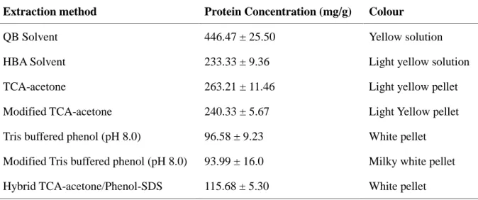
The dried leaves of F. deltoidea were pulverised by liquid nitrogen to obtain smaller size of plant tissue for higher surface area for extraction with the minimal proteolysis and protein degradation. Based on the results of Bradford assay, it was found that solvent based extraction, especially QB based solvent extraction produced the highest crude protein content, whereas phenol based method showed the lowest crude protein content (Table 1). The hybrid method of TCA-acetone/phenol-SDS did not improve the protein yield if compared to the single approach of TCA-acetone, but slightly improved the crude protein content if compared to the phenol based method. Phenol based method just selectively dissolved and purified protein from aqueous phase after extraction [21]. Therefore, phenol based method showed lower protein content than TCA-acetone method. The conventional TCA-acetone method was previously found to be effective to remove plant lipids and pigments [22]. Therefore, this method was applied in this study because the leaves of F. deltoidea are dark green and waxy on the leaf surface. Possibly, higher crude protein content in the method of TCA-acetone precipitation could be due to the reaction between lignin from plant leaves and Coomassie brilliant blue in Bradford reagent. Previous studies reported higher protein yield of TCA-acetone method than phenol based method for lignified tissues including leaves and roots [15, 22]. Lignin is an aromatic polymer which could co-precipitate with proteins in the TCA-acetone method, and thus leading to over estimation of protein yield in the Bradford assay. The Coomassie brilliant blue dye can bind to aromatic amino acid residues and aromatic compounds of lignins, and therefore producing false positive results [23].
Table 1. Crude protein concentration extracted by different methods for the leaves of Ficus
deltoidea
Extraction method Protein Concentration (mg/g) Colour
QB Solvent 446.47 ± 25.50 Yellow solution
HBA Solvent 233.33 ± 9.36 Light yellow solution
TCA-acetone 263.21 ± 11.46 Light yellow pellet
Modified TCA-acetone 240.33 ± 5.67 Light Yellow pellet
Tris buffered phenol (pH 8.0) 96.58 ± 9.23 White pellet
Modified Tris buffered phenol (pH 8.0) 93.99 ± 16.0 Milky white pellet
Hybrid TCA-acetone/Phenol-SDS 115.68 ± 5.30 White pellet
Only the young leaves were selected for this study of comparison because usually mature leaf would higher polyphenol content which might require tedious rinsing steps for removal. According to Gorg et al. [24], protein precipitation by TCA in acetone could increase protein concentration and improve contaminant removal. The wash with 10% TCA and acetone was more effective than that rinsing step with TCA or acetone alone [25]. The leaves of F. deltoidea are also fibrous, and therefore, phenol based method was also tried in the present study, since this method was reported to be suitable for recalcitrant plant tissue, as well as effective to remove phenolic compounds, carbohydrates, nucleic acids and salts from contaminating protein pellet [22].
In the following method, a hybrid technique which combined TCA-acetone precipitation and followed by phenol-SDS dissolution was used for protein extraction from F. deltoidea. Wang et al. [17] did mention that this hybrid method was suitable for aged leaf. This could be due to the fibrous structure of aged leaf with higher content of polyphenols. Therefore, the combination of precipitation and followed by protein dissolution could reduce contamination, and thus obtaining higher protein content. Previously, this hybrid approach was likely to be effective in dealing with protein extraction from recalcitrant tissue such as olive leaves [17], fruits [26] and sea grass Posidonia [27]. The addition of SDS as a solubilizing agent in the Tris buffered phenol was to recovery protein. SDS could disrupt the protein-nucleic acid interaction and inactivate ribonucleases, thus altering protein confirmation to cause protein losing its initial structure to be well dissolved into phenol phase [28]. Nevertheless, there was also study found that the addition of SDS did not improve protein yield [29, 30].
From the physical observation, the colour of protein solution from the solvent based extraction was darker (yellowish), and therefore it might contribute to the higher crude protein content. This is because plant pigments could interference the absorbance value, thus contributing to false positive result. Eze and Dumboff [30] reported that the addition of chlorophyll extract to the standard protein solution could increase the absorbance reading, nearly 20% in the Bradford assay. Therefore, it is noticed that the more colour intense protein solution or pellet, the higher crude protein content will be determined in the Bradford assay.
2.2 Protein quality based on electrophoretic gel
Although solvent based extraction produced the highest crude protein content, there is no major protein band was detected in electrophoretic polyacrylamide gel. The observation could be attributed to the high polyphenols and/or plant pigments contamination since the protein solution was yellow and solvent based extraction was less effective to remove the contaminants from the fibrous leaves of F. deltoidea. Therefore, solvent based extraction was unlikely to be suitable for protein extraction from the plant.
The molecular sizes of proteins extracted from the leaves of F. deltoidea using different extraction methods are listed in Table 2. Among the extraction methods, TCA-acetone and modified TCA-acetone were found to be less effective to obtain high number and quality of proteins. Only 9 and 6 protein bands are observed on the gel for TCA-acetone and modified TCA-acetone methods, respectively as presented in Fig 1. The repeated steps of grinding and rinsing in the modified TCA-acetone method resulted the loss of protein number from 9 to 6 protein bands, especially lower molecular weight of proteins. The resolution of protein bands from the TCA-acetone method showed smearing effect and light bands. This method might be more suitable for young growing vegetative tissue with less contaminants for co-extraction. Possibly, the incomplete re-solubilization of protein pellet in the sample buffer for electrophoresis. It is known that the major problem of TCA-acetone precipitation method is resolubilization of precipitated proteins [29].
Table 2. Molecular sizes of proteins extracted from the leaves of Ficus deltoidea and
separated by electrophoresis
Molecular size of protein bands by different extraction mtheods (kDa)
TCA-acetone Modified TCA-acetone Tris buffered phenol (pH 8.0) Modified Tris buffered phenol (pH 8.0) Hybrid TCA-acetone/phenol-SDS 113.48 ± 3.44 107.67 ± 3.26 102.16 ± 3.10 100.29 ± 1.19 100.29 ± 1.19 88.42 ± 1.05 88.42 ± 1.05 87.92 ± 2.67 78.61 ± 1.76 77.34 ± 0.92 77.34 ± 0.92 78.56 ± 2.38 75.61 ± 1.70 74.54 ± 2.26 72.16 ± 0.86 70.72 ± 2.14 64.72 ± 1.45 64.72 ± 1.45 63.67 ± 1.93 60.41 ± 1.83 55.58 ± 0.66 51.60 ± 1.56 47.28 ± 1.06 47.28 ± 1.06 46.70 ± 0.56 46.70 ± 0.56 46.45 ± 1.41 39.05 ± 0.88 39.05 ± 0.88 35.72 ± 1.08 34.75 ± 0.78 33.45 ± 0.45 33.89 ± 1.03 29.75 ± 0.67 32.16 ± 0.97 25.46 ± 0.57 26.06 ± 0.79 24.37 ± 0.29 24.38 ± 0.29 23.46 ± 0.71 20.97 ± 0.47 20.94 ± 0.25 21.12 ± 0.64 18.66 ± 0.42 18.46 ± 0.22 18.46 ± 0.22 17.12 ± 0.52
16.69 ± 0.20 16.69 ± 0.20 12.49 ± 0.38 12.64 ± 0.15 12.64 ± 0.15 11.24 ± 0.34 10.56 ± 0.13 10.56 ± 0.13 10.12 ± 0.31 8.58 ± 0.19 8.58 ± 0.19 8.23 ± 0.10 8.23 ± 0.10 8.20 ± 0.25
Fig.1. Electrophorestic separation of proteins extracted from the leaves of Ficus deltoidea
using different methods; M, marker; TA, TCA-acetone; MTA, modified TCA-acetone; TP, Tris buffered phenol; MTP, modified Tris buffered phenol; TA/PS, hybrid
TCA-acetone/phenol-SDS and QB, solvent based extraction; HBA, solvent based extraction
Similarly, phenol based method exhibited distortion bands and smearing effect on the gel. This could also contribute by contaminants pipetted from aqueous phase because no clear phenol phase separation was observed, even though sucrose was added into extraction to assist phase separation. Anyhow, phenol based extraction was found to be better than TCA-acetone method because higher protein number (13 protein bands) with good resolution of protein quality on the 12 % polyacrylamide gel. The use of larger ratio of sample to extraction buffer (1000 mg: 15 ml extraction buffer) and additional rinsing steps by chilled methanol in the modified Tris buffered phenol method further improve the quality of the gel image with more intense protein bands (Fig. 1). The larger ratio of sample to buffer might increase the efficiency of protein extraction and the additional rinsing by methanol to remove phenolic compounds could increase the purity of extracted protein for electrophoresis.
The hybridization of TCA-acetone and phenol-SDS was found to improve the number of protein bands significantly. There are 22 protein bands on the gel as presented in Fig.1. Proteins were resolved into distinct bands that extended a broad range of apparent molecular weight from 8.20 to 113.48 kDa. The procedures of TCA-acetone precipitation could eliminate phenolics, lipids and pigments, while phenol based extraction further reduced the contamination of polysaccharides, nucleic acids and salts. Hence, TCA-acetone precipitation can always be used as the starting protocol for plant protein extraction for the removal of pigments, lipids and phenolic compounds, and followed by selective protein dissolution to discard the remaining impurities such as carbohydrates, nucleic acids and salts. In good agreement with Saravanan and Rose [15] who suggested TCA-acetone and phenol based method can be applied to extract protein from a wide range of tissues with high efficiency of contamination removal. This method is also recommended for downstream process which requires high quality protein such as gel electrophoresis and liquid chromatography tandem mass spectrometry analysis.
2.3. Effect of pH for Tris buffered phenol on protein extraction
The effects of pH in Tris buffered phenol solution on plant protein extraction were also investigated since different pH values were applied in many previous studies. Significantly, the pH of Tris buffered phenol exhibited a remarkable effect on the efficiency of plant protein extraction in terms of protein yield and quality. The higher volume of extraction buffer (15 mL) appeared to produce lower protein concentration with lower standard deviation of triplicate data as presented in Table 3. Although 3 mL of Tris buffered phenol produced higher protein content, the standard deviations of the results are also higher than those results from higher volume of extraction buffer. Furthermore, the protein pellet was yellow in colour. Therefore, the higher protein concentration in the 3 mL of extraction system might be due to the contamination from plant impurity that leading to higher concentration of crude protein content.
Table 3. Effects of pH and volume of extraction buffer on protein concentration and colour
Tris buffered phenol
(mL)
pH
Protein concentration
(mg/g)
Colour of protein pellet
15 7.5 3.91 ± 0.40 White 8.0 10.81 ± 0.11 White 8.5 4.42 ± 0.44 White 3 7.5 23.33 ± 3.22 Yellow 8.0 9.82 ± 4.62 Yellow 8.5 8.85 ± 5.06 Yellow
The resolution of protein bands on the gel at pH 8.0 was found to be better than the other pH values as presented in Fig. 2. It is clear that less background interference at pH 8.0 for plant protein extraction from the leaves of F. deltoidea. The smaller volume of extraction buffer (3 mL) was found to produce poor resolution of 12 % polyacrylamide gel (Fig. 2). The observation supports the explanation of higher crude protein content which is attributed to the higher contamination of plant impurity in the 3 mL Tris buffered phenol system. The optimum value of pH 8.0 differed from the result reported by Isaacson et al. [16] who reported that protein could optimally be extracted from maize root, orange peel or tomato leaves at pH 7.5. Another study conducted by Tsugama et al. [29] agreed that the optimum pH was between 8.0-8.5 for protein extraction from recalcitrant plants such as banana (Musa spp.), apple (Malus domestica L.) and potato (Solanum tuberosum L.). The findings revealed that the optimum pH for protein extraction is highly relied on the protein structure and its chemical characteristics. The explanation has also proven that the efficiency of plant protein extraction is highly depended on the type of plant and plant tissue.
Fig.2. Polyacrylamide gel of proteins extracted from the leaves of Ficus deltoidea at different
pH values (7.5, 8.0 and 8.5) using 15 and 3 mL of Tris buffered phenol where M is the marker protein with the molecular size ranging from 10-220 kDa.
Protein is likely to be partitioned in phenol phase because of hydrogen bond interaction with peptide backbone [31]. On the other hand, the other plant metabolites such as polysaccharides, polyphenols and pigments would have weak interaction with protein in mild alkaline medium [29]. The mild alkaline condition would also inhibit protease activity and possibly neutralize acids released from vacuoles during plant cell lysis. To avoid complication during plant
protein extraction, EDTA and β-mercaptoethanol were added in the extraction buffer acting as
reducing agents to chelate metalloproteases and polyphenol oxidases, as well as to prevent protein oxidation. Furthermore, PMSF was added to act as a protease inhibitor. The utilization of potassium chloride was to increase protein solubility.
3. EXPERIMENTAL 3.1 Herbal plants
Experiments were carried out using the leaves of F. deltoidea sourced from Nursery Herba Pak Ali, Johor, Malaysia. The plant was cultivated in a mixture of sand and compos soil (1:1) and allowed to grow in natural glass house at Plant Biotechnology Laboratory, Faculty of
Bioscience and Medical Engineering, Universiti Teknologi Malaysia, 81310 Skudai, Johor Darul Takzim, Malaysia.
3.2 QB Solvent Based Extraction
QB method was carried according to the procedures described by Ni et al. [32] 100-150 mg of plant leaves were ground in a pre-cooled mortar. Approximately, 100-150 mg of ground tissue powder was incubated with freshly prepared 1 mL cooled extraction buffer (2M KPO4, 0.5M EDTA, 1% Triton X 100, 80% glycerol, 1M DTT and distilled water) and vortexed vigorously for 30 seconds. Lysate was centrifuged at 14000 rpm for 20 minutes at 4⁰C. The supernatant were collected and were transferred to new tube. If there is still tissue seen, lysate were spin for another 10 minutes at same speed and temperature. Supernatant were kept at -80⁰C for long term storage.
3.3 HBA Solvent Based Extraction
HBA method was carried according to the method described by Fan and Yuan [33]. 100-150 mg of plant material was ground in a precooled mortar in the presence of liquid nitrogen. Approximately 100-150 mg of ground tissue powder was incubated with freshly prepared 1 mL of cooled extraction buffer (100 mM Tris, pH 7.4, 10% sucrose, 5 mM EDTA, pH 8.0, 0.19%
EGTA, and freshly added 0.28% β-mercaptoethanol and 1mM PMSF). Mixture were vortexed
vigorously for 30 seconds and were incubated on ice for 15 minutes. Mixture were centrifuged at 15000 rpm for 5 minutes at 4⁰C. The supernatant were collected and were transferred to new tube. If there is still tissue seen, lysate were spin for another 10 minutes at same speed and temperature. Supernatant were kept at -80⁰C for long term storage.
3.4 TCA-acetone Precipitation Method
TCA-acetone precipitation was carried out according to the procedures reported by Damerval et al. [34] with some modifications. Approximately, 100 – 150 mg of plant leaves were ground in a pre-cooled mortar and pestle in the presence of liquid nitrogen. The ground leaves were
incubated with freshly prepared 2 mL of 10 % TCA, 0.07 % β-mercaptoethanol in cold acetone
for overnight. Then, the mixture was centrifuged at 10,000 xg for 15 min at 4 °C and the supernatant was discarded. The pellet was rinsed twice with ice-cold acetone with 0.07 %
β-mercaptoethanol until the pellet became colourless. The protein pellet was dried and
resuspended in SDS sample buffer.
In another trial, the method of TCA-acetone was modified by adding comprehensive rinsing steps before and after extraction as described in the method reported by Wang et al. [17] The ground leaves were subjected to cold acetone wash (2 mL) for twice within the process of tissue
grinding by liquid nitrogen to discard contaminants such as chlorophylls and polyphenols. A repeated step of grinding was to obtain finer tissue powder. This initial wash was carried out by vortexing the ground leaves in a tube for 30 s, and then centrifuged at 10,000 xg for 2 min at 4
⁰C. Subsequently, the fine powder was repeatedly rinsed with cold acetone with 10 % TCA to
remove water soluble contaminants until colourless decant was obtained. The wash was continued with cold aqueous with 10 % TCA and finally rinsed with 80 % acetone for twice. To ensure the rinsing steps are effective, the pellet was vortexed in the wash solution for 30 s and centrifuged at 10,000 xg for 2 min at 4 ⁰C. The pellet was then dried and resuspended in SDS sample buffer.
3.5 Phenol extraction
Alternatively, phenol extraction was carried out based on the procedures described by Faurobert et al. [18] Approximately, 100-150 mg of frozen leaf tissue was ground in presence of liquid nitrogen and extracted with 3 mL extraction buffer (0.7 M sucrose, 0.1 M KCl, 0.5 M Tris-HCl (pH 7.5, 8.0 and 8.5), 50 mM EDTA, 1 mM PMSF and 2 % β-mercaptoethanol. The mixture was incubated at 4 °C for 10 min under shaking condition. Tris buffered phenol (3 mL) was added to the mixture and incubated on a shaker for another 10 min at room temperature. The sample was centrifuged at 5,500 xg at 4 °C for 10 min after incubation. The phenol phase which is on the top layer was carefully drawn and re-extracted with another 3 mL of extraction buffer for 3 min. After centrifugation, the final phenol phase was collected, and 20 mL of precipitation solution (ammonium acetate (0.1 M) in chilled methanol) was added. The mixture was shaken by inverting and incubated overnight at -20°C. The pellet was harvested by centrifugation at 5,500 xg for 10 min at 4 °C. The pellet was rinsed again with chilled acetone trice and centrifuged at 5,500 xg for 5 min at 4 °C. Finally, the pellet was dried under vacuum and resuspended in SDS sample buffer.
In another trial, this phenol extraction method was modified by increasing the ratio of sample to extraction buffer volume from 100 mg sample : 3 ml extraction buffer to 1000 mg sample : 15 ml extraction buffer as described in the method reported by Isaacson et al. [16] Besides that, protein pellet was subjected to an additional rinsing step using ice cold methanol for twice to remove any impurity. Subsequently, the pH of Tris buffered phenol was varied from 7.5 to 8.5 in order to investigate the effect of pH on the efficiency of plant protein extraction.
3.6 Hybrid method of TCA-acetone/phenol extraction
This protocol was carried out according to the protocols reported by Wang et al. [17] who combined both TCA-acetone and phenol extraction. Approximately, 100-150 mg of plant
leaves was ground in a pre-cooled mortar and pestle in the presence of liquid nitrogen. The powdered tissue was transferred into a tube and rinsed with 10% TCA in acetone before centrifugation at 16,000 xg for 3 min at 4°C. The supernatant was discarded and the sample was rinsed with 80 % methanol plus 0.1 M ammonium acetate. The supernatant was removed after centrifugation at 16,000 xg for 3 min at 4°C. The sample was rinsed again with 80 % acetone and harvested by centrifugation. After air-dried under vacuum for 3 min, the residual acetone was removed. A 0.4-0.8 mL of Tris buffered phenol incorporated with SDS buffer (30 % sucrose, 2 % SDS, 0.1 M Tris-HCl (pH 8.0) and 5% β-mercaptoethanol) was added to the sample and mixed thoroughly before incubation for 5 min. Then, the mixture was centrifuged at 16,000 xg for 3 min at 4°C. The upper layer of phenol phase was transferred into a new tube. A precipitation buffer (ammonium acetate (0.1 M) in methanol) was added to recover the phenol phase and stored at -20°C for overnight. The pellet was harvested by centrifugation at 16,000 xg for 5 min at 4°C. The harvested pellet was rinsed with methanol and consequently with acetone. Finally, the pellet was dried and resuspended in sample buffer.
3.7 Bradford assay for protein content
The crude protein content of extract from the leaves of F. deltoidea was estimated by using Bradford assay [35]. The crude protein was dissolved in sample buffer, and 10 µL of the sample was diluted with 490 µL of deionized water and mixed with 500 µL of Bradford reagent. The solution was incubated for 5 min at room temperature. The blue colored solution was formed as a result of protein complex formation (protein-dye complex) under acidic condition. The intensity of blue colour was measured by an UV-Vis spectrophotometer (UV-1800, Shimadzu, Japan) at 595 nm. The total protein concentration was determined based on triplicate data. A serial of BSA solutions with the concentration ranged from 1 to 15 µg/mL was prepared to construct a calibration curve.
3.8 One-Dimensional Gel Electrophoresis
One dimensional SDS-PAGE was carried out according to the procedures described by Laemmli [36]. A 15 μL of protein sample (20 mg/mL) was mixed with 15 μL of SDS sample buffer and then heated at 95 oC for 5 min. The sample buffer consisted of 1 mL of 10 % (w/v) SDS, 1.2 mL of 0.5 M Tris-HCl (pH 6.8) and 0.5 mL of β-mercaptoethanol. A 5 mL of 50 % (v/v) glycerol was added and another 1 mL of 0.05 % (w/v) bromophenol blue (BPB) to give bluish colour. The mixture was topped up to 10 mL by deionized water. The denatured protein solution was cooled to 30 oC and then loaded onto a 12 % polyacrylamide gel (5 x 8.2 cm)
topped with a 5 % stacking gel. The whole gel was then transferred to a Mini-Protean II cell (Bio-Rad Laboratories GmbH, Munchen, Germany) for electrophoresis.
A wide range of protein mixture with the molecular weight ranged from 10-245 kDa was used to calibrate the gel. A 5 µL of protein marker solution was pipetted and loaded onto one of the wells on top of the stacking gel. Then, 20 µL of sample was loaded onto another well of the gel. The gel was placed into a running tank and the inner chamber was filled up with 1X running buffer for separation process. The complex mixture of protein sample was separated on the separation gel by regulating the voltage at 100 V for 20 min, and followed by 180 V for 70 min. Electrophoresis was completed once the dye reached the bottom of the gel. The gel was taken out to stain with 0.1 % (w/v) CBB solution in 50 % methanol and 10 % acetic acid for 30 min. The gel was subsequently immersed in 5 % acetic acid solution overnight to destain the blue colored background of the gel in a fume cupboard.
4. CONCLUSION
Plant protein extraction is a complicated and challenging work, mainly due to the broad range of secondary metabolites and low protein concentration in plant tissues which may be interfered by the presence of proteases. To the best of our knowledge, no single extraction protocol is effective to extract all proteins and for all plant species because of the diverse structure and stability of proteins. The present study revealed that the hybrid technique of TCA-acetone/phenol-SDS could be the most appropriate protein extraction method for the recalcitrant leaves of F. deltoidea. This is because this hyphenated technique could produce higher protein quantity and quality based on the Bradford assay and electrophoresis, respectively. Tris buffered phenol is the second appropriate buffer system in this study. Somehow, it is also important to note that the pH and buffer volume are critical factors which might interfere the protein quality and thus directly affecting protein quantitation.
5. ACKNOWLEDGEMENTS
The authors would like to thank Plant Biotechnology Laboratory, Faculty of Biosciences & Medical Engineering, Universiti Teknologi Malaysia, for allowing us to use their laboratory
equipment. This project was funded by the Ministry of Higher Education, Malaysia for the financial support using the Exploratory Research Grant Scheme, 4L114.
6. REFERENCES
[1] Salleh N, Ahmad VN. BMC Complement. Altern. Med. 2013, 13, 359, doi : 10.1186/1472-6882-13-359.
[2] Adam Z, Khamis S, Ismail A, Hamid M. Evidence-Based Complement. Altern. Med. 2012, 2012, 12, http://dx.doi.org/10.1155/2012/632763.
[3] Misbah H, Aziz A, Aminudin N. BMC Complement. Altern. Med. 2013, 13, 118, doi : 10.1186/1472-6882-13-118.
[4] Aris SRS, Mustafa S, Ahmat N, Jaafar FM, Ahmad R. Malaysian J. Anal. Sci. 2009, 13, 146–150.
[5] Hakiman M, Maziah M. J. Med. Plants Res. 2009, 3, 120–131.
[6] Adam Z, Ismail A, Khamis S, Mohd Mokhtar MH, Hamid M. Sains Malaysiana 2011, 40, 489–495.
[7] Oh MJ, Abdul Hamid M, Ngadiran S, Seo YK, Sarmidi MR, Park C S. Arch. Dermatol. Res. 2011, 303, 161–170, doi : 10.1007/s00403-010-1089-5.
[8] Abdulla MA, Abdul-Aziz Ahmed K, Abu-Luhoom FM, Muhanid M. Biomed. Res. 2010, 21, 241–245.
[9] Mitra SK, Walters BT, Clouse SD, Goshe MB. J. Proteome Res. 2009, 8, 2752–2767, doi : 10.1021/pr801044y.
[10]Santoni V, Bellini C, Caboche M. Planta 1994, 192, 557–566, doi : 10.1007/BF00203594. [11]Hurkman WJ, Tanaka CK. Plant Physiol. 1986, 81, 802–806.
[12]Meyer Y, Grosset J, Chartier Y, Cleyet‐Marel J. Electrophoresis 1988, 9, 704–712, doi : 10.1002/elps.1150091105.
[13]Vander Mijnsbrugge K, Meyermans H, Van Montagu M, Bauw G, Boerjan W. Planta 2000, 210, 589–598.
[14]Wang W, Scali M, Vignani R, Spadafora A, Sensi E, Mazzuca S, Cresti M. Electrophoresis 2003, 24, 2369–2375, doi : 10.1002/elps.200305500.
[15]Saravanan RS, Rose JKC. Proteomics 2004, 4, 2522–2532, doi : 10.1002/pmic.200300789.
[16]Isaacson T, Damasceno CMB, Saravanan RS, He Y, Catalá C, Saladié M, Rose JKC. Nat. Protoc. 2006, 1, 769–774, doi : 10.1038/nprot.2006.102.
[17]Wang W, Vignani R, Scali M, Cresti M. Electrophoresis 2006, 27, 2782–2786, doi : 10.1002/elps.200500722.
[18]Faurobert M, Pelpoir E, Chaïb J. Methods Mol. Biol. 2007, 355, 9–14, doi : 10.1385/1-59745-227-0:9.
[19]He CF, Wang YM. Plant Mol. Biol. Report. 2008, 26, 292–300, doi :
10.1007/s11105-008-0040-9.
[20]Chatterjee M, Gupta S, Bhar A, Das S. Int. J. Proteomics 2012, 2012, 10, http://dx.doi.org/10.1155/2012/536963.
[21]Wu X, Xiong E, Wang W, Scali M, Cresti M. Nat. Protoc. 2014, 9, 362–374, doi : 10.1038/nprot.2014.022.
[22]Rose JKC, Bashir S, Giovannoni JJ, Jahn MM, Saravanan RS. Plant J. 2004, 39, 715–733, doi : 10.1111/j.1365-313X.2004.02182.x.
[23]Sebastiana M, Figueiredo A, Monteiro F, Martins J, Franco C, Coelho AV, Vaz F, Simões T, Penque D, Pais MS. Springer Plus 2013, 2, 210, doi : 10.1186/2193-1801-2-210. [24]Görg A, Obermaier C, Boguth G, Csordas A, Diaz J, Madjar J. Electrophoresis 1997, 18,
328–337, doi : 10.1002/elps.1150180306.
[25]Song J, Braun G, Bevis E, Doncaster K. Electrophor. Int. J. 2006, 27, 3144–3151, doi : 10.1002/elps.200500921.
[26]Mazzuca S, Spadafora A, Filadoro D, Vannini C, Marsoni M, Cozza R, Bracale M, Pangaro T, Innocenti AM. J. Exp. Mar. Bio. Ecol. 2009, 374, 113–122,
http://dx.doi.org/10.1016/j.jembe.2009.04.010.
[27]Rio DC, Ares M, Hannon GJ, Nilsen TW. Cold Spring Harb. Protoc. 2010, 2010, pdb – prot5438, doi : 10.1101/pdb.prot5438.
[28]Carpentier SC, Witters E, Laukens K, Deckers P, Swennen R, Panis B. Proteomics 2005, 5, 2497–2507, doi : 10.1002/pmic.200401222.
[29]Tsugama D, Liu S, Takano T. Plant Methods 2011, 7, 1–7, doi : 10.1186/1746-4811-7-22. [30]Eze JMO, Dumbroff EB. Can. J. Bot. 1982, 60, 1046–1049, doi : 10.1139/b82-133. [31]Hochstrasser DF, Harrington MG, Hochstrasser AC, Miller MJ, Merril CR. Anal.
Biochem. 1988, 173, 424–435, doi : 10.1016/0003-2697(88)90209-6.
[32]Ni M, Dehesh K, Tepperman JM, Quail PH. Plant Cell 1996, 8, 1041–1059, http://dx.doi. org/10.1105/tpc.8.6.1041.
[33]Fan Z, Yuan L. Plant Biotechnol. J. 2010, 8, 308–315, doi :
[34]Damerval C, De Vienne D, Zivy M, Thiellement H. Electrophoresis 1986, 7, 52–54, doi : 10.1002/elps.1150070108.
[35]Bradford MM. Anal. Biochem. 1976, 72, 248–254, doi : 10.1016/0003-2697(76)90527-3. [36]Laemmli UK. Nature 1970, 227, 680–685, doi : 10.1038/227680a0.
How to cite this article:
Abdullah F I, Chua L. S and Rahmat Z. Comparison of protein extraction methods for the leaves of ficus deltoidea. J. Fundam. Appl. Sci., 2017, 9(2), 908-924.