HAL Id: hal-01601849
https://hal.archives-ouvertes.fr/hal-01601849
Submitted on 3 Jun 2020
HAL is a multi-disciplinary open access
archive for the deposit and dissemination of
sci-entific research documents, whether they are
pub-lished or not. The documents may come from
teaching and research institutions in France or
abroad, or from public or private research centers.
L’archive ouverte pluridisciplinaire HAL, est
destinée au dépôt et à la diffusion de documents
scientifiques de niveau recherche, publiés ou non,
émanant des établissements d’enseignement et de
recherche français ou étrangers, des laboratoires
publics ou privés.
Distributed under a Creative Commons Attribution - ShareAlike| 4.0 International
License
Exfoliated epithelial cells : a source of bioligical
information in preterm infants
Bertrand Kaeffer, Hélène Billard, Evelyne Gauvard, Anne Drouard, Gwenola
Le Drean, Arnaud Legrand, Patricia Parnet, Dominique Darmaun, Véronique
Gournay, Jean-Christophe Rozé
To cite this version:
Bertrand Kaeffer, Hélène Billard, Evelyne Gauvard, Anne Drouard, Gwenola Le Drean, et al..
Exfoli-ated epithelial cells : a source of bioligical information in preterm infants. The BioTechniques virtual
symposium, Aug 2013, NA, France. 2013. �hal-01601849�
Figure 1. NPAS2 expression in gastric exfoliated cells.
Cells were recovered from infants between Day 5 to Day-36
after birth (A, D; E: Background) and labeled for nuclei by Hoechst (blue head arrow) and NPAS2 (red head arrow, Santa Cruz, sc28708).
Exfoliated epithelial cells: a source of biological information in preterm infants.
Bertrand Kaeffer, Hélène Billard, Evelyne Gauvard, Anne Drouard, Gwénola Le Dréan, Arnaud Legrand, Patricia Parnet, Dominique Darmaun, Véronique Gournay, Jean-Christophe Rozé Address: Unité Mixte de Recherche-1280, INRA, University & Hospital of Mother and Child, Nantes, France
Results
Section: Cellular Analysis, The Modern Laboratory, Biotechniques 2013 Virtual Symposium
Introduction
Material & Methods
Exfoliated epithelial cells can be isolated from digestive fluids as a cellular mixture (senescent, apoptotic, proliferative, or quiescent cells). By quantifying the degree of exfoliation, i.e. by assessing the loss of epithelial cells or of their specific molecular compounds, it is possible to evaluate pharmacological or nutritional stress in clinical trials.
Written informed consents were obtained from both parents
to collect gastric fluid aspirates of 21 infants (Biocollection PROG/09/18).
Conclusions
Correspondence : Bertrand.Kaeffer@univ-nantes.fr
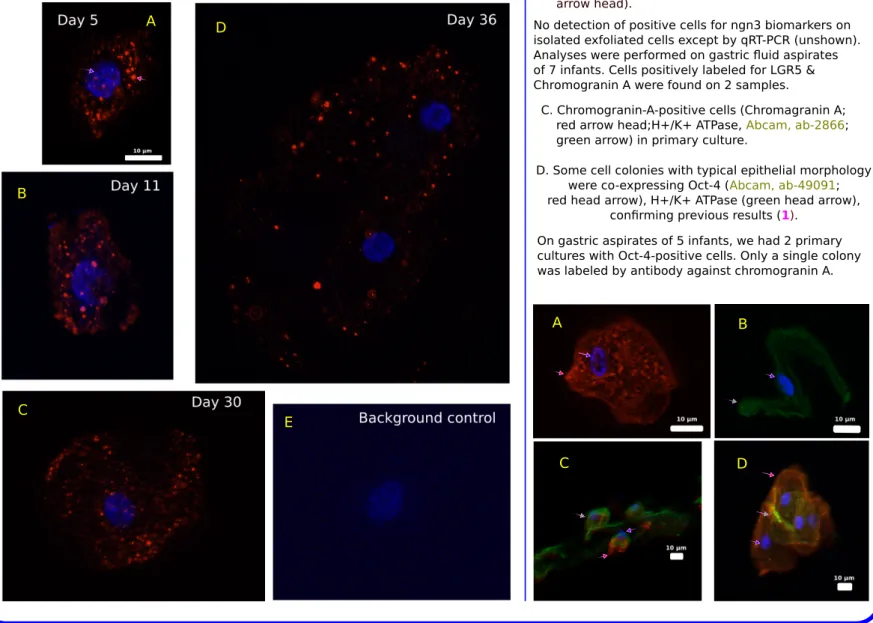
Figure 2. LGR5 & Chromogranin A expressions by gastric cells freshly isolated (A, B) or after 16 days of primary culture (C).
(1) Resuspended (10 cycles) in 10 ml Buffer (1 mM EDTA / 0.05 mM DTT in PBS0)
Perspectives
We intend to design a system allowing the co-detection of
messenger RNA and microRNA to evaluate the physiological state of infants. A B D C E
Fixed cells were immunolabeled & observed by confocal imaging We used a single procedure to isolate epithelial cells from
gastric fluid aspirates (1) or stools (2) of preterm infants. Here gastric fluids were:
(2) Centrifuged at 1300 rpm, 5 min, RT, to pellet cells (3) Rinsed in PBS0 and centrifuged
(4) Cell pellet is either fixed for microscopy or stored at -70°C
Stored cells were lysed for qRT-PCR
No detection of positive cells for ngn3 biomarkers on isolated exfoliated cells except by qRT-PCR (unshown). Analyses were performed on gastric fluid aspirates of 7 infants. Cells positively labeled for LGR5 & Chromogranin A were found on 2 samples.
(1) Along with previously described PERIOD1 & CLOCK (1), we show that NPAS2, an isoform of CLOCK, is expressed in parietal cells of preterm infants as reported in mouse stomach (3). NPAS2 is critical for adaptability to food restriction (7)
(2) Adaptation of primary cultured cells to a 3D long-term culture system (4) may be used to select epithelial cells from
the neuroendocrine lineage.
4. Barker et al., 2010 Stem Cell 6: 25–36
Transcriptomic analyses have been done on epithelial cells recovered from gastric fluid aspirates (2) and stools (2, 5, 6).
By taking advantage of the Triocapi cohort (NCT 0163 0278), gastric fluid aspirates of preterm infants receiving different Ibuprofen doses, integrative miRNA-mRNA profiling will be used to evaluate the degree of gastric exfoliation.
(1) (2)
A B
C D
D. Some cell colonies with typical epithelial morphology were co-expressing Oct-4 (Abcam, ab-49091; red head arrow), H+/K+ ATPase (green head arrow),
confirming previous results (1).
C. Chromogranin-A-positive cells (Chromagranin A; red arrow head;H+/K+ ATPase, Abcam, ab-2866; green arrow) in primary culture.
A. Membrane labeling of LGR5 (red head arrow; Santa Cruz sc-68580) of a typical epithelial cell (nucleus in blue, blue arrow head).
B. Chromogranin-A-positive cell (green head arrow, Spring Bioscience E1520). Nucleus in blue (blue arrow head).
On gastric aspirates of 5 infants, we had 2 primary cultures with Oct-4-positive cells. Only a single colony was labeled by antibody against chromogranin A.
Here we search for the expression of NPAS2, a CLOCK isoform & the expression of biomarkers of stem cell and of neuroendocrine lineage.
References
5. Chapkin et al., 2010 Am J Physiol Gastrointest Liver Physiol 298: G582–G589
6. Chandel et al., 2011 Pediatric Res 70(2): 153–158
1. Kaeffer et al., 2011 PLoS ONE 6(10): e25562
2. Kaeffer et al., 2007 Pediatr Res 62: 564–569
3. Mazzoccoli et al., 2012 Chronobiol. Int. 29: 1300-1311