UNIVERSITÉ DE MONTRÉAL
DÉPARTEMENT DE BIOCHIMIE ET MÉDECINE MOLÉCULAIRE
FACULTÉ DE MÉDECINE
“The small subunit of the mitoribosome from Andalucia
godoyi. Isolation and study of its protein composition”
Présenté par
José Angel Gonzalez Alcazar
Mémoire présenté à la Faculté de médecine en vue de l’obtention du grade de maîtrise
en biochimie
20 mars 2018
1
UNIVERSITÉ DE MONTRÉAL
DÉPARTEMENT DE BIOCHIMIE ET MÉDECINE MOLÉCULAIRE
FACULTÉ DE MÉDECINE
Ce mémoire intitulé:
“The small subunit of the mitoribosome from Andalucia
godoyi. Isolation and study of its protein composition”
Présenté par :
José Angel Gonzalez Alcazar
a été évalué par un jury composé des personnes suivantes :
Dr. Sebastian Pechmann Dre. Gertraud Burger Dr. Martin Schmeing
2
TABLE OF CONTENTS
1. INTRODUCTION ... 10
1.1 Mitochondrial genomes... 10
1.2 The most bacteria-like mtDNAs... 11
1.3 Prokaryotic translation ... 12
1.4 Translation in mammalian mitochondria ... 13
1.4 Differences of translation in bacteria, and mammalian and yeast mitochondria ... 15
1.5 Ribosome composition ... 17
1.6 The mammalian mitoribosome ... 18
1.7 The yeast mitoribosome ... 21
1.8 Mitoribosome composition in kinetoplastids ... 22
1.9 Mitoribosome evolution ... 23
1.9.1 Evolution of the mitoribosome structure... 23
1.9.2 Evolution of mitoribosome protein composition... 24
1.10 Andalucia godoyi ... 27
2. HYPOTHESIS ... 28
3. MATHERIALS AND METHODS ... 28
3.1 A. godoyi cell culture ... 28
3.2 Sucrose gradient purification of the small subunit of the mitoribosome (mt-SSU ribosome) ... 29
3.3 Validation of the enrichment of the mt-SSU-rRNA ... 32
3.3.1 RNA quantification... 32
3.3.2 Electrophoretic separation of rRNAs ... 32
3.3.3 Radiolabelling of probes ... 33
3.3.4 Northern blot hybridization ... 33
3.3.5 SDS-PAGE of mitoribosome proteins (mito-r-proteins) ... 35
3.4 Mass spectrometry analysis ... 35
3.5 Sequence analysis ... 37
4. RESULTS ... 37 4.1 Sucrose gradient purification of the A. godoyi small subunit mitoribosome
3
4.2 Validating the enrichment of A. godoyi mt-SSU-rRNA ... 39
4.3 Mass spectrometry analysis and comparison with predicted SSU mito-r-proteins ... 42
4.4 Additional proteins detected in the mt-SSU ribosomal fraction of Andalucia godoyi ... 47
4.5 Tracing the evolutionary history of SSU mitoribosomal proteins in Discoba 48 5. DISCUSSION ... 49
5.1 Technical challenges in the purification of A. godoyi mt-SSU ribosome ... 49
5.2 Identification and analysis of SSU mitoribosomal proteins from A. godoyi 49 5.3 The composition of A. godoyi mt-SSU ribosome... 50
5.4 Evolution of protein composition in the mt-SSU ribosome of Discoba ... 51
6. OUTLOOK AND FUTURE WORK ... 52
7. REFERENCES ... 54
8. APPENDICES ... 60
8.1 Materials ... 60
8.2 Solutions and buffers ... 63
8.3 Primers used in RT-PCR ... 74
8.4 Probes used in Northern blot ... 74
LIST OF FIGURES AND TABLES
Figure 1. Translation on mammalian mitochondria proceeds in four consecutive steps: Initiation, elongation, termination, and recycling ... 14Figure 2. Structural view of the bacterial ribosome ... 17
Figure 3. Differences of mRNA entrance site and polypeptide exit site in bacterial ribosomes and human mitoribosomes ... 20
Figure 4. Representation of the constructive evolution of mitoribosomes ... 24
Figure 5. Phylogenetic tree of eukaryotes used to predict the evolutionary history of mitoribosome proteins ... 25
Figure 6. Prediction of mito-r-proteins possessed by the LECA and its following evolution in Bikonts and Unikonts ... 26
Figure 7. Light micrographs of A. godoyi ... 27
Figure 8. Linear regression analysis performed for protein quantification ... 36
Figure 9. RNA extracted from fractions of a 5 ml 15-40% sucrose gradient ... 38
4
Figure 11. The enrichment of the 16S bt-rRNA and the 23S bt-rRNA was analyzed
by Northern blot ... 40
Figure 12. The enrichment of the mt-SSU-rRNA was quantified by Northern blot 41 Figure 13. The enrichment of the mt-LSU-rRNA was analyzed by Northern blot .. 41
Figure 14. Protein electrophoresis ... 42
Figure 15. Total number of proteins in the SSU of E. coli and the mt-SSU ribosome of select eukaryotes ... 46
Figure 16. Number of bacterial homolog proteins in the mt-SSU ribosome of different organisms ... 46
Figure 17. Number of eukaryotic-specific proteins in the mt-SSU ribosome of different organisms ... 47
Table 1. Summary of the composition in several characterized ribosomes ... 19
Table 2. Primers used for RT of rRNAs ... 31
Table 3. Primers pairs and number of cycles used to amplify rRNAs ... 31
Table 4. RNA quantification of samples used in Northern blot ... 32
Table 5. Probes used for Northern blot of rRNAs ... 34
Table 6. BSA standards used in the linear regression analysis for protein quantification... 36
Table 7. Mito-r-proteins inferred from the A. godoyi nuclear and mitochondrial genome sequences... 43
Table 8. Proteins in the small subunit of the mitoribosome ... 44
Table 9. Inference of the set of mito-r-proteins present in the mt-SSU ribosome of the last common ancestor of Discobaa ... 48
5
LIST OF ABBREVIATIONS
rRNA: ribosomal RNA tRNA: transfer RNA mRNA: messenger RNA
bt-rRNA: bacterial ribosomal RNA cyt-rRNA: cytosolic rRNA
mt-rRNA: mitochondrial ribosomal RNA mt-tRNA: mitocondrial transfer RNA mt-mRNA: mitocondrial Messenger RNA A: adenine
IF: initiation factor EF: elongation factor RF: release factor
mtIF: mitochondrial initiation factor mtEF: mitochondrial elongation factor mtRF: mitochondrial release factor SSU: small subunit
LSU: large subunit
bt-SSU : small subunit of the bacterial ribosome bt-LSU : large subunit of the bacterial ribosome cyt-SSU : small subunit of the cytosolic ribosome
6
cyt-LSU : large subunit of the cytosolic ribosome mt-SSU: small subunit of the mitochondrial ribosome mt-LSU: large subunit of the mitochondrial ribosome
bt-SSU-rRNA: rRNA of the small subunit of the bacterial ribosome mt-SSU-rRNA: rRNA of the small subunit of the mitoribosome cyt-SSU-rRNA: rRNA of the small subunit of the cytosolic ribosome bt-LSU-rRNA: rRNA of the large subunit of the bacterial ribosome mt-LSU-rRNA: rRNA of the large subunit of the mitoribosome cyt-LSU-rRNA: rRNA of the large subunit of the cytosolic ribosome GDP: guanosine-5'-diphosphate
GTP: guanosine-5'-triphosphate PTC: peptidyl transferase center OPR: Open reading frame
5-prime UTR: 5-prime untranslated region r-protein: ribosomal protein
mito-r-protein: mitoribosome protein PPR : pentatricopeptide repeat
PES : polypeptide exit site PAS : polypeptide accessible site
LECA: last eukaryotic common ancestor mtDNA: mitochondrial DNA
BSA: bovine serum albumin
7
ACKNOWLEDGMENTS
I want to thank Gertraud Burger for being my director and for the wonderful opportunity to work in her laboratory during my master’s project. I also thank M. Valach for his support during the optimization of laboratory protocols and W. Gray for providing functional annotations of nucleus-encoded proteins.
8
RÉSUMÉ
Les mitochondries proviennent d’une alphaprotéobactérie ingérée qui a développé une relation endosymbiote permanente avec son hôte. Par la suite, la plupart des gènes de l'endosymbionte ont été perdus ou migrés vers l'hôte, et le mitoribosome a été remodelé en raccourcissant les ARNr mitochondriaux (ARNr-mt) et en recrutant de nouvelles protéines ribosomiques mitochondriales (protéines-r-mt). Pourtant, comment exactement ce remodelage évolutionnaire a eu lieu reste spéculatif en raison du petit nombre d'études chez les eucaryotes primitifs. Ici, nous montrons que la petite sous-unité du mitoribosome d’Andalucia godoyi, un jakobid hétérotrophe flagellé avec le génome mitochondrial le plus primitif, a une proportion élevée de composants de type bactérien par rapport à son homologue dans les eucaryotes hautement divergents. Nous avons trouvé dans l'analyse par spectrométrie de masse que la petite sous-unité du mitoribosome d’A. godoyi contient plus de protéines ribosomiques bactériennes (21 protéines) que son homologue dans tout autre eucaryote (9 à 20 protéines). Bien que la petite sous-unité du mitoribosome d’A. godoyi représente un stade plus primitif du remodelage des mitoribosomes, certaines des protéines spécifiques à la mitochondrie sont déjà présentes (S25, S29, S33, S35, Rsm22 et Mrp10), or dans une moindre mesure que dans la petite sous-unité du mitoribosome de S. cerevisiae (16 protéines) et H. sapiens (13 protéines). En outre, la petite sous-unité du mitoribosome d’A. godoyi ne contient pas les protéines-r-mt S22, S26, S27, S31, S34, S36, S37, S38, S39, S41, S42, S43, S44, S45, S46, S48, et S49 - dont la plupart sont spécifiques aux eucaryotes. Nos résultats, en combinaison avec les caractéristiques de type bactérien dans les ARNr-mt d’A. godoyi, démontrent que cet organisme possède la petite sous-unité du mitoribosome la plus primitive connue à cette date. La caractérisation des mitoribosomes primitifs peut constituer un point de départ pour l'étude des étapes précoces et intermédiaires de l'évolution du mitoribosome.
Mots-clés : Andalucia godoyi, mitoribosome, petite sous-unité, évolution, jakobids, Discoba, Excavata.
9
ABSTRACT
Mitochondria originated from an ingested alpha-proteobacterium that developed a permanent endosymbiont relation with its host. Thereafter, most of the endosymbiont’s genes were lost or migrated to the host, and the mitoribosome was remodeled by shortening the mito-ribosomal RNAs (mt-rRNAs) and the recruitment of new mitochondrion-specific ribosomal proteins (mito-r-proteins). Yet, how exactly this evolutionary remodeling took place remains speculative due to the small number of studies in primitive eukaryotes. Here we show that the mt-SSU ribosome of A. godoyi, a free-living heterotrophic flagellated jakobid with the most primitive mitochondrial genome, has an elevated proportion of bacteria-like components compared to its
counterpart in highly diverged eukaryotes. We found by mass spectrometry analysis that the mt-SSU ribosome of A. godoyi contains more homologs of bacterial r-proteins (21 proteins) than its counterpart in any other eukaryote (from 9 to 20 proteins). Although the mt-SSU ribosome of A. godoyi represents a more primitive stage of mitoribosome
remodeling, some of the mitochondrion-specific proteins are already present (S25, S29, S33, S35, Rsm22, and Mrp10), yet to a smaller extent than in the mt-SSU ribosome of S. cerevisiae (16 proteins) and H. sapiens (13 proteins). Furthermore, the mt-SSU ribosome of A. godoyi lacks the mito-r-proteins S22, S26, S27, S31, S34, S36, S37, S38, S39, S41, S42, S43, S44, S45, S46, S48, and S49 -most of which are eukaryotic-specific. Our results, in addition to the bacteria-like features of A. godoyi’s mt-rRNAs, demonstrate that this organism possesses the most primitive mt-SSU ribosome known. The
characterization of primitive mitoribosomes may be a starting point for studying the early and intermediate steps in mitoribosome evolution.
Key words: Andalucia godoyi, mitoribosome, small subunit, evolution, jakobids, Discoba, Excavata.
10
1. INTRODUCTION
1.1 Mitochondrial genomes
The mitochondrion is a double-membrane bounded organelle that is present in most known eukaryotic organisms. This organelle originated from an ingested alpha-proteobacteria and is an integrative part of extant eukaryotes. The mitochondrion provides cellular energy, and is involved in other cellular tasks such as signalling, cellular differentiation, cell death, maintaining control of the cell cycle, and cell growth (1).
Mitochondria contain their own genome that consists of several copies of a single circular chromosome. However, mitochondrial genomes have diverged to such a degree that makes difficult to recognize their bacterial origin. For instance, the number of genes in mitochondrial genomes is three orders of magnitude smaller than in typical bacterial genomes, and gene organization does not follow operon rules. Moreover, a single-subunit enzyme performs transcription in mitochondria instead of a four subunit RNA polymerase as in bacteria.
Mitochondrial evolution in different eukaryotic lineages have resulted in a great diversity of sizes, content, and organization of mtDNAs. For instance, mitochondrial DNA (mtDNA) can vary in length from 6 to 2,400 kb and be present in many thousands of copies depending on the specie or cell type. Chromera velia contains the smallest mitochondrial genome, which harbors only 2 protein-coding genes (2). The most gene-rich mtDNAs containing ~100 genes are found in a recently discovered eukaryotic group, the jakobids, which will be discussed in more detail in later sections.
Extant mitochondrial genomes have lost most of their ancestral bacterial genes due to (i) deletion of genes that are no longer needed for survival of the endosymbiont and (ii) unidirectional transfer of genes to the nucleus (3, 4). Gene migration from mitochondria to the nucleus has been explained by the mutagenic nature of reactive oxygen species that arise from the electron transport chains, population genetic aspects, and the physical polarity of endosymbiosis creating a one-way street of gene transfer from lysed organelles
11
to the nucleus (5-8). Moreover, DNA transfer from an organelle to the nucleus does not have sequence-specific barriers (4, 9-11).
The mitochondrial genomes of all eukaryotes are thought to have retained genes that encode components of the electron transport chain due to the need of co-location for redox regulation, and to avoid the mistargeting of hydrophobic membrane proteins to the endoplasmic reticulum (4, 12, 13). Additionally, mitochondrial genomes encode ribosomal RNAs (rRNAs) and transfer RNAs (tRNAs) needed for the translation of mitochondrion-synthesized messenger RNAs (mt-mRNAs) (4). The rest of the mitochondrial proteins that are not mitochondrion-encoded are synthetized in the cytoplasm and then imported into the organelle (14).
The unidirectional transfer of genes from the mitochondrial genome to the nucleus seems to confer adaptive advantages to the eukaryotic cell too. For instance, the nuclear genes encoding mitochondrial proteins can be recombined by sex. Further, the mitochondrial DNA has a higher mutation rate than the nuclear DNA due to the oxidative stress that accumulates in mitochondria. Therefore, mutation in genes encoding mitochondrial proteins is reduced if these genes are nucleus-encoded (4).
1.2 The most bacteria-like mtDNAs
The mtDNA of jakobids, an order of eukaryotes in the megagroup of Discoba, appear to have the most bacteria-like mt-DNAs currently known due to their high number of encoded transcripts, gene organization, structure of mitochondrion-encoded rRNAs (mt-rRNAs), and bacteria-like regulatory elements of gene expression.
The jakobid mtDNAs contain gene clusters that are densely packed and their gene rearrangement is generally alike to the one of free-living alpha-proteobacteria (15).
The jakobid mtDNAs encode molecules not usually encoded in the mtDNA of most eukaryotes. For instance, a four-subunit RNA polymerase, the SecY complex, the 5S rRNA, and an structural RNA linked to the mitochondrial RNP complex (15, 16). Moreover, the
12
5S rRNA is likely integrated in the mitoribosomes of jakobids since it is expressed in these organelles (17).
The mt-rRNAs of nine jakobids strongly resemble to the typical models of E. coli 16S and 23S rRNAs. The universal core of mt-rRNAs shares a high degree of sequence identity between jakobids and its bacterial counterparts. Furthermore, the mitochondrion-encoded genes possess upstream Shine-Dalgarno-like motifs that match to the 3-prime-terminal pyrimidine-rich motif in the mt-rRNA of the small subunit of the mitoribosome (mt-SSU-rRNA) (15).
1.3 Prokaryotic translation
Translations is the process by which a ribosome reads the sequence of mRNAs to synthesize proteins (18).
Translation of genes in Prokaryotes has been widely investigated and described (19-25). Prokaryotic translation can be divided in four steps: initiation, elongation, termination, and recycling. Several interactions between tRNAs, mRNAs, rRNAs, and proteins occur in every translation step to ensure the synthesis of correct peptides at the right time. In initiation, the initiation factors IF1, IF2, and IF3 recruit the initiator fMet-tRNAfMet and the small 30S subunit (SSU) to an AUG start codon (characterized by an upstream Shine-Dalgarno sequence in the mRNA). Then, the AUG codon and the f-Met-tRNA are positioned to the P site of the ribosome, and the large 50S subunit (LSU) is stably associated before starting the elongation step. In elongation, the ribosome selects the cognate aminoacyl tRNAs specified by the mRNA sequence that is bound to the A site of the ribosome. Then, the tRNA cognate to the next mRNA codon is transported to the A site in complex with the elongation factor (Ef-Tu) and GTP, which is followed by peptide bond formation with the tRNA in the P site. Subsequently, the nascent polypeptide is transported to the P site, and the elongation factor G (EF-G) catalyzes the translocation of the two tRNAs and the mRNA by one codon. Thereafter, the tRNA in the P site is ejected through
13
the E site and the elongation step is repeated until the ribosome reaches a stop codon defined by UAA, UAG, or UGA. The release factors 1 and 2 (RF1 and RF2) recognize the stop codon and release the nascent polypeptide. Then, the dissociation of RF1 and RF2 is promoted by the release factor (RF3). Subsequently, EF-G and the recycling factor (RRF) help to dissociate the ribosome subunits and the remaining tRNA in the P site. Finally, the ribosome subunits can be recycled for another translation cycle.
1.4 Translation in mammalian mitochondria
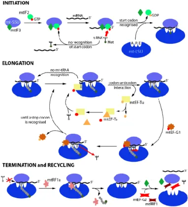
Translation of genes in mammalian mitochondria has been widely investigated (26-32). The mammalian mitochondria encode 9 monocistronic and two dicistronic mt-mRNAs. The mt-mRNAs have a modified codon usage with the standard UGA stop codon recognized as tryptophan and the standard AGA and AGG arginine codons being instead used as stop codons. The folding and excising of mt-tRNA structures is carried out by the mitochondrial tRNase Z ELAC2 and the mitochondrial RNase P respectively. The mitochondrial poly(A) polymerase adds a poly(A) end of approximately 50 nt to maturate every light-strand protein-encoding mt-mRNA. The translation of mt-mRNAs proceeds in three steps: initiation, elongation, termination and recycling (figure 1).
1.3.1 Initiation and elongation in mammalian mitochondrial translation
The small subunit of the mitoribosome (mt-SSU ribosome) recruits the mt-mRNA that is bound to the initiation factor mtIF3 (figure 1). This step blocks the reassociation of the mt-SSU with the large subunit of the mitoribosome (mt-LSU ribosome) (33). The mitoribosome recognizes either of three initiation triplets AUG, AUA, and AUU. Then, the initiation factor mtIF2:GTP recruits the f-Met-tRNA to the initiation triplets (34). Subsequently, the mt-SSU forms a complex with the mt-LSU. This prompts the hydrolysis of mtIF2-bound to GTP producing GDP and liberating the initiation factors mtIF2/mtIF3 from the mt-SSU.
14
Figure 1. Translation on mammalian mitochondria proceeds in four consecutive steps: Initiation, elongation, termination, and
recycling. The elongation factor mtEF-G1 interacts with the
mitoribosome, which leads to changes in the structural conformation of the mitoribosome. This liberates the mitochondrial transfer RNA (mt-tRNA) from the A-site and moves the dipeptidyl-tRNA into the P-site. Then, the deacylated mt-tRNA moves to the E-site (35, 36).
The elongation step is repeated until a stop codon is located in the A-site. Adapted from (33).
The elongation factor mtEF-Tu, a GTP and a charged mt-tRNA form the ternary complex (GTP:mtEF-Tu complex) that enters the A site (figure 1). Then, the mitoribosome hydrolyzes GTP to liberate GDP and mtEF-Tu. Subsequently, the peptidyl transferase centre (PTC) in the mt-LSU catalyzes the formation of the peptide bond. The dipeptidyl-tRNA moves to the P-site and a new deacylated mt-dipeptidyl-tRNA enters the A-site. The movement of the mt-tRNA and the mt-mRNA is guided by the mtEF-G factor during translocation
15
(37). Finally, the interaction between mtEF-Tu and the exchange factor mtEF-Ts re-establishes the GTP:mtEF-Tu complex.
1.3.2 Termination of mammalian mitochondrial translation and recycling of mitoribosomes
In humans, the mitochondrial release factor mtRF1 recognizes the stop codons in mitochondrial open reading frames (OPRs) (figure 1) (38). Then, the ester bond between the mt-tRNA and the final amino acid is hydrolyzed. Other mammalian species may have other additional mitochondrial release factors (39).
The mitochondrial release factors mtRRF1 and mtEF-G2 help the dissociation of the SSU and the LSU (figure 1). This releases the mRNA and the deacylated mt-tRNA (40, 41). Finally, mtRRF1 and mtEF-G2 are released to reinitiate the translation cycle in mitochondria.
1.4 Differences of translation in bacteria, and mammalian and yeast mitochondria
During translation initiation in bacteria, base pairing occurs between the Shine-Dalgarno sequence in the 5-prime untranslated region (5-prime UTR) located upstream of the start codon of the mRNA and the 3-prime end in the rRNA of the SSU. However, mammalian mt-mRNAs lack the prime UTR and all mt-mRNAs are devoid of the 5-prime cap structure (32, 42). Yeast mt-mRNAs usually contain the 5-5-prime UTRs that are targeted by transcript specific activators (32, 43, 44).
The mitochondrial factors that regulate translation initiation, elongation, termination, and recycling of mitoribosomes are nucleus-encoded and operate in concert with the mitoribosomes. Many of these factors have homologs in bacteria, but specialization occurred in response to the evolutionary changes in the mitochondrial
16
genetic code, mitoribosome structure, mitoribosome composition and nature of mitochondrion-encoded transcripts (42).
Initiation factors such as IF1, IF2, and IF3 determine the accuracy and effectiveness of translation in bacteria (32). Mammalian and yeast mitochondria have only two initiation factors compared to bacteria, mtIF2 (homolog of bacterial IF2) and mtIF3 (homolog of bacterial IF3) (45, 46). Although a mitochondrial homolog of IF1 is absent, mammalian IF2mt could provide structural compensation for the lack of IF1 (47, 48).
In bacteria, there is a high number of interactions between binding sites in r-proteins (L5, and L25), helices in rRNAs (h38, h76, h77, and h84), and the elbow region in tRNAs (49). Since every tRNA-binding site that is known from bacteria is present in the mammalian mitoribosome, one may think that elongation is the most conserved translational step. Yet, in contrast to bacterial tRNAs, mt-tRNAs in mammals attach to the LSU only by the acceptor stem due to the great diversity of elbow region shapes in the mt-tRNAs (32). In bacteria, EF-Tu delivers tRNA to the ribosome and participates in decoding, and translocation is catalyzed by the elongation factor EF-G (32, 50). Mammalian mitochondria have the same elongation factors as bacteria. However, mammalian mtEF-Tu and mtEF-Ts differ in structure from their bacterial homologs (51, 52). Moreover, bakers’ yeast mitochondria are lacking mtEF-Ts (32, 53).
The bacterial EF performs recycling of ribosomes too, whereas the RRF1mt and mtEF-G2 (an homolog of bacterial EF-G) perform recycling of mitoribosomes in mammalian and yeast mitochondria (41). In addition, a third mitochondrial dissociation factor, mtIF3 was proposed to play a role in the recycling of mitoribosomes in yeast and mammals (32).
A final difference is that stop codons in bacteria are recognized by two termination factors, RF1 and RF2, while mammalian and yeast mitochondrial translation systems only possess RF1 (54-56).
17
1.5 Ribosome composition
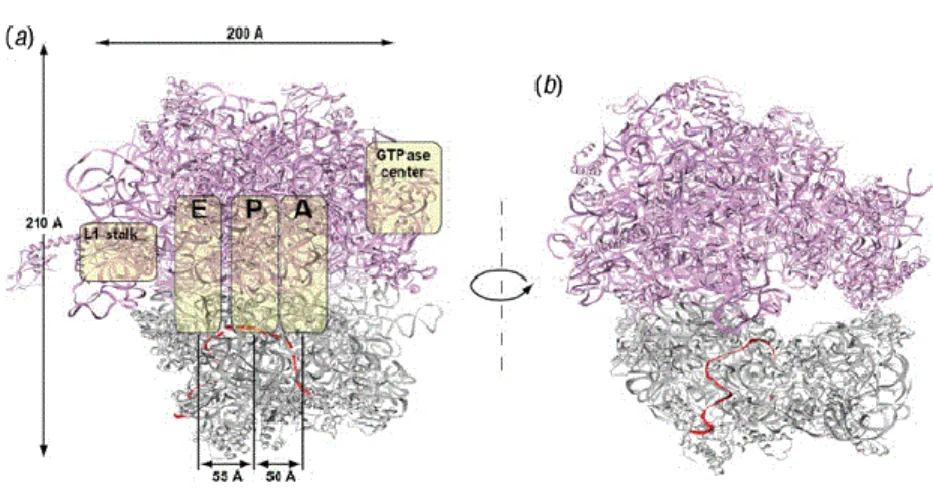
Ribosomes across the tree of life have a large subunit (LSU) and a small subunit (SSU) composed of r-proteins and rRNAs. These subunits work together to translate mRNAs into a polypeptide chain. All ribosomes share a conserved core structure mainly composed of rRNA and r-proteins localized near the ribosome surface (16). The interface between the LSU and the SSU has three binding sites for the tRNAs, the A site, the P site and the E site (figure 2) (57). The A site is the access point for the aminoacyl tRNA in the ribosome (except for the first aminoacyl tRNA that enters through the P site during translation initiation). The P site is where the peptide bond is formed, and the E site (exit site) is where the uncharged tRNA is ejected after its amino acid was used to form a peptide bond in the nascent polypeptide chain. As described above, the mRNA binds to the SSU and moves through the ribosome one codon a time during the elongation of the nascent polypeptide chain.
Figure 2. Structural view of the bacterial ribosome. (a) The 30S small subunit is
displayed in gray and the 50S large subunit in purple, and the mRNA is displayed in red. The yellow covers indicate the localization of the L1 stalks, the tRNA-binding sites (A, P, and E sites), and the GTPase center. The tRNAs bind to the ribosome at the A site, passes through the P site, and exits through the E site. (b) The tunnel of the
18
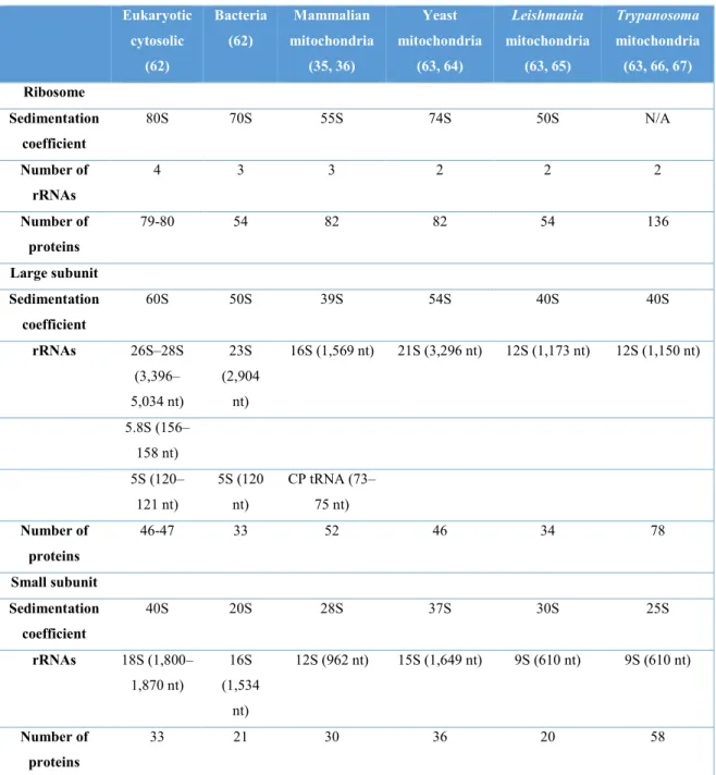
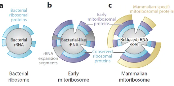
The different types of ribosomes differ in their RNA and protein content. For instance, eukaryotic (cytosolic) ribosomes have a rRNA: r-protein ratio that is close to one (50), whereas in bacterial ribosomes and in mammalian mitoribosomes, it is 7:3 and 3:7 respectively (33). The different types of ribosomes also differ in their sedimentation coefficient, length and number of rRNAs, and r-protein number (table 1).
1.6 The mammalian mitoribosome
The two subunits of bacterial ribosomes interact mainly via RNA:RNA bridges (58). In contrast, the intersubunit bridges of mammalian mitoribosomes are mainly composed by protein-protein and RNA-protein connections (35, 36). The 5S rRNA is absent from mammalian mitoribosomes and it is compensated by a tRNA-Val that forms several interactions with mitoribosome proteins (mito-r-proteins), which allows to mediate interactions between the mt-SSU and the mt-LSU (32, 49, 59, 60). The mito-r-proteins in the mammalian mt-LSU ribosome have contacts with an average of 4.9 neighbouring proteins whereas bacterial r-proteins have on average only 1.5 neighbors (49). Several mito-r-proteins, for instance S6, S16, S18, S25, L10, and L66, contain zinc-binding motifs that coordinate a single zinc ion between two proteins (16).
The mitoribosome recruited new proteins, and accumulated N- and C-terminal extensions of otherwise conserved proteins. In Mammalia, the mt-rRNA was reduced, with mito-r-proteins replacing the missing rRNA segments, and providing additional functions such as association to the inner mitochondrial membrane (35, 36, 61).
19
Table 1. Summary of the composition in several characterized ribosomes.
Eukaryotic cytosolic (62) Bacteria (62) Mammalian mitochondria (35, 36) Yeast mitochondria (63, 64) Leishmania mitochondria (63, 65) Trypanosoma mitochondria (63, 66, 67) Ribosome Sedimentation coefficient 80S 70S 55S 74S 50S N/A Number of rRNAs 4 3 3 2 2 2 Number of proteins 79-80 54 82 82 54 136 Large subunit Sedimentation coefficient 60S 50S 39S 54S 40S 40S rRNAs 26S–28S (3,396– 5,034 nt) 23S (2,904 nt) 16S (1,569 nt) 21S (3,296 nt) 12S (1,173 nt) 12S (1,150 nt) 5.8S (156– 158 nt) 5S (120– 121 nt) 5S (120 nt) CP tRNA (73– 75 nt) Number of proteins 46-47 33 52 46 34 78 Small subunit Sedimentation coefficient 40S 20S 28S 37S 30S 25S rRNAs 18S (1,800– 1,870 nt) 16S (1,534 nt) 12S (962 nt) 15S (1,649 nt) 9S (610 nt) 9S (610 nt) Number of proteins 33 21 30 36 20 58
Modified from (16). *; CP, central protuberance; N/A, not purified as a monosome.
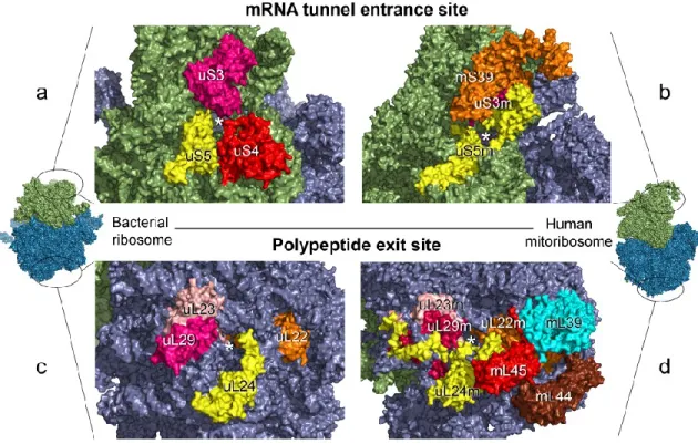
The mt-mRNA entrance channel in the mt-SSU ribosome of mammals is highly remodeled regardless of the conservation of its central core (33, 61, 68). The mt-SSU ribosome of mammals lacks the protein S4 and the C-terminus of the protein S3, which are critical for defining the ringed shape of the mRNA entrance channel in the bacterial SSU
20
(bt-SSU) (figure 3) (33). This lost is compensated in mammalian mitoribosomes by extensions in the proteins S5 and S39 (a PPR-containing protein with RNA-binding activity) located close to the entrance channel (figure 3) (33, 69, 70). Moreover, the absence of the anti-Shine-Dalgarno motif and the 5-prime UTR in mt-mRNAs, which are needed to align the mitoribosome with the start codon, is compensated by the protein S29 (figure 3) (33, 71).
Figure 3. Differences of mRNA entrance site and polypeptide exit site in bacterial ribosomes and
human mitoribosomes. The E. coli ribosome and the human mitoribosome are depicted left and right,
respectively. The black circles in the monosomes indicate the expanded region for visualization purposes. The position of the mRNA entrance site in the small subunit of E. coli ribosome (a) and human mitoribosome (b) is shown at the top of the image. The position of the exit site in the large subunit of E. coli ribosome (c) and human mitoribosome (d) is shown at the bottom. Prefixes are used
before or after protein names to indicate their nomenclature of origin: u + protein name refers to bacterial r-proteins, m + protein name to mito-r-proteins, u + protein name + m to proteins shared by
both bacterial ribosomes and mitoribosomes. *; r-proteins, ribosomal proteins; mito-r-proteins, mitoribosome proteins. Adapted from (33).
21
The exit channel in the mammalian mt-LSU ribosome contains the mitochondrion-specific proteins L39, L44, and L45 that may play a role in the synthesis of hydrophobic proteins (33). Moreover, the mito-r-protein L45 is predicted to mediate the attachment of the mitoribosome to the inner mitochondrial membrane (42, 61).
1.7 The yeast mitoribosome
Overall, the secondary structure of the 15S mt-rRNA resembles that of the bacterial 16S rRNA with discrepancies only at the periphery of the mitoribosome (72). The 5S rRNA is absent in yeast mitoribosomes as described before in the human mitoribosome, which is compensated by mt-rRNA expansion segments rather than tRNAs as in mammals (32). The yeast mitoribosome possess most of the intersubunit bridges characteristic of bacterial ribosomes (72). Moreover, the mt-SSU ribosome of yeast possesses more homologs of bacterial r-proteins than its mammalian counterpart (72). The mt-mRNA entrance channel lacks the large-scale remodeling seen in the mammalian mt-mRNA entrance channel. Furthermore, the decoding center in the yeast mt-SSU ribosome is composed of a loop in the mito-r-protein S12 and several nucleotides that have equivalents in the decoding centre of bacterial ribosomes (72).
Although the yeast mitoribosome possess an elevated number of bacteria-like features compared to the mammalian mitoribosome, it also contains features that are unique to yeast. For instance, yeast specific mito-r-proteins and nine additional mitochondrion-specific intersubunit bridges. The majority of the yeast mito-r-proteins with homologs in bacterial and mammalian mitoribosomes have N- and C-terminal extensions that increase the protein interconnectivity, but these terminal extensions are not conserved in structure, sequence, or length across homolog mito-r-proteins (72). The 3-prime end sequence of the 15S mt-rRNA lacks the anti-Shine-Dalgarno sequences and is covered by proteins in the body of the mt-SSU ribosome in contrast to the bacterial 16S rRNA that possesses an anti-Shine-Dalgarno sequence and is restricted to the exit channel (72). Moreover, the yeast mt-mRNA exit channel is flanked by a protuberance in the S42-S43 heterodimer and extensions in several mito-r-proteins (72). Furthermore, Mba1 (homolog of mammalian
22
L45) and several mt-rRNA expansion segments attach the yeast mitoribosome to the inner mitochondrial membrane (42, 73, 74).
1.8 Mitoribosome composition in kinetoplastids
The kinetoplastids are a group of flagellate protists from the megagroup Discoba (to which jakobids belong) whose mitoribosomes have been extensively characterized. T. brucei contains 133 mito-r-proteins and L. tarantolae more than 50 mito-r-proteins (66, 75). The mitoribosome of kinetoplastids has few bacterial and mammalian homologs, and most of its constituent proteins are either kinetoplastid-specific or organism-specific (66, 75, 76). Moreover, most of the conserved mito-r-proteins are far lengthier than their homologs in bacteria, yeast, and mammals due to N- and C-terminal extensions (60, 66, 77-79).
The 9S and the 12S mt-rRNAs of T. brucei and L. tarantolae mitoribosomes are shorter and have a minimal secondary structure compared to their bacterial counterparts (75, 80). The additional protein mass in mitoribosomes of kinetoplastids, and lengthier N- and C-terminal extensions in homolog proteins may counterbalance the highly reduced nature of their mt-r-rRNAs (66, 80). The same phenomena has been observed, but to a lesser extent in mammalian mitoribosomes (59).
The mitoribosome of T. brucei contains a high number of unique mt-r-proteins (105 out of 136 mito-r-proteins) that could be part of a larger “supercomplex” associated with the mitoribosome that possess additional functions. For instance, some of these proteins contain PPR and GTP binding motifs. Moreover, some of these proteins possess predicted activities such as GTPase, methyltransferase, peptidyl-prolyl isomerase, helicase activity, and chaperone function. These motifs and functions potentially play a role in ribosome assembly, RNA folding, mt-rRNA processing, protein assembly, protein-RNA interactions, and subunit structure stabilization (66, 81).
In L. tarantolae mitoribosome, the mt-mRNA entrance and the exit channels contain Leishmania-specific mito-r-proteins. Moreover, these mito-r-proteins replace the missing rRNA segments in the A, P, and E sites (76).
23
1.9 Mitoribosome evolution
1.9.1 Evolution of the mitoribosome structure
Endosymbiotic bacteria tend to accumulate mutations that reduce the stability of their rRNAs and most mitochondrial genomes have a higher A+T content than bacterial genomes, which confers a higher number of weak base pairs to the mt-rRNAs (82). Moreover, high mutation rates, small effective population sizes, and low rate of recombination in mitochondria cause slightly deleterious mutations in the mt-RNAs (83). Genome analysis of several model organisms showed that, compared to bacterial ribosomes, the protein mass of mitoribosomes is generally larger, while the length of most mt-rRNAs is similar or smaller. Although highly diverged eukaryotes have structurally reduced mt-rRNAs and an elevated number of mito-r-proteins, there is no strict correlation between the loss of mt-rRNA segments and the gain of mito-r-proteins (59).
It has been suggested that many mitoribosome proteins may have already been recruited early in eukaryotic evolution, converting the mitoribosome from “mt-rRNA rich and protein-poor” to “rRNA rich and protein-rich” (figure 4) (59). The reduction of mt-rRNA may have started only when metazoan diverged and resulted in “mt-mt-rRNA poor and protein-rich” mitoribosomes (figure 4).
Additionally, several N- and C-terminal extensions in mito-r-proteins occurred in different linages and separately, after the divergence of the last eukaryotic common ancestor (LECA), so that the length of homolog proteins can differ considerably.
24
Figure 4. Representation of the constructive evolution of mitoribosomes. a) Proteobacterial
ribosome, b) Early mitoribosome with additional rRNA expansion segments and early mitoribosome proteins. Adapted from (16).
1.9.2 Evolution of mitoribosome protein composition
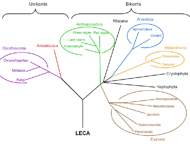
The evolutionary history of mito-r-proteins has been traced in the phylogenetic tree of eukaryotes (figures 5 and 6) by the identification of homologs of alpha-proteobacterial r-proteins and mito-r-r-proteins in a wide range of nuclear, mitochondrial, bacterial and archaeal genomes (63). The phylogenetictree based on mito-r-proteinsindicates that 54 mito-r-proteins (21 in the SSU, and 33 in the LSU) were likely present in the alpha-proteobacterial ancestor of the mitoribosome (figure 6). The protein S20 was apparently lost early in eukaryotic evolution, since it is absent in all analyzed eukaryotic genomes (figure 6).
25
Figure 5. Phylogenetic tree of eukaryotes used to predict the evolutionary history of mitoribosome proteins. *; LECA, last common eukaryotic ancestor. Adapted from (63).
A set of 27 alpha-proteobacterial mito-r-proteins was likely encoded in the mitochondrial genome of the last eukaryotic common ancestor (LECA) because it is still encoded in the mitochondrial genome of Reclinomonas americana (84), a jakobid protist that contained the largest mitochondrion-encoded gene set that was known at the time of the analysis. The other 26 alpha-proteobacterial mito-r-proteins not encoded in any available mitochondrial genome were predicted to be relocated to the host nuclear genome before the divergence of the major eukaryotic linages (figure 6). The migration of genes encoding alpha-proteobacterial r-proteins from the mtDNA to the nucleus probably started before the recruitment of eukaryotic-specific mito-r-proteins since gene migration from endosymbionts to the host is a common event in endosymbiosis. Then, nineteen eukaryotic-specific mito-r-proteins were apparently recruited before the divergence of the LECA, which added up to a total of 72 mito-r-proteins (63).
26
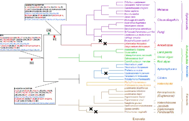
Figure 6. Prediction of mito-r-proteins possessed by the LECA and its following evolution in Bikonts and Unikonts. Red arrows designate losses of mito-r-proteins, whereas green arrows
designate gains. The mitochondrion-encoded mito-r-proteins of alpha-proteobacterial origin are shown in bold. Mitochondrion-encoded mito-r-proteins are shown in red, whereas nucleus-encoded mito-r-proteins are shown in black. Eukaryotic-specific mito-r-proteins are shown in blue italic font. The crosses indicate eukaryotic linages that have lost their mitochondria. *;
mito-r-proteins, mitoribosome proteins. Adapted from (63).
Considerable changes happened between the endosymbiosis and the diversification of present-day eukaryotic lineages because the LECA already possessed a remodeled mitoribosome and a quite developed genome reduction process after endosymbiosis (63). Losses and gains of mito-r-proteins are still ongoing as there are great differences between the mitoribosomes of closely related organisms (63). Gains and losses have occurred independently and repeatedly in different lineages. Moreover, there appears to be no general trend in protein dispensability since the losses have affected both alpha-proteobacterial and eukaryotic-specific mito-r-proteins (figure 6) (63).
27
1.10 Andalucia godoyi
The etymology of Andalucia refers to the region of Spain and it was named after José Godoy, a well-known philanthropist from Andújar, Spain (85). Andalucia godoyi is a member species of the jakobids.
Jakobids are small, bacterivorous, heterotrophic flagellates found in freshwater and marine habitats. They are unicellular eukaryotes typified by two flagella, one of which is guided posteriorly, and a devouring groove along the body utilized for uptake and ingestion of little particles and bacteria (86, 87). This group is a clade within Discoba and includes nine different genera: Andalucia, Velundella, Stygiella, Moramonas, Jakoba, Stomatochone, Stenocodon, Reclinomonas, and Histiona (85, 86, 88-92).
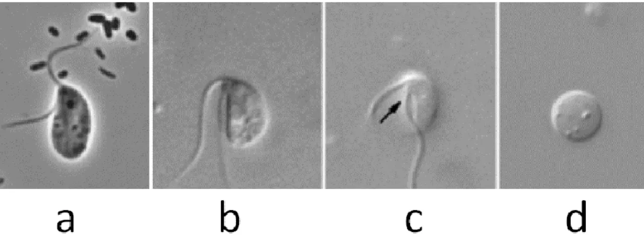
The two flagella of A. godoyi are inserted subapically and apically of its feeding groove (85) (figure 7). The cells have a size of 3–5 µl, and possess a paranuclear body and tubular mitochondrial cristae. The flagella are twice the length of the cell.
A. godoyi contains the largest mitochondrial gene set currently known (15). Moreover, its mtDNA encodes trnT, cox15 and rpL35 that are absent in the mtDNA of other jakobids.
Figure 7. Light micrographs of A. godoyi. a) Static cell, b) Mobile cell, c) Mobile cell, an arrow points the
28
2. HYPOTHESIS
In the course of eukaryotic evolution, the mitoribosome, has experienced losses of components originating from the ribosome of the bacterial ancestor and in turn has gained eukaryote-specific mito-r-proteins. Since the mitoribosome composition has been only investigated in highly diverged eukaryotes such as yeast, mammals, and kinetoplastids, the question arises when exactly and how this transition happened, gradually or as a wholesale restructuring. Since A. godoyi has the least diverged mitochondrial genome currently known, we expect that its mitoribosome represents an intermediate state in the transition from a bacteria-like ribosome to the mitoribosome as we know it from model organisms. Therefore, we hypothesize that the Andalucia mitoribosome has a higher number of bacteria-like proteins and fewer mitochondrion-specific r-proteins than its counterparts in the model organisms discussed above.
To test this hypothesis, we initially attempted to characterize entire mitoribosomes of this jakobid, but their purification proved extremely complicated. Therefore, we focused our investigation on the mt-SSU ribosome, which we succeeded to readily enrich. In fact, this subunit of the mitoribosome is more extensively remodeled than the mt-LSU ribosome, and thus is well suited for the study. Specifically, the objectives of this thesis are inferring the mitoribosome composition of A. godoyi from its genomic sequences, experimental characterization of its SSU ribosome, and comparison with the composition in the mt-SSU of model organisms discussed above.
3. MATHERIALS AND METHODS
3.1 A. godoyi cell culture
A. Simpson kindly provided Andalucia godoyi (85). We feed A. godoyi using Enterobacter aerogenes as a food source. We grew A. godoyi in plastic culture bottles containing 15 ml
29
of WCL medium at 20 °C for 2 weeks. Subsequently, we transferred the culture to 100 ml of WCL medium and incubated it as specified before. Then, we transferred it to 500 ml of WCL medium and incubated it as specified before. The cells were checked regularly under the microscope and more E. aerogenes was added when most of it was consumed by A. godoyi. The titer at the stationary phase was 1.875x10⁶ cells/ml.
We harvested protist cells when all bacteria were consumed. The culture was centrifuged in a GSA rotor at 8,000 g for 20 min at 4 °C. The supernatant was discarded, and the pellet resuspended in 1 ml of WCL medium. Then, the cells were centrifuged at 7969 g for 3 min and the supernatant discarded. The cells were resuspended in 1% DMSO and stored at – 80 °C until their utilization.
3.2 Sucrose gradient purification of the small subunit of the mitoribosome (mt-SSU ribosome)
We tested four cell-lysis buffers with different ratios of monovalent to divalent ion concentrations (appendixes 8.2.9 to 8.2.16) (93). Then, we selected the buffer A because it produced the lowest degree of mt-rRNA degradation.
Andalucia cells were resuspended in one volume of homogenization buffer A and lysed with two volumes of lysis buffer A, 1X EDTA-free protease inhibitor, and SUPERase·In™ (4 U/μL) to inhibit the degradation of the mt-rRNAs. The homogenate was mixed and then incubated on ice for 5 min followed by centrifugation at 18,000 g for 10 min. The supernatant was loaded on top of a 5 ml 15-40 % sucrose gradient and centrifuged at 45,900 rpm for 3 h at 4°C using an AH-650 swinging-bucket rotor. Gradient fractions of 250 μL were collected using a micropipette from the top.
The mt-SSU ribosome of A. godoyi is present in the gradient fractions enriched in mt-SSU-rRNAs because intact ribosomes contain r-proteins bound to rRNAs. RNA was extracted from every sucrose gradient fraction to analyze the migration pattern of mt-rRNAs. We added 5 sample volumes of homemade trizol substitute and vortexed for 15 sec followed by 5 min of incubation at room temperature (94). Then, we added the
30
equivalent of one sample volume of chloroform and shacked vigorously for 30 sec followed by 5 min of incubation at room temperature. Subsequently, the samples were centrifuged at 20,000 g for 15 min at 4 °C and the upper aqueous phase was collected. 3 µl of glycogen (5 mg/ml) and 1.1 sample volumes of isopropanol were added followed by incubation for 40 min at 4 °C. Subsequently, the samples were centrifuged at 20,000 g for 15 min at 4 °C and the isopropanol was discarded. 200 µl of cold 70% ethanol were added and the sample centrifuged at 20,000 g for 10 min at 4 °C. Then, the 70% ethanol was discarded, and the RNA pellet was resuspended in 5 µl of DEPC-treated H2O.
Every RNA sample was mixed with one sample volume of Thermo Scientific 2X RNA Loading Dye followed by incubation for 8 min at 80 °C and run at 110 mV in a 1 % agarose gel in 0.5x TBE. The run was stopped once the blue color of the loading dye was positioned 2.5 cm before the gel end. We used total RNA extracted from whole cells of A. godoyi as a positive control of mt-rRNA and cytoplasmic rRNA (cyt-rRNA), and total RNA extracted from Enterobacter as a control of contamination with bacterial rRNA.
We used the RNase-free DNase I from Roche® to digest the total RNA extracted from every sucrose gradient fraction. Briefly, 5 µl of total RNA were mixed with 5 µl of 1X reaction buffer, and 5 units of DNase I in a total volume of 50 µl followed by incubation at 37 °C for 30 min. Trizol RNA extraction was performed to remove the enzyme.
We utilized the avian myeloblastosis virus (AMV) reverse transcriptase from Roche to synthesize cDNA using rRNAs as a template. 0.8 µl of the above RNA sample were mixed with 0.5 µl of each 10 mM primer, and 1 mM of each dNTP followed by incubation at 80 °C for 2 min. Then, 1X of the reverse transcriptase buffer and one unit of the reverse transcriptase enzyme were added in a final volume of 10 µl followed by 1 h of incubation at 42 °C. Different primers were used to PCR-amplify each one of the ribosomal rRNAs in A. godoyi and E. aerogenes (table 2 and appendix 8.3).
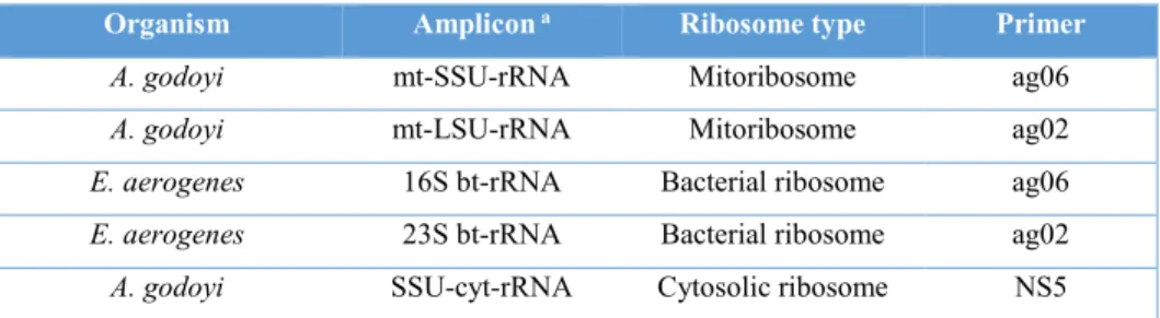
31
Table 2. Primers used for RT of rRNAs (see the appendix 8.3).
Organism Amplicon a Ribosome type Primer
A. godoyi mt-SSU-rRNA Mitoribosome ag06
A. godoyi mt-LSU-rRNA Mitoribosome ag02
E. aerogenes 16S bt-rRNA Bacterial ribosome ag06
E. aerogenes 23S bt-rRNA Bacterial ribosome ag02
A. godoyi SSU-cyt-rRNA Cytosolic ribosome NS5
a mt, mitochondrial; bt, bacterial; cyt, cytosolic; rRNA, ribosomal RNA.
We used the Q5® High-Fidelity DNA Polymerase set from New England Biolabs and followed the guidelines of the supplier. Briefly, 0.25 μl of the cDNA were mixed with 0.2 mM of each dNTP, 0.5 mM of each primer, 1X Q5 buffer, and 0.05 μl of Q5 DNA Polymerase (2000 units/ ml). The annealing temperature was calculated with the Tm
calculator v1.9.7 from the website of New England Biolabs
[https://tmcalculator.neb.com/#!/main], as 2X (A or T) + 4X (C or G) -5 °C. We used different primer pairs and number of PCR cycles to amplify the cDNA synthesised from different rRNA types (table 3 and appendix 8.3). The thermal cycles were: 98 °C for 2 min, 98 °C for 10 sec, annealing temperature for 10 sec, 72 °C for 15 sec, go to step 2 (98 °C for 10 sec), 72 °C for 2 min, and 4 °C forever.
Table 3. Primers pairs and number of cycles used to amplify rRNAs.
Amplicon Primer pairs b Annealing
temperature
PCR cycles
A. godoyi mt-LSU-rRNA ag01 + ag02 63 °C 22
A. godoyi mt-SSU-rRNA ag05 + ag06 68 °C 15
E. aerogenes 16S bt-rRNA ag06 + eb04 68 °C 15
E. aerogenes 23S bt-rRNA ag02 + eb01 66 °C 22
A. godoyi cyt-SSU-rRNA NS5fwd +
BMB-C-rev
70 °C 18
a mt, mitochondrial; bt, bacterial; cyt, cytosolic; rRNA, ribosomal RNA. b for primer sequences, see Appendix 8.3.
32
3.3 Validation of the enrichment of the mt-SSU-rRNA
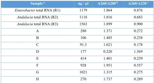
3.3.1 RNA quantification
We performed Northern Blot analysis of mt-rRNAs in order to quantify the enrichment of the mitoribosome subunits in sucrose gradient fractions. The RNA extracted from each sucrose gradient fraction was quantified in a NanoDrop machine before the electrophoretic separation of RNAs (table 4).
Table 4. RNA quantification of samples used in Northern blot.
Sample a ng / µl A260/A280 b A260/A230 c
Enterobacter total RNA (R1) 1179 1.864 0.876
Andalucia total RNA (R2) 1118 1.816 0.683
Andalucia total RNA (R3) 1563 1.899 0.900
A 280 1.371 0.272 B 106 1.485 0.238 C 91.3 1.621 0.178 D 177 0.220 1.569 E 414 1.401 0.239 F 928 1.951 0.557 G 1021 1.315 0.275 H 270 1.737 0.289
a A, gradient fractions 1-2 (see figure 9); B, 3-4; C, 5-6; D, 7-8; E, 9-10; F, 11-12; G, 13-14;
H, 15-16.
b A260/A280, Nucleic Acid 260/280 ratio calculated by dividing the absorbance of the
sample at 260 nm between the absorbance at 280 nm.
c A260/A280, Nucleic Acid 280/260 ratio calculated by dividing the absorbance of the
sample at 280 nm between the absorbance at 260 nm.
3.3.2 Electrophoretic separation of rRNAs
We conducted electrophoresis with the TT buffer (see appendix 8.2.18 for the composition) that was shown to improve the separation of long rRNAs (95). 4 µl of each RNA sample were mixed with 50% formamide, 1X of the TT buffer, 0.5 mM EDTA, and
33
0.02% bromophenol blue in a final volume of 8.5 µl followed by denaturation at 70 °C for 5 min. Subsequently, the samples were placed on ice and formaldehyde was added to a final concentration of 0.4 M. The samples were run at 6 V/cm in a 2 % agarose gel and a buffer containing 1.1% formamide and 1X TT buffer until the loading dye was positioned 2.5 cm before the gel end.
3.3.3 Radiolabelling of probes
We used the DNA probe ag03 for selectively highlighting the mt-SSU-rRNA, eb03 for 23S bt-rRNA, ag04 for mt-LSU-rRNA, and eb02 for bt-rRNA (table 5 and appendix 8.4). The oligonucleotides were radioactively labeled at their 5-prime end. For that, 2.5 μM of the oligonucleotide were mixed with 1X of the polynucleotide kinase (PNK) buffer, 2.5 of µCi/µl 6000 Ci/mmol [γ-32P]-ATP, and 0.5 U/μl of the T4 PNK in a final volume of 20 μl
followed by incubation for 45 min at 37 °C. Then, the enzyme was inactivated at 65 °C for 20 min. The unincorporated radioactive label was removed using NucAway™ Spin Columns according to the manufacturer’s instructions.
3.3.4 Northern blot hybridization
The agarose gel was rinsed in DEPC-treated H2O for 1 h and then soaked in 10X SSC
transfer buffer. The nylon membrane was first saturated with milli-Q H2O and then with
10X SSC buffer. We let RNA to transfer to the nylon membrane overnight in the Northern blot apparatus, and then we dried it at room temperature for a couple of min and marked the orientation of the membrane with a soft pencil. Subsequently, the membrane was dried for 2 h at 80 °C.
We qualitatively estimated the transfer efficiency by subtracting the RNA amount remaining in the agarose gel from the RNA amount that was loaded. The agarose gel was stained with 2.5 µl of ethidium bromide (10 mg/ml) while gently shaking for 15 min in 250
34
ml of 1X TT buffer. The picture was taken in a UV trans-illuminator to estimate the transfer efficiency.
Table 5. Probes used for Northern blot of rRNAs (see appendix 8.4).
Amplicon Probe Tm °C
mt-SSU-rRNA ag03 59.7 mt-LSU-rRNA ag04 61.4 16S bt-rRNA eb02 62.5 23S bt-rRNA eb03 64.4 *; mt, mitochondrial; bt, bacterial; rRNA, ribosomal RNA.
The nylon membrane was incubated with I) 2X SSC buffer for a moment at room temperature, II) hybridization solution for a moment at room temperature, III) pre-hybridization solution for 45 min at 65 °C.
10 µL of the labeled probe were mixed with 90 µL of distilled H2O and denatured
at 95 °C for 5 min followed by cooling on ice for 2 min. The DNA probes were added to the hybridization solution and hybridized overnight with the nylon membrane at the Tm of the DNA probes minus 5 to 10 °C (see table 5). Subsequently, the nylon membrane was incubated with I) 2X SSC and 0.1% SDS at room temperature for a moment, II) a volume of 2X SSC and 0.1% SDS equivalent to 3-5 volumes of the Pre-hybridization solution for 5 min at room temperature, III) a volume of 2X SSC and 0.1% SDS equivalent to 3-5 volumes of the Pre-hybridization solution for 15 min at the Tm of DNA probes minus 10 to 15 °C, IV) the step III was repeated.
Thereafter, the membrane was dried for 5 min at room temperature, wrapped in a plastic bag, exposed onto a phosphorimager-type screen (Imaging Screen-K; Kodak), and scanned after an exposure of several hours using Molecular Imager FX™ (BioRad).
35
3.3.5 SDS-PAGE of mitoribosome proteins (mito-r-proteins)
The samples were incubated in 0.1 M DTT, 1X SDS-PAGE loading buffer, and DEPC-treated H2O at 95 °C for 5 min and run in a 18% SDS-PAGE at 80 mV until the dye was
positioned 1 cm before the gel end. The SDS-PAGE was stained for 1 h using Coomassie Brilliant Blue G-250 in 45% methanol and 10% acetic acid while gently shaking. Subsequently, it was first incubated for 1 h with distaining solution while gently shaking and then overnight with the same solution. Finally, it was washed with milli-Q H2O and
the picture was taken using the Gel Doc™ EZ system.
3.4 Mass spectrometry analysis
Samples were concentrated as follows: We loaded fractions enriched in the mt-SSU ribosome on a 10-kDa Amicon® Ultra-0.5 centrifugal filter (Millipore Sigma) and performed centrifugation at 14,000 g for 30 min at 4 °C. The mito-r-proteins remained in the column and the eluate was discarded. The centrifugation in the Amicon® Ultra-0.5 filter unit was repeated until the entire sample was concentrated into 65 µL. Then, the column was placed upside down in a new centrifugation tube and the concentrated mito-r-proteins were recovered by centrifugation at 1,000 g for 2 min at 4 °C.
Proteins were precipitated as follows: The concentrated mito-r-proteins were mixed with UA buffer (for the buffer composition, see appendix 8.2.28) to a final concentration of 6M urea, and loaded on top of a 3-kDa Amicon® Ultra-0.5 centrifugal filter unit followed by centrifugation at 14,000 g until obtaining a sample volume of 100 μl. Then, the sample was mixed with 50 μl of distilled H2O and 250 μl of UA buffer followed by
centrifugation at 14,000 g until obtaining a sample volume of 100 μl. Afterwards, the sample was mixed with 100 μl of distilled H2O and 200 μl of UA buffer followed by
centrifugation at 14,000 g until obtaining a sample volume of 100 μl. Finally, the column was placed upside down in a new centrifugation tube and the concentrated mito-r-proteins were recovered by centrifugation at 1,000 g for 2 min at 4 °C.
36
Prior to submission to the MS analysis, we quantified the proteins by the Bradford method using a calibration curve of bovine serum albumin (BSA) (table 6 and figure 8). 1 µl of denatured proteins were mixed with 160 µl of Bio-Rad Protein Assay Dye Reagent, and 640 µl of milli-Q H2O followed by incubation for 5 min at room temperature. The
absorbance was measured at 595 nm.
Table 6. BSA standards used in the linear regression analysis for protein quantification.
1 µg/µl BSA 1 2 3 4 5 Abs at 595 nm 0,0695 0,1585 0,1826 0,2417 0,2641
Figure 8. Linear regression analysis performed for protein quantification. *; Y,
absorbance at 595 nm; X, µg of BSA; R2, R squared value.
In-solution trypsin digestion and LC-MS/MS analysis of the peptide mixture were performed at the proteomics platform of the IRCM (Institut de recherches cliniques de Montréal).
37
3.5 Sequence analysis
MS/MS samples were analyzed with MaxQuant v1.6.1.0 software using the custom database of A. godoyi proteins (based on the predicted mitochondrial and nuclear genes), assuming trypsin as the digestion enzyme (96). Spectra were searched with a fragment ion mass tolerance of 0.5 Da and a precursor ion tolerance of 20 ppm. Carbamidomethyl of cysteine was specified as a fixed modification. Oxidation of methionine, deamidation of asparagine or glutamine, phosphorylation of serine, threonine or tyrosine, and conversion of glutamine to pyrrolidonecarboxylic acid (PCA) were specified as variable modifications. Five or less per-peptide modifications were allowed. Minimum and maximum peptide lengths were set to 7 and 25 amino acid residues, respectively, with the molecular weight range from 700 to 4,600 Da. False discovery rate (FDR) for peptide-spectrum matches (PSMs) was set to 1%. As positive protein identifications were considered those for which at least one unique peptide could be assigned with a minimum identification probability above the calculated 1% FDR (false discovery rate).
4. RESULTS
The main objectives of this work were the prediction of the composition of A. godoyi mitoribosome using its genomic sequences, experimental characterization of the protein composition in its mt-SSU ribosome, and comparison against its counterpart’s composition in the model organisms discussed above.
4.1 Sucrose gradient purification of the A. godoyi small subunit mitoribosome
We estimated the enrichment of the small subunit of the mitoribosome (mt-SSU ribosome) based on the quantification of mt-SSU-rRNA in gradient fractions containing protein complexes, because free rRNA does not enter sucrose layers of concentration
38
superior to 10%. To analyze the enrichment of mt-SSU-rRNA bound to mito-r-proteins, RNA from the gradient fractions was extracted and migrated (see section 3.2).
The jakobid mitochondria is physically associated with other subcellular structures and there are not standardized protocols for its purification (87). Since preliminary purification experiments in our research group showed low yields of mitochondria, we chose to use the sucrose gradient ultracentrifugation to purify the mt-SSU ribosome of A. godoyi directly from the whole-cell lysates.
The agarose gel in figure 9 shows the RNA profile across the various sucrose gradient fractions. A. godoyi mt-SSU-rRNA (1588 nt) migrates indeed slightly above 1,500 nt, and it is observed in all pooled fractions. The enrichment of mt-SSU-rRNA starts in pool C, with nearly no other RNA species visible. In contrast, the yield is highest in pools F and G, which contain approximately equimolar quantities or even more of cytosolic rRNAs and mt-LSU-rRNA.
Figure 9. RNA extracted from fractions of a 5 ml 15-40% sucrose gradient. Every two subsequent fractions were pooled. *; mt,
mitochondrial; C1, total RNA from E. aerogenes; C2, total RNA from
A. godoyi; A, fractions 1-2; B, 3-4; C, 5-6; D, 7-8; E, 9-10; F, 11-12;
G, 13-14; H, 15-16; I, 17-18.
Since A. godoyi uses bacteria as a food source, we analyzed the presence of the 16S bt-rRNA to rule out contamination from bacterial ribosomes. The 16S bt-rRNA (1533 nt)
39
from the food bacterium has a very similar migration behaviour as A. godoyi mt-SSU-rRNA. To unambiguously distinguish the two SSU-rRNAs, we performed PCR amplification with specific primers on selected unpooled sucrose-gradient fractions. Figure 10 shows that contaminating SSU-rRNAs occur in fractions 8 and higher. The evidences of Figures 9 and 10 combined demonstrate that A. godoyi mt-SSU-rRNA is the purest in fractions 5 to 7.
Figure 10. Amplification of SSU-rRNAs in single gradient fractions. Primers ag06 + ag05 were used
for mt-SSU-rRNA, NS5-fwd + BMB-C-rev for cyt-SSU-rRNA, and ag06 + eb04 for 16S bt-rRNA (See appendix 8.3). *; M, marker; mt, mitochondrial; cyt, cytosolic; bt, bacterial. 7-10 individual gradient
fractions.
4.2 Validating the enrichment of A. godoyi mt-SSU-rRNA
Northern hybridization served as the final validation of mt-SSU ribosome enrichment in sucrose gradient fractions. For that, we used RNA extracted from the pooled gradient fractions (see table 4) and probes specific for the 16S bt-rRNA, 23S bt-rRNA, mt-SSU-rRNA, and the mt-LSU-rRNA (see the table 5 and the appendix 8.4). The fractions 5 and 6 were enriched in the mt-SSU ribosome of A. godoyi (see section 4.1).
40
A strong signal for 16S bt-rRNA and 23S bt-rRNA was detected using the probes eb02 and eb03 in Enterobacter total RNA (figure 11). We detected a weak signal in Andalucia total RNA and in the heavier gradient fractions, which may be due to non-specific cross hybridization of probes eb02 and eb03 with the cyt-rRNAs. Non-non-specific cross hybridization can be observed in the RNA marker as well. The gradient fractions 1-12 were free from bacterial ribosomes because we did not detect any hybridization signal in the fraction pools A-F (figure 11).
Figure 11. The enrichment of the 16S bt-rRNA and the 23S bt-rRNA was analyzed by Northern blot (see figure 9). The probes eb02 and eb03 were used for detection of 16S
bt-rRNA and 23S bt-bt-rRNA, respectively (See appendix 8.4). R1, Enterobacter total RNA; R2-R3, Andalucia total RNA; A-H, pooled gradient fractions (see the legend of the figure 9).
A strong signal representing the mt-SSU-rRNA was detected using the probe ag03 with Andalucia total RNA (figure 12). A weak non-specific cross hybridization can be detected in the RNA marker as well. We did not detect any hybridization signal in the fraction pools A-B. The mt-SSU ribosome is enriched in the pools C and D (fractions 5-8) because the peak of the mt-SSU-rRNA started at the fraction pool C and the strongest signal was detected in the pool D (figure 12). However, we chose only the pool C for the characterization of the mt-SSU ribosome to avoid contamination with Andalucia cyt-SSU-rRNA contained in the gradient fraction 8 (pool D) (see figure 9).
41
Figure 12. The enrichment of the mt-SSU-rRNA was quantified by Northern blot (see figure 9). The
probe ag03 was used for mt-SSU-rRNA detection (see appendix 8.4). R1, Enterobacter total RNA; R2-R3,
Andalucia total RNA; A-H, gradient fraction pools (see the legend of the figure 9).
A strong signal for the mt-LSU-rRNA was detected in Andalucia total RNA using the probe ag04 (figure 13), but not in fraction pools A-E. The upmost fraction containing the mt-LSU-rRNA is pool F and the strongest signal was detected in pool G (figure 13).
Figure 13. The enrichment of the mt-LSU-rRNA was analyzed by Northern blot (see figure 9).
The probe ag04 was used for 16S mt-rRNA detection (See appendix 8.4). R1, Enterobacter total RNA; R2-R3, Andalucia total RNA; A-H, gradient fraction pools (see the legend of the figure 9).
We finally analysed the profile of protein bands in the gradient fractions 5-6 (see figure 9), which turned out to differ considerably from the profile in the whole-cell lysate (figure 14). Moreover, the protein concentration in these gradient fractions appears to be smaller than in the whole-cell lysate. We infer that the concentration of cytoplasmic proteins is lower in the gradient fractions where the mt-SSU ribosome is enriched.
42
Figure 14. Protein electrophoresis. M, protein marker; 1, whole-cell lysate of A. godoyi;
2, proteins from fractions 5-6 (see figure 9). The grid of detected protein bands by the Image Lab™ software is shown on the right.
4.3 Mass spectrometry analysis and comparison with predicted SSU mito-r-proteins
It is known that small polar proteins can be easily lost during the purification of ribosomes (97). Therefore, we compared the protein composition in the mt-SSU ribosome of A. godoyi in silico.
Michael W. Gray kindly provided the annotation of nucleus-encoded mito-r-proteins of A. godoyi. Then, we compiled this protein set with previously annotated mitochondrion-encoded mito-r-proteins (15), strongly suggesting that Andalucia’s mitoribosome contains 69 mito-r-proteins, of which 28 belong to the mt-SSU ribosome and 41 to the mt-LSU ribosome (table 7).
43
Table 7. Mito-r-proteins inferred from the A. godoyi nuclear and mitochondrial genome sequences.
Small subunit (mt-SSU) Large subunit (mt-LSU)
S1 S25 L1 L24 S2 S29 L2 L25 S3 S33 L3 L27 S4 S35 L4 L28 S5 Rsm22 L5 L29 S6 Ppe1 L6 L31 S7 Mrp10 L9 L32 S8 L10 L33 S9 L11 L34 S10 L12 L35 S11 L13 L36 S12 L14 L38 S13 L15 L40 S14 L16 L41 S15 L17 L43 S16 L18 L45 S17 L19 L46 S18 L20 L49 S19 L21 L53 S21 L22 L54 S23 L23
*; mito-r-proteins, mitoribosome proteins.
Trypsin digestion and analysis by LC-MS/MS allowed detection of each mito-r-protein predicted to be part of the mt-SSU ribosome of A. godoyi (table 8) with at least two unique peptides in the gradient fraction enriched with the mt-SSU ribosome. The only exceptions were S25 and Ppe1, for which, in this experiment, we did not detect peptides. Still, an earlier MS experiment performed on lysates of a subcellular fraction enriched in mitochondria reported multiple peptides of the S25 protein (though none for Ppe1). Table 8 compares the sets of mito-r-proteins across different organism, employing the unified nomenclature for mito-r-proteins proposed by (98).