Université de Sherbrooke
Rôle du facteur de transcription Hnf4alpha dans la réponse de la cellule épithéliale intestinale face aux infections bactériennes
Par
Dianne Pupo Gómez Programme de biologie cellulaire
Mémoire présenté à la Faculté de médecine et des sciences de la santé en vue de l’obtention du grade de maître ès sciences (M.Sc.)
en biologie cellulaire
Sherbrooke, Québec, Canada 2021
Membres du jury d’évaluation
Pr François Boudreau, Département d’immunologie et de biologie cellulaire Pr Steve Jean, Département d’immunologie et de biologie cellulaire Pr Alfredo Menendez, Département de microbiologie et d’infectiologie
Rôle du facteur de transcription Hnf4alpha dans la réponse de la cellule épithéliale intestinale face aux infections bactériennes
Par
Dianne Pupo Gómez Programme de biologie cellulaire
Mémoire présenté à la Faculté de médecine et des sciences de la santé en vue de l’obtention du diplôme de maître ès sciences (M.Sc.) en biologie cellulaire, Faculté de médecine et des
sciences de la santé, Université de Sherbrooke, Sherbrooke, Québec, Canada, J1H 5N4
Les maladies inflammatoires de l'intestin (MIIs) sont un groupe de troubles chroniques qui touchent plus de 270 000 personnes au Canada. De plus, les MIIs sont des maladies multifactorielles dépendant de dérèglements génétiques, immunitaires et environnementaux. L'épithélium gastro-intestinal joue un rôle important en tant que barrière. De plus, il est bien reconnu qu'un défaut d'intégrité de la barrière et de ses fonctions peuvent être impliqués dans le développement de ces maladies. D'autre part, notre laboratoire a montré que la délétion conditionnelle du récepteur nucléaire HNF4α dans l'épithélium intestinal de souris peut conduire au développement d'une inflammation chronique intestinale. Cependant, l'impact de la perte de ce facteur transcriptionnel sur la barrière épithéliale est encore controversé. Dans ce mémoire, nous avons tenté d’évaluer l'impact de la délétion de HNF4α sur la barrière épithéliale lors d'infections bactériennes. La génération de mutantes HNF4α dans l'épithélium intestinal a été réalisée en croisant des animaux Hnf4aloxP/loxP C57BL/6avec un modèle murin Villin-CreERT2, dans lequel la délétion inductible du gène a été conditionnée par l'administration de tamoxifène chez la souri à un jeune âge adulte. Chez les souris Hnf4aΔIEC-ind, une augmentation du passage du FITC-dextran dans la circulation sanguine a révélé que l'épithélium intestinal présentait une augmentation de la perméabilité associée à la mutation. Il est intéressant de noter que l'infection par voie orale avec une souche de Salmonella Typhimurium déficiente pour un gène impliqué dans l’invasion n'a pas montré de différences significatives en termes de charge bactérienne détectée pour Hnf4aΔIEC-ind et les souris témoins. Puis, l'expression génique des cibles sélectionnées a été évaluée chez le modèle Hnf4aΔIEC-ind par qPCR. En ce sens, la perte de HNF4α semble affecter l'expression de différentes molécules des jonctions apicales, liées à la fois à la perméabilité paracellulaire (Cldn2) et au transport ionique (Cldn15). De même, nous prouvons que les souris Hnf4aΔIEC-ind montrent une expression accrue de gènes liés à la protection et à l'intégrité de la barrière épithéliale, tels que Retlnb, Muc2, ainsi que les peptides antimicrobiens Defa5 et Defa20, une observation plus marquée chez les animaux mâles. Les examens histologiques chez les souris Hnf4aΔIEC-ind ont démontré des augmentations du nombre et de la taille des cellules caliciformes, principalement au niveau de la crypte iléale. En plus, la délétion de Hnf4a a non seulement modifié le patron d'expression de la fucosylation dans l'iléon après l'infection, mais aussi, a semblé influencer l'expression des gènes associés à la spécification et à la différenciation des cellules sécrétoires. Globalement, nos résultats suggèrent que HNF4α pourrait jouer un rôle adaptatif important en tant que médiateur de la fonction de barrière épithéliale intestinale en présence d'infections bactériennes. Mots clés: HNF4α, épithélium intestinal, barrière, tamoxifène, infection, maladies inflammatoires de l'intestin (MIIs)
SUMMARY
Role of the transcription factor Hnf4alpha in intestinal epithelial cell response to bacterial infections
Dianne Pupo Gómez Cell biology Program
Master’s thesis presented to the Faculty of Medicine and Health Sciencies in view of obtaining a Master of Science (MSc.) in Cell Biology. Université de Sherbrooke,
Sherbrooke, Québec, Canada, J1H 5N4
Inflammatory bowel diseases (IBD) are a group of chronic disorders that affect more than 270,000 individuals in Canada. In addition, IBD are multifactorial diseases depending on genetic, immune and environmental dysregulations. The gastrointestinal epithelium plays an important role as a barrier. Also, it is well recognized that a defect in the integrity of the barrier and its functions may be involved in the development of these diseases. On the other hand, our laboratory has shown that the conditional deletion of HNF4alpha nuclear receptor (HNF4α) in the intestinal epithelium of mice can lead to the development of intestinal chronic inflammation. However, the impact of the loss of this transcriptional factor on the epithelial barrier is still controversial. The aim of this thesis was to evaluate the impact of Hnf4a deletion on the epithelial barrier during bacterial infections. The HNF4α mutants generation in the intestinal epithelium was achieved by crossing Hnf4aloxP/loxP C57BL/6 animals with a
Villin-CreERT2 mouse model, in which the inducible gene deletion was caused by the administration of tamoxifen in mice at young adult age. In Hnf4aΔIEC-ind mice, an increase in the passage of FITC-dextran into the blood circulation revealed that the intestinal epithelium presented an increase in permeability associated with the mutation. Interestingly, oral infection with an invasion-deficient Salmonella Typhimurium strain did not show relevant differences in terms of the bacterial load detected for Hnf4aΔIEC-ind and control littermate mice. Also, gene expression of selected targets was assessed in Hnf4aΔIEC-ind by qPCR. In this sense, the loss of HNF4α seemed to affect the expression of different molecules of the apical junctions, related to both paracellular permeability (Cldn2) and ion transport (Cldn15). Likewise, Hnf4aΔIEC-ind mice showed increased expression of genes related to the protection and integrity of the epithelial barrier, such as Retlnb, Muc2, as well as the anti-microbial peptides Defa5 and Defa20, an observation being more marked in male animals. Histological examinations showed that Hnf4aΔIEC-ind mice display increases in the number and size of goblets cells, mainly at the level of the ileal crypt. Furthermore, Hnf4a deletion not only modified the expression patterns of fucosylation in the ileum after infection, but also, seemed to influence the expression of genes associated with the specification and differentiation of secretory cells. Globally, these results suggest that HNF4α could play an important adaptive role as a mediator of the intestinal epithelial barrier function in the presence of bacterial infections.
Keywords: HNF4α, intestinal epithelium, barrier, tamoxifen, infection, inflammatory bowel diseases (IBD)
TABLE
OF
CONTENTS
RESUME ... iii
SUMMARY ... iv
TABLE OF CONTENTS ... v
LIST OF FIGURES ... vii
LIST OF TABLES ... viii
LIST OF ABBREVIATIONS ... ix
1. INTRODUCTION ... 1
1.1 Gastrointestinal tract ... 1
1.2 Intestinal barrier ... 2
1.2.1 Goblet cells and mucus ... 3
1.2.2 The junctional complexes as a physical barrier of the intestinal epithelium ... 9
1.3 Inflammatory Bowel Diseases ... 12
1.3.1 Intestinal barrier dysfunction in IBD ... 12
1.3.2 Microbiota in IBD ... 15
1.3.3 Genetic variants and IBD ... 19
1.4 HNF4A as a susceptibility gene in IBD and its controversial role in the control of the intestinal epithelial barrier. ... 21
1.5 Premises and hypothesis ... 23
2. MATERIALS AND METHODS ... 24
2.1 Animal model: the conditional tamoxifen-inducible Hnf4a knockout mouse model ... 24
2.2 Mouse genotyping ... 24
2.3 In vivo intestinal permeability assessment ... 25
2.4 Mouse sacrifice and tissue fixation methods ... 25
2.5 Staining ... 25
2.5.1 Hematoxylin & Eosin (H&E) ... 25
2.5.2 Alcian Blue – Periodic acid Schiff (AB-PAS) ... 26
2.6 Salmonella Typhimurium infection ... 27
2.7 RNA isolation ... 27
2.8 Reverse transcription ... 28
2.9 Quantitative real-time RT-qPCR ... 28
2.11 Fluorescence in situ hybridization (FISH) ... 31
2.12 Electron microscopy analysis ... 32
2.13 Extraction and quantification of proteins from animal tissue ... 32
2.14 IgA detection in the intestinal epithelium ... 33
2.15 Statistical analyses ... 33
3. RESULTS ... 34
3.1 Increased paracellular permeability of the intestinal mucosa in the absence of epithelial HNF4α. ... 34
3.2 Loss of HNF4α does not influence infection outcomes from attenuated Salmonella Typhimurium strain SB103 ... 36
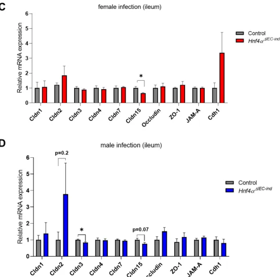
3.3 The apical junction complex appears to be modified in HNF4αΔIEC-ind mice ... 39
3.4 HNF4α intestinal epithelial deletion alters the expression of different gene transcripts related to the protection and integrity of the epithelial barrier after Salmonella infection. ... 40
3.4.1 Apical complex junctions ... 40
3.4.2 Cytokines and immune system modulators ... 42
3.4.3 Antimicrobial peptides ... 44
3.4.4 Intestinal sIgA production ... 45
3.4.5 Mucus layer ... 47
3.5 Hnf4aΔIEC-ind mice display increases in the small intestinal number and size of goblet cells after attenuated Salmonella infection. ... 48
3.6 Deletion of HNF4α in the ileum of male mice impact gene transcript levels associated with secretory cells specification and differentiation. ... 51
3.7 Loss of HNF4α seems to influence fucosylation patterns of infected female mice in the small intestine. ... 53
4. DISCUSSION ... 55 5. CONCLUSIONS ... 70 6. PERSPECTIVES ... 71 7. REMERCIEMENTS ... 73 8. REFERENCES ... 74 9. ANNEXES ... 105
Figure 1. Composition of the intestinal epithelium………3 Figure 2. Structure of intestinal goblets cells……….6 Figure 3. Distribution of the intercellular junctional complex among intestinal epithelial cells………...10 Figure 4. Different pathways and genes identified as risk factors in IBD pathology……...20 Figure 5. Intestinal permeability is increased in the absence of HNF4α………..35 Figure 6. Loss of HNF4α does not lead to increased susceptibility to Salmonella
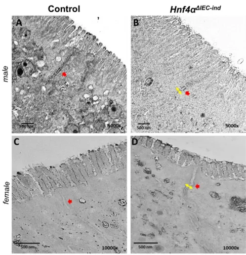
infection……….37 Figure 7. Distension of the apical complex junctions in the absence of HNF4α………39 Figure 8. Expression of cell junction-associated components in the intestine of mice deleted for Hnf4a………...41 Figure 9. Expression of immune response modulators from the intestinal epithelium of control and Hnf4aΔIEC-ind after infection with the attenuated Salmonella Typhimurium strain SB103.………...43 Figure 10. Profile of AMP genes expression in the ileum of Hnf4aΔIEC-ind mice during bacterial infection………..45 Figure 11. Quantification of total sIgA production in the intestine of mice after enteric infection……….46 Figure 12. Altered gene expression for intestinal epithelial goblet cells of Hnf4aΔIEC-ind mice after infection with invasive deficient Salmonella Typhimurium………..48 Figure 13. Histological and goblet cells analyses in the ileum of Hnf4aΔIEC-ind mice after Salmonella Typhimurium strain SB103 infection………..49
Figure 14. Histological and goblet cells analyses in the colon of Hnf4aΔIEC-ind mice after Salmonella Typhimurium strain SB103 infection………..50
Figure 15. Altered number and size of goblets cells in the ileum of infected Hnf4aΔIEC-ind mice………...51 Figure 16. Loss of HNF4α enhance the expression of genes associated with goblet cells differentiation in the ileum of Hnf4aΔIEC-ind infected male mice………...52 Figure 17. Loss of HNF4α promotes abnormal fucosylation in infected mice……….54
Annexes
Figure 1. Hnf4aΔIEC-ind male mice have a thick internal mucus layer………..112 Figure 2. Epithelial 2D-Monolayers established from intestinal organoids derived from
LIST
OF
TABLES
Table 1. Primers used for PCR genotyping………..24
Table 2. Tissue rehydration and dehydration protocol………...26
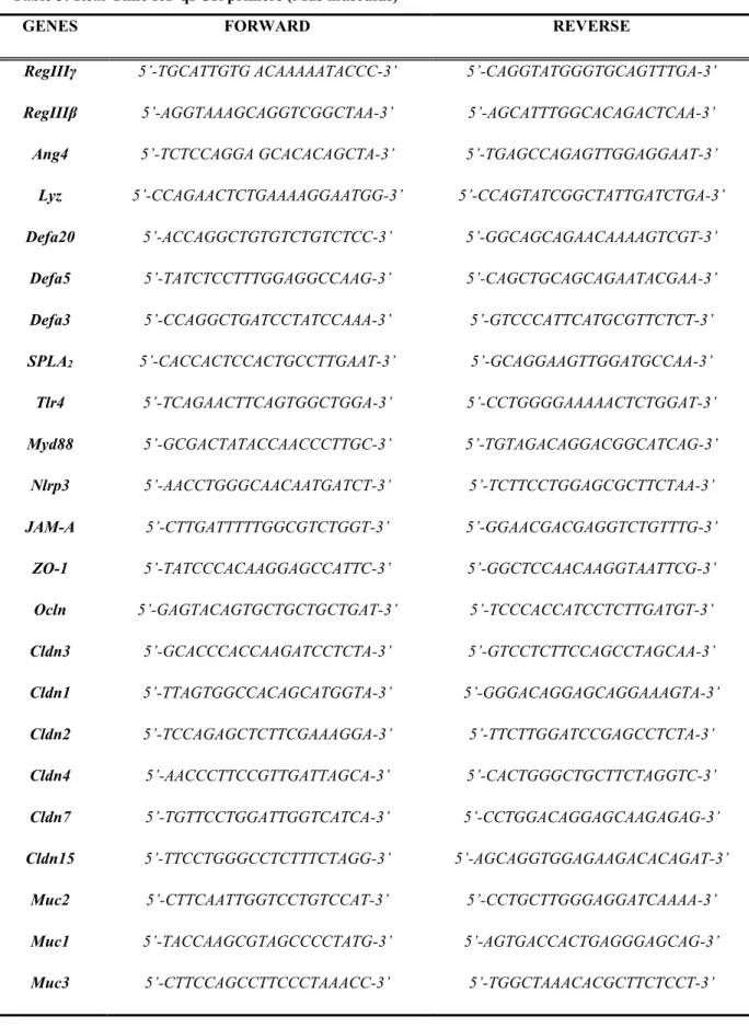
Table 3. Real-Time RT-qPCR primers (Mus musculus) ………...29
Table 4. Primary and secondary antibodies used for IF………31
Table 5. Fish probes primer sequences………...32
Annexes Table 1. (Supplemental materials and methods) Composition of the ad-FD+++ medium...110
Table 2. (Supplemental materials and methods) Composition of the WENR medium…..110
Table 3. (Supplemental results) Statistical analysis of the CFU obtained in mutant and control mice feces for each day of infection………...112
AIEC: adherent and invasive Escherichia coli AJ: adherens juntions
Ang4: angiogenin 4
ATOH1/MATH1: atonal homolog 1 AMP: antimicrobial peptides
BSA: Bovine serum albumin Casp 1: inflammatory caspase 1 CD: Crohn’s disease
CDH1: E-cadherin
CLDN: Claudin human gene or protein Cldn : Claudin mouse gene
cDNA: Complementary deoxyribonucleic acid CDX2: caudal-type homeobox 2
CFU: colony forming units
DAPI: 4’,6-diamidino-2-phenylindole DNA: Deoxyribonucleic acid
dNTPs: Nucleoside triphosphates Defa: α-Defensin
EDTA: Ethylenediaminetetraacetic acid ER: Endoplasmatic Reticulum
Erα/β: Estrogen receptor alpha or beta FCGBP: Fc fragment of IgG binding protein FISH: Fluorescence in situ hybridization
FITC-dextran: polymer conjugated to Fluorescein isothiocyanate FXR: Farnesoid X receptor
Fut2: fucosyltransferase 2
GATA4: GATA-binding family 4 GI: Gastrointestinal tract
GFI1: Growth factor independent 1 GR: Glucocorticoid receptor GsdmD: Gasdermin D
GWAS: Genome-wide association studies HES1: Hairy and enhancer-of-split 1
HNF4α: Hepatocyte nuclear factor 4 alpha (protein) HNF4A: human gene
Hnf4αΔIEC-ind: Inducible deletion of HNF4α from the intestinal epithelium of mice IBD: Inflammatory Bowel Disease
IEC: intestinal epithelial cells
IESC: intestinal epithelial stem cells IL: Interleukin
JAM: junctional adhesion molecules KLF4: Kruppel-like factor 4
LoxP: Locus of X-ing over PI LPS: Lipopolysaccharides M: microfold cells
MAPK: Mitogen-activated protein kinases MLCK: myosin light chain kinase
mRNA: Messenger ribonucleic acid Muc: mucin mouse gene or protein MUC: mucin human gene or protein NAF: Sodium fluoride
NF-kB: Nuclear factor-kappa B
NHE3: transporter Na+/ H+ exchanger isoform 3
NR: nuclear receptor Ocldn: occludin gene
PBS: Phosphate-buffered saline PCR: Polymerization reaction PFA: Paraformaldehyde sPLA2: phospholipase A2
pi: post-infection
PI3K: Phosphoinositide 3-kinase pNPP: p-nitrophenyl-phosphate
PPARγ: Peroxisome Proliferator-Activated receptors PRRs: Pattern recognition receptors
PSA: polysaccharide A PXR: Pregnane x receptor
RELMβ: Resistin-like molecule β (gene Reltnb) REGIII: Regenerating islet-derived protein III RNA: Ribonucleic acid
RT: Room temperature
RT-qPCR: Reverse transcription polymerase chain reaction sIgA: secretory immunoglobulin A
SCFA: short chain fatty acids
SGLT: Sodium-glucose co-transporter
SPDEF: SAM pointed domain containing ETS transcription factor TAM: tamoxifen
TBP: TATA binding protein
TER: transepithelial electrical resistance TF: transcriptional factor
TFF3: Trefoil factor 3 TJ: tight juntions
TLR: Toll-like receptors
TNFα: Tumor necrosis factor alpha UC: Ulcerative colitis
UEA-1: Ulex europaeus agglutinin 1 VDR: Vitamin D receptors
1.1 Gastrointestinal tract
The gastrointestinal tract (GI) constitutes the main organ related to digestive functions including absorption of nutrients, water and electrolytes transport, and secretion of proteins to the intestinal lumen in order to maintain the correct balance or homeostasis. Subsequent to the stomach (Kong et al., 2018), the small intestine includes the duodenum, jejunum, and ileum. The following large intestine is separated into the proximal colon, transverse colon, distal colon, and the rectum. The colonic content is made up of a mixture of bile, secreted mucus, and molecules that cannot be digested or absorbed. Also, the colon contains the highest proportion of the commensal microbiota that colonizes the intestine, thus directly influencing the development and maturation of the immune system functions. Taking into account the latter, it is known that the intestinal epithelium also represents an effective barrier against the invasion of microorganisms or infectious agents that can cause various diseases. Besides, it hinders the passage of molecules with potentially harmful functions in the body (Turner, 2009).
The wall of the intestinal epithelium is made up of four layers including the mucosa, submucosa, muscularis and serosa. Within these layers, the mucosa is divided into the muscularis-mucosa (composed of several thin layers of smooth muscle fibers located underlying the lamina propria and related to the ability of the mucosa to fold and move), the lamina propria (found below the epithelium that houses a large number of immune cells and connective tissue) and finally the epithelium. In the small intestine, the epithelial surface forms finger-like protrusions which are called villi and project towards the intestinal lumen, thus allowing to increase the contact surface between the food bolus and the absorptive cells (JN et al., 2010). However, these villi are lost in the anatomical structure of the colonic mucosa during fetal development. On the other hand, invaginations of the epithelium appear in the connective tissue at the base of the villi and are called crypts of Lieberkühn, which contain the stem cells that allow the intestinal cellular renewal every 3 to 7 days (Barker, 2014). Crypts are the basic unit in the colon as opposed to the crypt/villus axis of the small intestine.
1.2 Intestinal barrier
The intestinal epithelium is the largest mucosal surface of the body protecting from exposure to the commensal microbiota contained in the GI lumen. Measuring approximately 400 m2,
this epithelium is composed of a wide layer of specialized and polarized cells that are interconnected through their membranes and with the basement membrane through protein complexes. Pluripotent stem cells generate a group of cells known as transit-amplifying cells, which divide successively and give rise to 4 main types of well-differentiated intestinal epithelial cells (IEC): goblets cells, Paneth cells, enteroendocrine cells and absorptive cells. These cells migrate towards the tip of the villus (or the apex of the crypt in the colon) and develop specific functions in the epithelium. Paneth cells are located at the bottom of the crypt and are involved in the epithelium defense (production of antimicrobial factors such as, α- / β-defensins and lysozyme) as well as in stem cells maintenance (Gassler, 2017). Goblet cells are responsible to synthesize and secrete mucus, while enteroendocrine cells produce hormones and neuropeptides with important functions which differ along the length of the GI tract. Finally, absorptive cells represent 80% of total epithelial cells and are adapted for metabolic and digestive functions, as well as the development of the innate immune response due to the expression of specific receptors at their surface (Pott and Hornef, 2012; Van Der Flier and Clevers, 2009).
Other IEC types that play an important role in the defense are Tuft cells that have activity against helminths and microfold (M) cells that are located in specific regions of the small intestine and known as aggregated lymphoid follicles or Peyer's patches. M cells are associated with immunological vigilance and maturation through the recognition of luminal antigens or microorganisms and their subsequent presentation to the underlying immune cells (Peterson and Artis, 2014). In general, IEC and their functions allow the formation of a dynamic physical and biochemical barrier protecting the host.
Fig.1: Composition of the intestinal epithelium. The intestinal epithelium contains a single layer of
specialized cells (enterocytes, goblet cells, Paneth cells and enteroendocrine cells) that form a physical and biochemical barrier capable of separating the commensal microbiota from the immune system. These cells are continuously produced by the stem cells (IESC) found at the bottom of the crypt to proliferate, differentiate and migrate towards the tip of the villus (small intestine) or the crypt (colon), with the exception of Paneth cells. Goblet and Paneth cells belong to the secretory lineage and produce a group of factors (trefoil factor 3 or Tff3), proteins (mucins) and antimicrobial peptides (AMP). They constitute essential components of the mucus, a hydrogel that covers the epithelium and allows the exclusion of bacteria from the epithelial surface. Another essential factor in the mucus layer is secretory IgA (sIgA), which is synthesized by plasma cells and released into the intestinal lumen through a process of transcytosis. Microfold or M cells present in the region of the epithelium-associated lymphoid follicles are involved in the maturation of the immune system by sensing microbes and luminal antigens and transporting them across the barrier to dendritic cells and macrophages
(Peterson and Artis, 2014), with permission from Nature Reviews Immunology.
1.2.1 Goblet cells and mucus
Goblet cells are responsible to produce the mucus that is further organized as layers to act as the first line of defense of the epithelial barrier. Specifically, in the colon the mucus is observed as a semi-sterile space of approximately 100 µm and covers the intestinal epithelium. With the help of its hydrophobic and surfactant properties, mucus main function is to limit and prevent bacteria attachment and invasion from the intestinal lumen towards epithelial cells (Qin et al., 2008). It is well accepted that the small intestine contains one single thin mucus gel layer while the colon harbors two distinct mucus layers. The inner layer, also called glycocalyx, is a stratified, denser and sterile region, which is related to the protection, hydration, renewal and differentiation of the epithelium while promoting oral
tolerance (Ugolev and De Laey, 1973; Shan et al., 2013). On the other hand, the external or loose mucus layer covers the feces and provides a source of nutrients to the commensal bacteria found in this region. Furthermore, this layer contains antimicrobial peptides, water and secretory immunoglobulin A (sIgA) synthesized by plasma cells located in the lamina propria (Mathias et al., 2015; Johansson and Hansson, 2016). The mucus concentrates these factors close to the epithelium and therefore generates an antibacterial gradient from the epithelial cells to the lumen. This could explain, to some extent, why bacteria are not in contact with epithelial cells of the small intestine. Also, the outer layer expands 2 to 3 times in volume due to the activity of endogenous proteases, which in turn increase the size of the pores, so that bacteria can penetrate it. However, this penetrability is a dynamic process given that goblet cells continually renew the mucus layer, a process that pushes bacteria towards the lumen. It might explain why many pathogenic bacteria have developed motility features among their morphological characteristics in order to be able to swim against the mucus flow to overcome the diffusion barrier. Hence the mucus thickness of the small intestine may thus be the reason why pathogenic bacteria mainly infect this region when compared to the whole GI tract (McGuckin et al., 2011). It is also proposed that the commensal microbiota, represented by more than 1014 bacterial cells and approximately a thousand of identified
species, may influence the accessibility and penetrability of the mucus. However, the molecular mechanisms linked to this modulation have not been fully elucidated (Jakobsson et al., 2015).
The proportion of goblet cells along the intestine differs depending on the segment analyzed. Studies suggest a gradual increase throughout the epithelium, where the lowest being detected in the duodenum, while the highest number of goblet cells being in the colon (Karam, 1999). Moreover, different functional types of goblet cells are found in the intestine depending on their location within the small intestine or the colon (Gustafsson et al., 2012; Yu et al., 2010; Specian and Neutra, 1982; Johansson, 2012). The study of mucin turnover in the small intestine by in vivo labeling of O-glycans with N-azidoacetylgalactosamine, has revealed important differences in subpopulations of goblet cells in terms of rate for mucin biosynthesis and secretion. While, transmembrane MUC17 is similarly produced along the crypt-villus axis, the turnover of MUC2 is slower in crypt goblet cells when compared to those detected in the villi (Schneider et al., 2018).
Goblet cells derive from a common cell progenitor and their fate depends on the NOTCH signaling pathway. When the NOTCH pathway is activated, hairy and enhancer of split-1(HES1) is expressed, promoting an absorptive phenotype (Katoh and Katoh, 2007). However, the suppression of NOTCH triggers the rapid conversion of epithelial cells into cells of the secretory lineage, such as goblet cells, concomitant in turn with the activation of atonal homolog 1 (ATOH1 or MATH1) (Yang et al., 2001; Schonhoff et al., 2004; Gregorieff and Clevers, 2005; Radtke and Clevers, 2005; Kopan and Ilagan, 2009; VanDussen and Samuelson, 2010; Noah et al., 2011). Downstream of MATH1, other elements such as the growth factor independent 1 (GFI1), the SAM pointed domain containing ETS transcription factor (SPDEF) (Shroyer et al., 2005) and Krüppel-like transcription factor 4 (KLF4) (Zheng et al., 2009; Ghaleb et al., 2008), all contribute to goblet cell terminal differentiation.
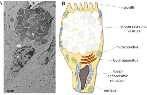
The cytoplasm of goblet cells is occupied by a structure known as distended theca which contains granules. In these vesicles is present a family of large glycoproteins called mucins, which constitute the main component of the mucus. Mucins have a polymeric structure and are attached to numerous side chains of hygroscopic and hydrophilic oligosaccharides (O-linked-oligosaccharide). Glycans contribute to the sticky binding sites formation in the hydrogel, thus trapping microorganisms to facilitate their subsequent elimination (Barr et al., 2013). In humans, more than 20 different mucin types have been detected, which are encoded by mucin genes represented as MUC, followed by a number associated to the chronological order in which they were identified. Mucins are classified into two structural groups: membrane-associated (or transmembrane) mucins, among which are MUC1, MUC3B, and MUC3A, the latter being specific to the intestinal epithelium, and secreted mucins such as MUC5AC, MUC5B, MUC6, MUC19 and MUC2 (Dhanisha et al., 2018). The synthesis process of these mucins as simple polypeptides passes through the endoplasmic reticulum and the Golgi apparatus, where they undergo different post-translational modifications to complete their maturation, such as N/O-glycosylation, fucosylation and addition of other chemical groups such as sulfates or acetyls (Faderl et al., 2015; Thomsson et al., 2012; Matsuo et al., 1997).
Fig. 2: Structure of intestinal goblets cells. A) Electron micrograph of ileal goblets cells. B) Schematic
presentation of different cellular components. (microvilli, V: mucin secreting vesicles, mitochondria, Golgi apparatus, ER: rough endoplasmic reticulum, n: nucleus).
MUC2 constitutes the most abundant secreted mucin in the mucus of both the small intestine and colon and plays a determining role in the organization of the hydrogel layer (Johansson et al., 2008). Several studies suggest that MUC2 has approximately 5100 amino acids and is located within 29.5 kb DNA fragment, which is found at chromosomal locus 11p15.5 (Dekker et al., 2002; Allen et al., 1998). The monomers of this glycoprotein contain two highly glycosylated core domains known as proline, threonine, and serine-rich mucin domains (PTS domains), the cysteine-rich N- and C-terminal regions with 1300 and 1000 amino acids respectively, and four D-domains of the von Willebrand factor. All these regions are folded and stabilized from the presence of numerous disulfide bonds, resulting in a viscous network of gel-forming mucins. The rodent homologs of human MUC2 display a motif of similar structure (Lang et al., 2007; Birchenough et al., 2015). After being processed in Golgi apparatus, the MUC2 glycoprotein is densely packed into the secretory granules due to the low pH and the high concentration of Ca2+ in these compartments, to then be released
by different routes: the constitutive (continuous secretion at basal levels) or stimulated Ca2+
-dependent pathways. The production and massive secretion of MUC2 by exocytosis during stimulated secretion can be initiated by different external stimuli such as hormones/neurotransmitters (prostaglandins, acetylcholine), microorganisms and their
products, cytokines (IL-22, IFN-γ) as well as reactive oxygen and nitrogen species. This process occurs through activation of secondary messengers such as Ca2+ (compound
exocytosis that allows the fusion and release of multiple mucin granules) or cyclic adenosine monophosphate (cAMP) which induces the release of simple granules (Kim and Ho, 2010; Songhet et al., 2011; Turner et al., 2013). Recent advances suggest a connection between mucin secretion by compound exocytosis and the formation of goblet cell-associated antigen passages directly activated by the action of acetylcholine on muscarinic receptors of these cells (Knoop and Newberry, 2018). After being produced, the small intestine mucus is not attached to the epithelium but easily carried along the GI by peristalsis. This phenomenon coupled to the fast mucus renewal contribute to maintaining the protection of the villi due to elimination of bacteria from the surface, in addition to the fact that there is a high concentration of antimicrobial factors in this zone (Ermund et al., 2013; Vaishnava et al., 2011). However, this effect is not observed in germ-free mice where MUC2 remains bound to goblet cells. In order to release this mucin from the production site, the proteolytic enzyme meprin β needs to be activated. This metalloprotease is located at the enterocyte apical membrane and is activated when exposed to bacteria, which allows its release and diffusion in the mucus. Thus, it appears that there is a close relationship between the production-release mechanisms of mucus and the presence of bacteria in the small intestine (Schütte et al., 2014). Other factors synthesized by goblet cells to ensure mucus quality are Fcg-binding protein (FCGBP) (directly linked to MUC2 to act as a cross-linker in the network of this protein), AGR2 (linked to goblet cell differentiation), CLCA1, the granulated zymogen 16 protein (ZG16) (linked to bacterial aggregation and prevention of diffusion into the mucus layer), Trefoil factor 3 (TFF3), and the resistin-like pro-inflammatory molecule RELMβ (Allaire et al., 2018). TFF3 is the second most abundant product in secretory vesicles and belongs to a family of small peptides highly expressed in the GI. Its activity is associated with structural integrity of the mucus and the protection and repair of epithelial tissues by inhibiting, for instance, apoptosis. This factor is also related to the migration of IECs and angiogenesis. Furthermore, it contributes to the innate immune response mediated by different receptors such as the Toll-like receptor family (Taupin and Podolsky, 2003; Podolsky et al., 2009; Kjellev, 2009). However, its antimicrobial activity has not been widely studied. In contrast, RELMβ is strongly induced apically during enteric infections such as in murine helminth
infection and IBD models. Its activity is linked to the chemotaxis inhibition of parasites. On the other hand, RELMβ participates in MUC2 secretion, T cells responses and during macrophages activation (Herbert et al., 2009; Bhinder et al., 2014; Nair et al., 2008). Thus, goblet cells contribute dynamically to the intestinal barrier functions. MUC2 is a crucial component for these functions and its depletion from the mucus layer contributes to the development of inflammatory diseases and cancer via permissive interactions between commensal bacteria and the epithelium (Velcich et al., 2002; Van der Sluis et al., 2006; Bergstrom et al., 2010; Tadesse et al., 2017; Johansson et al., 2014).
1.2.1.1 Antimicrobial components in the mucus
Goblet cells are not the only entities to ensure defense mechanisms and protection of the host against microorganism invasions. Other epithelial cell lineages such as enterocytes and Paneth cells are involved and produce a number of components with antimicrobial properties. Although enterocytes are capable of synthesizing some antimicrobial peptides (AMP), Paneth cells of the small intestine are the major source for the production of these molecules. These cells are characterized with granules filled with peptides and enzymes such as lysozyme, cathelicidins, α-defensins (cryptdins in mice), phospholipase A2 (sPLA2), etc,
(Gallo and Hooper, 2012; Bevins and Salzman, 2011; Bel et al., 2017). AMP induce bacterial lysis due to pore formation in the membrane of these microorganism. On the other hand, specific enzymes such as cryptdin 3 cause an increase in water secretion within the intestinal lumen, allowing physical detachment of bacteria near the epithelial surface. Furthermore, α-defensins (more than 20 cryptdin molecules in mice) are related to the adaptive immune response (Chu et al., 2012; Elphick and Mahida, 2005; Lencer et al., 1997). Other AMP such as REGIII (γ, β) molecules that belong to the C-type lectin family cause destabilization of the Gram-positive bacterial membrane by direct binding to phospholipids. More specifically, REGIIIγ has the main function of keeping the small intestinal epithelial surface sterile and free of bacteria (Vaishnava et al., 2011; Mukherjee et al., 2014).
sIgA is a fundamental molecule of the GI immune response and is produced by plasma cells of the lamina propria. sIgA synthesis contributes to the defense against intestinal pathogens and the control of the commensal microbiota, based on its ability to sequester antigens and microbes through a process known as immune exclusion. Murine models made deficient in sIgA synthesis showed a significant imbalance in the composition of the commensal bacterial
community. These observations suggest an important role for these antibodies in modulating the diversity and quantity of bacteria in the intestine. It has also been proposed that the highest sIgA response is obtained when bacteria reach the inner mucus layer, this being a powerful indication of subsequent penetration/invasion through the intestinal mucosa (Palm et al., 2014; Suzuki et al., 2004). sIgA is secreted to the mucosal surface as polymeric antibodies under stable conditions, more specifically as immunoglobulin dimers that interact with the polymeric immunoglobulin receptor (pIgR) on its way through epithelial cells toward the lumen. The expression of this receptor in the intestine can be regulated by cytokines such as IL-17 or by activation of signaling pathways such as NF-κB. Two antibody subclasses are distinguished in humans: IgA1 and IgA2, the latter being more stable to bacterial proteolytic degradation and therefore predominant in the mucosa (Cerutti, 2008; Mathias et al., 2015; Brandtzaeg, 2013; Pabst and Slack, 2020).
In summary, these multiple secretory strategies emerging from the epithelial barrier contribute to limiting or resolving pro-inflammatory events and to maintaining the integrity and a healthy state of the intestinal epithelium.
1.2.2 The junctional complexes as a physical barrier of the intestinal epithelium
The epithelial barrier controls ions, nutrients and water passage from the lumen to the organism and restrict translocation of luminal antigens such as microorganisms and their harmful derivatives. Transport in the epithelium occurs by three mains pathways: the transcellular pathway, the transporter-mediated pathway and the paracellular pathway. The fundamental characteristic of the paracellular pathway is to allow the passage of molecules by passive diffusion between the spaces of adjacent cells. Precisely, the function of the narrow physical barrier that controls and regulates the paracellular transport of various molecules is determined by a protein complex that forms the so-called intercellular junctions, made up of the tight junctions (TJ) in the most apical and lateral areas of the cells, followed by the adherens junctions (AJ), the desmosomes and the Gap junctions.
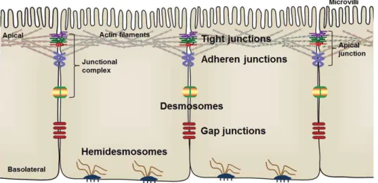
Fig. 3: Distribution of the intercellular junctional complex among intestinal epithelial cells. Tight junctions
are located in the most apical and basolateral area of IECs and are responsible for sealing the epithelial barrier. Adherens junctions interconnect with actin filaments, participate in cell signaling processes and, like the desmosomes, maintain cohesion and mechanical adhesion between cells. Hemidesmosomes connect the cell intermediate filaments to basal lamina, allowing cell anchoring and the interactions with other proteins. Gap junctions form tunnels between the cells for ions and water molecules transport (Zhu et al., 2018), with
permission from Taylor & Francis. 1.2.2.1 Tight junctions
TJ are formed by the assembly of a group of three families of cytosolic and transmembrane proteins: the Claudins (CLDN), the occludins (OCDLN), and the junctional adhesion molecules (JAM) (Luissint et al., 2016). In addition to limiting selective traffic through the paracellular barrier of the epithelium, TJ determine the correct polarity of IECs. They form a dynamic multiprotein complex programmed to open and seal the barrier during normal or threatening conditions from the environment. Among the components of TJ are the zonula occludens protein family members (ZO 1-3) and cingulin, whose functions are related to TJ stabilization via interaction with cytoplasmic proteins of the peripheral membrane and the actin cytoskeleton (Cereijido et al., 2007). Similarly, other proteins such as JAM, have a central role in the regulation of the epithelial barrier, migration and cell proliferation. While occludins have not been widely studied and some controversy exists in regard to their functional activity, they are involved in signal transduction processes and in strengthening the structural properties of TJ (Al-Sadi et al., 2010; Luissint et al., 2014 ; Luissint et al., 2016; Zhu et al., 2018).
TJ are mainly made up of CLDN, which play a key role in maintaining the paracellular barrier function. This family of approximately 27 isoforms, at least identified in mammals, are integral membrane proteins with unique characteristics in terms of expression and
distribution throughout the epithelial tissues (Cording et al., 2013). Many of the CLDN members have tensile or selective barrier-forming properties, allowing cell stability (CLND1, 3, 4, 5, 11, 14 and 18), while others are classified as leak CLDN (CLDN2, 10, 15 and 17), because they increase paracellular permeability, from the formation of channels or pores that allow the anions, cations, and water passage (Lee, 2015; Günzel and Yu, 2013). Different studies show that CLDN composition varies depending on age and the GI tract segment analyzed. Whereas some isoforms such as CLDN2 and CLDN15 are expressed mainly at the crypt base, others such as CLDN3, CLDN4, and CLDN7 are localized toward the luminal zone. Some studies suggest an apparent dissociation or alteration in the intestinal CLDNs during the development of inflammatory events, thus highlighting the importance of CLDNs for optimal IEC barrier function (Capaldo and Nusrat, 2015; Zeissig et al., 2007; Findley and Koval, 2009).
1.2.2.2 Adherens junctions
The AJ located below the TJ maintain mechanical bonding between adjacent cells and participate in the cellular signaling processes leading to the regulation of gene transcription (Takeichi, 2014). This adhesion is maintained by a transmembrane proteins complex including cadherins and nectins, which interact at the same time with other anchoring proteins (α- and β-catenins) while making a bridge with the actin filaments (Ivanov and Naydenov, 2013). E-cadherin constitutes one of the most important molecules among the members of the AJ. Its intestinal epithelial functions are not only limited to the cellular adhesive properties and desmosomes formation, but also impact cell migration and proliferation through the Wnt signaling in the intestinal crypt cells (Nelson and Nusse, 2004; Groschwitz and Hogan, 2009). Both TJ and AJ are regulated by kinases or phosphatases that induce phosphorylation events, thus controlling the interaction with other proteins and their enzymatic activity (Bertocchi et al., 2012; Garcia et al., 2018).
The imbalance in the intercellular junctional organization and therefore, a loss of the intestinal barrier functionality can lead to the appearance of different diseases, one of the most studied being inflammatory bowel diseases (IBD).
1.3 Inflammatory Bowel Diseases
IBD are a group of chronic disorders that cause inflammation of the GI tract. IBD are mainly represented by Crohn's disease (CD) and ulcerative colitis (UC), which are characterized with periods of active and remission phases. UC is characterized by diffuse inflammation of the colonic mucosa with the most common symptoms being bloody diarrhea. On the other hand, CD causes ulceration of different parts of the intestinal tissue, especially at the level of the terminal ileum and colon. CD is characterized by symptoms of diarrhea, abdominal pain, and weight loss. Although IBD can be diagnosed at any age, a huge majority of new cases are detected during adolescence or early adulthood (De Souza and Fiocchi, 2016).
The incidence of IBD is high worldwide with a higher prevalence in Western countries. Nevertheless, many newly industrialized or increasingly developing countries in Asia, Middle East, Africa, and South America are experiencing a rapid increase in the incidence of these diseases (Kaplan and Ng, 2017). Canada has one of the highest incidence/prevalence rates. In 2018, approximately 270,000 individuals were reported to be living with IBD in the country and this number could rise to approximately 400,000 by 2030. Although there are advances in elucidating the mechanisms that influence the development of these diseases, there are currently no accurate treatments that allow the cure of patients affected by IBD. The etiology or pathogenesis of IBD is attributed to the presence of complex interactions among a group of factors including genetic predisposition of the host, dysregulated immune response to environmental triggers (diet factors, alcohol, antibiotics, smoking, stress) and imbalance in the dynamic behavior of the commensal microbiota, known as dysbiosis (Abraham and Cho, 2009).
1.3.1 Intestinal barrier dysfunction in IBD
There is now clear evidence that integrity of the intestinal epithelial barrier is important in the development of IBD, since it constitutes the delimiting factor for exposure of the microbiota to the host immune system. Consequently, defects in intestinal mucosa homeostasis can trigger alteration of intestinal permeability. This will lead to antigens translocation and activation of signaling cascades causing apoptosis, erosion, and ulceration, which are considered as crucial steps in the initiation or development of chronic intestinal inflammatory disorders (Mankertz and Schulzke, 2007; Antoni et al., 2014; Zeissig et al., 2004; Schulzke et al., 2006). It has been shown that in UC or CD patients, there is an altered
expression of the proteins that make up the TJ (Takeuchi et al., 2004; Hollander, 1988; Schmitz et al., 1999; Prasad et al., 2005).
CLDN1, among the most studied CLDNs, regulates intestinal homeostasis and modulates the activation of the NOTCH signaling pathway. There are growing evidences that suggest a significant increase in the expression of CLDN1 in murine tissue in the presence of active inflammation, leading then to the activation of NOTCH and therefore the inhibition of goblet cells differentiation with the result of reducing MUC2 production and mucus formation (Poritz et al., 2011; Pope et al., 2014). Precisely, Gowrikumar et al. proposed that CLDN1 in IBD patients may constitute a possible marker of severity in colitis and malignancy by promoting susceptibility to colitis-associated cancer (Gowrikumar et al., 2019). Other studies suggest that in the intestinal mucosa of patients who develop CD, there is a change in the expression pattern of CLDN3, -5, and -8 (sealing process) as well as for the occludin protein (Zeissig et al., 2007; Goswami et al., 2014). Similarly, in UC, a decrease and delocalization of some TJ proteins, such as occludin, CLDN3, -4, and -7 was reported (Prasad et al., 2005; Oshima et al., 2008; Heller et al., 2005; Weber et al., 2008). Deficiency in CLDN7 results in dysregulation of the paracellular flow of small molecules such as microbial products (bacteria-derived N-formyl-L-methionyl-L-leucyl-L-phenylalanine peptides), causing inflammation in murine models. It also promotes the activation of certain metalloproteinases activating in turn signaling events such as the NF-κB pathway to finally induce the inflammatory process and the loss of cell-extracellular matrix interaction (Tanaka et al., 2015; Ding et al., 2012). On the other hand, CLDN12 was shown to be induced in IBD patients, especially into the ileum of CD patients (Lameris et al., 2013). This protein is involved in vitamin D-dependent Ca2+ permeability in the intestinal epithelium (Fujita et al.,
2008). Additionally, CLDN18 exhibits higher expression in tissues samples of UC patients, as well as in mice during experimental colitis (Zwiers et al., 2008; Dotti et al., 2017). Strand, an integral membrane protein of approximately 24.5 kDa, influences cell permeability and transepithelial electrical resistance (TER) due to the formation of channels allowing the transport of water and small cations such as Li+, Na+, and K+ (Rosenthal et al., 2010; Weber
et al., 2015). CLDN2 is mostly expressed in the GI epithelial tract in both humans and rodents at birth. However, its expression decreases rapidly afterward (Tamura et al., 2011; Holmes et al., 2006; Ong et al., 2020). CLDN2 becomes highly expressed during intestinal
inflammation and IBD accordingly to the severity of the disease (Lameris et al., 2013; Hering et al., 2012; Luettig et al., 2015). Increase expression of CLDN2 can trigger the alteration of the TJ, leading to the loss of solutes and fluids through the epithelial barrier, the appearance of symptoms such as diarrhea, and ultimately antigens translocation. Recently, Raju et al. showed that inactivation of the channels formed by CLDN2 attenuated immune-mediated colitis in mice (Raju et al., 2020).
Several studies indicate that cytokines also contribute to alteration of the intestinal epithelial barrier during IBD. Indeed, IL-13, TNFα and IFN-γ can stimulate expression of CLDN2 in
in vitro models and reduce expression of other barrier-forming proteins such as CLDN1, -5,
-7 or ZO-1 (Mankertz et al., 2009; Weber et al., 2010; Amasheh et al., 2009; Heller et al., 2005). In response to these inflammatory mediators, specific epithelial barrier disorders occur in association with TJ proteins trafficking in the membrane. For example, TNFα and INF-γ can activate the selective endocytosis phenomenon of occludin, CLDN1, CLDN4 and JAM-A (Utech et al., 2010; Bruewer et al., 2005; Marchiando et al., 2010). It was also observed that IL-6, an important pro-inflammatory mediator produced by both immune and epithelial cells, as well as IL-17, induce CLDN2 expression by activating different signaling pathways such as MEK/ERK or PI3K (Kinugasa et al., 2000; Suzuki et al., 2011). On the other hand, TJ assembly depends on myosin light chain kinase (MLCK) that is affected in IBD patients (Blair et al., 2006). MLCK activation and phosphorylation stimulates the redistribution of TJ proteins into subcellular compartments within enterocytes. Induced expression of MLCK caused by TNFα and IL-1β via the activation of NF-κB results in the enhancement of cell permeability as determined by in vitro assays (Ye and Ma, 2008; Al-Sadi et al., 2010). Both cytokines also promote the cleavage of other proteins belonging to the AJ and desmosomes, such as desmocholine 2 or E-cadherin. The latter represents a potential biomarker given that its proteolytic fragments can be detected in the inflamed mucosa of UC patients and experimental colitis models in mice (Kamekura et al., 2015; David and Rajasekaran, 2012).
Defects in the mucus layer can trigger the appearance of IBD. Polymorphism in some MUC genes that code for members of the mucin family have been shown to be in association with the pathogenesis of the disease (Moehle et al., 2006; McCole, 2014; Kyo et al., 2001). In addition, loss of functional MUC2 in mice leads to spontaneous appearance of colitis (Van
der Sluis et al., 2006; Heazlewood et al., 2008). Interestingly, Visschedijk et al. also observed the development of UC cases associated with the production of MUC2 rare variants (Visschedijk et al., 2016).
A decrease in the thickness and uniformity of the mucus layer in UC patients favor commensal microorganisms or their metabolite to come into contact with the epithelium. This can be due to the loss of goblet cells, as well as with the attenuation of their secretory functions that contribute to meeting microbial challenges. Besides, variations in secondary modifications of structural components of the hydrogel network, such as, for example, O-glycosylation or detection of Shorter O-linked oligosaccharide side chains in mucin can impact the quality of the gel. These changes have been correlated with the extent of the inflammatory status and disease activity in the intestine of UC patients (Longman et al., 2006; Strugala et al., 2008; Larsson et al., 2011; Johansson et al., 2014; Fu et al., 2011; Alipour et al., 2016; Van Der Post et al., 2019; Dotti et al., 2017). On the other hand, although a hyperproduction of mucins was confirmed in CD patients, changes in processes of glycosylation and sulphation or gel hydration were proposed as mechanisms contributing to the decrease in viscosity, greater bacterial penetrability and erosion of the mucus layer (Buisine et al., 2001; Abraham and Cho, 2009; Sun et al., 2016). Undoubtedly, the integrity of the mucus barrier constitutes a turning point in the protection and repair of the epithelium during inflammation. However, it is not entirely clear whether such defects are the cause or the consequence of a dysregulated immune system during the initiation of IBD.
1.3.2 Microbiota in IBD
The human intestine is colonized by thousands of microorganisms, mostly of bacteria, which establish mutual relationships by forming a complex microbial ecosystem. This symbiotic interaction between the host and commensal microbes and their metabolites will influence both the intestinal environment, as well as the physiology of many tissues and organs in the human body (Sender et al., 2016). Several evidences support that the intestinal microbiota plays an essential role in various processes such as the maturation of the immune system, the regulation of the neuroendocrine and nervous system response, as well as the initiation and development of various diseases (Heijtz et al., 2011; Uronis et al., 2009; De Filippo et al., 2010). Different studies have demonstrated the beneficial role of these microorganisms for human health. For example, some bacterial species can synthesize a group of essential
nutrients, such as vitamins, which are usually obtained through food (Magnúsdóttir et al., 2015). They can also degrade non-digestible materials from the diet, which generates a set of metabolites such as short-chain fatty acids (SCFA) with multiple positive effects on the body (Gill et al., 2018). In addition, under normal physiological conditions, the intestinal microbiota contributes to energy production in the host organism, integrity of the mucosa and also prevention against pathogenic microorganisms colonization.
The GI tract is relentlessly challenged by the luminal contents harboring innumerable microorganisms and food antigens (Rapozo et al., 2017). Also, genetic variation in the host can affect the microbiome towards dysbiosis (Hall et al., 2017; Imhann et al., 2018). Interestingly, alterations of the gut microbiota are seen in several intestinal adverse conditions, including irritable bowel syndrome, chronic idiopathic diarrhea, celiac disease, and IBD (Shanahan, 2007; Swidsinski et al., 2008; Quigley, 2011; D’Argenio et al., 2016; Eck et al., 2017). The persistence of pathogens in the intestine, an abnormally permeable mucosal barrier leading to excessive bacterial translocation, and the presence of dysbiosis in the commensal microbiota are some of the hypotheses raised to explain the pathogenesis of these diseases (De Hertogh et al., 2008; Kalischuk and Buret, 2010; Liu et al., 2020). Much of the evidences supporting a functional role of the microbiota in the development of chronic inflammatory disorders are from work with animal models, more specifically in germ-free or antibiotic-treated conditions. In germ-free mice with chemical or genetically induction of inflammation, the impact of the bacterial presence was exposed on the appearance or manifestation of symptoms similar to those found in IBD, when rodents were grown under conventional conditions (Khan et al., 2019). It is argued that dysbiosis is a cause as well as an outcome of IBD. Dysbiosis is characterized as the flowering of pathobionts and the loss of commensals or diversity, key elements to be considered in the search of mechanisms and therapies for IBD (Levy et al., 2017; Kostic et al., 2014).
Quantitative and qualitative changes in the composition of the gut microbiota was detected in CD and UC (Alam et al., 2020; Yilmaz et al., 2019; Lloyd-Price et al., 2019). A reduction of up to 25% of microbial genes was observed in IBD patients compared to healthy individuals (Qin et al., 2010). These microbial changes were mostly related to the loss of beneficial species belonging to genera such as Firmicutes or Bacteroides, or families such as
species belonging to Enterobacteriaceae (Escherichia coli) was showed (Chassaing and Darfeuillemichaud, 2011; Hedin et al., 2014; Hedin et al., 2016; Nguyen, 2011; Bien et al., 2013; Li et al., 2014; Gilbert et al., 2016). From metabolomic and taxonomic profile analysis of microbial communities in human feces from IBD patients, Franzosa et al. correlated a loss of taxonomic diversity with depletion of enzymes, chemicals, or differentially abundant metabolites during the manifestation of intestinal dysbiosis (Franzosa et al., 2019).
1.3.2.1 Intestinal microbiota and metabolites modulate the development and function of the immune system and play a critical role in IBD.
SCFA such as butyrate that are produced by intestinal bacteria regulate protective immunity and reduce tissue inflammation. A decrease in the expression and secretion of SCFA, which impair the metabolism of tryptophan, was related to the loss of the integrity of the intestinal barrier in IBD (Schirmer et al., 2019). Bacteria can metabolize tryptophan from the diet. Tryptophan metabolites can in turn interact with the aryl hydrocarbon receptor and attenuate inflammation. Wlodarska et al. reported that IBD patients have low levels of indoleacrylic acid (bacterial indole metabolites), whose function is to regulate mucus production and suppression of inflammatory cytokines (Wlodarska et al., 2017). Similarly, a bacterial imbalance from the reduction of strict anaerobic species, such as Clostridium groups IV and XIVa, against the expansion of aerobic or facultative aerobic bacteria, can lead to an intraluminal increase in oxygen and therefore the development of metabolic changes (inflammation) in the intestine. It is known that these Clostridium species are capable of generating antigenic signals such as retinoic acid that contributes to the suppressive immune response and anti-inflammatory activity from the induction of regulatory T cells dependent on transforming growth factor- (TGF-β), whose activation it is essential for intestinal homeostasis (Mucida et al., 2007; Albenberg et al., 2014; Atarashi et al., 2013; Furusawa et al., 2013). Some other species with great anti-inflammatory potential were also found affected in the commensal microbiota of UC or CD patients. For example, Roseburia,
Faecalibacterium prausnitzii are linked to IL-10 secretion and the production of a 15 kDa
protein with possible inhibitory properties of inflammation by suppressing the NF-κB pathway; Bacteroides fragilis synthesizes the immunomodulatory capsular polysaccharide A (PSA); sphingolipids protect against colitis episodes in mouse experimental models, and
finally Akkermansia muciniphila induces antigen-specific T cell responses and homeostatic IgG production (Liu et al., 2020; Guan, 2019).
1.3.2.2 Other bacteria associated with IBD
Microbes can reduce immune tolerance, leading to inappropriate activation of pathways designed to protect against pathogens invasion. Among the potentially pathogenic microbiota microorganisms related to IBD is the invasive proteobacterium Campylobacter concisus, which inhabits naturally in the oral cavity but can infect the GI tract by damaging the intestinal epithelial barrier through the production of cytokines such as TNFα (Mahendran et al., 2011; Mahendran et al., 2016). Another bacterium widely enriched in the mucosa of UC patients is Fusobacterium varium, whose mechanism of action is to invade IECs, a key event in triggering the inflammatory response (Sekizuka et al., 2017). Likewise, an abundant presence of Ruminococcus gnavus was detected in IBD patients. R. gnavus synthesizes and expresses an inflammatory glucorhamnan polysaccharide and also produces and metabolizes sialic 2,7-anhydro-Neu5Ac. This allows a better adaptation and nutritional competitive advantage of this microorganism in the mucus, due to its ability to break down mucins when compared to other bacteria (Nishino et al., 2018; Henke et al., 2019; Bell et al., 2019). The enterotoxigenic Bacteroides fragilis is also linked to IBD and colorectal cancer development. It promotes cell proliferation, the cleavage of E-cadherin that leads to increased mucosal permeability, and finally the secretion of pro-inflammatory interleukins (Zamani et al., 2017; Rhee et al., 2009). In addition, the invasive and adherent Escherichia coli (AIEC) is among the most studied microorganisms with the highest prevalence in samples from CD patients (Darfeuille-Michaud, 2002; Desilets et al., 2016; Prorok-Hamon et al., 2014). This bacterium colonizes the intestinal mucosa via the adhesin FimH localized at the end of the bacterial pilus or by invasion through type 1 pili, a crucial virulence factor for the recognition of the Carcinoembryonic antigen-related cell adhesion molecule 6 receptor expressed on IECs. Furthermore, AIEC can penetrate the epithelium via the M cells localized on the surface of the Peyer's patches, thus interacting with the immune cells of the lamina propria and inducing the inflammatory response. The infection, replication, and survival of this pathogen in macrophages allow the secretion of high levels of TNFα without inducing cell death (Lee et al., 2019; Palmela et al., 2018). On the other hand, some authors suggest the predominance and risk of suffering from chronic inflammatory processes, mainly UC, after successive
repetitions of infection with Salmonella. In a study carried out by Tripathi et al. a 79.7% positivity rate for specific Salmonella sequences in UC patients was identified by PCR compared to the 16.3% observed in healthy individuals (Tripathi et al., 2016). This suggests that the prevalence of Salmonella can be linked to the development of IBD. Similar conclusions were raised using Salmonella Typhimurium as a model of infection in mice made deficient for IL-10 or exposed to DSS during experimental colitis (Schultz et al., 2018). As additional pathogens linked to IBD, sulfate-reducing bacteria obtain their energy by oxidizing organic compounds or hydrogen while reducing sulfate to hydrogen sulfide (H2S),
which promote cleaving the disulfide bond in mucins and therefore favoring bacterial translocation (Ijssennagger et al., 2016). Some of these bacterial populations, such as
Desulfovibrio species, are overexpressed in samples of the intestinal mucosa and feces from
IBD patients (Mills et al., 2008; Verma et al., 2010; Loubinoux et al., 2002; Rowan et al., 2010). Finally, there are similar evidences to support a role for mycobiota dysbiosis in the pathogenesis of IBD. Specifically, Candida albicans were found to be abundantly expressed in CD and UC patients (Li et al., 2014; Mar et al., 2016).
1.3.3 Genetic variants and IBD
Genome-wide association studies (GWAS), as well as meta-analysis studies have become successful tools for the study of complex diseases including IBD. There are now more than 240 genetic risk loci identified for IBD, including some that are shared between CD and UC (De Lange et al., 2017; Uhlig and Muise, 2017). The clustering of these genes or loci involved with the onset or progress of these diseases shows important links with specific pathways including immune response, autophagy, cellular metabolic processes, defense, repair and intestinal epithelium barrier function (Khor et al., 2011; Graham and Xavier, 2020) (Fig. 4). Among the most studied genes are NOD2, ATG16L1, IRGM, 23R, CARD9, RNF186,
IL-17RA, and PRDM1, with many of them containing several variants associated with risk or
Fig. 4: Different pathways and genes identified as risk factors in IBD pathology. A) Sensing of
microorganisms by phagocytes that induce the activation of cytokines or oxidative stress genes capable of promoting inflammation. B) The intestinal epithelial barrier, the complex of cell junctions and genes involved in the mechanisms of repair and maintenance of cell integrity. C) The adaptive immune system (B, T and dendritic cells) that trigger the specific or memory response against microorganisms. D) Disturbance in fibroblast cells that contributes to the dynamic response to inflammatory events. E) Pathways and cellular stress factors (hypoxia, autophagy) related to inflammation and cell death pathways. F) Cytokines that regulate the interconnection between stromal cells, cells of the immune system and IEC. G) Formation of the inflammasome complex through the recognition of antigens or extracellular microorganisms and activation of pro-inflammatory cell death pyroptosis (Graham and Xavier, 2020), with permission from Nature.
1.3.3.1 Intestinal barrier genes and IBD
Some works suggest a strong association between genetic markers involved in the formation, maintenance, and regulation of the intestinal epithelial barrier and the etiopathology of IBD. Understanding of the mechanisms involved in these processes could allow a better diagnosis and classification of patients and the application of possible treatment therapies. CDH1 is associated with UC and encodes for E-cadherin, a component of AJ (McGovern et al., 2010).
CDH1 locus risk haplotypes rs12597188, rs10431923, and rs9935563 are associated with
(Muise et al., 2009). GNA12 (rs798502), which codes for the Gα12 protein, is also related with UC. This protein is a GTPase that is bound to the cellular membrane and interacts with accessory proteins such as ZO-1 to promote correct TJ assembly (Anderson et al., 2011; Meyer et al., 2002). Protein tyrosine phosphatase nonreceptor type 2 gene locus 18p11 variants (PTPN2; rs2542151, rs7234029) are associated with CD. A possible PTPN2-mechanisms of action in the disease is through the strong activation of transcriptional agents such as NF-κB (Consortium et al., 2007; Barrett et al., 2008; Weersma et al., 2009; Glas et al., 2012). IBD genetic associations were also made for genes that code for the mucins. For example, the MUC3 gene as well as rare alleles of the MUC3A gene inserted with an unusual number of 51bp repeat units were both associated with UC and CD (Kyo et al., 2001). A single nucleotide polymorphism (rs3180018 at 1q22) within the MUC1 gene was also confirmed as a risk locus for CD. Furthermore, overexpression of MUC1 with an hypoglycosylation status bringing more susceptibility to degradation was observed in IBD patients (Franke et al., 2010). Finally, Vancamelbeke et al. carried out a study to determine the high or low polygenic risk of association with CD or UC. From a set of 128 genes related to the intestinal barrier, they observed an enrichment of some of these genes in IBD samples. In addition to the modulated expression of HNF4A, they confirmed an important dysregulation of many genes of the epithelial barrier in the ileum and colon during the active phase of UC and CD, including CLDN1 and CLDN8. They also identified significant modulations in mucosal layer genes such as MUC1, whose dysregulation was also maintained during the inactive phase of the diseases (Vancamelbeke et al., 2017).
1.4 HNF4A as a susceptibility gene in IBD and its controversial role in the control of the intestinal epithelial barrier.
HNF4A is a single gene located on chromosome 20 in humans and encodes the transcription
factor hepatocyte nuclear factor 4-α (HNF4α). This transcription factor (TF) is a member of the superfamily of ligand-dependent nuclear receptors (NR2A1) and has twelve isoforms, divided into P1 or P2 promoter-driven isoform classes (Babeu and Boudreau, 2014; Bridgham et al., 2010). HNF4α is expressed in multiple organs including the liver, pancreas, stomach, kidneys and the epithelium of the whole intestine (Drewes et al., 1996; Jiang et al., 2003; Sladek et al., 1990). Many functions have been attributed to HNF4α such as lipid and glucose metabolism, regulation of cell proliferation and differentiation and regulation of key
components of cell junctions (Battle et al., 2006; Hayhurst et al., 2001; Babeu and Boudreau, 2014; Lili et al., 2016). Although its functions have been extensively studied in the liver, HNF4α was also showed to play an essential role in the morphogenesis of the GI tract. Hnf4a-null mice showed high lethality during embryogenic development with severe defects in visceral endoderm formation (Chen et al., 1994). Additionally, it was observed that Hnf4a deletion in the mouse embryonic colon led to disrupted crypt topology characterized by a decreased number of epithelial and mature goblet cells (Garrison et al., 2006). Some studies have linked HNF4α with IBD. A significant reduction in intestinal HNF4α levels was observed in both UC and CD human samples (Ahn et al., 2008; Darsigny et al., 2009). Coincidently, mice conditionally deleted for Hnf4a in the intestinal epithelium were found to be more sensitive to experimental colitis when exposed at young age (Ahn et al., 2008), while displaying signs of spontaneous chronic inflammation resembling human IBD at older age (Darsigny et al., 2009). Subsequently, GWAS studies identified the HNF4A gene locus as being significantly enriched from UC cohorts (Barrett et al., 2009; Van Sommeren et al., 2011; Jostins et al., 2012; Chellappa et al., 2016). Genetic variations were also detected in the P2 promoter of HNF4A (rs1884613) in association with childhood-onset of CD (Marcil et al., 2012).
HNF4α interacts with genes that participate in the formation of a narrow intestinal epithelial barrier and that are functionally involved in IBD. Parviz et al. showed that the loss of HNF4α in the fetal liver induces an important reduction in E-cadherin expression (Parviz et al., 2003). In contrast, colonic epithelial loss of HNF4α did not influence E-cadherin expression (Garrison et al., 2006). Using a tamoxifen-inducible Villin-Cre;Hnf4afx/fx conditional knockout mouse model, Cattin et al. showed an increase in IECs proliferation in association with the activation of Wnt/β-catenin signaling pathway (Cattin et al., 2009). They also reported an increase in intestinal permeability in these mutant mice. Coincidently, they observed a decrease in the expression of ZO-1, Cldn4, and Cldn7 gene transcripts and an increase in Cldn2 expression. Therefore, they reported a switch in E-cadherin localization from the membrane to the cytoplasm of IECs (Cattin et al., 2009).
Using a constitutive intestinal epithelial conditional knockout mouse model, Darsigny et al. showed that the loss of HNF4α decreased mucosal ion transport without affecting barrier permeability and this, at a moment preceding the spontaneous manifestation of IBD-related