Acute Stress Differentially Affects Aromatase
Activity in Specific Brain Nuclei of Adult Male and
Female Quail
Molly J. Dickens, Charlotte A. Cornil, and Jacques Balthazart
Groupe Interdisciplinaire de Génoprotéomique Appliquée Neurosciences, University of Liege, B-4000 Lie`ge, Belgium
The rapid and temporary suppression of reproductive behavior is often assumed to be an important feature of the adaptive acute stress response. However, how this suppression operates at the mech-anistic level is poorly understood. The enzyme aromatase converts testosterone to estradiol in the brain to activate reproductive behavior in male Japanese quail (Coturnix japonica). The discovery of rapid and reversible modification of aromatase activity (AA) provides a potential mechanism for fast, stress-induced changes in behavior. We investigated the effects of acute stress on AA in both sexes by measuring enzyme activity in all aromatase-expressing brain nuclei before, during, and after 30 min of acute restraint stress. We show here that acute stress rapidly alters AA in the male and female brain and that these changes are specific to the brain nuclei and sex of the individual. Specifically, acute stress rapidly (5 min) increased AA in the male medial preoptic nucleus, a region controlling male reproduc-tive behavior; in females, a similar increase was also observed, but it appeared delayed (15 min) and had smaller amplitude. In the ventromedial and tuberal hypothalamus, regions associated with female reproductive behavior, stress induced a quick and sustained decrease in AA in females, but in males, only a slight increase (ventromedial) or no change (tuberal) in AA was observed. Effects of acute stress on brain estrogen production, therefore, represent one potential way through which stress affects reproduction. (Endocrinology 152: 0000 – 0000, 2011)
I
t is often assumed that one key feature of the adaptive stress response in vertebrates is the quick and reversible suppression of reproductive behavior that allows the an-imal to escape and recover from the immediate threat of a stressor (1, 2). Perception of a noxious stimulus, a stressor, induces an increase in glucocorticoids [primarily cortico-sterone (CORT) in avian species] resulting from a cascade of signaling hormones through the hypothalamic-pitu-itary-adrenal axis (3). The acute stress response and ele-vation in glucocorticoids have been linked to suppression of sexual behaviors in mammals (4, 5), amphibians (6, 7), and birds (8), but how stress produces these behavioral effects at the mechanistic level remains unclear.Here, we investigate one possible mechanism underly-ing the influence of the stress response on reproductive behavior by focusing on stress-related changes in brain
aromatase activity (AA). The potential for rapid changes in AA in the male hypothalamic/preoptic area (HPOA) has been well established in the context of sexual interaction with a female (9, 10).
Aromatization of testosterone (T) into 17-estradiol (E2) is a critical element in the activation of most aspects of male reproductive behavior in mammalian and avian species (11). AA is controlled by mechanisms acting in two time domains: 1) in the long term (hours to days), the concentration and activity of the enzyme are controlled by genomic actions of steroids, which modulate the transcription of the aromatase gene; and 2) in the short term, the activity of existing enzy-matic molecules changes rapidly (minutes) after exposure in
vitro to phosphorylating conditions or neurotransmitters,
such as glutamate, or after exposure to and sexual interaction with a receptive female in vivo (12, 13).
ISSN Print 0013-7227 ISSN Online 1945-7170 Printed in U.S.A.
Copyright © 2011 by The Endocrine Society
doi: 10.1210/en.2011-1341 Received June 6, 2011. Accepted August 15, 2011.
Abbreviations: AA, Aromatase activity; CORT, corticosterone; E2, 17-estradiol; EIA,
en-zyme immunoassay; HPOA, hypothalamic/preoptic area; 3-HSD, 3-hydroxysteroid de-hydrogenase; mBST, medial portion of the bed nucleus of the stria terminalis; PAG, peri-acqueductal gray; POM, medial preoptic nucleus; T, testosterone; TnA, nucleus taeniae of the amygdala; Tub, tuberal; VMN, ventromedial.
N E U R O E N D O C R I N O L O G Y
Endocrinology, November 2011, 152(11):0000 – 0000 endo.endojournals.org 1
Interestingly, rapid up-regulation or down-regulation of brain estrogen content (via acute injection of E2or an aromatase inhibitor) modulates in parallel (stimulate or inhibit, respectively) the expression of sexual behavior in quail or mice (14 –16). Rapid changes in brain AA and consequently in local E2concentrations thus represent a plausible link between exposure to acute stress and inhi-bition of reproductive behavior.
To clarify the potential role of aromatase in linking the stress response and sexual behavior, we tested the tempo-ral effects of acute stress on AA in brain nuclei that contain dense populations of aromatase-expressing neurons (17). We identified anatomicaly discrete and sex-specific up and down-regulations of enzymatic activity that are likely to mediate at least in part effects of stress on reproductive behavior.
Materials and Methods
Animals
A total of 103 Japanese quail (Coturnix japonica) were used (females, n⫽ 51 and males, n ⫽ 52). This species has been well established as an important model species for studying the aro-matase system (11, 18). All experiments were conducted on 10-to 14-wk-old quail raised from eggs obtained from our breeding colony. All birds were maintained on a long day photoperiod (16 h light and 8 h dark) and kept gonadally intact. Birds were pro-vided with food and water ad libitum. Experiments complied with the Belgian laws on the Protection of Experimental Animals and were approved by the Ethics Committee for the Use of An-imals at the University of Lie`ge (protocol no. 1027).
Stress protocol
Birds of each sex were randomly assigned to one of five groups: baseline (represented here as 0 min), 5, 15, or 30 min of stress and 30 min of stress plus 30 min of poststress recovery (represented here as 60 min). In all stress groups (5, 15, 30, and 60 min), quail were restrained for the specified duration in a restraint cage (similar to the cage described in Ref. 19) made of a metal cylinder (10-cm diameter ⫻ 16-cm length) that was shown in preliminary tests to cause a significant elevation in plasma CORT concentrations. Birds in the 60-min group were placed for 30 min in the restraint cage and then released to their home cage and sampled 30 min later.
At the specified time for each group, blood samples (be-tween 0.1 and 0.5 ml) were collected from both brachial and jugular veins to allow an assessment of potential differences between peripheral and central hormone concentrations, re-spectively (20). Immediately after blood sample collection, birds were killed and the brain dissected from the skull and rapidly frozen on dry ice. These samples were also stored at ⫺80 C until assayed.
Blood samples and brains were collected as quickly as pos-sible, with all samples (in the order of brachial, jugular, and brain) obtained and brain placed on dry ice within 4 min from the time the bird was removed from the cage. Latency between
re-moval from the restraint cage and sample collection was on av-erage 1 min, 1 min 40 sec, and 3 min for brachial or jugular blood and brain, respectively (maximum lower than 2 min, 3 min 30 sec, and 4 min, respectively). Follow-up studies have demon-strated that patterns in AA observed here do not differ from those seen in birds immediately killed and not subjected to blood sam-pling. Birds were not anesthetized to prevent neurochemical changes and additional time under stress. All sampling was con-ducted in the morning between 0900 and 1200 h.
Micropunch dissections
To determine how AA in individual brain nuclei responded to stress, the following brain nuclei were targeted for collection using a modified Palkovits punch technique (21) that had been validated for use with quail (22, 23): the medial preoptic nucleus (POM), medial portion of the bed nucleus of the stria terminalis (mBST), nucleus taeniae of the amygdala (TnA), ventromedial (VMN) and tuberal (Tub) hypothalamus, and the periacqueduc-tal gray (PAG). Tissue collection was guided by the knowledge of the exact location of these populations of aromatase-expressing cells (17) and the micropunch technique is thoroughly described in Ref. 23. Punches were expelled into a frozen Eppendorf tube using a cold air filled syringe, kept frozen, and maintained at⫺80 C until assayed.
AA assay
As described in Cornil et al. (23), micropunches were homog-enized in 120 l of ice-cold buffer [150 mMKCl, 1 mM Na-EDTA, and 10 mMTris-HCl (pH 7.2)] using a glass pestle spe-cifically fitting 1.5-ml Eppendorf tubes. AA was determined by measuring the production of tritiated water associated with the conversion of [1-3H]-androstenedione into estrone (24, 25).
This assay has been thoroughly described in published studies, most recently in Cornil et al. (23). Samples were randomly as-signed to and run within nine assays, each containing multiple internal controls [HPOA homogenates expressing high (male) or low (female) AA]. The intraassay coefficient of variability was less than 5.3%, whereas the interassay coefficient of variation was less than 4.5%.
To account for differences in the amount of tissue actually obtained from the micropunches, results were corrected for pro-tein concentrations. A commercially available Coomassie Plus Protein Assay reagent (Pierce, Rockford, IL) was used to deter-mine protein content using 10l of the micropunch homoge-nates (duplicates of 5l). All results are, therefore, expressed as a measure of pmol of aromatization product ⫻ h⫺1 ⫻ mg
protein⫺1.
CORT and T enzyme immunoassays (EIA)
Circulating concentrations of steroids were assayed using EIA kits (Cayman Chemical Co., Ann Arbor, MI). First, we val-idated each kit using a procedure similar to that described in Washburn et al. (26). Two pooled samples were used, one with high expected concentrations (stressed individuals for CORT and males for T) and one with low expected concentrations (baseline samples for CORT and females for T). To test for par-allelism, these samples were serially diluted and compared with the standard curve of the assay. For CORT, we used unextracted plasma samples as well as samples that had been extracted from the plasma using dichloromethane (for details, see below). We
determined that, at lower dilution factors, substances in the plasma interfere with the assay such that extraction is required to obtain an accurate measurement. We also tested the accuracy of the assays by adding known amounts of steroids to these samples. These tests demonstrated that these assays accurately and reliably quantify CORT and T in extracted plasma samples from quail. The cross-reactivity for the CORT antiserum as re-ported by Cayman is 100% with CORT, 11% for 11-dehydro-corticosterone, 7% for 11-deoxy11-dehydro-corticosterone, and less than 1% for other steroids. The cross-reactivity for the T antiserum as reported by Cayman is 140% with 19-nortestosterone, 100% for T, 27% for 5␣-dihydrotestosterone, 19% for 5-dihydrotes-tosterone, and less than 5% for all other steroids.
CORT was assayed by EIA in both brachial and jugular sam-ples. CORT was extracted from the protein component of the plasma using dichloromethane and dried at 37 C under a stream of nitrogen gas. Samples were reconstituted to a 1:20 dilution using EIA buffer supplied with the kit. Samples were run in du-plicate and randomly assigned to one of five plates. The intraas-say coefficient of variability was less than 5.5%, and the coef-ficient of variation between plates was less than 9%. Sensitivity of the EIA as determined from the lowest reliable value detectable when samples were diluted at a 1:20 dilution factor was lower than 0.75 ng/ml.
T was assayed by EIA in jugular samples after extraction by the same protocol as described before for CORT. The dried steroid was also reconstituted to a 1:20 dilution using EIA buffer supplied with the kit. Samples were assayed in duplicate and randomly assigned to one of three plates, which were all run in the same assay. For each plate, the intraassay coefficient of variability was less than 3.5%, and the coefficient of vari-ation between plates was less than 15%. Sensitivity of the EIA as determined from the lowest reliable value detectable when samples were diluted at a 1:20 dilution factor was lower than 0.25 ng/ml.
Statistical analysis
All measures of AA and plasma steroid concentrations were analyzed with Prism, version 4.0b (2004; GraphPad
Software, Inc., San Diego, CA) or with SuperA-NOVA (Abacus Concepts, Inc., Berkeley CA) by appropriate two- or three-way ANOVA. When appropriate, additional paired comparisons were carried out using the post hoc Bonferroni test. Individual steroid concentrations and AA in spe-cific brain nuclei were compared by linear regres-sion analyses using JMP, verregres-sion 5 (2005; SAS Institute, Cary, NC).
Data points were removed if histological analysis revealed that punches had been performed at an er-roneous anatomical location (each punch quantified on a four-point qualitative scale from 0 to 3 with only those with totals of zero or near zero removed), if technical issues were noted for the assay (such as very low protein content in the AA assay or noted sample problems in the EIA) or if results could be considered as outliers (more than twoSDaway from the mean for each group). The number of data de-leted for these reasons was small (less than 5% for each analysis) and evenly distributed in experimen-tal groups and sexes. All data are presented by their mean⫾SEM.
Results
Effects of acute stress on AA
We investigated three aspects of the AA response to stress: time course, sex specificity, and anatomical speci-ficity. Data were analyzed for each nucleus by a two-way ANOVA with the sex of the subjects and the different durations of stress (time) as independent factors. As ex-pected from previous studies, there were clear sex differ-ences in the level of AA in most brain nuclei with males consistently showing elevated AA compared with females. Specifically, these sex differences were significant in the POM (F1,86⫽ 42.1, P ⬍ 0.0001), mBST (F1,88⫽ 22.59,
P ⬍ 0.0001), VMN (F1,81 ⫽ 12.36, P ⫽ 0.0007), Tub (F1,80⫽ 16.89, P ⬍ 0.0001), and TnA (F1,80⫽ 5.99, P ⫽ 0.015) but not in the PAG (F1,90⫽ 0.06, P ⫽ 0.81).
Inspection of overall trends in the graphed data indi-cates that changes in AA due to stress varied according to the brain region and sex. Although in the POM AA in-creased in response to stress in both sexes (Fig. 1), in the hypothalamic nuclei, AA increased transiently (VMN) or showed no change (Tub) in response to stress in males but decreased in response to stress in females (Fig. 2). Addi-tionally, AA did not appear to be reliably affected by stress in the two aromatase-expressing nuclei outside of the HPOA (TnA and PAG) (Fig. 3). These qualitative changes were largely confirmed by the statistical analyses. Changes of AA in POM and mBST
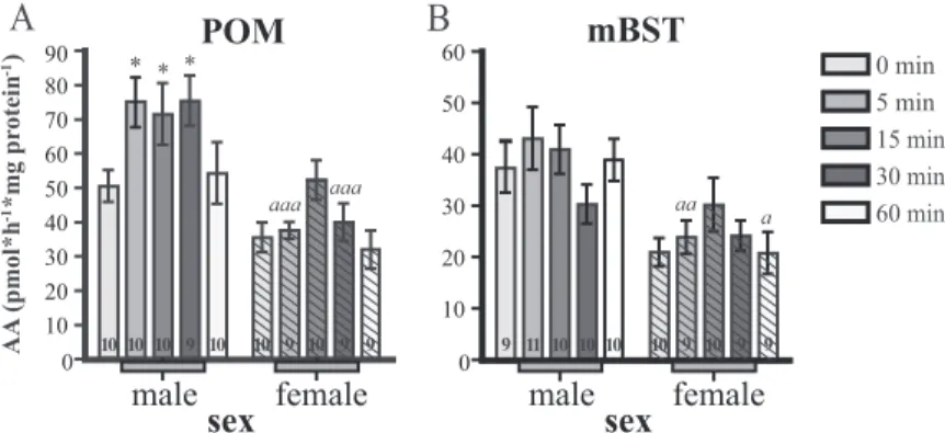
In the POM (Fig. 1A), there was an overall significant effect of stress duration on AA (F4,86⫽ 3.56, P ⫽ 0.01) and * 0 10 20 30 40 50 60 70 80 90 0 10 20 30 40 50 60 * * aaa aaa aa a AA (pmol*h *mg pr otein ) -1 -1 0 min 5 min 15 min 30 min 60 min
A
B
male femalesex male female sex
POM mBST
10 10 10 9 10 10 9 10 9 9 9 11 10 10 10 10 9 10 9 9
FIG. 1. Effects of restraint stress on AA (in pmol/h䡠mg protein) in (A) the POM and (B) the mBST of male and female quail (open and hashed columns, respectively). The different sampling time points are represented by different gray levels as indicated in the legend. The gray bar under the x-axis indicates time points during which birds were exposed to restraint stress. Results from post hoc Bonferroni tests are represented as asterisks when comparing each time with the control, 0 min time point and by a letter (a) when comparing time points between sexes. Single symbols represent P⬍ 0.05; double symbols represent P ⬍ 0.01; triple symbols represent P ⬍ 0.001. Sample sizes are indicated in each column.
no significant interaction between sex and stress duration (F4,86⫽ 1.39, P ⫽ 0.24), suggesting that exposure to stress similarly affects (increases) AA in both males and females. Because there was a duration and sex effect, we ran Bonfer-roni post hoc tests comparing AA at different time points with the baseline values (zero time point) and found signif-icant differences in males at 5, 15, and 30 min of stress, whereas parallel comparisons indicated no significant ence in females, suggesting that there may be subtle differ-ences in how male and female AA responds to stress in the POM. Additionally, post hoc Bonferroni tests revealed spe-cific differences between males and females at 5 and 30 min. In the mBST (Fig. 1B), there was neither a significant effect of stress duration (F4,88⫽ 1.52, P ⫽ 0.20) nor an interaction between sex and stress duration (F4,88⫽ 1.4,
P⫽ 0.24). Because there were overall sex differences in AA
(see before), we ran post hoc analyses to deter-mine which time points were driving this effect and found statistical differences between males and females at 5 and 60 min, suggesting only minor sex differences between the time course of AA changes in response to stress.
To summarize, in the male POM, the increase of AA was rapid (within 5 min), persisted for the whole duration of the stress period (30 min), and returned to baseline at 60 min. Although the two-way ANOVA suggested that changes were sim-ilar in the female POM, graphical inspection sug-gests that increases were less prominent, delayed, and not fully synchronized with the stress period (e.g. numerical increase in the female POM and apparent return to baseline before end of stress at 30 min). Numerically, similar patterns of increases in AA were observed in mBST, but these apparent changes were not statistically significant.
Changes of AA in VMN and Tub
Qualitative observations of the average data suggest that in both aromatase-expressing nuclei of the hypothal-amus, AA showed similar responses to stress such that in the VMN and Tub, stress resulted in a marked and pro-longed decrease of AA in females but a slight transient increase or lack of effect in males. These effects were largely confirmed by statistical analyses.
In the VMN (Fig. 2A), although there was no significant overall effect of stress duration (F4,81⫽ 1.63, P ⫽ 0.17), there was a significant interaction between sex and stress duration (F4,81⫽ 2.69, P ⫽ 0.04), indicating that di-rectionality of stress-induced AA change is different in males and females. Bonferroni post hoc tests indicated that AA was significantly increased at 15 min in males only, which may account for the significant difference between males and fe-males at that time point.
Changes of AA in the Tub (Fig. 2B) were qualitatively similar to those in the VMN: al-though there was no significant effect of stress duration (F4,79⫽ 0.75, P ⫽ 0.56), there was a significant interaction between sex and stress duration (F4,79⫽ 2.96, P ⫽ 0.025), again sug-gesting that the directionality or magnitude of stress-induced AA change is different in males and females. Qualitatively, it appears that the decrease of AA in the female Tub occurs rap-idly (within 5 min) and is maintained beyond stressor cessation. Post hoc tests indicated that the decrease was significant for the 15-, 30-,
AA (pmol*h *mg pr otein ) -1 -1 0 min 5 min 15 min 30 min 60 min
A
B
0 2.5 5.0 7.5 10.0 12.5 15.0 17.5 20.0 22.5 0 1.5 3.0 4.5 6.0 7.5 9.0 10.5 12.0 13.5 15.0 a * * * * a a male femalesex male female sex
VMN Tub
10 10 9 9 10 9 9 9 10 9 9 10 10 9 10 9 9 9 9 8
FIG. 2. Effects of restraint stress on AA (in pmol/h䡠mg protein) in (A) the VMN hypothalamus and (B) the Tub hypothalamus of male and female quail (open and hashed columns, respectively). The different sampling time points are represented by different gray levels as indicated in the legend. The gray bar under the x-axis indicates time points during which birds were exposed to restraint stress. Results from post hoc Bonferroni tests are represented as asterisks when comparing each time with the control, 0 min, time point and a letter (a) when comparing time points between sexes. The symbols correspond to P⬍ 0.05. Sample sizes are indicated in each column. AA (pmol*h *mg pr otein ) -1 -1 0 min 5 min 15 min 30 min 60 min
A
B
0 0 1.0 2.0 3.0 4.0 1.0 2.0 3.0 4.0 male femalesex male female sex
TnA PAG
10 10 9 7 9 9 9 8 8 9 10 11 10 10 10 10 10 10 10 9
FIG. 3. Effects of restraint stress on AA (in pmol/h䡠mg protein) in (A) the TnA and (B) the PAG in male and female quail (open and hashed columns, respectively). The different sampling time points are represented by different gray levels as indicated in the legend. The gray bar under the x-axis indicates time points during which birds were exposed to restraint stress. No significant sex difference and no effect of stress were observed in both nuclei. Sample sizes are indicated in each column.
and 60-min time points. This decrease resulted in a sig-nificant sex difference at 15 and 30 min.
To summarize, the qualitative patterns of AA changes in the VMN and Tub suggest that 30 min of restraint stress continuously inhibit AA in females and that enzymatic activity fails to return to baseline at 30-min poststressor. In contrast, only transient increases (VMN) or no change (Tub) in AA was observed in males. Statistical analysis, however, only demonstrates a differential average AA tween sexes (see before) and a significant interaction be-tween sex and stress duration. This observed pattern of differential changes in males and females contrasts with the results observed in the POM and mBST.
Changes of AA in TnA and PAG
In the TnA, there was no significant effect of stress duration (F4,80⫽ 1.95, P ⫽ 0.11) and no significant in-teraction between stress duration and sex (F4,80⫽ 0.38,
P⫽ 0.82) (Fig. 3A). Similarly, in the PAG, no significant
effect of stress duration (F4,90⫽ 1.45, P ⫽ 0.22) and no interaction between stress duration and sex (F4,90⫽ 1.08,
P⫽ 0.37) on AA could be detected (Fig. 3B).
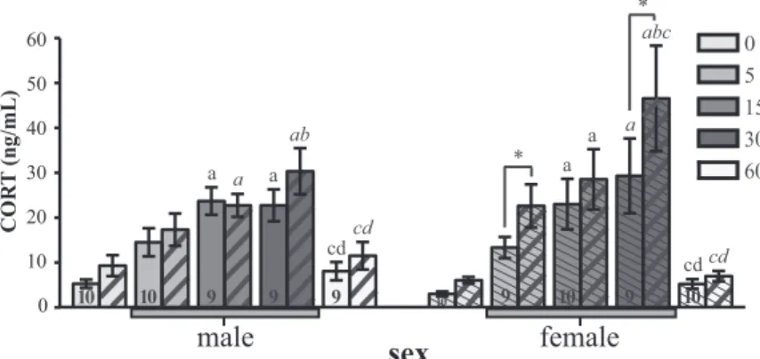
Stress-induced changes in CORT
CORT concentrations were first analyzed using a three-way ANOVA with the independent factors: sex, duration of stress, and location from which the blood sample was taken (jugular vs. brachial) (Fig. 4). As expected, stress exposure resulted in increased CORT concentrations on average in both sexes (effect of time: F4,81⫽ 15.24, P ⬍ 0.0001). However, there was no overall effect of sex on
relative CORT concentrations (F1,81⫽ 0.56,
P ⫽ 0.46), and there was no interaction
be-tween sex and stress duration (F4,81⫽ 1.25,
P⫽ 0.29), indicating that males and females
respond to stress with the same pattern of CORT release.
Overall, CORT concentrations were signif-icantly different between jugular and brachial samples (F1,81⫽ 41.49, P ⬍ 0.0001), and there was an interaction between the location of blood sampling and stress duration (F4,81⫽ 4.56, P⬍ 0.005). There was also a nearly sig-nificant interaction between sex and bleeding location (F1,81⫽ 3.40, P ⫽ 0.069), but the sec-ond-order interaction between sex, location, and stress duration was not significant (F4,81⫽ 1.54, P⫽ 0.20).
Jugular and brachial samples were collected at each time point to obtain some indication of CORT production in the brain. Although the brachial sample was always collected before the jugular sample, there was no correlation between the difference in an individual’s jugular to bra-chial CORT concentration and the time that elapsed be-tween each sample (r2⫽ 0.009, F
1,88⫽ 0.81, P ⫽ 0.37). We then separated data from the two sexes and ran two-way ANOVA on males and females separately to fur-ther assess the differences between jugular and brachial samples with independent factors: stress duration and sampling location. Data from males alone revealed signif-icant effects of location (F1,41⫽ 8.02, P ⫽ 0.007) and duration of stress (F4,41 ⫽ 7.63, P ⬍ 0.0001) with no interaction between these factors (F4,41⫽ 1.15, P ⫽ 0.35), indicating that differences between sampling locations are similar at different time points. Bonferroni post hoc tests were then used to determine at which time points CORT concentrations were significantly affected by stress (for a given sampling location) and also to test when significant differences between sampling locations were present. These tests indicated that jugular and brachial CORT con-centrations were significantly elevated at the 15- and 30-min time points, but no significant difference between lo-cations could be detected at all times considered (see details in Fig. 4).
The two-way ANOVA of female data similarly identi-fied overall effects of location (F1,40⫽ 27.6, P ⬍ 0.0001) and stress duration (F4,40⫽ 8.03, P ⬍ 0.0001), but here, there was a significant interaction between these two fac-tors (F4,40⫽ 3.81, P ⫽ 0.01). Post hoc tests revealed that this interaction is essentially driven by significant differ-ences between jugular and brachial samples at 5 and 30 min. In addition, Bonferroni post hoc tests analyzing
0 5 15 30 60
male female
sex
COR T (ng/mL) 0 10 20 30 40 50 60 a a ab a a a abc a * * cd cd cd cd 10 10 9 9 9 10 9 10 9 10
FIG. 4. Changes in CORT concentrations in plasma collected from both the brachial
(open, male; thin hash, female) and jugular (thick hash) veins across sampling times in males and females. The gray bar under the x-axis indicates time points during which birds were exposed to restraint stress. Post hoc results for two-way ANOVA run for each sex separately are indicated as letters in normal font (brachial) and italicized font (jugular). Significant differences (P⬍ 0.05) between sampling times within a sex and sample location are indicated by the letters a– e for the comparisons with time points 0, 5, 15, 30, and 60 min, respectively. Significant differences at P⬍ 0.05 between jugular and brachial samples at the same time point are represented by an asterisk. Samples are paired (both jugular and brachial from each individual), and sample sizes are only indicated in brachial columns.
which time points were different revealed significant in-creases in plasma CORT at 15 and 30 min for both loca-tions. In agreement with the presence of a difference be-tween jugular and brachial samples, at 30 min, CORT concentrations in jugular samples were elevated by com-parisons with the 0-, 5-, and 15-min samples, whereas in brachial samples, only the first of these comparisons (vs. 0 min) was significant.
Overall, these data confirm the effects of stress on CORT concentrations and, interestingly, demonstrate that CORT concentrations in the jugular vein are consis-tently higher than in the brachial plasma. This elevation suggests local production of CORT in the brain (27) that varies as a function of the duration of exposure to stress in a sex-specific manner.
Stress-induced changes in T
As expected, a two-way ANOVA identified an overall significant sex difference in T concentrations measured in the jugular plasma (F1,81⫽ 55.81, P ⬍ 0.0001) (Fig. 5). In addition, stress duration had a significant effect on T con-centrations (F4,81⫽ 3.20, P ⫽ 0.01) with no interaction between stress duration and sex (F4,81⫽ 0.47, P ⫽ 0.76). Bonferroni post hoc tests comparing T at different time points with the baseline values (zero) revealed significance at P⬍ 0.05 in males at 15, 30, and 60 min of stress; no significant decrease was detected for females.
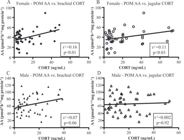
Correlations between changes in brain AA and steroid plasma concentrations
Because CORT release during stress may directly affect AA, we investigated whether stress-induced changes in plasma CORT were correlated to changes in brain AA. We first computed the correlations between plasma CORT concentrations and AA levels in the four nuclei where
sig-nificant changes in AA had been observed (POM, mBST, VMN, and Tub). Correlations were calculated separately for male and female data, because AA was sexually dif-ferentiated in these nuclei, and we wanted to avoid mixing variance due to sex differences to variance related to dif-ferences in plasma CORT concentrations.
As Table 1 and Fig. 6 demonstrate, in females, there was a weak positive correlation between POM AA and CORT in both sample locations, but there were no correlations in the other brain nuclei. In males, there were no significant correlations between brachial or jugular CORT and AA in any brain nuclei.
Due to the potential link between changes in T and changes in AA, we also calculated correlations between these data sets. In males and females no significant corre-lation between T concentrations and AA in any of the brain regions investigated could be detected (Table 1).
There was also no significant correlation between CORT and T concentrations in males (brachial: r2 ⫽ 0.0008, F1,42⫽ 0.03, P ⫽ 0.85; jugular: r2⫽ 0.051, F 1,42⫽ 2.29, P ⫽ 0.14) or females (brachial: r2⫽0.034,F 1,44⫽1.57,P⫽0.22; jugular: r2⫽ 0.043, F1,44⫽ 1.98, P ⫽ 0.17). Discussion
We demonstrate for the first time, to our knowledge, that AA in the adult quail brain is rapidly modulated by stress in an anatomically and sex-specific manner. The fast time course of these effects precludes enzymatic changes that would result from catabolism and de novo synthesis of the enzyme and provides further evidence for nongenomic modification of the enzyme activity in vivo. The differen-tial responses to the timing of the stressor (quick and re-versible in males, sustained beyond stressor cessation in females) suggest a potential mechanism for sex differences in reproductive sensitivity to stressors.
Rapid effects of stress on steroid synthesis
Changes in AA due to acute stress have not been re-ported for other species, but acute stress has been shown to rapidly affect other steroid synthesizing enzymes in the avian brain. In male songbirds, acute stress decreases jug-ular dihydroepiandrosterone, a precursor for androstene-dione, demonstrating that stress either decreases synthesis of this hormone or increases metabolism in the brain (20). Activity of 3-hydroxysteroid dehydrogenase (3-HSD), the enzyme that converts dihydroepiandrosterone into an-drostenedione, is also rapidly modified by acute stress in songbirds in a sex-specific manner. In males, which have relatively lower baseline 3-HSD activity, no changes in enzymatic activity due to stress are detected, whereas
0
male female
sex
0 5 15 30 60 Testoster one (ng/mL) 0.5 1.0 1.5 2.0 2.5 3.0 3.5 4.0 4.5 * * * 9 9 10 8 8 10 9 10 8 10FIG. 5. Changes in jugular T concentrations in male and female quail.
T concentration was significantly higher in males than in females and affected by stress in both sexes. Post hoc results for two-way ANOVA run for each sex separately are represented as asterisks when comparing each time with the control, 0 min, time point at P⬍ 0.05. Sample sizes are indicated in each column.
females, which have relatively higher baseline 3-HSD activity, experience a significant suppression of this en-zymatic activity after acute stress (28). Therefore, stress can rapidly and differentially affect the brain synthesis of sex steroids that are the substrate of AA in males and females.
Stress-induced changes of AA in POM and mBST In quail, like in mammals, POM and mBST are key sites of steroid action in the activation of male sexual motiva-tion and copulatory behavior (29, 30), and T acmotiva-tion in these nuclei requires its aromatization into an estrogen (11, 31). Note, however, that these nuclei are
heteroge-neous, and POM, for example, is also an important site for the regulation of temperature (32, 33) and food intake (34). Brain estrogens affect behavior by slow genomic mechanisms, but more rapid effects (10 – 45 min) have also been reported (14 –16). The rapid, localized stress-in-duced changes in AA observed in our study should thus have an impact on male sexual behavior.
The direction of the changes in AA observed here is counterintuitive given the assumption that acute stress af-fects reproductive behavior negatively (1), and aromatase in this brain region is critical in the stimulation of male reproductive behavior, even after short latencies ranging between 15 and 30 min (15). However, increased AA after
AA (pmol*h *mg pr otein ) -1 -1
A
B
0 20 40 60 80 100 0 20 40 60 80 0 20 40 60 80 AA (pmol*h *mg pr otein ) -1 -1 0 20 40 60 80 100 0 20 40 60 80 100 120 140 AA (pmol*h *mg pr otein ) -1 -1C
0 20 40 60 80 100 120 140 AA (pmol*h *mg pr otein ) -1 -1D
0 20 40 60 CORT (ng/mL) CORT (ng/mL) CORT (ng/mL) 0 20 40 60 CORT (ng/mL)Female - POM AA vs. brachial CORT Female - POM AA vs. jugular CORT
Male - POM AA vs. brachial CORT Male - POM AA vs. jugular CORT
2 r =0.16 p<0.01 2 r =0.11 p<0.03 2 r =0.07 p=0.06 2 r =0.002 p=0.92
FIG. 6. Linear regression analysis for female POM AA vs. CORT measured in the brachial (A) and jugular (B) veins and for male POM AA vs. CORT
concentration measured in the brachial (C) vs. jugular (D). Samples from all groups were included in the analysis. R2and P values are indicated in the box.
TABLE 1. Correlation between circulating CORT or T concentrations and AA in brain nuclei where enzymatic activity was affected by restraint stress
Sex Sample location POM mBST VMN Tub
Female Brachial CORT r2⫽ 0.16, P ⬍ 0.01 r2⫽ 0.014, P ⫽ 0.45 r2⫽ 0.0004, P ⫽ 0.90 r2⫽ 0.049, P ⫽ 0.16
Jugular CORT r2⫽ 0.11, P ⬍ 0.03 r2⫽ 0.027, P ⫽ 0.29 r2⫽ 0.0003, P ⫽ 0.91 r2⫽ 0.0039, P ⫽ 0.21
Jugular T r2⫽ 0.0001, P ⫽ 0.95 r2⫽ 0.012, P ⫽ 0.48 r2⫽ 0.0003, P ⫽ 0.92 r2⫽ 0.028, P ⫽ 0.27
Male Brachial CORT r2⫽ 0.07, P ⫽ 0.06 r2⫽ 0.005, P ⫽ 0.68 r2⫽ 0.00002, P ⫽ 0.97 r2⫽ 0.006, P ⫽ 0.60
Jugular CORT r2⫽ 0.002, P ⫽ 0.92 r2⫽ 0.04, P ⫽ 0.15 r2⫽ 0.003, P ⫽ 0.71 r2⫽ 0.076, P ⫽ 0.06
Jugular T r2⫽ 0.006, P ⫽ 0.87 r2⫽ 0.001 P ⫽ 0.82 r2⫽ 0.0003, P ⫽ 0.67 r2⫽ 0.082, P ⫽ 0.07 Correlations were calculated separately for males and females, and the table shows the coefficients of determination representing the percentage of variance (r2
stress as observed here has also been detected during a preliminary study in quail (11) as well as in follow-up studies (Dickens, M. J., unpublished data). In female rats, stress increased E2concentrations within an hour in the paraventricular nucleus and within 10 min in the jugular blood, although the origin of this increase (brain or ovary) was not identified (35). Interestingly, the increases of AA observed here after stress contrast with the decrease of preoptic AA observed in male quail immediately after a sexual encounter (9, 10); thus, stress and sexual interac-tions have opposite effects.
The counterintuitive nature of stress-induced increases in AA in the POM could be explained by three distinct hypotheses reflecting the direction of changes in estradiol concentrations taking place in vivo before brains are col-lected and assayed for AA. 1) In vitro AA measurements directly reflect increases in local estradiol in vivo. In this scenario, we would predict the functional output to be an increase in reproductive behavior. Although increases in sexual behavior after stress are unexpected, studies have shown this phenomenon; male rats exposed to a tail pinch stressor show an increase in reproductive effort immedi-ately after the stress (36). 2) In vitro AA measurements reflect no changes in local estradiol in vivo, because the stress-induced increases in enzymatic activity is balanced by stress-induced decreases in the enzymatic substrate, T. In this case, we would predict the output to be no change in reproductive behavior even if minor changes in behav-ior could result from the slight difference in the time course of changes in AA and T concentration. This balance may reflect an adapted physiological strategy to preserve sex-ual behavior despite stress exposure; such a trade-off be-tween survival and reproduction is seen in a range of spe-cies when considering stress effects on the gonadal axis (2). 3) In vitro AA measurements actually reflect decreases in local estradiol in vivo. In this scenario, we would predict the output to be a decrease in reproductive behavior. Ob-serving opposite effects in vitro as those potentially oc-curring in vivo could result from a rebound effect as dis-cussed for the diametrically opposite effects observed after copulation in Ref. 9. The increased AA could reflect the build-up of the enzyme due to the in vivo stress-induced inhibition of E2release and correlatively of the exocytosis of presynaptic vesicles containing aromatase. Such a build up of potentially active enzyme would thus result in an increased AA quantified in vitro.
We are currently testing these hypotheses by studying the behavioral output after the same stress protocol. Irre-spective of the mechanisms leading to rapid increases of AA in the POM of stressed subjects and of their function, the present data clearly demonstrate that in vivo AA in the POM is affected by stress within min.
Stress-induced changes of AA in VMN and Tub In female rodents, action of estrogens in the VMN is clearly important for the activation of lordosis behavior (37, 38), and aspects of this behavior rely on nongenomic actions of estradiol (39, 40). Estrogen action in the VMN is also important for female sexual behavior in birds: es-tradiol benzoate implants in the VMN activate female courtship behavior, including sexual crouch in ovariecto-mized female doves (41). In female quail specifically, sex-ual interactions with a male increase Fos expression in the VMN and Tub, and the degree of activation is correlated with proceptive behavior (42).
It is usually assumed that the E2activating lordosis be-havior has an ovarian origin, and as a result, the role of aromatase in the female brain is rarely considered (see Ref. 43). Why E2should be produced locally in the brain while ample production takes place in the ovary has not been addressed but might reflect a need for higher local con-centrations to activate estrogen-sensitive signaling cas-cades (14, 44). In female musk shrew, an induced-ovula-tor, T aromatization in the VMN was shown to be critical for female sexual behavior (45), and in ovariectomized hamster, the activation of lordosis by T is blocked by in-jection of an aromatase inhibitor (46). In rats, restraint stress-induced inhibition of lordosis can be reversed by systemic treatment with estrogens (47).
Although considerable evidence links stress and CORT with reproduction, few studies have investigated the un-derlying physiological mechanisms, especially in females. Stress-induced depression of localized estrogen produc-tion in the VMN and Tub, as suggested by our data, may inhibit female sexual behavior, and the observed sustained suppression of AA beyond stressor cessation may result in a disruption of the hypothalamo-pituitary-gonadal axis and possibly ovulation. Interestingly, the prominent stress-induced decrease in hypothalamic AA was observed in females but not in males. The VMN is also an important site for control of food anticipation (48), whereas the Tub plays a regulatory role in the food intake-energy regula-tion-sleep interaction (49). However, given that, in fe-males, this region is critically important for reproductive physiology and behavior, it may be a more important tar-get for the acute stress response system to tartar-get repro-ductive behavior in the female brain, whereas in the male brain, the POM is a more important target for acute stress to affect reproductive behavior. Therefore, the sex-spec-ificity of the time course of stress effects in these regions (the sustained decrease in hypothalamic AA in the female compared with the transient change in the POM of males) may explain why reproduction in females is more sensitive to environmental cues than in males (50).
Mechanisms controlling fast changes in AA and their anatomical specificity
In vitro, AA can be decreased within min by
calcium-dependent phosphorylation processes presumably con-trolled by neurotransmitters such as glutamate or by K⫹ -induced neuronal depolarizations (13, 51). Stress--induced changes in AA were associated here with a major increase in plasma CORT concentrations that could be a key factor affecting AA via changes in its phosphorylation status.
However, individual CORT concentrations were not correlated with brain AA (except for a weak correlation in female POM), and changes in systemic CORT alone can-not explain the sex and anatomical specificity of the en-zymatic changes described here, unless CORT has differ-ential access to specific brain nuclei in males and females which appears unlikely. Jugular CORT concentrations were, however, higher overall than in brachial samples with significance at specific time points, suggesting that CORT synthesis takes place in the brain and is affected by acute stress. Individual brain regions may thus be exposed differently to locally elevated CORT concentrations (52). Stress might additionally modify the activity of other neu-ropeptides or neurotransmitters (CRH, vasotocin, norepi-nephrine, etc.) that could affect AA in an anatomically specific manner. Alternatively, stress might impact, via a systemic increase in CORT concentration, neurotransmit-ter activity of brain regions that could differentially affect AA in multiple targets based on the nature of the connec-tions between these regions (glutamatergic vs. ␥-aminobu-tyric acidergic projection). These notions could be tested by researching whether aromatase neurons express recep-tors for glucocorticoids, CRH, or vasopressin and by func-tional experiments assessing the effects on local AA of stereotaxic injections of these compounds.
Irrespective of underlying mechanisms, the present study shows that acute stress induces rapid changes in brain AA in an anatomically discrete and sex-specific man-ner. These enzymatic changes suggest a mechanistic link between the stress response and reproductive behavior that may reflect the differential regulation underlying how males and females respond to stressful stimuli.
Acknowledgments
Address all correspondence and requests for reprints to: Molly J. Dickens, University of Lie`ge, GIGA Neurosciences, Research Group in Behavioral Neuroendocrinology, 1 Avenue de l’Hopital (B36), 4000 Lie`ge, Belgium. E-mail: [email protected].
This work was supported by the National Science Foundation International Research Fellowship IRFP 0910495 (to M.J.D.), the National Institutes of Health Grant R01 NIH/MH50388, the
Belgian Fonds de la Recherche Fondamentale Collective Grant 2.4537.09, and the University of Lie`ge (Fonds Spe´ciaux 2009) (to J.B.). C.A.C. is Fonds de la Recherche Scientifique-Fonds National de la Recherche Scientifique Research Associate.
Disclosure Summary: The authors have nothing to disclose.
References
1. Wingfield JC, Maney DL, Breuner CW, Jacobs JD, Lynn S,
Ra-menofsky M, Richardson RD 1998 Ecological bases of
hormone-behavior interactions: the emergency life history stage. Am Zool 38:191–206
2. Wingfield JC, Sapolsky RM 2003 Reproduction and resistance to stress: when and how. J Neuroendocrinol 15:711–724
3. Sapolsky RM, Romero LM, Munck AU 2000 How do glucocorti-coids influence stress responses? Integrating permissive, suppressive, stimulatory, and preparative actions. Endocr Rev 21:55– 89 4. Bian J, Wu Y, Liu J 2005 Breeding behavior under temporal risk of
predation in male root voles (Microtus oeconomus). J Mammal 86: 953–960
5. Cecconello AL, Raineki C, Sebben V, Lucion AB, Sanvitto GL 2010 Effect of acute stress on sexual behavior in female rats: participation of the central angiotensinergic system. Behav Brain Res 207:429 – 433
6. Moore FL, Boyd SK, Kelley DB 2005 Historical perspective: hor-monal regulation of behaviors in amphibians. Horm Behav 48:373– 383
7. Moore FL, Miller LJ 1984 Stress-induced inhibition of sexual be-havior: corticosterone inhibits courtship behaviors of a male am-phibian. Horm Behav 18:400 – 410
8. Lynn SE, Stamplis TB, Barrington WT, Weida N, Hudak CA 2010 Food, stress, and reproduction: short-term fasting alters endocrine physiology and reproductive behavior in the zebra finch. Horm Be-hav 58:214 –222
9. Cornil CA, Dalla C, Papadopoulou-Daifoti Z, Baillien M, Dejace C,
Ball GF, Balthazart J 2005 Rapid decreases in preoptic aromatase
activity and brain monoamine concentrations after engaging in male sexual behavior. Endocrinology 146:3809 –3820
10. de Bournonville C, Ball GF, Balthazart J, Cornil CA Rapid changes of aromatase activity in discrete brain regions following social in-teractions. 6th International Meeting Steroids and Nervous System, Torino, Italy, 2011 (Abstract 147)
11. Balthazart J, Cornil CA, Charlier TD, Taziaux M, Ball GF 2009 Estradiol, a key endocrine signal in the sexual differentiation and activation of reproductive behavior in quail. J Exp Zool Part A 311:323–345
12. Balthazart J, Baillien M, Charlier TD, Cornil CA, Ball GF 2003 Multiple mechanisms control brain aromatase activity at the genomic and non-genomic level. J Steroid Biochem Mol Biol 86: 367–379
13. Balthazart J, Baillien M, Ball GF 2006 Rapid control of brain aro-matase activity by glutamatergic inputs. Endocrinology 147:359 – 366
14. Cornil CA, Dalla C, Papadopoulou-Daifoti Z, Baillien M,
Baltha-zart J 2006 Estradiol rapidly activates male sexual behavior and
affects brain monoamine levels in the quail brain. Behav Brain Res 166:110 –123
15. Cornil CA, Taziaux M, Baillien M, Ball GF, Balthazart J 2006 Rapid effects of aromatase inhibition on male reproductive behaviors in Japanese quail. Horm Behav 49:45– 67
16. Taziaux M, Keller M, Bakker J, Balthazart J 2007 Sexual behavior activity tracks rapid changes in brain estrogen concentrations. J Neurosci 27:6563– 6572
1995 Critical re-examination of the distribution of aromatase-im-munoreactive cells in the quail forebrain using antibodies raised against human placental aromatase and against the recombinant quail, mouse or human enzyme. J Chem Neuroanat 8:267–282 18. Ball GF, Balthazart J 2010 Japanese quail as a model system for
studying the neuroendocrine control of reproductive and social be-haviors. ILAR J 51:310 –325
19. Hazard D, Couty M, Gue´mene´ D 2007 Characterization of CRF, AVT, and ACTH cDNA and pituitary-adrenal axis function in Jap-anese quail divergently selected for tonic immobility. Am J Physiol Regul Integr Comp Physiol 293:1421–1429
20. Newman AE, Pradhan DS, Soma KK 2008 Dehydroepiandrosterone and corticosterone are regulated by season and acute stress in a wild songbird: jugular versus brachial plasma. Endocrinology 149:2537– 2545
21. Palkovits M 1973 Isolated removal of hypothalamic or other brain nuclei of the rat. Brain Res 59:449 – 450
22. Schumacher M, Balthazart J 1987 Neuroanatomical distribution of testosterone-metabolizing enzymes in the Japanese quail. Brain Res 422:137–148
23. Cornil CA, Ball GF, Balthazart J, Charlier TD 2011 Organizing effects of sex steroids on brain aromatase activity in quail. PLoS ONE 6:e19196
24. Roselli CE, Resko JA eds. 1991 In vitro assay of aromatase activity in the central nervous system. Chur, Switzerland: Harwood Aca-demic Publishers
25. Baillien M, Balthazart J 1997 A direct dopaminergic control of aro-matase activity in the quail preoptic area. J Steroid Biochem Mol Biol 63:99 –113
26. Washburn BE, Millspaugh JJ, Morris DL, Schulz JH, Faaborg J 2007 Using a commercially available enzyme immunoassay to quan-tify testosterone in avian plasma. Condor 109:181–186
27. Newman AE, Chin EH, Schmidt KL, Bond L, Wynne-Edwards KE,
Soma KK 2008 Analysis of steroids in songbird plasma and brain by
coupling solid phase extraction to radioimmunoassay. Gen Comp Endocrinol 155:503–510
28. Soma KK, Alday NA, Hau M, Schlinger BA 2004 Dehydroepiandro-sterone metabolism by 3-hydroxysteroid dehydrogenase/⌬5-⌬4 isomerase in adult zebra finch brain: sex difference and rapid effect of stress. Endocrinology 145:1668 –1677
29. Balthazart J, Taziaux M, Holloway K, Ball GF, Cornil CA 2009 Behavioral effects of brain-derived estrogens in birds. Ann NY Acad Sci 1163:31– 48
30. Balthazart J, Absil P, Ge´rard M, Appeltants D, Ball GF 1998 Ap-petitive and consummatory male sexual behavior in Japanese quail are differentially regulated by subregions of the preoptic medial nucleus. J Neurosci 18:6512– 6527
31. Balthazart J, Baillien M, Cornil CA, Ball GF 2004 Preoptic aroma-tase modulates male sexual behavior: slow and fast mechanisms of action. Physiol Behav 83:247–270
32. Dacks PA, Krajewski SJ, Rance NE 2011 Ambient temperature and 17-estradiol modify Fos immunoreactivity in the median preoptic nucleus, a putative regulator of skin vasomotion. Endocrinology 152:2750 –2759
33. Boulant JA 2006 Neuronal basis of Hammel’s model for set-point thermoregulation. J Appl Physiol 100:1347–1354
34. Santollo J, Torregrossa AM, Eckel LA 2011 Estradiol acts in the medial preoptic area, arcuate nucleus, and dorsal raphe nucleus to reduce food intake in ovariectomized rats. Horm Behav 60:86 –93
35. Liu J, Hu P, Qi XR, Meng FT, Kalsbeek A, Zhou JN 2011 Acute restraint stress increases intrahypothalamic oestradiol concentra-tions in conjunction with increased hypothalamic oestrogen recep-tor and aromatase mRNA expression in female rats. J Neuroen-docrinol 23:435– 443
36. Leyton M, Stewart J 1996 Acute and repeated activation of male sexual behavior by tail pinch: opioid and dopaminergic mecha-nisms. Physiol Behav 60:77– 85
37. Pfaff DW, Schwartz-Giblin S, McCarthy MM, Kow LM 1994 Cel-lular and molecular mechanisms of female reproductive behaviors. In: Knobil E, Neill JD, eds. The physiology of reproduction. 2nd ed. New York: Raven Press; 107–220
38. Blaustein JD 2009 Feminine reproductive behavior and physiology in rodents: integration of hormonal, behavioral, and environmental influences. In: Pfaff DW, Arnold AP, Etgen AM, Fahrbach SE, Rubin RT, eds. Hormones, brain and behavior. San Diego: Academic Press; 68 –107
39. Micevych PE, Mermelstein PG 2008 Membrane estrogen receptors acting through metabotropic glutamate receptors: an emerging mechanism of estrogen action in brain. Mol Neurobiol 38:66 –77 40. Kow LM, Pfaff DW 2004 The membrane actions of estrogens can
potentiate their lordosis behavior-facilitating genomic actions. Proc Natl Acad Sci USA 101:12354 –12357
41. Gibson MJ, Cheng MF 1979 Neural mediation of estrogen-depen-dent courtship behavior in female ring doves. J Comp Physiol Psy-chol 93:855– 867
42. Meddle SL, Foidart A, Wingfield JC, Ramenofskyand M, Balthazart
J 1999 Effects of sexual interactions with a male on Fos-like
immu-noreactivity in the female quail brain. J Neuroendocrinol 11:771– 784
43. Lephart ED 1996 A review of brain aromatase cytochrome P450. Brain Res Rev 22:1–26
44. Balthazart J, Ball GF 2006 Is brain estradiol a hormone or a neu-rotransmitter? Trends Neurosci 29:241–249
45. Veney SL, Rissman EF 2000 Steroid implants in the medial preoptic area or ventromedial nucleus of the hypothalamus activate female sexual behaviour in the musk shrew. J Neuroendocrinol 12:1124 – 1132
46. Hsu CH 1990 Blockade of lordosis by androst-1,4,6-triene-3,17-dione (ATD) and tamoxifen in female hamsters primed with testos-terone propionate. Horm Behav 24:14 –19
47. White S, Uphouse L 2004 Estrogen and progesterone dose-depend-ently reduce disruptive effects of restraint on lordosis behavior. Horm Behav 45:201–208
48. Ribeiro AC, LeSauter J, Dupre´ C, Pfaff DW 2009 Relationship of arousal to circadian anticipatory behavior: ventromedial hypothal-amus: one node in a hunger-arousal network. Eur J Neurosci 30: 1730 –1738
49. Fort P, Salvert D, Hanriot L, Jego S, Shimizu H, Hashimoto K, Mori
M, Luppi PH 2008 The satiety molecule nesfatin-1 is co-expressed
with melanin concentrating hormone in tuberal hypothalamic neu-rons of the rat. Neuroscience 155:174 –181
50. Ball GF, Ketterson ED 2008 Sex differences in the response to en-vironmental cues regulating seasonal reproduction in birds. Phil Trans R Soc B 363:231–246
51. Balthazart J, Baillien M, Ball GF 2001 Rapid and reversible inhi-bition of brain aromatase activity. J Neuroendocrinol 13:63–73 52. Newman AE, Soma KK 2009 Corticosterone and
dehydroepiandro-sterone in songbird plasma and brain: effects of season and acute stress. Eur J Neurosci 29:1905–1914