To cite this version :
Nesterenko, Alla and Alric, Isabelle and Violleau, Frédéric and Silvestre,
Françoise and Durrieu, Vanessa A new way of valorizing biomaterials: the use
of sunflower protein for 1 a-tocopherol microencapsulation. (2013) Food
Research International, vol. 53 (n° 1). pp. 115-124. ISSN 0963-9969
O
pen
A
rchive
T
OULOUSE
A
rchive
O
uverte (
OATAO
)
OATAO is an open access repository that collects the work of Toulouse researchers and
makes it freely available over the web where possible.
This is an author-deposited version published in :
http://oatao.univ-toulouse.fr/
Eprints ID : 9566
To link to this article :
DOI:10.1016/j.foodres.2013.04.020
URL :
http://dx.doi.org/10.1016/j.foodres.2013.04.020
Any correspondance concerning this service should be sent to the repository
administrator:
[email protected]
A new way of valorizing biomaterials: The use of sunflower protein for
α-tocopherol microencapsulation
A. Nesterenko
a,b, I. Alric
a,b, F. Violleau
c, F. Silvestre
a,b, V. Durrieu
a,b,⁎
aUniversité de Toulouse, INP-ENSIACET, LCA (Laboratoire de Chimie Agro-Industrielle), F-31030 Toulouse, France bINRA, UMR 1010 CAI, F-31030 Toulouse, France
cUniversité de Toulouse, INP-EI Purpan, Département Sciences Agronomiques et Agro-alimentaires, UPSP/DGER 115, F-31076 Toulouse, France
a b s t r a c t
Keywords: Sunflower protein α-Tocopherol Enzymatic hydrolysis N-acylation Microencapsulation Spray-dryingBiopolymer based microparticles were efficiently prepared from sunflower protein (SP) wall material and α-tocopherol (T) active core using a spray-drying technique. Protein enzymatic hydrolysis and/or N-acylation were carried out to make some structural modifications to the vegetable protein. Native and hydrolyzed SP were characterized by Asymmetrical Flow Field-Flow Fractionation (AsFlFFF). Results of AsFlFFF confirmed that size of proteinic macromolecules was influenced by degree of hydrolysis. The effect of protein modifications and the influence of wall/core ratio on both emulsions and microparticle properties were evaluated. Concerning emulsion properties, enzymatic hydrolysis involved a decrease in viscosity, whereas acylation did not significant-ly affect emulsion droplet size and viscosity. Microparticles obtained with hydrosignificant-lyzed SP wall material showed lower retention efficiency (RE) than native SP microparticles (62–80% and 93% respectively). Conversely, acyla-tion of both hydrolyzed SP and native SP allowed a higher RE to be reached (up to 100%). Increasing T concen-tration increased emulsion viscosity, emulsion droplet size and microparticle size, and enhanced RE. These results demonstrated the feasibility of highly loaded (up to 79.2% T) microparticles.
1. Introduction
Due to environmental considerations, research activity in the area of biodegradable and biocompatible polymers is still increasing. Among these polymers, proteins extracted from vegetables (pea, soy, wheat, sunflower, rice, barley, oat) have demonstrated good functional and amphiphilic properties such as water solubility, gelling properties, emulsifying and foaming capacity (Nunes, Batista, Raymundo, Alves, & Sousa, 2003). Vegetable proteins represent an excellent wall-forming material due to their good film-forming and barrier properties, capabil-ity for adhesion and resistance to oils and organic solvents (De Graaf, Harmsen, Vereijken, & Monikes, 2001; Nesterenko, Alric, Silvestre, & Durrieu, 2013). Thus, these relatively inexpensive, biocompatible and biodegradable biopolymers offer an interesting alternative to animal-based proteins and petroleum-derived polymers, especially for use as wall materials for microencapsulation in the food industry, cosmetics, pharmaceutics or in biodegradable material formulations.
Sunflower proteins represent the major component of oil cake after sunflower oil extraction. The defatted sunflower flour consists of about 20–40% crude protein, this value being highly dependent on sunflower variety and extraction conditions. In proteins extracted from sunflower seeds, four fractions are mainly present: globulins
range from 40 to 90%, albumins account for about 10–30% of total proteins and the two fractions glutelins and prolamins are present in minor quantities (Gonzalez-Perez & Vereijken, 2007). In contrast to soybean proteins, which are widely used in food and non-food ap-plications, sunflower proteins are mainly used for animal feed. The principal limitation of sunflower extracts for food applications is the presence of relatively high amounts of phenolic compounds, especial-ly chlorogenic acid and caffeic acid, which affect protein quality. Nevertheless, in recent years, interest in improving the production of phenolic free sunflower protein compounds has increased signifi-cantly (Pickardt et al., 2009; Salgado et al., 2012). This agricultural by-product with reduced phenolic component content could be useful for food applications because sunflower proteins, like soybean proteins, possess interesting functional and physico-chemical properties such as solubility, emulsifying, foaming and gelling capacities (Gonzalez-Perez & Vereijken, 2007; Gonzalez-Perez, Vereijken, Koningsveld, Gruppen, & Voragen, 2005; Molina, Petruccelli, & Anon, 2004; Patino et al., 2007). Microencapsulation permits the formation of a physical barrier between the external medium and sensitive core materials for their protection, particularly against moisture, pH and oxidation. It is also used for controlled release of active molecules, formulation stability enhancement, and flavor and taste masking. Sensitive core micropar-ticles can be produced by various processes such as spray-drying, spray-cooling/chilling, fluidized bed, coacervation, gelation, solvent evaporation, extrusion methods, and supercritical fluid expansion (Benita, Hoffman, & Donbrow, 1985; Dubey, Shami, & Bhasker,
⁎ Corresponding author at: Université de Toulouse, INP-ENSIACET, LCA (Laboratoire de Chimie Agro-Industrielle), F-31030 Toulouse, France.
2009; Gouin, 2004). The use of vegetable proteins as wall-forming materials in microencapsulation, reflects the present “green” trend in the pharmaceutical, cosmetics and food industries. Among the possible plant extracted proteins for microencapsulation wall material, soybean proteins, pea proteins and cereal proteins are the most studied ones (Nesterenko et al., 2013). Two techniques are mainly used for active material encapsulation with these vegetable proteins: spray-drying and coacervation. Various studies have proved the ability of vegetable protein wall materials to efficiently protect hydrophilic and hydropho-bic active materials. Nevertheless, microencapsulation efficiency and microparticle size are affected by different parameters, such as wall and core material concentrations, pH of media, temperature, use of ad-ditives and encapsulation technique.
Enzymatic, chemical or physico-chemical modifications can be used to significantly improve functional properties of vegetable pro-teins, in order to make these raw materials more suited to current mi-croencapsulation techniques. Vegetable protein hydrolyzates from soybean, pumpkin, sunflower, chickpea and rapeseed have shown various interesting solubility, foaming and emulsifying properties compared to the native proteins (Chabanon, Chevalot, Framboisier, Chenu, & Marc, 2007; Lamsal, Jung, & Johnson, 2007; Martinez, Baezaa, Millan, & Pilosof, 2005; Patino et al., 2007; Vastag, Popovic, Popovic, Krimer, & Pericin, 2010; Yust, Pedroche, Millan-Linares, Alcaide-Hidalgo, & Millan, 2010). The production of hydrolyzed pro-teins by sequential action of exoprotease is considered an effective way of obtaining protein with defined characteristics (Kong, Guo, Cao, & Zhang, 2008), and alcalase is an endopeptidase used for pro-duction of proteins with improved functional and nutritional proper-ties (Junyaprasert, Mitrevej, Sinchaipanid, Broome, & Wurster, 2001). Protein acylation also influences their physico-chemical functionali-ties. Attachment of hydrocarbon chains may modify the charge and structural properties of water-soluble proteins, increasing their hydro-phobicity, thus improving their surface functionality, emulsifying and foaming properties, offering a range of possibilities for the development of new applications (Matemu, Kayahara, Murasawa, Katayama, & Nakamura, 2011; Wong, Nakamura, & Kitts, 2006).
The aim of the present work was to propose a new application for sunflower proteins (SP) as wall material for microencapsulation, more specifically for α-tocopherol (T, used as a hydrophobic mole-cule model) microparticle preparation. The effect of SP modifications on their microencapsulation properties compared to native proteins was evaluated. Proteinic chains were modified by enzymatic hydroly-sis (from 6% to 24%) and/or N-acylation (fatty acid chain length of 8, 12 or 16 carbon atoms). Modified and native proteins were then used for T encapsulation with the spray-drying technique. In the con-text of “green” chemistry, all preparations and modifications were carried out without use of chemical catalysts, surfactants or organic solvents (Aider, 2010; Vilkhu, Mawson, Simons, & Bates, 2008). Influ-ence of SP structural modifications on emulsion properties (viscosity, oil droplet size), microencapsulation efficiency, size and morphology of microparticles obtained, was particularly investigated. In order to increase the T proportion in microparticles, different wall material/ active core ratios were also tested.
2. Materials and methods
2.1. Materials
Sunflower protein was provided and prepared by CVG (Dury, France) as follows: extraction from defatted sunflower flour at pH 9.0 and 50 °C, separation by centrifugation, purification by precip-itation at pH 4.5, neutralization and spray-drying. All other chemicals were of analytical grade. α-Tocopherol, alcalase (protease from
Bacillus licheniformis, 2.4 U/g), sodium hydroxide, hydrochloric acid,
octanoyl chloride, dodecanoyl chloride, hexadecanoyl chloride, Tween-80, monopotassium phosphate, dipotassium phosphate and
cyclohexane (HPLC grade) were purchased from Sigma (Saint-Quentin Fallavier, France).
2.2. Protein characterization
Sunflower protein was analyzed for proximate composition — moisture, lipid, ash and protein contents. The moisture content and the ash content were found by heating a sample of the air-dried ma-terial in an oven at 105 and 550–600 °C respectively to constant weight. The protein content was determined by the Kjeldahl method (N × 6.25) and lipid content by conventional Soxhlet extraction in cyclohexane for 7 h. These analyses were carried out in triplicate.
Sunflower protein solubility profiles were determined according to the method described byZheng, Yang, Tnag, Li, and Ahmad (2008). Pro-tein powder was mixed with deionized water (3% w/w), and the mix-ture pH was adjusted to 1.0–13.0 with 4 M NaOH or 4 M HCl. Protein solution was stirred for 1 h at room temperature, and at 70 °C, chosen to ensure higher solubility without protein denaturation, because the major SP globulin fraction (11S of helianthinin) still resists thermal denaturation at 90 °C (Gonzalez-Perez et al., 2004). After stirring, the suspensions were centrifuged at 10,000 rpm for 15 min (Sigma Laborzentrifugen, Osterode, Germany). The soluble protein fraction in the supernatant was analyzed by the Kjeldahl method and solubility (S%, w/w) was calculated from the following equation:
S %ð Þ ¼protein content in the supernatant
total protein content in solution $ 100 ð1Þ
2.3. Protein modifications 2.3.1. Enzymatic hydrolysis
The enzymatic hydrolysis was carried out according to the method described byKong et al. (2008). An aqueous solution of SP (5% w/w) was prepared and incubated in a bath at 50 °C for 15 min. When the protein solution reached 50 °C, enzyme was added (alcalase/protein ratio 0.002–0.025 U/g). During hydrolysis, the pH was maintained at 7.0 by adding a 4 M NaOH solution. Reaction time was varied from 10 to 120 min, and stopped by adjusting the pH to 4.5 with a 4 M HCl. Following the reaction period, the mixture was cooled, adjusted to pH 7.0 and heated at 95 °C for 10 min for enzyme deactivation. Then the mixture was freeze-dried using a Cryo-Rivoire equipment at 20 Pa (Cryonext, Saint Gely du Fesc, France) and stored at 4 °C (SP1, SP2, SP3, SP4) or kept for the acylation (SP5).
The degree of hydrolysis (DH) was evaluated using the o-phthaldialdehyde method (OPA) (Church, Swaisgood, Porter, & Catignani, 1983; Goodno, Swaisgood, & Catignani, 1981). OPA reacts with primary amino groups i.e. N-terminal and lysine residues of proteins, forming a chromophore with an UV-absorbance optimum at 340 nm. Measurements were made with an UV spectrometer (UV-1800, Shimadzu, Kyoto, Japan). The DH was calculated from the following equation:
DH %ð Þ ¼ðNh−N0Þ Nt−N0
ð Þ$ 100 ð2Þ
where Nhis the molar quantity of amino groups per gram of partially hydrolyzed protein, N0the molar quantity of amino groups per gram of non-hydrolyzed protein, and Nt the molar quantity of amino groups per gram of totally hydrolyzed protein. All analyses were car-ried out in triplicate.
2.3.2. N-acylation
N-acylation reactions were carried out on SP or SP hydrolyzates (acylation with hydrolyzed SP was started in the same reaction medi-um after hydrolysis and enzyme deactivation) using octanoyl chloride
(C8), dodecanoyl chloride (C12) or hexadecanoyl chloride (C16), fol-lowing the Schotten–Baumann reaction (Rondel, Alric, Mouloungui, Blanco, & Silvestre, 2009). SP solution (5% w/w, pH 10.0) was pre-pared in deionized water and the fatty acid chloride was slowly added to this mixture. The molar ratio C8 (C12, C16)/NH2of protein used for reaction was 1, to obtain the samples SP6 (SP7, SP8) respec-tively. The solution was stirred for 30 min at room temperature, and then for 180 min at 50 °C. During acylation, the pH was maintained at 10.0 by addition of a 4 M NaOH solution. Then the mixture was freeze-dried and the powder stored at 4 °C.
The degree of acylation (DA) was evaluated using the OPA method: DA %ð Þ ¼ðn−naÞ
n $ 100 ð3Þ
where n is the number of amino groups per gram of non-hydrolyzed or partially hydrolyzed protein, and nathe number of amino groups per gram of acylated protein. Analyses were performed in triplicate.
2.4. Fractionation of sunflower proteins by Asymmetrical Flow Field-Flow Fractionation
For size distribution characterization of native and hydrolyzed sunflower proteins, Asymmetrical Flow Field-Flow Fractionation (AsFlFFF) was used. The protein-based samples were prepared at a concentration of 0.5% w/w in deionized water at pH 9.0 which corre-sponds to highest protein solubility. The AsFlFFF apparatus was an Eclipse 2 System (Wyatt Technology Europe, Dernbach, Germany) connected to an Agilent 1100 UV detector (Agilent Technologies, Waldbronn, Germany) used for quantitative detection. The absor-bance was measured at 280 nm and the UV extinction coefficient value for sunflower proteins was fixed at 3.21 × 10³mL/g × cm. An Agilent 1100 Series Isocratic Pump with an in-line vacuum degasser and an Agilent 1100 Autosampler delivered the carrier flow and han-dled sample injection into the AsFlFFF channel. An in-line filter with a 0.1 μm pore size (VVLP, Millipore, Darmstadt, Germany) was placed between the pump and the channel. The eluent used for analysis was de-ionized water at pH 9.0 filtered at 0.1 μm before use. The AsFlFFF channel used had a trapezoidal geometry with a 490 μm spac-er thickness (surface area 22.17 cm2, volume 1.086 cm3
). The ultrafil-tration membrane forming the accumulation wall was made of regenerated cellulose with a cut-off at 10 kDa (Wyatt Technology Europe, Dernbach, Germany).
After injection of a 50 μL sample solution (at 0.2 mL/min for 3 min) and a focus period (1.0 mL/min for 1 min), elution (separation) was started at the 12th min of analysis. For separation, channel flow rate was fixed at 1 mL/min and the cross-flow rate was variable. Elution mode was started at a cross-flow rate of 2.5 mL/min for 4 min, and then reduced linearly for 10 min to a rate of 0.2 mL/min. Elution was stopped at the 26th min of analysis.
During analysis, AsFlFFF separates particles/molecules according to differences in diffusion coefficient D, which can be converted to the hy-drodynamic radius Rhusing the Stokes–Einstein relationship. According toYohannes, Wiedmer, Tuominen, Kinnunen, and Riekkola (2004)Rh can be determined using the retention time and the following experi-mental parameters:
Rh¼ kTV
0
πηVcrw2t0
$ tr ð4Þ
where k is the Boltzmann constant, T the temperature, V0the void vol-ume (volvol-ume of separation channel), η the eluent viscosity, Vcrthe cross-flow rate, w the channel thickness, t0the void time and t
rthe detected retention time.
The percentage of non-fractionated particles/molecules with a size equal or less than 10 kDa (which passed through the membrane
during analysis) in each sample, was calculated using the recovery % (the difference between injected and detected mass).
2.5. Emulsion preparation and characterization
Sunflower protein (SP) aqueous solution (8% w/w, pH 8.5) was prepared at 70 °C for 1 h under constant mechanical stirring (1000 rpm) in order to allow maximum protein solubilization. T was mixed with protein solution to obtain the preparation in which the SP/T ratio was 2/1 or 11.5% of total solids (the ratio of solid mass, i.e. SP and T, to total emulsion mass). The pre-emulsion obtained was circulated twice through a high-pressure homogenizer (APV Systems, Albertslund, Denmark) at 50 MPa. The preparation with native SP was named SP0; with hydrolyzed SP — SP1, SP2, SP3, and SP4; with hydrolyzed and acylated SP — SP5; and with acylated SP — SP6, SP7, and SP8, for a molar ratio of fatty acid chloride (C8, C12, and C16 respectively) on NH2of 1.
Different wall/core ratios of 1/1, 1/2 and 1/4 (14.8% w/w, 20.7% w/w and 30% w/w of total solids in emulsion) were also prepared using na-tive SP as a wall material to obtain samples SP0′, SP0″ and SP0‴ respec-tively. The influence of the wall/core ratio on the microencapsulation process was also studied after SP acylation (molar ratio C12/NH2of 0.5). Samples SP9, SP9′ and SP9″ were obtained when wall/core ratios were 2/1, 1/1 and 1/2 respectively. For all these samples, protein solu-tion concentrasolu-tion was also 8% w/w but a different quantity of T was added.
Oil droplet diameters and size distribution in emulsions were measured using laser diffraction (Zetasizer Nano-ZS, Malvern Instru-ments, Worcestershire, UK). To avoid multiple scattering effects, emulsions were diluted 100 times with deionized water before mea-surements. A relative refractive index ηoil/ηwater= 1.12 (ηoil= 1.49, ηwater= 1.33) was used for the particle size distribution calculations, assuming that all droplets were spherical in shape. The volume parti-cle diameter (D43or Dv) was calculated as the mean of three readings per sample.
To complete the information obtained by laser granulometry, emulsions were visualized using an Eclipse E600 optical microscope (Nikon, Sendai, Japan), linked to a digital video camera (DXM1200, Nikon, Sendai, Japan) at a magnification of 1000×.
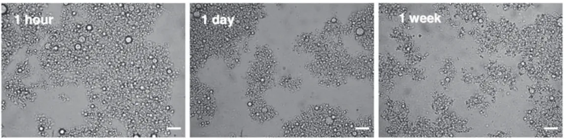
Light scattering and optical microscopy were used to check good dispersion of T in the protein solution, emulsion stability and droplet size uniformity. For this, sample SP0 was analyzed 1 h, 1 day and 1 week after homogenization.
Apparent viscosity of all emulsions after homogenization was de-termined at 20 °C and shear stress variation between 0 and 1 N/m2 for 3 min, using a Rheometer CSL100 (Carri-Med Ltd., Dorking, UK) with a 6 cm diameter plate-cone geometry and 0.035 rad angle. All emulsions were characterized as Newtonian fluids.
2.6. Microparticle preparation and characterization
Freshly homogenized emulsions were spray-dried in a Mini Spray Dryer B-290 (Büchi, Flawil, Switzerland) under stable process condi-tions as follows: inlet air temperature at 124 ± 4 °C and outlet at 74 ± 4 °C, drying air flow rate of 470 L/h, liquid feed flow rate of 0.33 L/h and aspiration of 100%. Microparticles were collected from the container, shut hermetically in opaque packaging and stored at 4 °C. The yield of spray-drying was defined as follows:
Spray % drying %ð Þ ¼ mP
mSPþT$ 100 ð5Þ
where mP is the mass of collected powder, and mSP + T the initial mass of solid content added in emulsion including SP and T.
The amount of T in the prepared microspheres was defined according toFaria, Mignone, Montenegro, Mercadante, and Borsarelli
(2010)using UV/VIS spectroscopy. Initially, an absorbance vs. concen-tration calibration curve was prepared with T dissolved in cyclohexane and analyzed at λmaxof 298 nm. Next, 5 mg of microspheres containing the T to be determined was dissolved in 10 mL of cyclohexane. The so-lution was stirred for 10 min and filtered through a 0.2 μm PTFE mem-brane filter. Previously, SP insolubility in cyclohexane had been verified, and absorbance of SP solution filtered through a 0.2 μm filter at 298 nm was equal to 0. The absorbance of the solution was measured using an UV spectrometer (UV-1800, Shimadzu, Kyoto, Japan) at 298 nm. This procedure was carried out in triplicate. Retention efficiency (RE) char-acterizing the efficiency of the microencapsulation process, was calcu-lated as the percent ratio of estimated T content in obtained particles or in obtained powder (Texp) over theoretical T content (Ttheo):
RE %ð Þ ¼Texp
Ttheo$ 100 ð6Þ
The difference between Texpand Ttheo was caused by T loss during spray-drying. Loading efficiency (LE), corresponding to T content per 100 g of powder, was calculated as:
LE %ð Þ ¼mTexp
mm $ 100 ð7Þ
where mT
expis the estimated mass of T in microparticles, and mmthe
mass of the analyzed sample of microparticles.
Particle size distribution of powder obtained was determined by the scattering pattern of a transverse laser light using the Scirocco 2000 equipment (Malvern Instruments, Worcestershire, UK). The vol-ume particle diameter (D43or Dv) was calculated as the mean of three measurements per sample.
Microparticle morphology was examined by scanning electron mi-croscopy (SEM). The particles were deposited on conductive double-faced adhesive tape and sputter-coated with silver. In order to examine the inner structure of prepared microparticles, the powder was frozen in liquid nitrogen and broken up in a mortar. SEM observations were performed with a LEO435VP scanning electron microscope (LEO Elec-tron microscopy Ltd., Cambridge, UK) operated at 8 kV.
2.7. In vitro release of α-tocopherol from sunflower protein based microparticles
The release rate of T from microparticles was evaluated using a 0.05 M phosphate buffer in saline solution at pH 7.4. To improve the solubility of T in aqueous medium, the release solution contained Tween-80 (0.5% w/v) (Luo, Zhang, Whent, Yu, & Wang, 2011; Yoo, Song, Chang, & Lee, 2006). The sample of microparticles was incubat-ed in the release mincubat-edium at a concentration of 0.3% w/v. The release was monitored at ambient temperature under shaking, at a speed of 100 rpm for 24 h. Because SP swelled up in the water, a homoge-neous microparticle dispersion formed. At the scheduled time, 5 mL of this microparticle dispersion was withdrawn from the release me-dium. The sample was centrifuged at 10,000 rpm for 10 min to sepa-rate protein microparticles from the aqueous solution. Released T was extracted from aqueous solution with cyclohexane and analyzed using UV absorbance measurement at 298 nm.
2.8. Thermogravimetric analysis (TGA)
TGA was carried out using a TGA/DSC Q600 from TA Instruments (New Castle, US) at a linear heating rate of 10 °C/min. The weight of all samples was kept within 9–10 mg in a platinum pan. Temperature range was from 20 to 1100 °C. Thermal stability of wall material (SP), active core (T) and prepared microparticles (SP0) was plotted on TG graphs.
2.9. Statistical analysis
The experimental data was statistically analyzed using Minitab 16 software (State College, USA). A one-way analysis of variance (ANOVA) was performed to determine significant differences (P b 0.05) between the samples. Tukey's test was adopted as the multiple comparison procedure.
3. Results and discussion
3.1. Sunflower protein characterization
The SP powder used as a matrix material for T microencapsulation was essentially proteins — 73.5%, but also 1.6% lipids, 3% ash and 6% moisture. The carbohydrate content — 15.9% was calculated as the difference between 100% and the combined percentage of crude pro-tein, lipid, ash and moisture.
According to the solubility profiles of SP (Fig. 1), the lowest solu-bility was observed at the protein's isoelectric point (pH 4.5). The in-creased protein solubility at acidic and alkaline pH values can be explained by protein functionality, affected by the equilibrium be-tween protein–solvent and protein–protein interactions (Mo, Zhong, Wang, & Sun, 2006). SP have negative net charges (COO−) at alkaline pH values and positive net charges (NH3+) at acidic pH values which results in repulsion between polymeric chains and an increase in protein solubility (Fennema, 1993). In this case, the high solubility of wall material is essential to ensure efficient microencapsulation (Gharsallaoui, Roudaut, Chambin, Voilley, & Saurel, 2007). Thus, before microencapsulation experiments the SP was solubilized at pH 8.5 and 70 °C.
3.2. Sunflower protein modifications
The degree of SP modifications is presented inTable 1. The increas-ing DH was influenced by hydrolysis reaction time and enzyme/protein ratio. The highest DH of 24% was obtained for a maximal reaction time of 120 min and an enzyme/SP ratio of 0.025 U/g. During the protein ac-ylation reaction, the chloride acid reacts preferentially with the protein amino functions, because of their higher reactivity in aqueous media than the carboxylic ones. The fatty acids unreacted with SP (C8, C12 and C16) were hydrolyzed to the respective fatty acid salts, which have surfactant properties. Thus, they were maintained in the mixture to give additional emulsion stabilization.
The increase in fatty acid chain length from C8 to C16 influenced the degree of acylation, which decreased from 50% to 39% respectively. This can be explained by the decrease of chloride reactivity with increasing chain length. As C8 was more reactive than C16, it interacted faster and more efficiently with protein amino groups. A difference degree of acylation with C12 between intact (SP7) and hydrolyzed (SP5) pro-teins of 46% and 56% respectively, was observed. After SP hydrolysis, the number of free amino groups accessible to interact with fatty acids increased, resulting in an increase in the degree of acylation.
3.2.1. AsFlFFF analysis of native and hydrolyzed sunflower proteins
The influence of degree of protein hydrolysis on their size distribu-tion was studied using AsFlFFF, a well known technique for protein frac-tionation and characterization (Daqiq, Fellows, Bekes, & Lees, 2007; Glantz, Hakansson, Lindmark-Mansson, Paulsson, & Nilsson, 2007; Guyomarc'h, Violleau, Surel, & Famelart, 2010; Kang, Moon, & Lee, 2011; Rbii, Surel, Brambati, Buchert, & Violleau, 2011). Fractograms illustrated inFig. 2correspond to native SP (SP0) and SP hydrolyzed at different levels (SP1–SP4). The higher retention time of particles/ molecules of intact proteins compared to SP1–SP4 samples reflects their larger hydrodynamic radius (Rh). The Rhof SP0 particles/molecules ranges from 20 to 100 nm with the major fraction at 40–50 nm. After hydrolysis, the Rhvalues were reduced, which leads to the decrease in the retention of smaller protein particles/molecules (12–14 min com-pared to 13–15 min for non-hydrolyzed proteins).
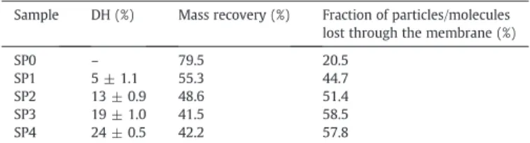
The intensity of the SP0 UV signal is higher than those of hydro-lyzed proteins due to the higher quantity of fractionated particles/ molecules. Moreover, the UV signal intensity and the quantity of frac-tionated particles/molecules decrease with an increase in the degree of hydrolysis. As shown inTable 2, the recovery mass of samples de-creased with protein hydrolysis. At the same time, the fraction of small particles/molecules with a molecular weight equal to or less than 10 kDa, which corresponds to the membrane's porosity, in-creased from 20.5% for native proteins to 44–58% for hydrolyzed SP. This behavior is mainly attributable to the presence of short proteinic
chains after hydrolysis. At hydrolysis degree higher than 13%, the ma-jority of SP chains in solution have a molecular weight less than 10 kDa (Rh≈ 2 nm).
3.3. Sunflower proteins/α-tocopherol emulsion characterization 3.3.1. Native sunflower proteins/α-tocopherol emulsion characterization
One of the most important factors leading to efficient microencapsu-lation of active compounds is their good dispersion in the wall material solution. In the case of hydrophobic core microencapsulation, an oil-in-water emulsion must be prepared before the encapsulation step (Augustin, Sanguansri, & Bode, 2006; Nesterenko, Alric, Silvestre, & Durrieu, 2012; Rascon, Beristain, Garcie, & Salgado, 2011; Rusli, Sanguansri, & Augustin, 2006; Yu, Wang, Yao, & Liu, 2007). It has been shown that retention of active material during spray-drying could be in-creased by reducing the mean emulsion droplet size of oil during the emulsification step (Rusli et al., 2006). To prepare a fine emulsion be-fore spray-drying, high pressure homogenization is widely used. In the case of a protein based emulsion, the intense mechanical forces un-dergone by globular proteins and oil droplets during homogenization, promote oil droplet dispersion and protein structure modification (Dickinson, 2001; Rampon, Riaublanc, Anton, Genot, & McClements, 2003). Protein chains become unfolded, which causes exposure of polar and non-polar regions making them more surface-active. Further-more, these proteins adsorbed on the oil droplet surface, provide good protection against coalescence and give physical stability to the emul-sion. Thus, to obtain a stable and uniform emulsion of SP and T, the mix-ture was circulated twice through the high-pressure homogenizer at 50 MPa.
As shown inFig. 3, the emulsion obtained with SP and T was com-posed of a homogeneous dispersion of T droplets surrounded by proteinic chains. The mean volume diameter of T emulsion droplets
Table 1
Modification degree of sunflower proteins. Sample name Nature of modification Hydrolysis time (min) Enzyme/SP (U/g) DH (%) DA (%) SP1 Hydrolysis 15 0.002 5 ± 1.1d – SP2 Hydrolysis 20 0.005 13 ± 0.9c – SP3 Hydrolysis 40 0.015 19 ± 1.0b – SP4 Hydrolysis 120 0.025 24 ± 0.5a – SP5A Hydrolysis + acylation C12 15 0.002 5 ± 1.1d 56 ± 0.5a SP6A Acylation C8 – – – 50 ± 1.1b SP7A Acylation C12 – – – 46 ± 1.6c SP8A Acylation C16 – – – 39 ± 0.5d SP9B Acylation C12 – – – 40 ± 0.6d a–dDifferent letters in the same column indicate a statistical difference between the
mean values (P b 0.05).
A Molar ratio fatty acid chloride/NH 2of 1. B Molar ratio fatty acid chloride/NH
2of 0.5.
Fig. 2. Hydrodynamic radius and UV signal (280 nm) as a function of analysis time for different samples of hydrolyzed SP. Table 2
Mass recovery and fraction lost through the membrane determined by AsFlFFF for na-tive and hydrolyzed sunflower proteins.
Sample DH (%) Mass recovery (%) Fraction of particles/molecules lost through the membrane (%) SP0 – 79.5 20.5
SP1 5 ± 1.1 55.3 44.7 SP2 13 ± 0.9 48.6 51.4 SP3 19 ± 1.0 41.5 58.5 SP4 24 ± 0.5 42.2 57.8
after 1 h, 1 day and 1 week remained 1.3 μm, 1.5 μm and 1.9 μm re-spectively (Fig. 4). A small increase in oil droplet size was observed, due to the coalescence of light particles with time. These results con-firmed the stability of formed SP/T oil-in-water emulsions and the homogeneity of T droplets.
3.3.2. Modified sunflower proteins/α-tocopherol emulsion characterization
The different emulsion characterizations are summarized inTable 3. Enzymatic hydrolysis and acylation of proteinic chains affected emul-sion properties such as oil droplet size and viscosity. All emulemul-sions with modified proteins have lower viscosity than the native SP/T emul-sion (SP0). The reduction in protein chain length by hydrolysis resulted in the drop in emulsion viscosity for samples SP1, SP2, SP3 and SP4. This could be connected to the relation between polymer molecular weight and its viscosity, usually described using the Mark–Houwink–Sakaruda equation. The same behavior was observed for soy proteins in a previ-ous study (Nesterenko et al., 2012) and even for other biopolymers (Avaltroni, Bouquerand, & Normand, 2004; Burkus & Temelli, 2003). The presence of small proteinic chains after hydrolysis with improved surfactant properties, favored T dispersion and prevented droplet coalescence. Thus, formation of homogeneous emulsions with small T droplets (0.7–1.7 μm) was observed.
Conversely, hydrophobic chains attached to water-soluble pro-teins by acylation resulted in an increase in the amphiphilic character of the macromolecules. The presence of these surface-active chains explained the decrease in emulsion viscosity observed for samples SP5, SP6, SP7 and SP8 compared to SP0. To conclude, both hydrolyzed and acylated SP were efficient polymers for forming stable and uni-form oil-in-water emulsions with T (Fig. 5). Lower protein agglomer-ation in the case of modified SP compared to native SP was observed. This can be attributed to the improved surfactant properties of modified proteins as well as to the lower viscosity of the emulsions obtained, and thus higher mobility of proteinic chains.
3.4. α-Tocopherol microencapsulation with sunflower proteins 3.4.1. Microencapsulation with hydrolyzed sunflower proteins
The results of T microencapsulation are presented inTable 3. The microparticles obtained by spray-drying were analyzed to determine their size and morphology, retention efficiency (RE) and loading effi-ciency (LE). The spray-drying yield for all emulsions varied from 60% to 70%, but was not significantly influenced by SP modifications. The effect of SP modifications on microparticles was particularly relevant for RE values. The increase in the degree of hydrolysis from 0% (SP0) to 24% (SP4) resulted in the decrease of microencapsulation efficiency from 92.6% to 61.8% respectively. This can be related to the AsFlFFF re-sults that confirmed the decrease in SP size with increasing degree of hydrolysis. Due to insufficient hydrolyzed protein chain length, this wall material cannot produce a sufficiently strong structural matrix to encapsulate T. Thus, higher amounts of T could be on the microparticle surfaces, giving agglomeration, confirmed by the in-crease of mean microparticle size from 6.9 μm (SP0) to 27.3 μm (SP4) and lower spray-drying yields. However, a small DH of protein allowed microparticles with satisfactory retention efficiency (80.2– 81.9%) to be obtained.
3.4.2. Microencapsulation with acylated SP
Grafting of fatty acid chains by acylation to both hydrolyzed (SP5) and native (SP6, SP7 and SP8) proteins involved an increase in
Fig. 3. View of SP/T oil-in-water emulsion (SP0) 1 h, 1 day and 1 week after high-pressure homogenization. Scale bar — 10 μm.
Fig. 4. Droplet size distributions in SP0 emulsion 1 h, 1 day and 1 week after high-pressure homogenization.
Table 3
Properties of spray-dried SP powder, SP/T emulsions and SP/T spray-dried microparticles.
Sample nameA Emulsion droplet size (μm) Emulsion viscosity (mPa·s) Spray-drying yield (%) REB(%) LEC(%) Particle size (μm)
SP0 1.3 ± 0.05b 36.1 ± 0.04 70 92.6 ± 1.8b 30.5 ± 0.6b 6.9 ± 0.03h SP1 1.7 ± 0.07a 5.2 ± 0.03 67 80.2 ± 1.1c 26.5 ± 0.4c 6.9 ± 0.08h SP2 1.6 ± 0.05a 4.0 ± 0.08 66 81.9 ± 1.5c 27.0 ± 0.5c 12.0 ± 0.12d SP3 0.8 ± 0.04de 3.6 ± 0.04 61 65.7 ± 3.1d 21.7 ± 1.3d 21.8 ± 0.13b SP4 0.7 ± 0.05ef 2.9 ± 0.02 60 61.8 ± 2.8d 20.4 ± 0.9d 27.3 ± 0.04a SP5 0.6 ± 0.05f 6.3 ± 0.03 60 99.8 ± 1.7a 32.9 ± 0.6a 16.8 ± 0.09c SP6 0.9 ± 0.04d 11.7 ± 0.03 68 97.0 ± 1.7ab 32.0 ± 0.5ab 8.5 ± 0.13e SP7 1.1 ± 0.03c 12.6 ± 0.02 67 100.8 ± 1.6a 33.3 ± 0.6a 7.3 ± 0.09g SP8 1.3 ± 0.04b 18.0 ± 0.06 67 99.8 ± 1.3a 33.0 ± 0.4a 7.9 ± 0.12f a–hDifferent letters in the same column indicate a statistical difference between the mean values (P b 0.05).
A SP0: non-modified sunflower proteins; SP1, SP2, SP3 and SP4: hydrolyzed sunflower proteins; SP5: hydrolyzed and acylated with C12 sunflower proteins; SP6, SP7 and SP8:
acylated with C8, C12 and C16 sunflower proteins, respectively.
B RE: retention efficiency determined by UV spectroscopy. C LE: loading efficiency or α-tocopherol content per 100 g of powder.
macromolecule hydrophobic properties. Proteinic matrix chains be-came more surface active and their affinity with hydrophobic core material increased. For this reason, higher RE values were observed when an acylated protein matrix was used compared to native pro-teins (97–100.8% and 92.6% respectively). Thus, acylation of both na-tive and hydrolyzed SP enhanced their functional properties and favored microencapsulation of hydrophobic active substances. Both of these phenomena, the decrease of RE after protein hydrolysis and the improvement in process efficiency after protein acylation, have already been observed for soybean proteins in a previous study (Nesterenko et al., 2012). Moreover, SP was the more efficient wall material in both native and modified states compared to soybean extracted proteins.
The mean particle diameter values obtained in this work were coher-ent with the expected range for microparticles produced by spray-drying (Favaro-Trindade, Santana, Monterrey-Quintero, Trindade, & Netto, 2010; Ortiz, Mauri, Monterrey-Quintero, & Trindade, 2009).
3.5. Wall/core ratio influence on the microencapsulation process
Different contents of active material in microparticles could be needed, depending on their applications (Elizondo et al., 2011). In order to evaluate the influence of T concentration on the microencap-sulation process, wall/core material ratios of 2/1 (SP0, SP9), 1/1 (SP0′, SP9′), 1/2 (SP0″, SP9″) and 1/4 (SP0‴) were studied. Data obtained from these experiments is shown inTable 4. For both native and acyl-ated SP, the increase in amount of T in the emulsion resulted in a
rise in emulsion droplet size and viscosity. The change in T droplet di-ameter from 1.3 μm to 2.2 μm for native SP and from 0.8 μm to 1.1 μm for acylated SP could be due to higher coalescence with increasing oil content. In prepared emulsions, SP concentration was kept constant at 8% w/w, but different amounts of T were added. Thus, creaming ve-locity decreased with higher droplet concentration (Chanamai & McClements, 2000; Robins, 2000) and total solid content of prepara-tions increased. Thus, the viscosity of formed emulsions was higher. The same phenomena have been observed for the emulsions with sunflower oil in pectin solution (Kawakatsu, Tragardh, & Tragardh, 2001), soy oil in whey protein solution (Reiffers-Magnani, Cuq, & Watzke, 1999) or corn oil in myofibrillar protein solution (Zorba, 2006).
A significant fall in spray-drying yield from 70–70.4% to 56–59.7% was observed with increasing wall/core ratio, due to the higher amount of T on particle surfaces. Additionally, the agglomeration ef-fect induced by the presence of surface oil resulted in an increase in mean microparticle sizes from 6.9 μm to 42.2 μm for native SP and from 7.7 μm to 24.7 μm for acylated SP. The displacement of size dis-tributions towards the higher values observed onFig. 6, confirms par-ticle agglomeration with addition of T. Surprisingly, the increase in amount of T in the emulsion also had a positive effect on the RE, which reached 99.9% for the highest SP/T ratio. The results obtained demonstrated that SP allows the production of relatively low to very highly loaded T microparticles, with loads from 30.5% to 79.2% for native SP and from 32.9% to 65.7% for acylated SP. This result is es-pecially interesting for conversion of liquid materials in free-flowing
Fig. 5. View of SP/T oil-in-water emulsions with SP modified by hydrolysis and/or acylation; 1 h after high-pressure homogenization. Scale bar — 10 μm.
Table 4
Properties of SP/T based emulsions and spray-dried microparticles with different wall/core ratios.
Sample name Ratio wall/core Emulsion droplet size (μm) Emulsion viscosity (mPa·s) Spray-drying yield (%) REA(%) LEB(%) Particle size (μm)
SP0C 2/1 1.3 ± 0.04c 36.0 ± 0.04 70 92.6 ± 1.8b 30.5 ± 0.6 6.9 ± 0.30d SP0′ 1/1 1.5 ± 0.06b 61.0 ± 0.07 69 93.4 ± 1.6ab 46.7 ± 0.8 8.3 ± 0.29cd SP0″ 1/2 2.2 ± 0.04a 96.0 ± 0.6 63 99.7 ± 3.2a 65.8 ± 2.1 8.8 ± 0.47c SP0‴ 1/4 2.2 ± 0.05a 159.0 ± 1.6 56 99.0 ± 3.5ab 79.2 ± 2.8 42.2 ± 1.17a SP9D 2/1 0.8 ± 0.04e 9.5 ± 0.03 70.4 99.5 ± 2.4a 32.9 ± 0.8 7.7 ± 0.20cd SP9′ 1/1 0.9 ± 0.03e 16.0 ± 0.61 68.7 98.7 ± 1.6ab 49.4 ± 0.8 8.6 ± 0.50c SP9″ 1/2 1.1 ± 0.05d 42.1 ± 0.68 59.7 99.9 ± 2.2a 65.7 ± 1.2 24.7 ± 0.71b a–eDifferent letters in the same column indicate a statistical difference between the mean values (P b 0.05).
A RE: retention efficiency determined by UV spectroscopy. B LE: loading efficiency or α-tocopherol content per 100 g of powder.
C SP0, SP0′, SP0″, and SP0‴: samples prepared with non-modified sunflower proteins and α-tocopherol. D S9, S9′, and S9″: samples prepared with sunflower proteins acylated by C12 (DA of 40%) and α-tocopherol.
powders. The relevance of this work is further confirmed when compared to literature values, where loading efficiency of microparti-cles prepared by spray-drying varied from 10 to 50% (Charve & Reineccius, 2009; Favaro-Trindade et al., 2010; Ortiz et al., 2009; Pierucci, Andrade, Farina, Pedrosa, & Rocha-Leao, 2007; Wang, Tian, & Chen, 2011).
The morphology of spray-dried microparticles was observed by SEM. Particles were characterized by spherical morphology and smooth surface without fissures or disruptions. T was contained in a porous interior structure surrounded by a continuous proteinic shell. The microstructure of particles seemed to change when T con-centration in the powder increased (Fig. 7). This can be attributed to a high density of oil droplets in the initial emulsion. Therefore dur-ing the spray-drydur-ing step, T droplets surrounded by SP form a contin-uous and porous network inside the microparticle.
3.6. Release profile of T from SP based microparticles
The in vitro release behavior of T loaded in SP microparticles was studied in a release medium made from a phosphate buffer and a
surfactant (Tween-80). The release profiles of T encapsulated by na-tive SP (SP0) and acylated with C12 proteins (SP7), are shown in Fig. 8. These kinetic studies demonstrated that T was efficiently protected by SP encapsulation in an aqueous media. The release rate is gradual and very similar for both native and modified SP micropar-ticles (about 50% within 12 h). After a 24 h period, the final amount of T released from SP microparticles based on native and acylated protein, was about 63% and 71% respectively.
3.7. Thermal analysis
The thermogravimetric (TG) weight loss and derivative of weight loss (DTG) graphs for SP, T and SP/T microparticles (SP0) are shown onFig. 9. The 100% mass loss for T with complete decomposition, oc-curred in the range between 220 and 380 °C with maximal weight loss at 370 °C. No degradation of T was observed below 200 °C. This be-havior confirmed that the core material used is not affected by the 120 °C spray-drying temperature. In addition, during the spray-drying process the particle temperatures reached approximately 70 °C due to cooling through water evaporation.
Fig. 6. Spray-dried microparticle size distributions of SP/T with different wall/core ra-tios (SP0 — 2/1, SP0′ — 1/1, SP0″ — 1/2 and SP0‴ — 1/4).
Fig. 7. Scanning electron micrographs of SP0 and SP0‴ microparticles (external and internal structures).
Fig. 8. Kinetic release profiles of T encapsulated by native sunflower proteins (SP0) and acylated with C12 sunflower proteins (SP7).
The weight loss of SP and SP0 microparticles began at 100 °C and can be attributed to the evolving of adsorbed water. The decomposi-tion of encapsulated T was observed after 400 °C because of a visible fall in the SP0 TG graph compared to that for SP. The degradation of SP0 microparticles is divided into two main steps. The first step at 280 °C corresponds to SP decomposition and the second step, be-tween 400 °C and 450 °C, represents T evolving from the microparti-cles. SP degradation takes place at temperatures above 200 °C with the maximal weight loss (more than 60%) between 200 and 450 °C. Concerning the 500 to 1000 °C temperature range, progressive degra-dation of SP and SP/T microparticles was observed. This thermal be-havior is very similar to soy protein microparticles loaded with T (Nesterenko et al., 2012).
4. Conclusions
As demonstrated in this paper, proteins extracted from sunflower seeds can be efficiently used for microencapsulation of hydrophobic substances using a spray-drying technique. Enzymatic hydrolysis and/or acylation of SP were also used in order to enhance the func-tional properties of this wall material.
Increase in degree of hydrolysis resulted in reduced oil droplet size in the emulsion and in decreased retention efficiency (from 92.6 to 61.8% for non-modified and 24% hydrolyzed sunflower pro-teins respectively). This could be due to short protein chains that do not form a strong structural matrix protecting T efficiently during spray-drying. The AsFlFFF results showed that for a DH higher than 13% the majority of SP chains in solution have a molecular weight smaller than 10 kDa. Nevertheless, a low DH of SP allows production of microparticles with relatively high microencapsulation efficiency (80.2–81.9%). On the other hand, attachment of hydrophobic chains by acylation to sunflower protein (native or hydrolyzed), improved its surface-active properties and hydrophobicity. The affinity between
wall and hydrophobic core material was enhanced and retention effi-ciency increased up to 100.8% for C12 acylated protein based micro-particles. But this modification has no significant influence on the release profile of active material from the microparticles.
Increase in amount of T resulted in higher emulsion droplet size, emulsion viscosity and diameter of microparticles. This study demon-strated that microparticles prepared from SP can be efficiently loaded with hydrophobic active material (α-tocopherol) up to 79% w/w. The work clearly shows that proteins extracted from sunflower oil-cake represent a new, natural and original matrix material for applications in microencapsulation. This could be an interesting way to valorize this agricultural by-product for various potential applications. Future research will focus on improving of release study with SP based microparticles.
References
Aider, M. (2010). Chitosan application for active bio-based films production and poten-tial in the food industry: Review. Food Science and Technology, 43, 837–842. Augustin, M. A., Sanguansri, L., & Bode, O. (2006). Maillard reaction products as
encapsulants for fish oil powders. Journal of Food Science, 71(2), 25–32. Avaltroni, F., Bouquerand, P. E., & Normand, V. (2004). Maltodextrin molecular weight
distribution influence on the glass transition temperature and viscosity in aqueous solutions. Carbohydrate Polymers, 58, 323–334.
Benita, S., Hoffman, A., & Donbrow, M. (1985). Microencapsulation of paracetamol using polyacrylate resins (Eudragit Retard), kinetics of drug release and evaluation of kinetic model. Journal of Pharmacy and Pharmacology, 37(6), 391.
Burkus, Z., & Temelli, F. (2003). Determination of the molecular weight of barley β-glucan using intrinsic viscosity measurements. Carbohydrate Polymers, 54, 51–57. Chabanon, G., Chevalot, I., Framboisier, X., Chenu, S., & Marc, I. (2007). Hydrolysis of
rapeseed protein isolates: Kinetics, characterization and functional properties of hydrolysates. Process Biochemistry, 42, 1419–1428.
Chanamai, R., & McClements, D. J. (2000). Dependence of creaming and rheology of monodisperse oil-in-water emulsions on droplet size and concentration. Colloids
and Surfaces, 172, 79–86.
Charve, J., & Reineccius, G. A. (2009). Encapsulation performance of proteins and tradi-tional materials for spray dried flavors. Journal of Agricultural and Food Chemistry,
57, 2486–2492.
Church, F. C., Swaisgood, H. E., Porter, D. H., & Catignani, G. L. (1983). Spectrophotometric assay using o-phthaldialdehyde for determination of proteolysis in milk and isolated milk proteins. Journal of Dairy Science, 66, 1219–1227.
Daqiq, L., Fellows, C. M., Bekes, F., & Lees, E. (2007). Methodologies for symmetrical-flow field-symmetrical-flow fractionation analysis of polymeric glutenin. Journal of Texture Studies,
38, 273–296.
De Graaf, L. A., Harmsen, P. F. H., Vereijken, J. M., & Monikes, M. (2001). Requirements for non-food applications of pea proteins. A review. Nahrung/Food, 45(6), 408–411. Dickinson, E. (2001). Milk protein interfacial layers and the relationship to emulsion
stability and rheology. Colloids and Surfaces, 20, 197–210.
Dubey, R., Shami, T. C., & Bhasker, R. K. U. (2009). Microencapsulation technology and application. Defence Science Journal, 59(1), 82–95.
Elizondo, E., Sala, S., Imbuluzqueta, E., González, D., Blanco-Prieto, M. J., Gamazo, C., et al. (2011). High loading of gentamicin in bioadhesive PVM/MA nanostructured microparticles using compressed carbon-dioxide. Pharmaceutical Research, 28, 309–321.
Faria, A. F., Mignone, R. A., Montenegro, M. A., Mercadante, A. Z., & Borsarelli, C. D. (2010). Characterization and singlet oxygen quenching capacity of spray-dried microcapsules of edible biopolymers containing antioxidant molecules. Journal of
Agricultural and Food Chemistry, 58, 8004–8011.
Favaro-Trindade, C. S., Santana, A. S., Monterrey-Quintero, E. S., Trindade, M. A., & Netto, F. M. (2010). The use of spray drying technology to reduce bitter taste of ca-sein hydrolysate. Food Hydrocolloids, 24, 336–340.
Fennema, O. R. (1993). Food chemistry. New York: Marcel Dekker Inc.
Gharsallaoui, A., Roudaut, G., Chambin, O., Voilley, A., & Saurel, R. (2007). Application of spray-drying in microencapsulation of food ingredients: An overview. Food
Research International, 40, 1107–1121.
Glantz, M., Hakansson, A., Lindmark-Mansson, H., Paulsson, M., & Nilsson, L. (2007). Revealing the size, conformation and shape of bovine casein micelles and aggre-gates with asymmetrical flow field-flow fractionation and multi-angle light scat-tering. Langmuir, 26, 12585–12591.
Gonzalez-Perez, S., & Vereijken, J. M. (2007). Sunflower proteins: Overview of their physicochemical, structural and functional properties. Journal of the Science of
Food and Agriculture, 87, 2173–2191.
Gonzalez-Perez, S., Vereijken, J. M., Koningsveld, G. A., Gruppen, H., & Voragen, A. (2005). Physicochemical properties of 2S albumins and the corresponding protein isolate from sunflower (Helianthus annuus). Journal of Food Science, 70, 98–103. Gonzalez-Perez, S., Vereijken, J. N., Merck, K. B., Koningsveld, G. A., Gruppen, H., &
Voragen, A. (2004). Conformational states of sunflower (Helianthus annuus) helianthinin: Effect of heat and pH. Journal of Agricultural and Food Chemistry, 52, 6770–6778.
Goodno, C. C., Swaisgood, H. E., & Catignani, G. L. (1981). A fluorimetric assay for avail-able lysine in proteins. Analytical Biochemistry, 115, 203–211.
Fig. 9. TG and DTG graphs of sunflower proteins (SP), α-tocopherol (T) and SP/T micro-particles (SP0).
Gouin, S. (2004). Microencapsulation: Industrial appraisal of existing technologies and trends. Trends in Food Science and Technology, 15, 330–347.
Guyomarc'h, F., Violleau, F., Surel, O., & Famelart, M. H. (2010). Characterization of heat-induced changes in skim milk using asymmetrical flow field-flow fraction-ation coupled with multiangle laser light scattering. Journal of Agricultural and
Food Chemistry, 58, 12592–12601.
Junyaprasert, V. B., Mitrevej, A., Sinchaipanid, N., Broome, P., & Wurster, D. E. (2001). Effect of process variables on the microencapsulation of vitamin A palmitate by gelatin-acacia coacervation. Drug Development and Industrial Pharmacy, 27(6), 561–566.
Kang, D. Y., Moon, J. M., & Lee, S. (2011). Comparison of size-exclusion chromatography and flow field-flow fractionation for separation of whey proteins. Bulletin of the
Korean Chemical Society, 32, 1315–1320.
Kawakatsu, T., Tragardh, G., & Tragardh, C. (2001). The formation of oil droplets in a pectin solution and the viscosity of the oil-in-pectin solution emulsion. Journal of
Food Engineering, 50, 247–254.
Kong, X. Z., Guo, M. M., Cao, D., & Zhang, C. M. (2008). Enzymatic preparation of immunomodulating hydrolysates from soy proteins. Bioresource Technology, 99, 8873–8879.
Lamsal, B. P., Jung, S., & Johnson, L. A. (2007). Rheological properties of soy protein hy-drolysate obtained from limited enzymatic hydrolysis. Food Science and Technology,
40, 1215–1223.
Luo, Y., Zhang, B., Whent, M., Yu, L., & Wang, Q. (2011). Preparation and characteriza-tion of zein/chitosan complex for encapsulacharacteriza-tion of α-tocopherol, and its in vitro controlled release study. Colloids and Surfaces, 85, 145–152.
Martinez, K. D., Baezaa, R. I., Millan, F., & Pilosof, A. M. R. (2005). Effect of limited hydro-lysis of sunflower protein on the interactions with polysaccharides in foams. Food
Hydrocolloids, 19, 361–369.
Matemu, A. O., Kayahara, H., Murasawa, H., Katayama, S., & Nakamura, S. (2011). Improved emulsifying properties of soy proteins by acylation with saturated fatty acids. Food Chemistry, 124, 596–602.
Mo, X., Zhong, Z., Wang, D., & Sun, X. (2006). Soybean glycinin subunits: Characterisation of physicochemical and adhesion properties. Journal of Agricultural and Food Chemistry,
54, 7589–7593.
Molina, M. I., Petruccelli, S., & Anon, M. S. (2004). Effect of pH and ionic strength mod-ifications on thermal denaturation of the 11S globulin of sunflower (Helianthus
annuus). Journal of Agricultural and Food Chemistry, 52, 6023–6029.
Nesterenko, A., Alric, I., Silvestre, F., & Durrieu, V. (2012). Influence of soy protein's structural modifications on their microencapsulation properties: α-Tocopherol microparticles preparation. Food Research International, 48, 387–396.
Nesterenko, A., Alric, I., Silvestre, F., & Durrieu, V. (2013). Vegetable proteins in micro-encapsulation: A review of recent interventions and their effectiveness. Industrial
Crops and Products, 42, 469–479.
Nunes, M. C., Batista, P., Raymundo, A., Alves, M. M., & Sousa, I. (2003). Vegetable pro-teins and milk puddings. Colloids and Surfaces, 31, 21–29.
Ortiz, S. E. M., Mauri, A., Monterrey-Quintero, E. S., & Trindade, M. A. (2009). Production and properties of casein hydrolysate microencapsulated by spray drying with soy-bean protein isolate. Food Science and Technology, 42, 919–923.
Patino, J. M. R., Conde, J. M., Linaresa, H. M., Jimenez, J. J. P., Sanchez, C. C., Pizones, V., et al. (2007). Interfacial and foaming properties of enzyme-induced hydrolysis of sunflower protein isolate. Food Hydrocolloids, 21, 782–793.
Pickardt, C., Neidhart, S., Griesbach, C., Dube, M., Knauf, U., & Kammerer, D. R. (2009). Optimisation of mild-acidic protein extraction from defatted sunflower (Helianthus
annuus L.) meal. Food Hydrocolloids, 23, 1966–1973.
Pierucci, A. P. T. R., Andrade, L. R., Farina, M., Pedrosa, C., & Rocha-Leao, M. H. M. (2007). Comparison of a-tocopherol microparticles produced with different wall materials:
Pea protein a new interesting alternative. Journal of Microencapsulation, 24, 201–213.
Rampon, V., Riaublanc, A., Anton, M., Genot, C., & McClements, D. J. (2003). Evidence that homogenization of BSA-stabilized hexadecane-in-water emulsion induces structure modification of the nonadsorbed protein. Journal of Agricultural and
Food Chemistry, 51, 5900–5905.
Rascon, M. P., Beristain, C. I., Garcie, H. S., & Salgado, M. A. (2011). Carotenoid retention and storage stability of spray-dried paprika oleoresin using gum Arabic and soy protein isolate as wall materials. Food Science and Technology, 44, 549–557. Rbii, K., Surel, O., Brambati, N., Buchert, A. M., & Violleau, F. (2011). Study of gelatin
re-naturation in aqueous solution by AFlFFF-MALS: Influence of a thermal pretreat-ment applied on gelatin. Food Hydrocolloids, 25, 511–514.
Reiffers-Magnani, C. K., Cuq, J. L., & Watzke, H. J. (1999). Composite structure formation in whey protein stabilized O/W emulsions. 1. Influence of the dispersed phase on viscoelastic properties. Food Hydrocolloids, 13, 303–316.
Robins, M. M. (2000). Emulsions — Creaming phenomena. Current Opinion in Colloid &
Interface Science, 5, 265–272.
Rondel, C., Alric, I., Mouloungui, Z., Blanco, J. -F., & Silvestre, F. (2009). Synthesis and properties of lipoamino acid-fatty acid mixtures: Influence of the amphiphilic structure. Journal of Surfactants and Detergents, 12, 269–275.
Rusli, J. K., Sanguansri, L., & Augustin, M. A. (2006). Stabilization of oils by microencap-sulation with heated protein–glucose syrup mixtures. JAOCS, 83, 965–971. Salgado, P. R., Drago, S. R., Ortiz, S. E. M., Petruccelli, S., Andrich, O., González, R. J., et al.
(2012). Production and characterization of sunflower (Helianthus annuus L.) protein-enriched products obtained at pilot plant scale. Food Science and Technology,
45, 65–72.
Vastag, Z., Popovic, L., Popovic, S., Krimer, V., & Pericin, D. (2010). Hydrolysis of pump-kin oil cake protein isolate and free radical scavenging activity of hydrolysates: Influence of temperature, enzyme/substrate ratio and time. Food and Bioproducts
Processing, 88, 277–282.
Vilkhu, K., Mawson, R., Simons, L., & Bates, D. (2008). Applications and opportunities for ultrasound assisted extraction in the food industry — A review. Innovative
Food Science and Emerging Technologies, 9, 161–169.
Wang, R., Tian, Z., & Chen, L. (2011). A novel process for microencapsulation of fish oil with barley protein. Food Research International, 44, 2735–2741.
Wong, P. Y. Y., Nakamura, S., & Kitts, D. D. (2006). Functional and biological activities of casein glycomacropeptide as influenced by lipophilization with medium and long chain fatty acid. Food Chemistry, 97, 310–317.
Yohannes, G., Wiedmer, S. K., Tuominen, E. K. J., Kinnunen, P. K. J., & Riekkola, M. L. (2004). Cytochrome c–dimyristoylphosphatidylglycerol interactions studied by asymmetrical flow field-flow fractionation. Analytical and Bioanalytical Chemistry,
380, 757–766.
Yoo, S. H., Song, Y. B., Chang, P. S., & Lee, H. G. (2006). Microencapsulation of α-tocopherol using sodium alginate and its controlled release properties. International Journal of
Biological Macromolecules, 38, 25–30.
Yu, C., Wang, W., Yao, H., & Liu, H. (2007). Preparation of phospholipid microcapsules by spray drying. Drying Technology, 25, 695–702.
Yust, M. M., Pedroche, J., Millan-Linares, M. C., Alcaide-Hidalgo, J. M., & Millan, F. (2010). Improvement of functional properties of chickpea proteins by hydrolysis with immobilised alcalase. Food Chemistry, 122, 1212–1217.
Zheng, H. G., Yang, X. Q., Tnag, C. H., Li, L., & Ahmad, I. (2008). Preparation of soluble soybean protein aggregates (SSPA) from insoluble soybean protein concentrates (SPC) and its functional properties. Food Research International, 41, 154–164. Zorba, O. (2006). The effects of the amount of emulsified oil on the emulsion stability