Cell migration controls key morphogenic events that shape the nervous system, ranging from neural tube closure to brain formation. During cerebral cortex development, cell migration is essential to set a precise temporal and spatial distribution pattern of neural cells that further engage in dynamic crosstalk to coordinate their maturation. Cerebral cortical activity relies on neural circuits formed of two main classes of neurons: excitatory projection neurons (PNs) that migrate along radial glia (RG) fibres within the cortex and inhibitory interneurons (INs) that originate from the ventral fore-brain and reach the cortex along two stereotypical tan-gential routes1. These neuronal classes include multiple
subtypes for which proper laminar positioning and bal-anced integration into neural networks are determinant factors for cortical function. Cortical cytoarchitectonics reflects interplays between cell extrinsic cues and intrin-sic mechanisms that coordinate the migration of neu-rons from their birthplace to a final destination, where they assemble into functional circuits. Most of these sig-nals are involved in the control of cytoskeletal elements and their regulators to support dynamic shape changes underlying cell motility and allocation to ad hoc cortical layers2. Migrating neurons not only receive important
cues that direct their navigation and differentiation into the cortex but also influence morphogenetic events occurring in the vicinity of their migratory path. Recent reports have placed glia, and in particular microglia, the resident macrophage of the brain, as essential play-ers for cortical morphogenesis via regulation of brain wiring and IN migration in the cortical wall. However, despite their prominent roles in cortical development, the migration pattern of the glial cells that transiently
or permanently populate the cerebral cortex remains largely unexplored3,4 Here, we review the migration
strategies adopted by neural cells to navigate in the cortical wall and offer perspectives for the roles of cell migration in the formation of the cerebral cortex. We also discuss how bringing together quantitative experi-mental analyses with mathematical modelling fosters the discovery of new mechanisms of cortical morphogene-sis5. Furthermore, this Review sheds light on recent
tech-nological developments that advance our understanding of human cerebral cortical morphogenesis and help us decipher how cell migration deficits can interfere with this process in brain pathology.
Cell migration in cortical development
Cell migration is an important process that allows dis-tinct cell types generated in different brain regions to settle in the cerebral cortex during embryogenesis. In mice, transient cell populations start colonizing the dor-sal forebrain at embryonic day 10.5 (E10.5), and these cells guide the later migration and placement in the developing cortex of neurons generated between E11.5 and E18.5. In addition, most glial cells invade the corti-cal wall concurrently with neurons (Fig. 1a), with which
some establish crosstalk. Cell migration strategies
Discontinuous migration of cortical interneurons. Cortical INs (cINs) exist in different shapes and forms, and their progenitors initiate interneuron specification in the ganglionic eminences (GEs) at the onset of cortico-genesis and before engaging in tangential migration6,7.
Transcriptional programmes are largely conserved
Cytoarchitectonics The cellular composition of a biological tissue. Cortical wall
Part of the dorsal forebrain that corresponds to the
presumptive cerebral cortex. Interneuron specification Cellular process engaging a precursor to self-autonomously acquire functional and morphological features of interneurons when placed in a neutral environment.
Cell migration promotes dynamic
cellular interactions to control cerebral
cortex morphogenesis
Carla G. Silva
1,2, Elise Peyre
1,2and Laurent Nguyen
1*
Abstract | The cerebral cortex is an evolutionarily advanced brain structure that computes higher
motor, sensory and cognitive functions. Its complex organization reflects the exquisite cell
migration and differentiation patterns that take place during embryogenesis. Recent evidence
supports an essential role for cell migration in shaping the developing cerebral cortex via direct
cellular contacts and spatially organized diffusible cues that regulate the establishment of its
cytoarchitecture and function. Identifying the nature of the crosstalk between cell populations
at play during brain development is key to understanding how cerebral cortical morphogenesis
proceeds in health and disease.
1GIGA-Stem Cells, University
of Liège, CHU Sart Tilman, Liège, Belgium.
2These authors contributed
equally: Carla G. Silva, Elise Peyre
*e-mail: lnguyen@uliege.be
https://doi.org/10.1038/ s41583-019-0148-y
among GE progenitors in mice. However, a limited set of genes expressed differentially in cIN progenitors prime the cells for individual differentiation and maturation trajectories into different interneuron subtypes, which further consolidate during their migration towards the cerebral cortex6,7. cINs are mainly generated in the
medial and caudal GEs (MGE and CGE, respectively) and are repelled from these regions by diffusible cues such as SLIT1, netrin 1 and ephrin A5 (reFs8–10). They
navigate tangentially along distinct migratory streams that are dynamically remodelled during development11.
The choice of a migratory path correlates, to some extent, with the temporal origin and fate of cINs12 and
is determined by permissive and chemoattractant cues, such as neuregulin 1 and CXCL12 (reFs13,14). The
direc-tion of migradirec-tion also relies on the dynamic branching of the leading process15 and polarity reversal16. By the end of
embryogenesis, cINs disperse in the cortical plate (CP) using their primary cilium to sense sonic hedgehog (SHH) gradients17,18 and undergo radial and/or oblique
migration to reach their cortical layers19 and
function-ally connect to partners. The migration of cINs in the forebrain parenchyma is discontinuous and alternates advancements with pauses. At the molecular level, this processes is paced by cytoskeleton-generated forces where microtubules recruit organelles and actomyosin SP neurons CR cells Microglia Corridor cells OPC cINs PNs
E10.5 E11.5 E12.5 E13.5 E14.5 E15.5 E16.5 E17.5 E18.5 Birth
a b TC fibre RG cell TH Bipolar PN Multipolar PN SP neuron 1 2 3 4 5 6 7 St2 St1 CR cell Microglial cell Corridor cell Blood vessel cIN
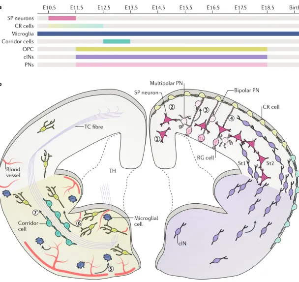
Fig. 1 | Migration strategies used by neural cells to colonize the mouse developing cerebral cortex. a | Timeline of generation and migration of cells that contribute to cortical morphogenesis. b | Subplate (SP) neurons (step 1) and Cajal-Retzius (CR) cells (step 2) migrate at the onset of corticogenesis and settle under the cortical plate or the pia, respectively. Different subtypes of CR cells are generated during development (light blue and pale green). Projection neurons (PNs; light pink) are generated by radial glia (RG) cells and are first multipolar. They undergo a non-directed migration and become bipolar to engage in locomotion on RG cell fibres (step 3). They reach their final location by detaching from these fibres and undergoing a final somal translocation. The cortical interneurons (cINs) are born in the subpallium and migrate tangentially along defined streams (St1 and St2) that are running in the cortical wall before reaching their ad hoc cortical layer (step 4). Additional cell populations reach the cortex during development and include the microglia, which mostly use vessels to enter the forebrain (step 5). They disseminate into the forebrain parenchyma using amoeboid-like movements. Other populations that reach the cortex by tangential migration include the
oligodendrocyte precursors (OPCs; lime green) that are born in the ventral forebrain (step 6). Corridor cells are located in the subpallium and migrate ventrally to allow the progression of thalamocortical (TC) fibres (step 7). Arrows represent the direction of migration of microglia (dark blue). E, embryonic day ; TH, thalamus.
Leading process Principal neurite of a migrating cell located ahead of the soma that contributes to its navigation along a migration path. Polarity reversal inversion of the polarity of a migrating cell along its moving axis.
contractions drive nucleokinesis20. The pausing time of
cINs represents around 40% of the migration process and requires a cyclic reduction in myosin light chain kinase (MLCK) activity via its processing by the cyto-solic carboxypeptidase 1 (reF.5). Pauses during migration
are not synchronized, thus generating heterogeneity of movement within the cIN population to regulate their subpallial sorting and cortical invasion. The regulated sorting of cINs shapes cortical morphogenesis via a crosstalk with dorsal cortical progenitors, the nature of which remains to be determined5.
As the migration of cINs into the cortex takes place in parallel with the production and migration of other cell types (Fig. 1b), it would be interesting to decipher
whether mechanisms generating heterogeneity of cell movement are also at play during the colonization of the cortex by other cell populations.
Multimodal migration of projection neurons. PN fate diversity is controlled by successive transcriptional waves in cortical progenitors21. At the onset of corticogenesis,
PNs spread radially throughout the entire cortical wall and reach their final position by somal translocation. At mid-corticogenesis, the subsequent waves of newborn PNs have no basal attachment to the pia and undergo a complex migration process. They first become multipo-lar and move in random directions22 before
repolar-izing to initiate locomotion along RG fibres23. The
multipolar-to-bipolar transition, the exact function of which remains elusive, is orchestrated by several tran-scription factors which, together with cytoplasmic and membrane effectors, promote the dynamic rearrange-ment of the cytoskeleton and centrosome positioning during migration24. PNs also undergo moderate
nucleo-kinesis during locomotion, a process driven by actomyo-sin contraction, endocytosis and cell adhesion events25.
PNs anchor their leading process in the marginal zone at the end of their locomotion to undergo a final somal translocation to settle at their final position16(Fig. 1b).
Pioneering migration of transient neurons during cere bral cortex development. Several neural cell popula-tions that reach the cortex by tangential migration are eliminated during brain maturation in rodents. These include the transient glutamatergic neurons composed of the Cajal-Retzius (CR) cells, the subplate (SP) cells and the CP transient (CPT) neurons. SP cells and CPT neurons are born in the rostral medial telencephalic wall and ventral pallium/pallium-subpallium bound-ary (PSB), respectively26–28. Tangentially migrating SP
neurons follow a trajectory parallel to the pia to reach the preplate (PP) and later settle under the CP26,29,30. SP
neurons generate the first axons that project out of the cortex and are postsynaptic targets for afferent thalamo-cortical (TC) axons31,32. Additionally, SP neurons guide
corticofugal projections, shape intracortical connectiv-ity and promote the formation of cortical networks in rodents and primates33–36. A fraction of non-tangentially
migrating SP neurons are generated by RG cells and migrate along their radial processes26. Some reports
suggest that SP neurons are still detected during the early postnatal periods both in rodents and primates37,38.
CPT neurons migrate tangentially into the cortex and exert non-cell-autonomous modulation of cortical neurogenesis by regulating the cell cycle length and proliferation rate of cortical progenitors28. The larger
transient population is represented by the CR cells that colonize the brain before CP formation and disappear by the end of the first postnatal week in mice39. In rodents,
CR cells are generated in three distinct regions of the forebrain — the hem, the PSB and the septum40 — and
disperse homogeneously under the cortical surface via contact-repulsion migration41(Fig. 1b). Mathematical
models show that the distribution of CR cells origi-nating from different locations is regulated by contact repulsion and by several intrinsic properties, such as the onset, speed and directionality of migration42. However,
in the human cortex, the classification of CR cells is more complex and includes the transient CR (tCR) and the permanent CR (pCR) cells43. pCR cells might share
a function with the postnatal rodent CR cells by con-tributing to the organization of cortical layering and cir-cuitry44. It is worth noting that the maintenance of these
cells in the human cortex during adulthood is subject to controversy as they share morphological features with some GABAergic interneurons, and co-detection of their specific markers (for example, TBR1 and calretinin) is found only in the hippocampus45.
Glial cell migration in the developing cerebral cortex. Despite their number, neurons represent only a fraction of cells that compose the cortex and its functional units. Microglia, originating from yolk sac myeloid progenitors, form one large non-neuronal population that invades the forebrain at the onset of neurogenesis46. These cells
con-tribute to developmental functions that go beyond their phagocytic activity47,48. Upon entrance in the dorsal
fore-brain, microglia begin to colonize from the pial surface, following dorsal-to-ventral and rostral-to-caudal gradi-ents49,50(Fig. 1b). CX
3CL1—CX3CR1 signalling is required
for their infiltration, proliferation and distribution in the developing brain. Microglia constitute a self-sustained population that proliferates during late gestation and postnatal periods. The exponential increase in micro-glia number after birth raises the possibility that blood monocytes or bone-marrow-derived cells might also reach the brain and add or substitute embryonically generated microglia46. At early stages of neurogenesis
and during adulthood, microglia often associate with progenitors and dying cells50–52. Apoptotic cell clearance
is a driving force for microglia invasion53. Colonization
of the CP by microglia requires RG processes and blood vessels, takes place from E16 onwards and follows an inside-out gradient48,54. Once they reach the cortical
wall, these cells are attracted to the ventricular zone (VZ) and subventricular zone (SVZ) by the CXCL12 released by intermediate progenitors (IPs), which activates CXCR4 or CXCR7 receptors in microglia55. Additional
molecules have been reported to recruit or facilitate microglia migration within the forebrain (for exam-ple, colony-stimulating factor 1 (CSF1), interleukin-34 (IL-34), VEGFR1 acting in synergy with CSF1 in the SVZ, superoxide ions and metalloproteinases (MMP8 and MMP9)) (reviewed in reF.56).
Nucleokinesis
Forward displacement of the nucleus in the leading process of a migrating neuron driven by dynamic changes of the cytoskeleton and in particular actomyosin contraction taking place around the nucleus. in moving neurons, the nucleokinesis is cyclic and paces their migration. Subpallial sorting Biological process contributing to the sorting of cells born in the ventral forebrain (the subpallium) into the dorsal forebrain (the pallium).
Another important pool of glial cells that colonize the cerebral cortex concurrently with cINs are the oligoden-drocyte precursor cells (OPCs), most of them becoming the myelinating cells of the CNS3. In the mouse, the first
wave of OPCs is generated at E11.5 in the MGE and the anterior entopeduncular area. This wave of OPCs is fol-lowed by a second one, born in the lateral GE (LGE) and CGE at E15.5 (Fig. 1a,b), and a third one arising after birth
within the cortical wall3. Experimental ablation showed
that OPCs from different lineages can compete for fore-brain territories3. The navigation of OPCs in the cortical
parenchyma requires the decoding of gradients of attrac-tive cues, such as PDGFRα57, and repulsive ones, such as
the BMPs secreted by the meninges58. In addition, OPCs
may interact with vessels via WNT-dependent signalling to colonize the pallium and later migrate from deep to more superficial layers in the cortex59. However, OPCs
can migrate in vitro in biological preparations devoid of endothelial cells60, suggesting that vessels might be only
one of the many substrates used by these cells to migrate. Interestingly, their association with blood vessels prevents their differentiation59. Despite accumulating studies
focus-ing on OPC motility, the exact migration mode adopted by the OPCs from the different waves remains undescribed.
The cerebral cortex is thus composed of distinct neu-ronal and non-neuneu-ronal cell types, the study of the diver-sity of which is currently being revisited by single-cell biology6,7,61,62. These cells settle either transiently or
per-manently (depending on type) in the cortical wall after having used distinct migration strategies adapted during evolution to face the physical constraints appearing in larger and more complex brains (Box 1).
Cellular crosstalk promotes morphogenesis Cerebral cortex arealization
The cortex is not homogeneous in size and function along the anteroposterior axis. Its tangential organi-zation segments the cortical surface into primary and
secondary motor, sensory (visual, somatosensory or auditory areas) and associative areas. Each field has a specific pattern of connection that generates cortical net-works producing behaviours. In primates, higher-order functions correlate with the expansion of cortical areas responsible for integrative functions and higher-order processing such as Broca’s area, composed of the soma-tosensory cortex, motor cortex and premotor cortex and controlling speech in humans63. Neocortical arealization is
primarily the result of the action of morphogens secreted early by patterning centres in proliferative cell popu-lations. Molecules such as BMP, FGF, SHH and WNT allow the establishment of the dorsoventral and anter-oposterior axes and in turn generation of the different cortical fields64. In addition to these morphogenic
gra-dients, postmitotic populations interplay to shape the tangential cytoarchitecture of the cortex. One of these populations is the CR cells, which have an important role for cortical patterning. CR cells born in distinct fore-brain regions differ in their molecular identity40. Each
subpopulation migrates from its birthplace to invade the cortical surface and colonize distinct domains of the cor-tex65. Interestingly, CR cells migrate in the vicinity of RG
cell fibres and disseminate before the onset of cortical neurogenesis. Loss of CR cells impairs the regionaliza-tion of the neuroepithelium65; however, the molecular
dialogue existing between CR and RG cells remains to be identified.
The modification of the migration speed of one CR neuron subpopulation leads to a change in the corti-cal surface covered by each subpopulation. In turn, this change leads to a proportional change in the size of higher-order cortical territories normally associated with a particular subtype of CR cells42.
Other populations of postmitotic cells can also influence cortical arealization. For example, patterning of the different areas of the sensory cortex results from the regulation and input from TC axons66–68. Removal or
increase of some TC afferent fibres results in postnatal alteration of the size of the primary visual cortex69,70 but
has little effect during development71. The exact nature of
the signalling triggered by the TC fibres in the somato-sensory cortex remains elusive, but interaction with postnatal PNs through cadherin has been suggested72.
Cerebral cortex lamination
The vertical lamination of the cortex depends on accu-rate spatial positioning and interaction between tran-sient (RG cells, CR cells and some SP neurons) and permanent (cINs, PNs and glia) cell populations within the extracellular matrix (Box 2). When RGs switch to a
neurogenic mode of division (around E12.5 in mice), CR cells have already covered the entire cortical surface26.
Together with the SP neurons positioned above the pro-liferative VZ, they form the PP, an instructive structure for migration and positioning of PNs73. Newborn PNs
experience a slow multipolar migration until they reach SP neurons, with which they establish transient glutama-tergic synapses that generate NMDA receptor-mediated signal transmission and Ca2+ entry73(Fig. 2). Ca2+
tran-sients are important for the morphological conversion of newborn PNs into fast bipolar locomoting PNs along Box 1 | cell migration in the cortex across evolution
the laminar organization of the cortex along its dorsoventral axis and the expansion of its layers are novel acquisitions of evolution126, which required adaptation of the
migration behaviour of resident cell populations. For example, somal translocation of early projection neurons (PNs) is mammalian-specific and has been introduced during evolution to efficiently convey positional information from the ventricular zone (vZ) to the cortex127. Later during corticogenesis, sibling PNs move laterally before locomoting
on radial glia fibres, thereby contributing to cortical expansion84,128. Despite originating
from different progenitors, cortical interneurons (ciNs) from sauropsids and mammals integrate the pallium via a conserved tangential migration129. Guidance cues are
preserved, but transplantation of non-mammalian ciNs into the mammalian cortex showed that they cannot enter the cortical plate130. similar to rodents, primate ciNs
originate from ganglionic eminences (Ges) and undergo tangential migration131. recent
studies identified novel late-migrating ciNs generated in the subventricular zone and targeting the frontal lobe of young children. their delayed incorporation into the cerebral cortex may support the extension of plasticity periods in humans132. the
refinement of some human cortical functions required the formation of novel circuits, and migration of neurons from the Ges to the dorsal thalamus may have synchronized the co-evolution of the frontal cortex and the thalamic nuclei133,134. the formation
of thalamocortical (tC) connections relies on corridor cells that have remarkably conserved guidance properties across species. During evolution, a reorientation of corridor cells from the lateral Ge to the non-permissive medial Ge opened a novel internal path for thalamic axons135,136. this evolutionary step might have been essential
for improving tC communication.
Anterior entopeduncular area
region of the ventral forebrain that hosts progenitors of gABAergic and cholinergic interneurons as well as oligodendrocyte progenitor cells.
Neocortical arealization Biological process that organizes the tangential subdivision of distinct neocortical fields responsible for computing higher cerebral functions.
Morphogens
Non-uniformly distributed molecules that govern biological processes contributing to morphogenesis in a developing organism. Multipolar migration Cell migration mode characterized by the continuous extension and retraction of new neurites from the soma.
RG fibres73. However, bipolar conversion of PNs occurs
in reelin-signal-deficient mouse models74, where SP
neu-rons are misplaced under the pia75, suggesting that this
process might involve several mechanisms.
CR cells are located under the pia and release reelin, which controls the repolarization of migrating PNs76 and
later instructs them to stop their migration76,77(Fig. 2).
Once released, reelin forms a positive gradient from the uppermost layer of the cortex (layer I), where CR cells reside. The release of reelin from CR cells allows newborn PNs to migrate beyond their older siblings and build an inside-out cortex. However, removal of the cortical hem, a source of CR cells, does not generate cortical layer abnormalities78, suggesting that the
func-tion of these cells might be replaced by other cell types or structures. Importantly, these cells play additional functions such as specifying the identity of the distal dendritic compartment of upper-layer neurons79 and
controlling the timing of migration of cINs in the cor-tex80. They also integrate maturing neuronal networks,
receiving excitatory and inhibitory inputs and signal to pyramidal neurons through long-range horizontal axonal projections81.
Together with SP neurons, CR cells shape cortical architecture in synchrony with RG cells to produce an appropriate number of PNs that populate the cortical layers and acquire the appropriate molecular identity. The vertical organization of the cortex is translated into a columnar organization of neurons that share an over-lapping sensory receptive field. Neurons composing a minicolumn are densely interconnected and respond synchronously to electric stimulation82. The
mini-columns are functional units and have been proposed to reflect the radial migration of clonally related PNs orig-inated from a single RG cell83. However, minicolumns
are not strictly clonally related, and the first phase of multipolar migration allows a certain degree of lateral spreading84. This leads PNs of different origins to
inter-mingle and allows the column to be populated by correct neuronal subtypes.
Cellular crosstalk in corticogenesis
The activity of the cerebral cortex relies on functional networks composed of excitatory PNs and inhibitory cINs, which are born from distinct pools of forebrain progenitors. The laminar distribution of cINs depends on cues provided by PNs85, and the neuronal excitatory–
inhibitory (E/I) ratio is established during the matu-ration of the cortex and is later stabilized by synaptic mechanisms86. An altered proportion of PNs and cINs
leading to a disruption of the E/I balance has been associ-ated with epilepsy87 and psychiatric disorders (reviewed
in reF.88). Multiple cellular and molecular mechanisms
are at play during the maturation of the cortex to con-trol the bona fide proportion of PNs and cINs that are connecting to each other in the cortical wall. Indeed, the number of migrating cINs reaching the cortical wall during development exceeds the final number, but the E/I balance is further adjusted via regulated apoptosis of supernumerary cINs89. Their elimination relies on
the early establishment of local cortical circuits where PNs regulate cIN survival through a synaptic-dependent mechanism90,91. Recent studies have highlighted
addi-tional molecular crosstalk between cortical progenitors and migrating neurons that contribute to the establish-ment of the cortical cytoarchitectonic. The developing cortical wall is composed of IPs that serve as transient amplifying progenitors to generate most superficial PNs. In addition, IPs promote the migration of cINs in the path between the intermediate zone and the SVZ by releasing diffusible signals, among which the chemo kine CXCL12 signals to CXCR4 receptors expressed at the surface of cINs14,92(Fig. 2). The controlled migration of
cINs in streams canalizes their navigation and may con-centrate morphogenic signals released by them. Indeed, while migrating in the vicinity of IPs, cINs release diffus-ible cues that modulate IP proliferation, thereby affect-ing the production of superficial PNs5(Fig. 2). Thus, IPs
and migrating cINs establish important bidirectional crosstalk to control their position and numbers during the formation of the cerebral cortex. It is worth noting that migrating cINs also control the biology of OPCs via the release of fractalkine, which stimulates their differentiation without affecting their proliferation93.
Early migrating PNs control cortical progenitor fate via multiple non-cell-autonomous mechanisms. For example, SIP1 expression in early-born PNs prevents premature expression and release of NT3 and FGF9 that signal back to cortical progenitors to promote advance-ment of the sequential production of upper-layer PNs, deep-layer PNs and glia94(Fig. 2). Another important
signalling initiated by newborn PNs goes through their ability to sense WNT7 and express Jagged 1 (JAG1) as a result of the activation of a non-canonical WNT sig-nalling. According to their proximity with RG cells, PNs further mediate a JAG1-activation of Notch to control the sequential production of PNs and glia95(Fig. 2).
Migrating glia shape cortical circuits
During development, microglia control cortical progen-itor numbers via phagocytosis. Moreover, they eliminate some progenitors after the neurogenic period51,
sug-gesting that microglia also regulate gliogenesis in the Box 2 | The extracellular matrix is a key regulator of neural cell movements
all brain cells face complex and changing extracellular microenvironments. the extracellular matrix (eCM) constitutes the first line of interaction between neural stem cells, migrating cells and postmitotic neurons with their environment137. the eCM
forms a basal lamina around the brain and its vasculature138,139 and has a fundamental
role in the formation and function of the blood–brain-barrier and in the establishment of proper cortical lamination140,141. impaired layering can result from disturbed
interaction between radial glia (rG) cells and the cortical marginal zone (MZ)142.
Moreover, disruption of β1 integrin or other components of the basement membranes of the meninges leads to rG death, which correlates with a decreased brain size143.
the ventricular zone also contains receptors for cell adhesion that are fundamental to anchor and stabilize rG apical processes144,145. Cell adhesion proteins are used by
migrating neurons to form adhesive interactions with other neurons, glial cells and the eCM. these interactions are mainly mediated by integrin signalling with some specificity in different types of neuron. For example, interneurons use α3β1 integrin to sense and respond to netrin 1, a ligand contributing to a permissive tangential path for migrating cortical interneurons (ciNs) in the MZ146. additional contributions from eCM
molecules have been demonstrated, such as chondroitin-4-sulfate acting in concert with semaphorin 3a to guide migrating ciNs147. However, it remains unknown whether
spatiotemporal changes in eCM composition actively contribute to control key cellular events that shape cerebral cortical morphogenesis.
cortex. Microglia have functions beyond phagocytosis by trimming non-myelinated axons, contacting synaptic elements and regulating spine density in postnatal peri-ods and engulfing and/or promoting synaptic contact formation (reviewed in reF.4). While migrating,
micro-glia might also contribute to blood vessel formation as suggested by their close association with endothelial cells96–98, and microglia control cortical invasion of cINs48
and influence OPC maturation99. Strikingly, not much
is known about the migration properties of OPCs and in particular about those generated in the subpallium3.
In the early postnatal period, some OPCs can establish
synaptic contacts with neurons and receive GABAergic inputs that are important to control their population size100,101. However, it remains uncertain whether these
early networks established between PNs or cINs and OPCs involve the ventrally generated OPCs.
These examples support the existence of abundant dynamic crosstalk among neural cells invading and settling into the cortex to support its morphogenesis. However, the complexity of such crosstalk in vivo limits a full understanding, and in silico modelling has recently been introduced to shed new light on the regulatory mechanisms at play. a b c CP SP SVZ VZ CP SP SVZ VZ CP IZ VZ RG cell SP neuron Multipolar PN Bipolar PN CR cell Ca2+ NMDAR Reelin Intermediate progenitor cell U pper -lay er neur ons Deep-lay er neur ons NTF3 FGF9 WNT7A Notch JAG1 cIN
Transient glutamatergic synapse
Diffusible cues
CXCL12
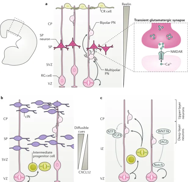
Fig. 2 | cellular crosstalk shapes cerebral cortical morphogenesis. a | At the onset of corticogenesis, projection neurons (PNs) that are born in the ventricular zone (VZ) by asymmetric division of radial glia (RG) cells are multipolar. They migrate through the subventricular zone (SVZ) towards subplate (SP) neurons and establish transient glutamatergic synapses with these neurons. NMDA receptor (NMDAR)-induced Ca2+ entry contributes to the morphological conversion
into bipolar cells, which then migrate on RG cell fibres through the cortical plate (CP). Cajal-Retzius (CR) cells are located at the top of the CP and release reelin, which establishes a gradient (grey triangle) contributing to the repolarization of PNs and the arrest of their migration. b | Migrating cortical interneurons (cINs, purple) and intermediate progenitors (IPs, lime green) in the cortical wall engage in bidirectional crosstalk (including diffusible cues such as secreted CXCL12), which controls the recruitment of cINs and the proliferation of IPs. By regulating IP proliferation, cINs regulate the final numbers of upper-layer PNs (light pink). c | Newborn PNs expressing the transcription factor SIP1 (light pink cells) control the fate of the RG daughter cells via the release of NT3 and FGF9 as well as the activation of a Notch-dependent signalling pathway in RG cells. These mechanisms control the sequential generation of PNs for deep and upper layers as well as glia arising later from dividing cortical progenitors. IZ, intermediate zone.
Understanding cortical cytoarchitectonics In vitro modelling
Many different in vitro assays have been used to charac-terize neural migration behaviour and its biochemical and molecular regulation (Fig. 3). Such assays include live
imaging of brain organotypic slices or explants to follow neuronal migration102, the Boyden chamber assay and
microfluidic devices to study the migration of distinct cell types under the chemoattraction of selective mol-ecules5,18. Interestingly, as novel biomaterials develop,
these assays are increasingly being revisited as new tools for the study of neuronal migration. Bioscaffolds mim-icking the extracellular matrix can be engineered into compartmented space and nanotopography in culture by using synthetic materials such as polyethylene glycol (PEG) or hydrogels. Moreover, photoactive materials are being used to print proteins and generate local mechan-ical forces on the substrate or guide local cell seeding103.
PEG hydrogel membranes can also be photopatterned in situ to generate microfluidic environments, constituting a flexible tool to study cell migration104.
Understanding how human brain cytoarchitecture is established is a major challenge of developmental biol-ogy. The limiting factors are the complex organization of the human brain and the poor accessibility to human tissue. Emerging culture systems are being made availa-ble to generate simple to complex human self-organized 3D structures named organoids, where understanding
of cell population interactions becomes possible in vitro. Organoids are generated by culturing human pluripotent stem cells in suspension, which aggregate and self-organize to form 3D structures with features of developing brains in vivo105. Together with technical
advances such as CRISPR–Cas9 to produce isogenic cells lines by genome editing, tissue clarification techniques and light sheet microscopy for large-scale 3D imaging, organoids have become a critical tool to study human cell population interactions and tissue morphogenesis. The ‘assembloid’ is a recent technological advancement of organoids to study human neuronal migration106,107.
It consists of a fusion of brain organoids patterned with morphogen addition in their culture media to generate different brain regions of interest or brain regions that cannot be properly specified together106,108. Human brain
assembloids could help to decipher whether migrating cINs control the proliferation of IPs as shown in rodents5
and how cIN subtype specification6,7 and survival91 are
controlled. Future improvements should allow the func-tional assembly of a wide range of distinct brain regions to understand how they influence each other to generate cortical cytoarchitecture and neuronal circuits. In silico modelling
Modelling helps in understanding the developmental processes in many different ways. Modelling can be used at different scales — from isolated mechanisms that have
Boyden chamber assay Cell chemotaxis assay based on a chamber of two medium-filled compartments separated by a microporous membrane.
Nanotopography specific surface features of biomaterials generated at the nanoscopic scale. Subpallium Pallium SVZ VZ Ventricle In silico modelling
data acquisition Single-cell tracing
Single-cell transcriptomics cIN
Intermediate PC
Histology analysis (tissue clearing) Time-lapse
analysis
Microfluidic migration assay
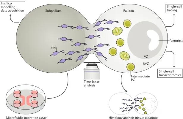
Fig. 3 | innovative tools to study neuronal migration of human-derived neurons. Assembloids are fused organoids patterned towards subpallial and pallial fates. On dissociation and culture in a microfluidic device, they can be used to follow neuron migration from its subpallial to pallial entity and thus untangle cell-autonomous from non-cell-autonomous components. Time-lapse analysis of assembloids might enable assessment of the impact of interneuron migration on proliferation of ventricular zone (VZ)-like and subventricular zone (SVZ)-like progenitors and their cell cycle exit. This analysis can be supported by in silico modelling. Cell diversity and maturation in assembloids could be assessed by coupling single-cell tracing with single-cell transcriptomics and the progressive establishment of its cytoarchitecture followed by immunostaining upon tissue clearing. cIN, cortical interneuron; PC. progenitor cell.
an impact at the single-cell level to models allowing the representation of an entire cell population or organs. Models predict phenotypic outcomes upon modifica-tion of input parameters and give rise to new testable hypotheses. Models have some limitations: small-scale models simplify complex processes in a limited develop-mental window to test specific hypotheses isolated from larger-scale and more complex phenotypes. On the other hand, multiscale and integrative models render devel-opmental processes from molecular signalling to tissue and organ formation possible, but they require a large amount of data and extensive acquired knowledge.
Comprehensive modelling has been used to under-stand the establishment of cortical cytoarchitecture, neuronal migration and cell population interactions. Cortical development has been simplified in silico to represent neurogenesis and the establishment of its radial organization. To understand PN production, experimen-tal parameters such as duration of the cell cycle, rate of cell cycle exit and cell survival have been used to derive mathematical models that can predict neuronal output across species109,110. Later, these models were refined by
integrating novel data acquired from the developing (for example, number of progenitor cells) and mature brain (for example, number and distribution of PNs) of different species111. The upgraded model predicts not
only neuronal output, layer formation and cell progeni-tor dynamics across species but also the spatiotemporal particularities of cortical formation, such as increased upper-layer occupancy with increased neuronal output and spatial neuronal density variation across the pri-mate cortex. Mathematical models accounting for cell proliferation and neuronal production have shown how the perturbation of proliferation regulators and timing of neurogenesis affect neuronal output, cortical thick-ness and surface size (for example, upon LHX2 muta-tion)112. Building on these studies by adding adequate
progenitor cell types, cortical development models have also demonstrated the strong adaptability of cortical progenitors to cope with postmitotic neuronal cell death during corticogenesis by reducing IP cell cycle length and by promoting self-renewing divisions113. Other
models have been established to predict how neuronal migration is regulated in the cortex. The radial migra-tion of PNs was also modelled using knowledge of a sim-plified network of known migration regulators: GABA, reelin, doublecortin (DCX) and LIS1. This model reca-pitulated the different phases of PN migration from the apical surface and their multipolar-to-bipolar transition and locomotion across neuronal layers. The model also predicted migration defects upon LIS1 and DCX loss of function114. On a larger scale, mathematical models
can envision how cells migrate and how their movement influences other cell populations and cortical develop-ment. Studying the dissemination of CR cells under the cortical surface is a good example of a successful appli-cation of this type of approach41,42. As the repartition
of CR cell subtypes during cortical development influ-ences the identity of the cortical domain they occupy, deciphering the parameters regulating their migration and final repartition is essential to understanding how cortical cytoarchitecture is generated. Several migration parameters of CR cells were measured, and random walk and contact repulsion were also differently weighted to mimic the average displacement speed of each CR cell subtype. These data were integrated into a model able to predict how the average speed and directionality of migration differentially influenced the final distribution of each CR subtype at the surface of the cortex. Another example is the modelling of cortical invasion by cINs during development. Parameters of cIN migration, such as pausing and nucleokinesis, influence the number of cINs entering the developing mouse cortex. Shifting the stereotypical biphasic cIN migration mode of division towards a continuous displacement was predicted to lead to a higher homogeneity of cell movement and in turn to a larger recruitment of cINs invading the cortex5.
In silico models are evolving and being built upon with the addition of new data and recently acquired knowledge. For instance, models will improve by inte-grating interactomes and proteomic and transcriptomic screens at the population and single-cell level (Box 3).
The ultimate goal of mathematical modelling in this field is to represent cell population dynamics in sufficient detail so as to correctly predict the outcome of diverse perturbations occurring during cortical development. Once validated experimentally, such a model system will likely be relevant to understanding pathologies and to the design of therapeutic strategies.
Cell migration in psychiatric disorders
Each year, about 165 million people in Europe are affected by mental illness, representing a major cause of morbidity and impaired quality of life in affected individuals. The current view is that a large proportion of these disorders take root during brain formation via deviation from normal developmental trajectories. Although most of these neurodevelopmental disorders have a strong genetic basis, the precise aetiology of the Box 3 | studying migration properties at the single-cell level
the understanding of the mechanisms governing cerebral cortical development has recently benefited from the blooming of single-cell biology. while accumulating data are being generated by single-cell transcriptomics to uncover cell diversity in the cerebral cortex6,7,61,62, clonal analysis has been used to label isolated progenitors and
monitor their progeny. Clonal tracing was originally performed by infecting tissues with a low titre of retroviruses and lentivirus or by expressing plasmids coding for a fluorescent reporter148,149. technological developments were recently made, including
the use of endogenous retroelements such as long interspersed nuclear element 1 (LiNe1) to trace cell lineage in humans150 as well as a transposon-based technique such
as the CLoNe method to target single neural progenitors in mice and chickens151.
Genetic animal models include the Brainbow mouse, in which clonally related cells are labelled in a wide range of colours depending on the random expression of different fluorophores upon Cre recombination152. MaGiC (multi-addressable
genome-integrative colour) is a genome-integrative transposon-based strategy derived from the Brainbow concept allowing labelling of different subcellular compartments153.
another genetic approach named mosaic analysis with double markers (MaDM) has recently been developed to more precisely map the lineage of mouse cortical progenitors and the migration of their progenies154. this genetic toolbox can also be
engineered at the locus of the gene of interest and coupled with knockout strategies to distinguish cell-autonomous and non-cell-autonomous regulations155. these genetic
tools advance our knowledge and shed some new light on cell migration processes in the brain. For example, it has been suggested that clonally related cortical interneurons are located in close proximity156,157, but a combination of MaDM with barcode viral
infections showed that these cells disperse over long distances in the cortex158. Random walk
Cell migration mode characterized by constant changes of movement angle.
condition often remains unknown. Thus, a major health challenge is to better understand their pathophysiolog-ical mechanisms to improve diagnosis and develop new pharmacotherapies that may ultimately lead to preclin-ical trials. Some genes linked to common neurodevel-opmental disorders, such as autism spectrum disorder (ASD) and schizophrenia, regulate cortical neuron devel-opment, integration and their functional assembly in cortical circuits. For instance, recessive loss-of-function mutations in CNTNAP2 lead to a rare but highly penetrant form of ASD in humans characterized by a migration deficit of PNs115. Analyses conducted in
Cntnap2-knockout mice116 not only support this
obser-vation but also reveal a reduction in cIN number117,118.
FMR1 encodes FMRP, which is an mRNA-binding
pro-tein involved in translational regulation. FMR1 mutation leads to fragile X syndrome, which accounts for 2–3% of autism cases119. Similarly to the loss of function of
CNTNAP2, FMRP deficit results in reduced migration of PNs into the CP120 and is also characterized by a lack of
cINs121. Although more work is needed to understand
the precise developmental sequence of pathological events leading to ASD, experimental analyses conducted with these animal models suggest that an early disrup-tion of neuronal migradisrup-tion leads to abnormal cortical circuit assembly and function. Interestingly, research in animal models has shown that reversal of genetically based cortical malformation arising from neuronal migration defects is possible at early postnatal stages by reactivating specific molecular pathways. This finding is exemplified by the partial rescue of subcortical band heterotopia via re-expression of DCX in a rat model of PN migration defect122. As the first ASD symptoms are
identifiable in the early months of childhood when the brain is plastic, this suggests that re-engineering the early postnatal cortex might be possible.
Recent technological developments of human orga-noid cultures105 open the path to foster our
understand-ing of the pathophysiological mechanisms underlyunderstand-ing ASD by using human-derived models. The recent gen-eration of brain assembloids106,108 uncovered a novel
migration deficit of cINs in Timothy syndrome, a mono-genic disease associated with ASD and epilepsy106. The
field of human organoids is growing at high pace and benefits from regular cell culture improvements123
and technological developments. As the in vitro mod-elling is getting closer to human brain development, human organoid cultures hold promise for untangling novel cellular crosstalk and movements underlying cor-tical morphogenesis in health and disease. Future tech-nical improvements are required to increase the cellular diversity of organoids and their controlled growth over longer periods, allowing further neuronal differentiation and maturation of functional neural circuits. Long-term culture of organoids is currently limited by the poor dif-fusion of oxygen, nutrients and waste. Thus, improved methods such as 3D culture using microcarriers could prolong culture to increase the differentiation potential and increase cell-type diversity. Microencapsulation is another methodological approach to improve the oxygenation and delivery of nutrients and growth factors to the culture124.
Conclusion and perspectives
The complexity of the cerebral cortex cytoarchitectonic has long been fascinating biologists. Despite accumu-lating knowledge obtained with complementary in vitro and in vivo models, we do not fully understand the spa-tial and temporal coordination of mechanisms driving cerebral cortical morphogenesis. Recent progress sug-gests that migration not only is required for allocating final cell positioning in the cortex but, in some instances, also generates specific crosstalk for instructing local morphogenetic events via physical contact and/or release of diffusible cues. These processes underlie not only early corticogenesis steps, such as the rate of progenitor proliferation, but also the regulation of long-range com-munication between distant brain structures, initiation and refinement of synaptic communication and organ-ization of higher-order areas responsible for cognition and complex behaviours.
By the time neurons wire together, some cortical cells have been eliminated in a regulated fashion. Although these cells actively contribute to morphogenetic cross-talk during development, the reason for their short life in the cortex after birth remains unclear but is often found to be conserved across evolution. This conser-vation suggests that their programmed elimination is required for the final maturation steps of the cortex and should be considered when generating large-scale modelling of neocortical microcircuitry125.
Despite considerable progress made during the past decade, we still do not have a comprehensive view on how cell migration contributes to morphogenesis, ulti-mately leading to the establishment of the complex cortical cytoarchitecture. Recent technological develop-ments, such as single-cell biology, simplified 3D culture and in silico modelling for brain development, should soon improve the resolution of analyses to answer the remaining questions and likely raise novel ones. For instance, the nature of mechanisms of cell diversification in the brain and the exact contribution of intrinsic and environmental factors to this process remain unclear.
Recent single-cell analyses have revealed the exist-ence of molecular signatures for newborn cINs that are otherwise difficult to identify because of their numer-ous subtypes, at times owing to the small size of their population or the late expression of landmark markers. This discovery should help monitor the migration of dif-ferentiating cINs in more detail and thus help to clarify whether the migration pattern of cINs as a whole pop-ulation is homogeneous or whether migration patterns are specific to cIN subtypes. Moreover, neurons and glia undergo stereotypic migration patterns to reach their final position in the cortex, and it remains unknown whether such behaviour is intrinsically regulated or mainly dependent on environmental cues and/or cell– cell interaction dynamics. The implementation of novel in silico models and predictive analyses will help us in deciphering to what extent neuronal migration is auton-omously and/or non-autonauton-omously driven at different developmental milestones and in distinct areas of the developing cerebral cortex.
1. Marin, O. & Rubenstein, J. L. Cell migration in the forebrain. Annu. Rev. Neurosci. 26, 441–483 (2003).
2. Heng, J. I., Chariot, A. & Nguyen, L. Molecular layers underlying cytoskeletal remodelling during cortical development. Trends Neurosci. 33, 38–47 (2010). 3. Kessaris, N. et al. Competing waves of
oligodendrocytes in the forebrain and postnatal elimination of an embryonic lineage. Nat. Neurosci. 9, 173–179 (2006).
4. Thion, M. S. & Garel, S. On place and time: microglia in embryonic and perinatal brain development.
Curr. Opin. Neurobiol. 47, 121–130 (2017). 5. Silva, C. G. et al. Cell-intrinsic control of interneuron
migration drives cortical morphogenesis. Cell 172, 1063–1078 (2018).
This work demonstrates the existence of a crosstalk between migrating cINs and PN progenitors to control the output of upper-layer neurons.
6. Mayer, C. et al. Developmental diversification of cortical inhibitory interneurons. Nature 555, 457–462 (2018).
7. Mi, D. et al. Early emergence of cortical interneuron diversity in the mouse embryo. Science 360, 81–85 (2018).
8. Hamasaki, T., Goto, S., Nishikawa, S. & Ushio, Y. A role of netrin-1 in the formation of the subcortical structure striatum: repulsive action on the migration of late-born striatal neurons. J. Neurosci. 21, 4272–4280 (2001).
9. Marin, O. et al. Directional guidance of interneuron migration to the cerebral cortex relies on subcortical Slit1/2-independent repulsion and cortical attraction.
Development 130, 1889–1901 (2003).
10. Zimmer, G. et al. Ephrin-A5 acts as a repulsive cue for migrating cortical interneurons. Eur. J. Neurosci. 28, 62–73 (2008).
11. Metin, C., Baudoin, J. P., Rakic, S. & Parnavelas, J. G. Cell and molecular mechanisms involved in the migration of cortical interneurons. Eur. J. Neurosci.
23, 894–900 (2006).
12. Lim, L. et al. Optimization of interneuron function by direct coupling of cell migration and axonal targeting.
Nat. Neurosci. 21, 920–931 (2018).
13. Flames, N. et al. Short- and long-range attraction of cortical GABAergic interneurons by neuregulin-1.
Neuron 44, 251–261 (2004).
14. Tiveron, M. C. et al. Molecular interaction between projection neuron precursors and invading interneurons via stromal-derived factor 1 (CXCL12)/ CXCR4 signaling in the cortical subventricular zone/ intermediate zone. J. Neurosci. 26, 13273–13278 (2006).
15. Martini, F. J. et al. Biased selection of leading process branches mediates chemotaxis during tangential neuronal migration. Development 136, 41–50 (2009).
16. Nadarajah, B., Brunstrom, J. E., Grutzendler, J., Wong, R. O. & Pearlman, A. L. Two modes of radial migration in early development of the cerebral cortex.
Nat. Neurosci. 4, 143–150 (2001).
17. Baudoin, J. P. et al. Tangentially migrating neurons assemble a primary cilium that promotes their reorientation to the cortical plate. Neuron 76, 1108–1122 (2012).
18. Higginbotham, H. et al. Arl13b in primary cilia regulates the migration and placement of interneurons in the developing cerebral cortex. Dev. Cell 23, 925–938 (2012).
19. Tanaka, D. H., Maekawa, K., Yanagawa, Y., Obata, K. & Murakami, F. Multidirectional and multizonal tangential migration of GABAergic interneurons in the developing cerebral cortex. Development 133, 2167–2176 (2006).
20. Bellion, A., Baudoin, J. P., Alvarez, C., Bornens, M. & Metin, C. Nucleokinesis in tangentially migrating neurons comprises two alternating phases: forward migration of the Golgi/centrosome associated with centrosome splitting and myosin contraction at the rear. J. Neurosci. 25, 5691–5699 (2005). 21. Telley, L. et al. Sequential transcriptional waves direct
the differentiation of newborn neurons in the mouse neocortex. Science 351, 1443–1446 (2016). 22. Tabata, H. & Nakajima, K. Multipolar migration:
the third mode of radial neuronal migration in the developing cerebral cortex. J. Neurosci. 23, 9996–10001 (2003).
23. Noctor, S. C., Martinez-Cerdeno, V., Ivic, L. & Kriegstein, A. R. Cortical neurons arise in symmetric and asymmetric division zones and migrate through specific phases. Nat. Neurosci. 7, 136–144 (2004).
24. LoTurco, J. J. & Bai, J. The multipolar stage and disruptions in neuronal migration. Trends Neurosci.
29, 407–413 (2006).
25. Kawauchi, T. et al. Rab GTPases-dependent endocytic pathways regulate neuronal migration and maturation through N-cadherin trafficking. Neuron 67, 588–602 (2010).
26. Barber, M. & Pierani, A. Tangential migration of glutamatergic neurons and cortical patterning during development: lessons from Cajal-Retzius cells.
Dev. Neurobiol. 76, 847–881 (2016). 27. Pedraza, M., Hoerder-Suabedissen, A.,
Albert-Maestro, M. A., Molnar, Z. & De Carlos, J. A. Extracortical origin of some murine subplate cell populations. Proc. Natl Acad. Sci. USA 111, 8613–8618 (2014).
28. Teissier, A. et al. A novel transient glutamatergic population migrating from the pallial-subpallial boundary contributes to neocortical development.
J. Neurosci. 30, 10563–10574 (2010).
29. Bayer, S. A. & Altman, J. Development of layer I and the subplate in the rat neocortex. Exp. Neurol. 107, 48–62 (1990).
30. Hoerder-Suabedissen, A. & Molnar, Z. Molecular diversity of early-born subplate neurons. Cereb. Cortex
23, 1473–1483 (2013).
31. Allendoerfer, K. L. & Shatz, C. J. The subplate, a transient neocortical structure: its role in the development of connections between thalamus and cortex. Annu. Rev. Neurosci. 17, 185–218 (1994). 32. Molnar, Z., Adams, R. & Blakemore, C. Mechanisms
underlying the early establishment of thalamocortical connections in the rat. J. Neurosci. 18, 5723–5745 (1998).
33. Kanold, P. O., Kara, P., Reid, R. C. & Shatz, C. J. Role of subplate neurons in functional maturation of visual cortical columns. Science 301, 521–525 (2003). 34. McConnell, S. K., Ghosh, A. & Shatz, C. J. Subplate
pioneers and the formation of descending connections from cerebral cortex. J. Neurosci. 14, 1892–1907 (1994).
35. Zhao, C., Kao, J. P. & Kanold, P. O. Functional excitatory microcircuits in neonatal cortex connect thalamus and layer 4. J. Neurosci. 29, 15479–15488 (2009).
36. Meinecke, D. L. & Rakic, P. Expression of GABA and GABAA receptors by neurons of the subplate zone in developing primate occipital cortex: evidence for transient local circuits. J. Comp. Neurol. 317, 91–101 (1992).
37. Kostovic, I. & Rakic, P. Developmental history of the transient subplate zone in the visual and somatosensory cortex of the macaque monkey and human brain. J. Comp. Neurol. 297, 441–470 (1990).
38. Woo, T. U., Beale, J. M. & Finlay, B. L. Dual fate of subplate neurons in a rodent. Cereb. Cortex 1, 433–443 (1991).
39. Hevner, R. F., Neogi, T., Englund, C., Daza, R. A. & Fink, A. Cajal-Retzius cells in the mouse: transcription factors, neurotransmitters, and birthdays suggest a pallial origin. Brain Res. Dev. Brain Res. 141, 39–53 (2003).
40. Bielle, F. et al. Multiple origins of Cajal-Retzius cells at the borders of the developing pallium. Nat. Neurosci.
8, 1002–1012 (2005).
41. Villar-Cervino, V. et al. Contact repulsion controls the dispersion and final distribution of Cajal-Retzius cells.
Neuron 77, 457–471 (2013).
42. Barber, M. et al. Migration speed of Cajal-Retzius Cells modulated by vesicular trafficking controls the size of higher-order cortical areas. Curr. Biol. 25, 2466–2478 (2015).
This study shows that vesicular trafficking controls the cortical distribution of distinct CR cells, which influences the size and wiring of different cortical areas.
43. Meyer, G. & Gonzalez-Gomez, M. The heterogeneity of human Cajal-Retzius neurons. Semin. Cell Dev. Biol.
76, 101–111 (2018).
44. Meyer, G. & Gonzalez-Gomez, M. The subpial granular layer and transient versus persisting Cajal-Retzius neurons of the fetal human cortex. Cereb. Cortex 28, 2043–2058 (2018).
45. Abraham, H. & Meyer, G. Reelin-expressing neurons in the postnatal and adult human hippocampal formation. Hippocampus 13, 715–727 (2003). 46. Ginhoux, F. & Garel, S. The mysterious origins of
microglia. Nat. Neurosci. 21, 897–899 (2018). 47. Schafer, D. P. et al. Microglia sculpt postnatal neural
circuits in an activity and complement-dependent manner. Neuron 74, 691–705 (2012).
48. Squarzoni, P. et al. Microglia modulate wiring of the embryonic forebrain. Cell Rep. 8, 1271–1279 (2014).
This work suggests that the embryonic invasion of the brain by microglia controls the wiring of some forebrain circuits by affecting axonal growth and interneuron positioning.
49. Arnold, T. & Betsholtz, C. The importance of microglia in the development of the vasculature in the central nervous system. Vasc. Cell 5, 4 (2013).
50. Ashwell, K. The distribution of microglia and cell death in the fetal rat forebrain. Brain Res. Dev. Brain Res.
58, 1–12 (1991).
51. Cunningham, C. L., Martinez-Cerdeno, V. & Noctor, S. C. Microglia regulate the number of neural precursor cells in the developing cerebral cortex. J. Neurosci. 33, 4216–4233 (2013). 52. Sierra, A. et al. Microglia shape adult hippocampal
neurogenesis through apoptosis-coupled phagocytosis. Cell Stem Cell 7, 483–495 (2010). 53. Casano, A. M., Albert, M. & Peri, F. Developmental
apoptosis mediates entry and positioning of microglia in the zebrafish brain. Cell Rep. 16, 897–906 (2016). 54. Swinnen, N. et al. Complex invasion pattern of the
cerebral cortex bymicroglial cells during development of the mouse embryo. Glia 61, 150–163 (2013). 55. Arno, B. et al. Neural progenitor cells orchestrate
microglia migration and positioning into the developing cortex. Nat. Commun. 5, 5611 (2014).
56. Xavier, A. L., Menezes, J. R., Goldman, S. A. & Nedergaard, M. Fine-tuning the central nervous system: microglial modelling of cells and synapses.
Phil. Trans. R. Soc. B 369, 20130593 (2014). 57. Frost, E. E., Zhou, Z., Krasnesky, K. & Armstrong, R. C.
Initiation of oligodendrocyte progenitor cell migration by a PDGF-A activated extracellular regulated kinase (ERK) signaling pathway. Neurochem. Res. 34, 169–181 (2009).
58. Choe, Y., Huynh, T. & Pleasure, S. J. Migration of oligodendrocyte progenitor cells is controlled by transforming growth factor beta family proteins during corticogenesis. J. Neurosci. 34, 14973–14983 (2014).
59. Tsai, H. H. et al. Oligodendrocyte precursors migrate along vasculature in the developing nervous system.
Science 351, 379–384 (2016).
60. Paez, P. M. et al. Golli myelin basic proteins regulate oligodendroglial progenitor cell migration through voltage-gated Ca2+ influx. J. Neurosci. 29,
6663–6676 (2009).
61. Marques, S. et al. Transcriptional convergence of oligodendrocyte lineage progenitors during development. Dev. Cell 46, 504–517 (2018). 62. Pollen, A. A. et al. Molecular identity of human outer
radial glia during cortical development. Cell 163, 55–67 (2015).
63. Krubitzer, L. The magnificent compromise: cortical field evolution in mammals. Neuron 56, 201–208 (2007).
64. Hasenpusch-Theil, K., Watson, J. A. & Theil, T. Direct interactions between Gli3, Wnt8b, and Fgfs underlie patterning of the dorsal telencephalon. Cereb. Cortex
27, 1137–1148 (2017).
65. Griveau, A. et al. A novel role for Dbx1-derived Cajal-Retzius cells in early regionalization of the cerebral cortical neuroepithelium. PLOS Biol. 8, e1000440 (2010).
66. Dehay, C., Horsburgh, G., Berland, M., Killackey, H. & Kennedy, H. Maturation and connectivity of the visual cortex in monkey is altered by prenatal removal of retinal input. Nature 337, 265–267 (1989). 67. Polleux, F., Dehay, C., Goffinet, A. & Kennedy, H.
Pre- and post-mitotic events contribute to the progressive acquisition of area-specific connectional fate in the neocortex. Cereb. Cortex 11, 1027–1039 (2001).
68. Borello, U., Kennedy, H. & Dehay, C. The logistics of afferent cortical specification in mice and men.
Semin. Cell Dev. Biol. 76, 112–119 (2017). 69. Vue, T. Y. et al. Thalamic control of neocortical area
formation in mice. J. Neurosci. 33, 8442–8453 (2013).
70. Chou, S. J. et al. Geniculocortical input drives genetic distinctions between primary and higher-order visual areas. Science 340, 1239–1242 (2013). 71. Garel, S., Yun, K., Grosschedl, R. & Rubenstein, J. L.
The early topography of thalamocortical projections is shifted in Ebf1 and Dlx1/2 mutant mice. Development
129, 5621–5634 (2002).
72. Egusa, S. F. et al. Classic cadherin expressions balance postnatal neuronal positioning and dendrite dynamics to elaborate the specific cytoarchitecture of the mouse cortical area. Neurosci. Res. 105, 49–64 (2016).