Journal of Veterinary Diagnostic Investigation 2014, Vol. 26(1) 136 –140
© 2014 The Author(s) Reprints and permissions: sagepub.com/journalsPermissions.nav DOI: 10.1177/1040638713515480 jvdi.sagepub.com
Brief Research Reports
Neospora caninum, the causative agent of neosporosis, is an apicomplexan intracellular parasite causing paresis in dogs and abortion in cattle.2,3 Studies of the domestic life cycle of
the parasite have shown that dogs are both intermediate and definitive hosts, whereas cattle are natural intermediate hosts.2
Diagnosis of neosporosis can be achieved using histol-ogy, immunohistochemical staining, polymerase chain reac-tion, and serology.4 Serological tests have the advantage that
they can be applied antemortem, but suffer from not being able to discriminate between latent and acute infections.4
Serological techniques for the specific detection of bovine and canine antibodies to N. caninum include the indirect fluorescent antibody test (IFAT),2 immunoblotting (IB),1 the
direct agglutination test (DAT),17 and a variety of
enzyme-linked immunosorbent assays (ELISAs).5 The accepted
ref-erence method for serological diagnosis is IFAT,6 with IFAT
having been used in many species and generally considered the “gold standard” when evaluating new methods.2–4
However, use of whole tachyzoite antigen in any sero-logical assay may result in high background absorbance values and cross-reactivity with antibodies against related parasites such as Toxoplasma gondii.5 Compared with the
native antigens, recombinant antigens are easily produced in large quantities and can be readily standardized for diagnostic
assays. In addition, their use may minimize the risk of cross-reactivity with other parasite species.15
The molecular search for diagnostic antigens for N. cani-num infection has focused on the identifying immunodomi-nant antigens that are recognized by sera from infected animals. In this sense, the dense granule proteins, NcGRA7 and NcGRA6, of N. caninum were shown to be effective candidates to diagnose N. caninum infection in cattle when used in ELISA.9–11
In contrast to the other serological techniques (e.g., ELISA, IFAT, IB), which are laborious and time-consuming and require specialized expertise and equipment, the latex agglutination test (LAT) is a technique that is very simple to carry out, making it suitable for clinical or field applica-tions.18 In the present study, the performance of a LAT using
recombinant NcGRA6 was evaluated for the serological detection of specific antibodies to N. caninum and compared 515480VDIXXX10.1177/1040638713515480Neospora caninum serological methodsGhalmi et al.
research-article2014
From the High National Veterinary School of Algiers, Algiers, Algeria (Ghalmi, Azzag); the Scientific Institute of Public Health, Brussels, Belgium (China); the Faculty of Veterinary Medicine, University of Liege, Liege, Belgium (Ghalmi, Losson); and the Beltsville Agricultural Research Center, U.S. Department of Agriculture, Beltsville, MD (Jenkins).
1Corresponding Author: Farida Ghalmi, Ecole Nationale Supérieure
vétérinaire d’Alger El Harrach, Algiers, Algeria. fghalmi@yahoo.fr
Comparison of different serological methods
to detect antibodies specific to Neospora
caninum in bovine and canine sera
Farida Ghalmi,
1Bernard China, Mark Jenkins, Naouelle Azzag, Bertand Losson
Abstract. Neospora caninum is an apicomplexan parasite responsible for paresis in dogs and abortion in cattle worldwide. Dogs serve as a definitive host, while cattle serve as intermediate host. Many different methods have been developed to detect specific antibodies present in cattle and dog serum. In the present study, the dense granule protein NcGRA6 was incorporated in a latex beads agglutination test (LAT), and compared to other serological methods, including enzyme-linked immunosorbent assay, the direct agglutination test, the immunoblot, and the indirect fluorescent antibody test (IFAT). Using the IFAT as the reference method, 100 sera isolated from Algerian cattle and 100 sera isolated from Algerian dogs, both possibly infected with N. caninum, were used to evaluate the LAT. The sensitivity, specificity, and kappa index were calculated for each host species and assay. For dog sera, the sensitivity and the specificity of the LAT was 76% and 100%, respectively. The McNemar test showed that the LAT was not significantly different from IFAT (P > 0.05). For cattle sera, the sensitivity and the specificity of the LAT were 60% and 100%, respectively. The McNemar test indicated that the LAT was significantly different from IFAT (P < 0.01) and that the LAT was only positive for cattle sera with titers of 1:800 or greater, indicating that LAT can be used for cattle in a clinical context. As well, the LAT has the advantage of being easy and rapid to perform compared to the other assays.
with other serological techniques for the detection of specific antibodies to N. caninum in cattle and dogs in Algeria.
The complementary DNA encoding the GRA6 protein of N. caninum was cloned in frame with an upstream polyhisti-dine tag sequence in a commercial plasmida to give the
pHisGRA6 plasmid. This plasmid was introduced by electro-poration into Escherichia coli BL21(DE3)pLysS cells.b The
recombinant cells were selected on trypticase soy agar pla-tesc containing 150 µg/ml of ampicillin. Colonies were
ana-lyzed by extracting plasmid DNA using a mini DNA kit,d
followed by an enzymatic restriction of the plasmid DNA using restriction enzyme digestion with BamHI and KpnI endonucleases.e The restriction products were analyzed by
agarose gel electrophoresis to check for the presence of a band of 4.4 kb corresponding to the vector and a band of 0.95 kb corresponding to the GRA6 insert.
In order to induce the production of the recombinant GRA6 protein, a 500-ml trypticase soy brothc containing
50 µg/ml of kanamycin was inoculated with a single colony of E. coli BL21 (pHisGRA6). Isopropyl β-d
-1-thiogalactopyranoside (IPTG)f was added to the culture at a
final concentration of 0.5 mM when the culture optical den-sity at 600 nm reached 0.5. An aliquot was removed after 1, 2, 3, and 4 hr of induction, and analyzed by sodium dodecyl sulfate–polyacrylamide gel electrophoresis (SDS-PAGE). In order to identify the recombinant protein, a commercially sourced antibodyg (that recognizes the peptide
Asp-Leu-Tyr-Asp-Asp-Asp-Lys), an anti-polyhistidine monoclonal antibody,h and a N. caninum–positive cattle serum6 were
used in a Western blot as previously described.6
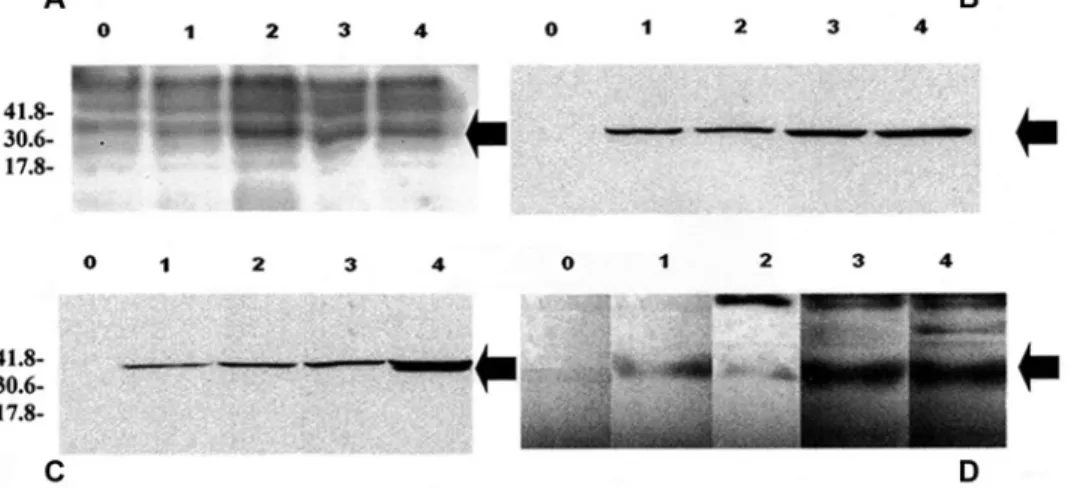
In the production process of the recombinant GRA6 pro-tein, a 33-kDa protein was clearly induced (Fig. 1A). In order to confirm that the 33-kDa protein was the recombinant GRA6 protein, Western blots with either monoclonal
antibodies specific to the tag peptide (Fig. 1B, 1C) or a poly-clonal sera positive for N. caninum (Fig. 1D) were used. As expected, in each case, the 33-kDa protein was detected. The tagged protein was purified using, successively, 2 different commercial kitsi,j in order to remove the remaining residual bands (Fig. 2). The concentration of the protein suspension was determined by the Lowry methodk and adjusted to 0.5 mg/ml in phosphate buffered saline (PBS).l
Figure 1. Induction of the recombinant (r)GRA6 protein. Lane 0: no induction; lane 1: 1-hr induction; lane 2: 2-hr induction; lane 3:
3-hr induction; lane 4: 4-hr induction. A, result of sodium dodecyl sulfate–polyacrylamide gel electrophoresis. B, immunoblot with the anti-Xpress monoclonal antibody. C, immunoblot with anti-polyhistidine monoclonal antibody. D, immunoblot with an anti–Neospora caninum polyclonal serum. The arrow indicates the rGRA6 protein. Molecular weight standards, in kilodaltons (kDa), are indicated.
Figure 2. Sodium dodecyl sulfate–polyacrylamide gel
electro-phoresis of the purified recombinant (r)GRA6 protein. Lane 1: total protein extract prior to affinity purification; lane 2: protein banding profile after purification with a commercial kiti; lane 3: protein
banding profile after purification with the second commercial kit.j The arrow indicates the rGRA6 protein. Molecular weight
Ghalmi et al. 138
Latex beads were coated with the purified recombinant GRA6 protein as previously described.16 In brief, 1 ml of
10% ml latex beadsm (0.8 µm in diameter) was washed
3 times in 5 ml of carbonate buffer (pH 9.6; 2,500 × g for 30 min). The pellet was suspended in 3 ml of carbonate buf-fer and 1.75 ml of the GRA6 recombinant protein (approxi-mately 0.9 mg) and incubated for 3 hr at 37°C and then over-night at 4°C. The sensitized beads were centrifuged and suspended in 5 ml of carbonate buffer containing 5% sucrose and 0.3% bovine serum albumin to block the nonoccupied sites. The beads were then incubated for 30 min at 37°C, centrifuged, and washed twice in PBS (pH 7.4), and sus-pended in 3 ml of PBS. The proficiency of the coating was controlled using IFAT coated beads and a positive and a negative anti–N. caninum polyclonal serum. The epifluores-cence was restricted to classical IFAT background with the nonimmune serum, and the epifluorescence was bright and clearly positive with the immune serum (data not shown). The sera were used to agglutinate the coated latex beads. A droplet (10 µl) of beads was mixed with a droplet (10 µl) of sera on a glass slide with a bacteriological loop. After 5 min, the result was observed. The strength of agglutination was graded as – (no visible aggregated beads), + (some aggre-gated beads with a majority of nonaggreaggre-gated beads), ++ (equal amount of aggregated and non-aggregated beads), or +++ (majority of aggregated beads). Latex agglutination test was then compared with other serological tests for N. cani-num antibodies using dog and bovine sera.
A total of 100 serum samples collected from March to June 2009 from Algerian dogs were analyzed. Likewise, serum samples from 100 Algerian cattle were collected from April to August 2009 and used in the LAT evaluation. For dog sera, an ELISA previously described14 and based on
sonicated tachyzoites was used (termed ELISA-U). A previ-ously validated commercial ELISAn,7 (termed ELISA-X) was
also used. The commercial ELISA-X is a sandwich ELISA based on recombinant NcSRS2 antigen. For cattle sera, a commercial ELISAo (termed ELISA-H) was used because it
was a well-validated assay.19 An additional commercial
ELISA-Xn was also used on cattle sera.7 The DAT was
per-formed as previously described.17 An IB assay based on total
tachyzoite protein extract was used as previously described.7,8
For the purpose of the current study, sensitivity (Se) and specificity (Sp) were defined as the relative Se and the rela-tive Sp of the tested assay in comparison to the reference assay (IFAT). The Youden index (Y)12 (in %) is the sum of
the Sp (in %) and the Se (in %) minus 100: Y = (Se + Sp) – 100. The value ranges between 0% and 100%. The kappa index (κ) is a statistical measure of inter-rater agreement and by extension to interassay agreement and was calculated and evaluated as previously described.12 The strength of the
agreement for the kappa coefficient was evaluated as fol-lows: ≤0 = poor; 0.01–0.20 = slight; 0.21–0.40 = fair; 0.41– 0.60 = moderate; 0.61–0.80 = substantial; and 0.81–1.00 = almost perfect. The McNemar test12 was applied to the
sero-logical test results, and the P values were calculated. The test was considered as significantly different from the reference test when P < 0.05.
Among the 100 tested canine sera, 25 were positive (at the dilution 1:50) and 75 were negative by IFAT. Of the 5 assays evaluated, sensitivities were between 60% and 88%, with ELISA-X having the highest Se. The Se of the LAT (76%) was intermediate. Specificities were high for all assays, including LAT, being greater than or equal to 99% (Table 1). None of the serological tests, with the exception of ELISA-U, were significantly different from IFAT (P > 0.05).
Table 1. Comparison of serological methods used to detect antibodies specific to Neospora caninum in bovine and canine sera.*
Sera†/Method Sensitivity (%) Specificity (%) Youden index (%) Kappa index (agreement evaluation) McNemar test (P value) Dog
ELISA-X 88 99 87 0.89 (almost perfect) 0.62
ELISA-U 60 100 60 0.69 (substantial) <0.01
DAT 72 99 71 0.77 (substantial) 0.08
LAT 76 99 75 0.68 (substantial) 0.13
IB 85 100 85 0.92 (almost perfect) 0.13
Cattle
ELISA-H 85 98 83 0.85 (almost perfect) 0.13
ELISA-X 77 100 77 0.81 (almost perfect) <0.01
DAT 65 100 65 0.69 (substantial) <0.01
LAT 60 100 60 0.64 (substantial) <0.01
IB 100 100 100 1 (almost perfect) 1.00
* ELISA = commercial enzyme-linked immunosorbent assay; DAT = direct agglutination test; LAT = latex agglutination test; IB = immunoblot. Both ELISA-Ho and ELISA-Xn are commercial assays that were used as per the manufacturer’s instructions. The third assay, ELISA-U, was used as previously
described.14
† In the dog sera, 25 were positive (at the dilution 1:50) by the reference method—the indirect fluorescence antibody test (IFAT)—while 75 were negative by IFAT. In the cattle sera, 40 were positive by IFAT (at the dilution 1:200) and 60 were negative by IFAT.
For bovine sera, 40 sera were positive in IFAT at the dilu-tion 1:200, 32 at diludilu-tion 1:400, 24 at diludilu-tion 1:800, and 11 at dilution 1:1,600. The Se of the IB was 100%, indicating that all the positive sera in IFAT were also positive in IB. The other methods were less sensitive than the IB method, with LAT having a 60% Se (Table 1). For LAT, only strongly positive sera (titer 1:800 or higher) were positive. The Se results were better because all of the methods but the ELISA-H showed a Sp of 100%, indicating that all sera negative in IFAT were also scored as negative with the other techniques. The combination of the Sp and the Se reflected by the Youden index indicated that the ideal score of 100% was reached by IB, while the other techniques obtained a score ranging from 83% (ELISA-H) to 60% (LAT). Finally, the kappa index gave the same ranking with the IB displaying 100% correlation with IFAT, and ELISA tests giving a score higher than 80%. The agglutination methods (DAT and LAT) on the other hand obtained a kappa index lower than 70%. According to these results, the McNemar test indicated that DAT, LAT, and ELISA-X methods were significantly different from IFAT (P < 0.05).
The detection of antibodies directed to N. caninum is undertaken worldwide both in dogs and in cattle.5 The
refer-ence method for the detection of antibodies directed to N. caninum is the IFAT.2 The drawbacks to IFAT are that the
method requires cell cultures and fluorescence microscopy, and expertise in discerning positive and negative samples. Moreover, only a few samples can be tested on a single slide. The commercial ELISA showed good Se (>75%) and excel-lent Sp (approximately 100%) values. Others have stated that ELISA is a viable alternative to IFAT for the detection of N. caninum antibodies in animals because a large number of sera can be tested on a single ELISA plate.4 However, while
a number of ELISA methods have been developed and com-mercialized,4 the technique requires a multi-well plate reader
for analyzing reactions, thus limiting its use in the field or in poorly equipped laboratories in developing countries.
Immunoblotting is mainly used as a confirmatory method1
and provides additional information such as the molecular weight of the reactive antigens. From a technical point of view, this method suffers from a need for native antigen preparation, SDS-PAGE and electrophoretic blotting, and finally antigen detection. Standardization of such a method is also difficult because antigen preparations can vary from one experiment to another.4 In the current study, the IB method
had high Sp and Se, but may be a poor primary screening method because of the low numbers of samples that can be analyzed at one time. Immunoblotting represents an excel-lent method to corroborate clinical findings and results of other serological assays.
The DAT involves the agglutination of whole tachyzoites, thus requiring free tachyzoites produced in cell cultures.17 As
expected, the DAT method displayed specificities and sensi-tivities similar to LAT. The preparation of the tachyzoites in this technique seems to be crucial and maybe difficult to standardize in laboratories not familiar with this technique.
In contrast, the use of latex beads coated with a recombi-nant antigen offers another way to perform agglutination. The LAT is a simple, rapid method, which makes it suitable for clinical or field applications. A key issue in the development of a LAT is the selection of the antigen to bind to the beads. Immunodominant antigens are the best candidates because they induce high titers of specific antibodies. The advantage of this method is that coating beads requires very few basic materials, primarily latex beads and purified proteins. The agglutination test requires only a solid support, the coated beads, and the serum to be tested. Therefore, this method can be used in basic clinical laboratories and in the field.
In the present work, the dense granule protein GRA6 was used because it appears to be an immunodominant N. cani-num antigen.10,13 The results indicated that the Se of the LAT
ranged from 60% to 76% for dog and cattle sera, respec-tively. On the other hand, the Sp of the LAT was nearly 100% for both canine and bovine sera. The high Sp and mod-erate Se indicates that LAT is an adequate test for corrobo-rating clinical findings of neosporosis. Overall, the results indicated that the LAT with NcGRA6 is a rapid, simple, and inexpensive diagnostic test that may be suitable for detecting N. caninum–specific antibodies in dogs and cattle infection under field conditions. Improvements in the linking of the NcGRA6 protein to beads and increasing the binding signal may improve the Se of LAT.
Sources and manufacturers
a. pTrcHisB plasmid, Invitrogen Corp., Carlsbad, CA. b. Life Technologies, Carlsbad, CA.
c. Tryptic soy agar (TSA), tryptic soy broth (TSB); Sigma-Aldrich NV/SA, Bornem, Belgium.
d. Pure Yield plasmid Miniprep, Promega Benelux BV, Leiden, The Netherlands.
e. Fermentas GmbH, St. Leon-Rot, Germany.
f. IPTG, Isopropyl β-D-1-thiogalactopyranoside; Sigma-Aldrich NV/SA, Bornem, Belgium.
g. Anti-Xpress antibody, Invitrogen Corp., Carlsbad, CA. h. Monoclonal anti-polyhistidine clone HS-1, Sigma-Aldrich
NV/SA, Bornem, Belgium.
i. MagneHis protein purification system, Promega Benelux BV, Leiden, The Netherlands.
j. QIAexpress Ni-NTA Fast start kit, Qiagen Benelux BV, Venlo, The Netherlands.
k. DC protein assay, Bio-Rad Laboratories NV, Nazareth, Belgium. l. Phosphate buffered saline, BioReagent, pH 7.4, for molecular
biology; Sigma-Aldrich NV/SA, Bornem, Belgium.
m. Latex beads (0.8 µm in diameter), Sigma-Aldrich NV/SA, Bornem, Belgium.
n. Bio-X trousse ELISA Neospora caninum, Bio K 192; Bio-X Diagnostics SPRL, Jemelle, Belgium.
o. ELISA HerdChek, IDEXX Laboratories, Westbrook, ME.
Declaration of conflicting interests
The author(s) declared no potential conflicts of interest with respect to the research, authorship, and/or publication of this article.
Ghalmi et al. 140
Funding
The author(s) disclosed receipt of the following financial support for the research, authorship, and/or publication of this article: This work was supported by a grant of the Algerian Ministry of Higher Education Teaching and the Belgian Technical Cooperation.
References
1. Atkinson R, Harper PA, Reichel MP, Ellis JT: 2000, Progress in the serodiagnosis of Neospora caninum infections of cattle. Parasitol Today 16:110–114.
2. Dubey JP: 2003, Review of Neospora caninum and neosporo-sis in animals. Korean J Parasitol 41:1–16.
3. Dubey JP, Lindsay DS: 1996, A review of Neospora caninum and neosporosis. Vet Parasitol 67:1–59.
4. Dubey JP, Schares G: 2006, Diagnosis of bovine neosporosis. Vet Parasitol 140:1–34.
5. Dubey JP, Schares G, Ortega-Mora LM: 2007, Epidemiology and control of neosporosis and Neospora caninum. Clin Microbiol Rev 20:323–367.
6. Ghalmi F, China B, Ghalmi A, et al.: 2012, Study of the risk factors associated with Neospora caninum seroprevalence in Algerian cattle populations. Res Vet Sci 93:655–661.
7. Ghalmi F, China B, Kaidi R, Losson B: 2009, Evaluation of a SRS2 sandwich commercial enzyme-linked immunosorbent assay for the detection of anti–Neospora caninum antibodies in bovine and canine sera. J Vet Diagn Invest 21:108–111. 8. Ghalmi F, China B, Kaidi R, Losson B: 2009, First epidemiological
study on exposure to Neospora caninum in different canine popu-lations in the Algiers District (Algeria). Parasitol Int 58:444–450. 9. Howe DK, Crawford AC, Lindsay D, Sibley LD: 1998, The
p29 and p35 immunodominant antigens of Neospora caninum tachyzoites are homologous to the family of surface antigens of
Toxoplasma gondii. Infect Immun 66:5322–5328.
10. Jenkins MC, Fetterer R, Schares G, et al.: 2005, HPLC purifica-tion of recombinant NcGRA6 antigen improves enzyme-linked
immunosorbent assay for serodiagnosis of bovine neosporosis. Vet Parasitol 131:227–234.
11. Jenkins MC, Wouda W, Dubey JP: 1997, Serological response over time to recombinant Neospora caninum antigens in cattle after a neosporosis-induced abortion. Clin Diagn Lab Immunol 4:270–274.
12. Kirkwood BR, Sterne JAC: 2003, Matched studies. In: Essential medical statistics, 2nd ed., pp. 214–223. Blackwell Science, Malden, MA.
13. Lally NC, Jenkins MC, Dubey JP: 1996, Evaluation of two
Neospora caninum recombinant antigens for use in an
enzyme-linked immunosorbent assay for the diagnosis of bovine neo-sporosis. Clin Diagn Lab Immunol 3:275–279.
14. Lasri S, De Meerschman F, Rettigner C, et al.: 2004, Comparison of three techniques for the serological diagnosis of Neospora caninum in the dog and their use for epidemiologi-cal studies. Vet Parasitol 123:25–32.
15. Louie K, Sverlow KW, Barr BC, et al.: 1997, Cloning and characterization of two recombinant Neospora protein frag-ments and their use in serodiagnosis of bovine neosporosis. Clin Diagn Lab Immunol 4:692–699.
16. Madhusudana SN, Saraswati S: 2003, Development and evalu-ation of a latex agglutinevalu-ation test for rabies antibodies. J Clin Virol 27:129–135.
17. Packham AE, Sverlow KW, Conrad PA, et al.: 1998, A modi-fied agglutination test for Neospora caninum: development, optimization, and comparison to the indirect fluorescent-anti-body test and enzyme-linked immunosorbent assay. Clin Diagn Lab Immunol 5:467–473.
18. Sukthana Y, Chintana T, Supatanapong W, et al.: 2001, Predictive value of latex agglutination test in serological screening for Toxoplasma gondii. Southeast Asian J Trop Med Public Health 32:314–318.
19. Wu JT, Dreger S, Chow EY, Bowlby EE: 2002, Validation of 2 commercial Neospora caninum enzyme linked immunosor-bent assays. Can J Vet Res 66:264–271.