SPOR Proteins Are Required for Functionality of Class A
Penicillin-Binding Proteins in Escherichia coli
Manuel Pazos,
aKatharina Peters,
aAdrien Boes,
bYalda Safaei,
cCalem Kenward,
cNathanael A. Caveney,
dCedric Laguri,
eEefjan Breukink,
fNatalie C. J. Strynadka,
cJean-Pierre Simorre,
eMohammed Terrak,
bWaldemar Vollmer
aaCentre for Bacterial Cell Biology, Biosciences Institute, Newcastle University, Newcastle upon Tyne, United Kingdom
bInBioS–Centre d'Ingénierie des Protéines, Liège University, Liège, Belgium
cBiochemistry and Molecular Biology and Centre for Blood Research, The University of British Columbia, Vancouver, British Columbia, Canada
dDepartment of Molecular and Cellular Physiology, Stanford University School of Medicine, Stanford, California, USA
eUniversity of Grenoble Alpes, CNRS, CEA, IBS, Grenoble, France
fMembrane Biochemistry and Biophysics, Department of Chemistry, Faculty of Science, Utrecht University, Utrecht, The Netherlands
Manuel Pazos and Katharina Peters contributed equally to this work. Author order was determined alphabetically.
ABSTRACT
Sporulation-related repeat (SPOR) domains are present in many
bacte-rial cell envelope proteins and are known to bind peptidoglycan. Escherichia coli
contains four SPOR proteins, DamX, DedD, FtsN, and RlpA, of which FtsN is essential
for septal peptidoglycan synthesis. DamX and DedD may also play a role in cell
divi-sion, based on mild cell division defects observed in strains lacking these SPOR
do-main proteins. Here, we show by nuclear magnetic resonance (NMR) spectroscopy
that the periplasmic part of DedD consists of a disordered region followed by a
ca-nonical SPOR domain with a structure similar to that of the SPOR domains of FtsN,
DamX, and RlpA. The absence of DamX or DedD decreases the functionality of the
bifunctional transglycosylase-transpeptidase penicillin-binding protein 1B (PBP1B).
DamX and DedD interact with PBP1B and stimulate its glycosyltransferase activity,
and DamX also stimulates the transpeptidase activity. DedD also binds to PBP1A and
stimulates its glycosyltransferase activity. Our data support a direct role of DamX
and DedD in enhancing the activity of PBP1B and PBP1A, presumably during the
synthesis of the cell division septum.
IMPORTANCE
Escherichia coli has four SPOR proteins that bind peptidoglycan, of
which FtsN is essential for cell division. DamX and DedD are suggested to have
semiredundant functions in cell division based on genetic evidence. Here, we solved
the structure of the SPOR domain of DedD, and we show that both DamX and
DedD interact with and stimulate the synthetic activity of the peptidoglycan
syn-thases PBP1A and PBP1B, suggesting that these class A PBP enzymes act in concert
with peptidoglycan-binding proteins during cell division.
KEYWORDS
SPOR domain, cell division, peptidoglycan, peptidoglycan synthases
T
he peptidoglycan (PG) sacculus is an essential net-like polymer that surrounds the
cytoplasmic membrane in most bacteria (1, 2). Although elastic, the sacculus is rigid
enough to maintain the shape of a bacterial cell and protect it from bursting due
to turgor. In Escherichia coli, the PG sacculus forms a thin, mostly single layer in
the periplasm. PG is composed of linear glycan strands made of alternating
N-acetylglucosamine and N-acetylmuramic acid residues which are connected by
short stem peptides containing
L- and
D-amino acids (3). The glycan strands are
polymerized from lipid II precursor by glycosyltransferases. The most abundant peptide
cross-links connect
D-Ala at position 4 of one peptide with meso-diaminopimelic acid
Citation Pazos M, Peters K, Boes A, Safaei Y,
Kenward C, Caveney NA, Laguri C, Breukink E, Strynadka NCJ, Simorre J-P, Terrak M, Vollmer W. 2020. SPOR proteins are required for functionality of class A penicillin-binding proteins in Escherichia coli. mBio 11:e02796-20.
https://doi.org/10.1128/mBio.02796-20.
Editor Kimberly A. Kline, Nanyang
Technological University
Copyright © 2020 Pazos et al. This is an
open-access article distributed under the terms of theCreative Commons Attribution 4.0 International license.
Address correspondence to Manuel Pazos, [email protected], or Waldemar Vollmer, [email protected]. This article is a direct contribution from Waldemar Vollmer, a Fellow of the American Academy of Microbiology, who arranged for and secured reviews by William Margolin, McGovern Medical School, and Juan Hermoso, Spanish National Research Council- CSIC.
Received 2 October 2020 Accepted 6 October 2020 Published
RESEARCH ARTICLE
Molecular Biology and Physiology
crossm
® 3 November 2020
on November 3, 2020 at UNIV DE LIEGE
http://mbio.asm.org/
during growth and cell division (8, 9). Many components of the elongasome and
divisome complexes are known, but the molecular mechanisms by which these
com-plexes function in the cell are largely unknown (6, 7).
The divisome synthesizes the cell division septum and separates the two daughter
cells. The cytosolic tubulin homolog FtsZ localizes first at the future cell division site and
scaffolds the recruitment of the other cell division proteins, initially by a
diffusion-and-capture mechanism (10). Multiple short FtsZ filaments treadmill around the cell (Z-ring),
attached to the cytoplasmic membrane by ZipA and FtsA. PBP1A and PBP1B localize to
these developing division sites to insert new PG at the lateral walls in a process known
as preseptal PG synthesis or PIPS (PBP3-independent PG synthesis) (11, 12). Preseptal
PG synthesis takes place before septation and constriction are observed (13). Later
during septation, FtsQLB and FtsN are required for the activation of septal PG synthesis
(14–16), which is catalyzed by FtsW (glycosyltransferase [GTase]), PBP3 (transpeptidase
[TPase]) and PBP1B or PBP1A (GTase and TPase) (17–20). Separation of the two
daughter cells requires the hydrolysis of septal PG mainly by the amidases AmiA, AmiB,
and AmiC, which remove stem peptides to form denuded glycan strands (21). The
recruitment of EnvC (activator of AmiA and AmiB) and NlpD (activator of AmiC) to
preseptal positions is essential for the temporal and spatial regulation of the amidase
activity (22, 23) and therefore for correct cell constriction and separation. Lytic
trans-glycosylases (LT) and
DD-endopeptidases also contribute to septal PG hydrolysis; these
cleave within the glycan strands and hydrolyze
DD-cross-links, respectively (24, 25).
There are many nonessential proteins, often with unknown or seemingly redundant
functions for sacculus growth, and these might be necessary to ensure robust growth
and maintenance of the integrity of the sacculus under changing environmental
conditions (26). Here, we focus on a family of proteins containing a SPOR
(sporulation-related repeat) domain. These SPOR proteins bind peptidoglycan and are widely
conserved among bacteria (27). Recent structural work explained their ability to bind
denuded glycan strands (28). In E. coli, the SPOR proteins localize to division septa when
amidases are present and show a stronger septal localization signal in mutants lacking
lytic transglycosylases, supporting their binding to denuded glycan strands (29). E. coli
contains four SPOR proteins, DamX, DedD, RlpA, and FtsN, of which only FtsN is
essential for cell division and viability (Fig. 1) (30, 31). None of the E. coli SPOR proteins
have been reported to have an enzymatic activity, but the RlpA homologue in
Pseu-domonas aeruginosa is an LT that acts on denuded glycan strands (32). The
best-characterized SPOR protein is FtsN, which interacts with peptidoglycan (33), the cell
division protein FtsA (34, 35), and the septal PG synthases PBP3 and PBP1B, stimulating
both GTase and TPase synthetic activities of the latter (36). Genetic evidence supports
a role for both DamX and DedD in cell division. The deletion of damX or dedD either has
no detectable phenotype or causes mild cell chaining, but the lack of both genes
results in a more severe cell division defect and filamentation (30, 31). The
overpro-duction of DamX inhibits cell division and, consequently, leads to cell filamentation and
death (37). DedD becomes essential in cells containing an FtsN version lacking the
SPOR domain (FtsN
slm117), a partially functional allele that supports cell viability (30).
The SPOR domain of DedD is dispensable, but its transmembrane region and the
adjacent periplasmic residues appear to be important for its function (38).
on November 3, 2020 at UNIV DE LIEGE
How DamX and DedD affect cell division and what their function is during septation
are currently unknown. In this work, we provide the first genetic and biochemical
evidence supporting a direct role of DamX and DedD in enhancing the activity of PBP1B
and, in the case of DedD, the activity of PBP1A.
RESULTS
Solution-state structure of E. coli DedD. SPOR domain protein structures have
been determined for two of the four proteins in E. coli: FtsN (PDB ID
1UTA
) (39) and
DamX (PDB ID
2LFV
) (40). Additionally, structures of the P. aeruginosa homologue of
RlpA, both in the apo form and in complex with denuded glycans (PDB IDs
6I05
,
6I09
,
6I0N
, and
6I0A
) (28), and the sporulation-specific CwlC from Bacillus subtilis (PDB ID
1X60
) (41) have been determined. In the context of SPOR proteins that exist in E. coli,
only DedD has not been structurally characterized. We therefore decided to pursue the
structure of E. coli DedD via nuclear magnetic resonance (NMR) spectroscopy to provide
structural context for our functional study of the role of SPOR proteins in E. coli.
An NMR-based approach was required due to the predicted combination of both
structured and intrinsically disordered regions in DedD (38). The
1H-
15N correlation
spectrum of
15N-labeled DedD (residues 28 to 220 with a single transmembrane region
removed) displayed both disperse peaks and intense narrow peaks with a very low
1H
chemical shift dispersion, confirming the presence of structured and unstructured
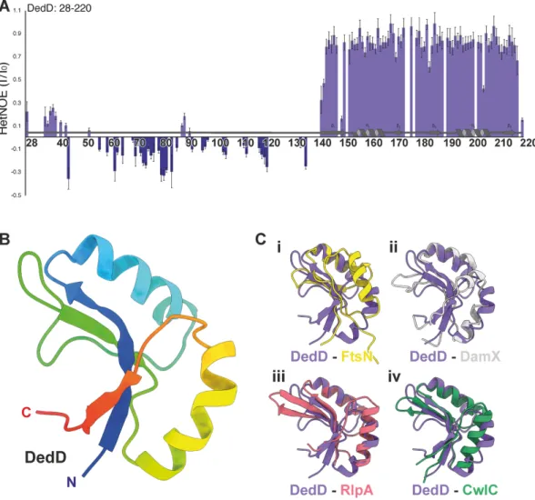
regions (Fig. 2A; Table S1 and Fig. S1, S2, and S3).
1H-
15N nuclear Overhauser effect
(NOE) relaxation measurements displayed positive NOE values (
⬎0.6) for residues 144
to 220, in agreement with the presence of a globular and stable SPOR domain (Fig. 2A).
Residues 36 to 141 produced low or negative NOE values, which indicate fast motion,
and residues 142 and 143 showed transitional values between the structured and
unstructured regions of DedD (Fig. 2A). Therefore, we confirm that the solution-state
structure of DedD is that of a structured SPOR domain tethered to the inner membrane
via an unstructured and flexible linking region.
For the ordered and folded region of DedD, we were able to fully determine the
structure. We found that residues 143 to 220 form a canonical SPOR domain consisting
of a four-stranded antiparallel
-sheet flanked on one side by a pair of ␣-helices
(Fig. 2B). On the fold level, we observe high structural similarity among all five SPOR
domains that have been determined. At an atomistic level, we observe that DedD has
backbone root mean square deviation (RMSD) values of 1.1, 1.4, 0.9, and 1.0 Å across 19,
17, 25, and 38 trimmed residues for FtsN, DamX, RlpA, and CwlC, respectively (Fig. 2C).
This level of atomistic variability is mostly observed in the pair of
␣-helices that act to
scaffold the
-sheet, while the -sheet itself is more structurally conserved. This is
FIG 1 SPOR domain containing proteins in E. coli. (Left) Domains of each protein and their defining residues according to UniProt and
Pfam. Potential␣-helices (GOR secondary structure prediction method version IV) (53) are represented as light gray bands and the
predicted coiled-coil as a double-headed arrow. TMD, transmembrane region; SPOR, SPOR domain; SP, signal peptide; DPBB_1, double-psi beta barrel; N, amino terminus; C, carboxyl terminus. (Right) Schematic representation of the SPOR proteins and the PG synthases PBP1A and PBP1B. X, DamX; A, RlpA; D, DedD; N, FtsN.
Role of SPOR Proteins in E. coli ®
on November 3, 2020 at UNIV DE LIEGE
http://mbio.asm.org/
perhaps unsurprising, as the
-sheet region was seen to form the majority of the
binding interface in the liganded structures of P. aeruginosa RlpA (28). To further
unravel the potential similarities and differences in the binding mode of DedD in
comparison to other SPOR proteins, we generated a model of liganded DedD. In this
model, we superimposed the apo NMR structure of DedD on the glycan-liganded
crystal structure of RlpA (PDB ID
6I0A
) (28). We see that the binding mode is very similar
to that of RlpA, and as previously proposed for CwlC and FtsN models (28). This binding
involves key conserved residues in the exposed basic, electropositive binding cleft
(Fig. S4), such as Q147 (Q270 in RlpA), as well as analogous residues, such as L151 in
place of F274 of RlpA. However, the modeled liganded DedD lacks the additional
contributions from the nonconserved W365 and W416, as observed in the proposed
model for DamX binding of the ligand (28). Overall, the observed conservation of the
SPOR domains and structural organization suggest a shared functional overlap and
interactions with other proteins involved with peptidoglycan and cell division,
partic-ularly the PG synthases.
The absence of SPOR proteins increases the sensitivity to cefsulodin. We
hypothesized that the SPOR proteins are required for the correct functionality of PBP1B
FIG 2 Structure of E. coli DedD. Heteronuclear1H-15N NOE (HetNOE) ratios between saturated and reference experiments for
DedD. (A)1H-15N heteronuclear NOE of15N-labeled DedD (residues 28 to 220) with secondary structuring based on the final
structure. Residues within the structured region (K142 to N220) are ordered on the picosecond-to-nanosecond timescale, as
indicated by an NOE value of⬎0.6, which is clearly differentiated from the flexible C-terminal region (D28 to K142). (B)
Structured regions are depicted as a ribbon diagram with secondary structure elements shown, colored in rainbow from the
N terminus (blue) to the C terminus (red). The SPOR domain regions of (i) E. coli FtsN (yellow; PDB.ID1UTA) (39), (ii) E. coli DamX
(gray; PDB.ID2LFV) (40), (iii) P. aeruginosa RlpA (red; PDB.IDs6I05) (28), and (iv) B. subtilis CwlC (green; PDB.ID1X60) (41) aligned
to the structured SPOR domain region of E. coli DedD (C). In each case, DedD is in purple.
on November 3, 2020 at UNIV DE LIEGE
in E. coli, based on the decrease in fitness of the single-knockout mutants when grown
in the presence of cefsulodin, as revealed by a chemical genomics screening (42).
Cefsulodin is a
-lactam with high affinity for PBP1A and therefore a suitable tool to
assess the functionality of PBP1B, as E. coli requires at least one of these class A PBPs
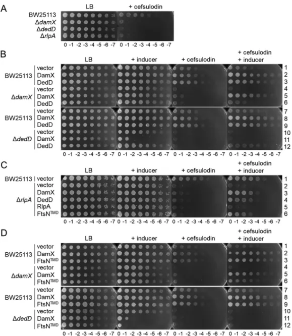
for viability (43). First, we confirmed the increased susceptibility to cefsulodin of the
mutants with single knockouts of DamX, DedD, and RlpA (Fig. 3A), suggesting that all
three proteins enhance the functionality of PBP1B in the cell. Complementation of the
knockout strains by expression of the plasmid-borne genes showed that
oligohistidine-tagged DamX and DedD restored the wild-type level of cefsulodin resistance (Fig. 3B,
rows 5 and 12). The overexpression of the oligohistidine-tagged RlpA did not
comple-ment the mutation (Fig. 3C, row 5), suggesting that the tag interfered with the function
FIG 3 The absence of SPOR domain proteins increases the sensitivity to cefsulodin. (A) Plate spotted with tenfold serial
dilutions of the indicated strains in the presence or absence of cefsulodin. (B) Overproduction of plasmid encoded DamX or DedD in wild-type, ΔdamX, or ΔdedD cells in the presence or absence of cefsulodin and/or inducer. (C) Overproduction
of plasmid-encoded DamX, DedD, RlpA, or DamX with the transmembrane region of FtsN (FtsNTMD) in wild-type or ΔrlpA
cells in the presence or absence of cefsulodin and/or inducer. (D) Overproduction of plasmid encoded DamX or FtsNTMD
in wild-type, ΔdamX, or ΔdedD cells in the presence or absence of cefsulodin. For all panels: cefsulodin was used at 30 mg
ml⫺1, the inducer sodium salicylate was used at 10 mg ml⫺1.
Role of SPOR Proteins in E. coli ®
on November 3, 2020 at UNIV DE LIEGE
http://mbio.asm.org/
of RlpA. A polar effect of the rlpA deletion on adjacent genes is unlikely, because the
overexpression of DamX or DedD largely restored cefsulodin resistance (Fig. 3C, rows 3
and 4). Our further investigations focused mainly on DamX and DedD.
To study the possible redundant roles of DamX and DedD, we also tested the effect
of their overexpression in wild-type cells and mutants lacking other SPOR proteins. The
expression of plasmid-borne damX or dedD increased the resistance to cefsulodin in
wild-type cells (Fig. 3B, rows 2, 3, 8, and 9), again supporting their positive effect on the
functionality of PBP1B. In the absence of RlpA, the overproduction of DamX or DedD
partially restored the resistance to cefsulodin (Fig. 3C, rows 3 and 4). DedD
overpro-duction could partially complement the absence of damX (Fig. 3B, row 6), but,
inter-estingly, DamX overproduction was lethal in ΔdedD cells (Fig. 3B, row 11). Together,
these results suggest that both DamX and DedD are functionally semiredundant but
the ratio of DamX to DedD is critical in the cell.
We then aimed to express different truncated versions of DamX to identify the
region of the protein required for the observed effect on cefsulodin sensitivity (Fig. S5).
However, the truncated DamX versions were unstable in the cell, and the overproduced
proteins could not be detected by Western blot analysis using antibodies against the
oligohistidine tag (Fig. S6). We were able to detect an overproduced DamX version
containing the transmembrane region of FtsN instead of its own. Cells overproducing
this DamX version enhanced the resistance to cefsulodin similarly to wild-type DamX
(Fig. 3C, row 6, and Fig. 3D, rows 3 and 6; Fig. S5 and S6) and showed similar toxicity
in ΔdedD cells (Fig. 3D, row 12).
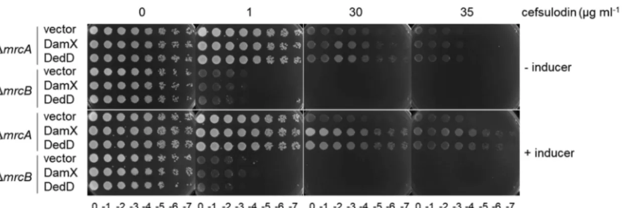
Because the cell requires at least one of the two main class A PBPs for survival, we
overproduced DamX or DedD in cells lacking either PBP1A or PBP1B. We used
cefsu-lodin at a concentration of 1
g ml
⫺1for cells lacking PBP1B (mrcB) and 30
g ml
⫺1for
cells lacking PBP1A (mrcA). The overproduction of DamX or DedD increased the
resistance to cefsulodin of cells lacking PBP1A (mrcA) but not of cells lacking PBP1B
(mrcB) (Fig. 4), supporting the notion that a higher level of DamX or DedD enhances the
functionality of PBP1B.
PBP1A and/or PBP1B are required to incorporate new peptidoglycan at the lateral
cell wall of the future cell division site during the preseptal cell elongation phase (11).
The SPOR domains of DamX and DedD are also recruited to these preseptal positions,
forming ring-like structures (30). Preseptal PG synthesis is most pronounced in cells
treated with aztreonam, which inhibits PBP3, the essential TPase required for the
septum formation at the division site. We tested whether the MIC of aztreonam is
altered when the functionality of PBP1B is reduced. Cells lacking PBP1B showed higher
susceptibility to aztreonam than cells lacking PBP1A, as previously reported (44), but
the absence of DamX did not alter the susceptibility to aztreonam in cells lacking PBP1A
or PBP1B (Fig. S7). Together, these results show that the reduced functionality of PBP1B
FIG 4 DamX and DedD enhance cellular PBP1B functionality. Tenfold serial dilutions of ΔmrcA or ΔmrcB cells overproducing the
full-length plasmid-encoded DamX or DedD proteins were spotted on a plate at different cefsulodin concentrations. Inducer, 10g
ml⫺1sodium salicylate.
on November 3, 2020 at UNIV DE LIEGE
in the damX mutant manifests specifically in the presence of cefsulodin and is
inde-pendent of PBP3 activity.
Interactions between SPOR proteins and PBP1A or PBP1B. To study the
inter-action between SPOR proteins and the class A PBPs, protein couples were coexpressed
from a pETDuet plasmid into the membranes of the E. coli host, solubilized with DDM
(N-dodecyl
-
D-maltoside) detergent and copurified by affinity chromatography on a
nickel-nitrilotriacetic acid (NTA) column, making use of an N-terminal His tag on the
SPOR protein. The purified fractions were labeled with Bocillin FL and analyzed by
sodium dodecyl sulfate-polyacrylamide gel electrophoresis (SDS-PAGE) followed by
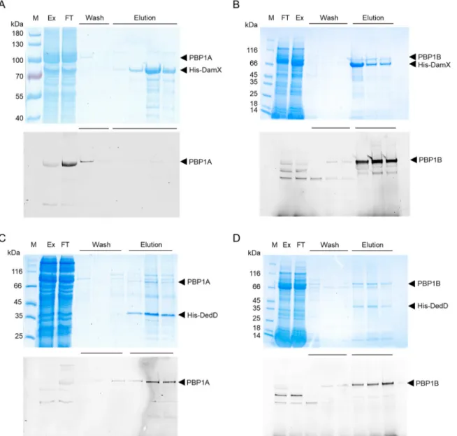
fluorescence imaging and Coomassie blue staining. The results show that PBP1B
coeluted with His-DamX, while PBP1A was found only in the wash fraction (Fig. 5A and
B). This suggest that DamX binds PBP1B and that the binding of DamX to PBP1A is
either weak or absent. His-DedD copurified with both PBP1A and PBP1B, indicating that
it interacts with both PBPs (Fig. 5C and D).
FIG 5 Interactions between SPOR proteins and class A PBPs. The proteins were coexpressed in E. coli and copurified on a nickel affinity
column with a His tag on the SPOR protein used as bait. The PBPs were labeled using Bocillin FL followed by analysis of the samples by SDS-PAGE, fluorescence imaging (bottom), and Coomassie blue staining (top). The expressed protein pairs are indicated by arrowheads: His-DamX and PBP1A (A), His-DamX and PBP1B (B), His-DedD and PBP1A (C), and His-DedD and PBP1B (D). M, protein marker; Ex, protein extract fraction; FT, flowthrough fraction. The wash and elution fractions are indicated with horizontal lines.
Role of SPOR Proteins in E. coli ®
on November 3, 2020 at UNIV DE LIEGE
http://mbio.asm.org/
SPOR proteins stimulate PBP1A and PBP1B. We next tested if the SPOR proteins
affect the GTase and TPase activities of PBP1A and PBP1B using three different in vitro
PG synthesis assays. The first assay quantifies the consumption of fluorescent
dansyl-lipid II. Both SPOR proteins had different, mild effects on the two synthases. DedD
increased the GTase rate of PBP1A 1.8-
⫾ 0.3-fold, but DamX had no effect (Fig. 6A).
DedD and DamX stimulated the GTase of PBP1B to similar extents (DedD, 2.6-
⫾
0.5-fold; DamX, 2.0-
⫾ 0.3-fold) (Fig. 6B). RlpA shows no effect on the GTase activity of
PBP1A or PBP1B (Fig. 6A and B).
To estimate the TPase activity we quantified the percentage of cross-linked peptides
present in the reaction products of an endpoint assay. Radiolabeled lipid II was used as
the substrate, and the products were separated by high-pressure liquid
chromatogra-phy (HPLC). We observed no significant changes in the percentage of cross-links
produced by PBP1A or PBP1B (Fig. 7A and B, respectively) in the presence or absence
of a SPOR protein.
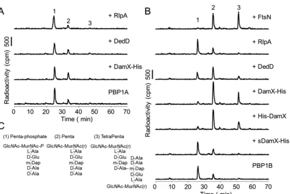
We also tested the effect of the SPOR proteins on the activities of PBP1A and PBP1B
when the synthases were present at low concentration, presumably in a less active
monomeric state (19, 36). Using the HPLC-based endpoint assay, we observed no effect
on the activity of PBP1A by any of the SPOR proteins (Fig. 8A). In the case of PBP1B, we
observed that the addition of DedD increased the monomeric GTase product peak
(Penta; compound 2) but less so the cross-linked GTase-TPase product peak
(TetraPenta; compound 3) (Fig. 8B). The presence of DamX increased both Penta and
TetraPenta products, resulting in an almost complete consumption of the lipid II
substrate (Fig. 8B). A soluble version of DamX lacking the transmembrane region
(sDamX) did not stimulate the GTase and TPase activities of PBP1B (Fig. 8B), suggesting
that the transmembrane region of DamX is required for stimulation. The stimulation of
PBP1B by DamX took place regardless of the N- or C-terminal position of the His tag in
FIG 6 Effect of SPOR proteins on the GTase activity of PBPs. Consumption of fluorescent lipid II by the
GTase activity of PBP1A (A) or PBP1B (B) in the presence of the indicated proteins. The GTase rates are shown as the decrease in fluorescence over time. Values are means and standard deviations from three independent experiments, after normalizing to the values for PBP1A or PBP1B alone. Student’s t test
(two-tailed) was used for statistical analysis (*, P⬍ 0.05; **, P ⬍ 0.01). Moe., moenomycin.
on November 3, 2020 at UNIV DE LIEGE
the purified protein. We confirmed the previously reported stimulation of PBP1B by
FtsN, which was stronger than the stimulation of PBP1B by DamX, as observed by the
higher consumption of the lipid II substrate (Fig. 8B, compound 1) and higher
abun-dance of the TetraPenta product (compound 3). The structures of the main reaction
products are shown in Fig. 8C.
We also assessed the GTase activity of PBP1B using fluorescently labeled lipid II
substrate, in the presence of ampicillin, and separated the produced glycan strands by
SDS-PAGE. We observed an increase in the amount of glycan strand products with
DamX but not RlpA or DedD (Fig. 9A). DamX with the His tag at the C terminus and
DamX with the His tag at the N terminus gave similar results, but the DamX version
without the transmembrane region did not stimulate PBP1B (sDamX) (Fig. 9B). Again,
FtsN stimulated the GTase activity of PBP1B more strongly than DamX, consistent with
our results obtained with the other assays (Fig. 9A). As expected, control reaction
mixtures containing RlpA, DedD, or the different DamX proteins in the absence of
PBP1B showed no GTase products (Fig. 9A and B), excluding the presence of
contam-inating GTases in the purified protein samples. To summarize, our activity assays
showed that DamX and DedD stimulate the activity of PBP1B and, in the case of DedD,
also the activity of PBP1A.
DISCUSSION
This work identified a direct role for the SPOR proteins DamX and DedD in the
function of PG synthases. The absence of DamX or DedD decreased the functionality of
PBP1B in the cell, leading to an increase in the susceptibility to cefsulodin, which
primarily inhibits PBP1A. We present the structure of the SPOR domain of DedD and
modeled its binding to glycan chains. Our biochemical data highlight the connection
between PG synthases and PG-binding proteins during cell division. Presumably, this
connection contributes to stabilizing the inward-growing septal PG layer. The
de-FIG 7 Effect of SPOR proteins on the TPase activity of PBPs. Representative HPLC chromatograms of
PBP1A (A) and PBP1B (B) in vitro PG synthesis reactions using radioactive lipid II as the substrate in the presence of the indicated proteins. The synthesized PG was digested with cellosyl, reduced with sodium borohydride, and analyzed by HPLC. TPase activities (right) were determined by the percentage of peptides in cross-links present in the reaction products. The values are means and standard deviations from three independent experiments.
Role of SPOR Proteins in E. coli ®
on November 3, 2020 at UNIV DE LIEGE
http://mbio.asm.org/
creased functionality of PBP1B in the dedD and damX mutants was reverted by the
ectopic expression of the respective genes from a plasmid, excluding a polar effect. Our
cellular viability data support a semiredundant role for DamX and DedD in enhancing
the functionality of PBP1B, and the effect is also seen in wild-type cells. An explanation
FIG 8 Effects of SPOR proteins on the GTase and TPase activity of PBPs at low concentrations. Representative HPLC chromatograms of PBP1A (A) and PBP1B (B) in vitro PG synthesis reactions using radioactive lipid II as the substrate in the presence of the indicated proteins. The synthesized PG was digested with cellosyl, reduced with sodium borohydride, and analyzed by HPLC. Peak 1 is generated from glycan strand ends and unreacted lipid II, peak 2 is a GTase product, and peak 3 is a GTase/TPase product. (C) Structures of the main products of the in vitro synthesis reactions.
FIG 9 Effects of SPOR proteins on the GTase activity of PBPs at low concentrations. SDS-PAGE analysis
of glycan strands synthesized by PBP1B GTase activity at low concentration in the presence of the different SPOR domain containing proteins (A) and the different DamX constructs (B). Reaction mixtures were incubated at 37°C for 1 h, using a mixture of unlabeled and ATTO 550-labeled lipid II as the substrate, in the presence of the indicated interacting proteins. The numbers refer to disaccharide units.
on November 3, 2020 at UNIV DE LIEGE
for these effects might come from our biochemical data, which show that DedD and
DamX both have a small but significant stimulatory effect on the GTase and TPase
activities of PBP1B.
The previously reported DamX-associated phenotypes are all linked to cell division.
A strong overproduction of DamX leads to cell filamentation and death (37). The
additional deletion of chromosomal damX in the background of a dedD mutant causes
severe filamentation (30), which can be reversed by mild overproduction of DamX (31).
Our results show that mild overproduction of DamX improves the viability of
cefsulodin-challenged cells with the exception of ΔdedD cells, in which the mild
overproduction of DamX was toxic. However, DedD overproduction improved the
viability of all strains in the presence of cefsulodin and was not toxic in ΔdamX cells.
These results suggest that an optimal ratio of DedD to DamX is important to maintain
cell viability and that DedD is capable of “neutralizing” the toxicity of increased DamX
levels. Presumably, DamX and DedD have semiredundant roles in the stimulation of
class A PBPs despite having otherwise different, but complementary, roles during cell
division. Both proteins interact with FtsQ in a bacterial two-hybrid assay (30, 31), and
the absence of DamX improves the viability of ftsQ(ts) cells under nonpermissive
conditions, suggesting that DamX antagonizes FtsQ function (31). Perhaps DedD
protects FtsQ from being antagonized by DamX at the division site, explaining why a
mild increase of DamX is toxic only when DedD is absent. In our activity assays, DedD
slightly stimulated not only PBP1B but also the GTase of PBP1A, which might explain
the more severe cell division defects in the absence of DedD than in the absence of
DamX. Because the absence of DedD is better tolerated in cells lacking PBP1A than
PBP1B (38) and the simultaneous lack of PBP1A and PBP1B is lethal (43), we hypothesize
that DedD is more specific for the function of PBP1A and DamX is more specific for that
of PBP1B.
All SPOR proteins localize at the cell division site in E. coli, but the timing seems to
be different for each of them. In case of FtsN, a small fraction of the total cellular protein
amount is recruited to the division site by an interaction with FtsA before septum
synthesis begins (35). FtsN-FtsA connect the cytosolic FtsZ ring with PBP1A and PBP1B
to direct preseptal PG synthesis (12). Despite their similar structures (Fig. 2), their SPOR
domains also show different localization patterns. Unlike the SPOR domains of FtsN and
RlpA, those of DamX and DedD are able to form ring-like structures in the absence of
constriction (30).
The localization of DedD at the division site and its functionality are significantly
diminished but not abolished in the absence of its SPOR domain, which renders the
N-terminal transmembrane and adjacent residues essential (38). Since mutations in the
transmembrane region abolish the recruitment of a SPOR less DedD to the division site
(38), it is tempting to speculate that the interaction of DedD with the class A PBPs takes
place through the transmembrane regions of interacting proteins and that such
interaction contributes to the recruitment of DedD to the division site and to the
regulation of septation.
DamX contains a large and likely folded cytoplasmic domain, together with the
largest periplasmic linker region of all the inner membrane SPOR proteins. DamX
localizes at the division sites in an FtsZ-dependent and FtsA-, FtsQ-, PBP3-, or
FtsN-independent manner (31). The spatial and temporal pattern of DamX localization,
together with its positive effect on PBP1B, is consistent with a role during the synthesis
of preseptal PG. The SPOR domain is needed to efficiently target DamX to the division
site and to cause cell division inhibition when DamX is overproduced (40). Together,
these results suggest that DedD and DamX might recognize and bind to specific
structures in PG present at the division site before the septum synthesis starts, which
could be either denuded glycan strands (28) or perhaps the 1,6-anhydro ends of glycan
strands (33, 36). Later, during septation, SPOR proteins provide a connection between
PG synthases and the inward-growing septal PG, which may function to stabilize the
constricting cell envelope and/or regulate PG synthesis. It remains to be seen in future
Role of SPOR Proteins in E. coli ®
on November 3, 2020 at UNIV DE LIEGE
http://mbio.asm.org/
thiogalactopyranoside) to the cell culture, which was further incubated for 4 h at 37°C. Cells were
harvested by centrifugation (6,200⫻ g, 15 min, 4°C), and the pellet was resuspended in buffer I (25 mM
Tris-HCl, 1 M NaCl [pH 7.5]). After addition of 200M phenylmethylsulfonylfluoride (PMSF), a 1-in-1,000
dilution of protease inhibitor cocktail (Sigma-Aldrich) and DNase, the cells were disrupted by sonication
(Branson Digital). The cell lysate was centrifuged (130,000⫻ g, 60 min, 4°C), and the supernatant was
incubated overnight with 4 ml of nickel-nitrilotriacetic acid (Ni-NTA) Superflow (Qiagen), which had been pre-equilibrated in buffer I containing 20 mM imidazole, at 4°C with gentle stirring. The resin was poured into a gravity column and washed with 25 volumes of wash buffer (25 mM Tris-HCl, 1 M NaCl, 10 mM
MgCl2, 20 mM imidazole [pH 7.5]). Bound protein was eluted with elution buffer (25 mM Tris-HCl, 1 M
NaCl, 10 mM MgCl2, 400 mM imidazole [pH 7.5]). The Ni-NTA-eluted protein was dialyzed against 1 liter
of dialysis buffer I (25 mM Tris-HCl, 500 mM NaCl, 10 mM MgCl2[pH 7.5]) for 30 min; 500 ml of dialysis
buffer I was replaced with 500 ml dialysis buffer II (25 mM Tris-HCl, 300 mM NaCl, 10 mM MgCl2[pH 7.5])
and further dialyzed for 30 min. Restriction-grade thrombin (4 U ml⫺1; Merck Millipore, Darmstadt,
Germany) was added to remove the oligohistidine tag during overnight dialysis against 1 liter of dialysis
buffer II at 4°C. The sample was diluted 1:1 with buffer no salt (25 mM Tris-HCl, 10 mM MgCl2[pH 7.5])
and applied in AKTA A buffer (25 mM Tris-HCl, 10 mM MgCl2, 150 mM NaCl [pH 7.5]) to a 5-ml HiTrap Q
HP column using an ÄKTA Prime system (GE Healthcare Bio-Sciences) for anion-exchange
chromatog-raphy (flow rate, 1 ml min⫺1). Although a gradient from 150 mM to 1 M NaCl was applied, the protein was
present in the flowthrough and wash. The protein was dialyzed against 3 liters of storage buffer (25 mM
HEPES-NaOH, 500 mM NaCl, 10 mM MgCl2, 10% glycerol [pH 7.5]) and stored at⫺80°C.
(ii) His-DamX. LOBSTR cells containing plasmid pPZW23 were grown in 2 liters of LB medium
supplemented with kanamycin at 37°C to an OD578of 0.4 to 0.5. Protein overproduction was induced by
addition of 0.5 mM IPTG to the cell culture, which was further incubated for 4 h at 37°C. Cells were
harvested by centrifugation (6,200⫻ g, 15 min, 4°C), and the pellet was resuspended in buffer I (25 mM
Tris-HCl, 1 M NaCl [pH 7.5]). After addition of 200M PMSF, a 1-in-1,000 dilution of protease inhibitor
cocktail (Sigma-Aldrich) and DNase, the cells were disrupted by sonication (Branson Digital), and the cell
lysate was centrifuged (130,000⫻ g, 60 min, 4°C). The supernatant was discarded, and the membrane
pellet was resuspended in extraction buffer (25 mM Tris-HCl, 1 M NaCl, 2% Triton X-100 reduced, 10 mM
MgCl2, 10% glycerol [pH 7.5]) and incubated overnight with mixing at 4°C. Resuspended sample was
centrifuged (130,000⫻ g, 60 min, 4°C), and the supernatant was incubated with Ni-NTA Superflow
(Qiagen) as described for RlpA. The protein did not bind to the resin and was collected from the flowthrough. The flowthrough containing the protein was dialyzed against 3 liters of dialysis buffer I
(25 mM Tris-HCl, 150 mM NaCl, 10 mM MgCl2[pH 7.5]) overnight. The sample was applied in AKTA A
buffer (25 mM Tris-HCl, 10 mM MgCl2, 150 mM NaCl, 0.2% Triton X-100 reduced [pH 7.5]) to a 5-ml HiTrap
Q HP column using an ÄKTA Prime system (GE Healthcare Bio-Sciences), and the protein was collected in the flowthrough. The protein was dialyzed against 3 liters of dialysis buffer II (10 mM sodium acetate,
150 mM NaCl, 10 mM MgCl2[pH 4.8]) overnight. The sample was applied in dialysis buffer II containing
0.2% Triton X-100 reduced to a 5-ml HiTrap SP HP column using an ÄKTA Prime system (GE Healthcare
Bio-Sciences) for cation-exchange chromatography (flow rate, 1 ml min⫺1). The protein was eluted using
a gradient from 150 mM to 2 M NaCl. Protein-containing fractions were dialyzed against 3 liters of storage
buffer (25 mM HEPES NaOH, 10 mM MgCl2, 150 mM NaCl, 10% glycerol [pH 7.5]) and stored at⫺80°C.
(iii) DamX-His and sDamX-His. LOBSTR cells containing plasmid pPZW26 and pPZW27 were grown
in 2 liters of LB medium supplemented with kanamycin at 37°C to an OD578of 0.4 to 0.5. Protein
overproduction was induced by addition of 0.5 mM IPTG to the cell culture which was further incubated
for 4 h at 37°C. Cells were harvested by centrifugation (6,200⫻ g, 15 min, 4°C), and the pellet was
resuspended in buffer I (25 mM Tris-HCl, 1 M NaCl [pH 7.5]). After addition of 200M PMSF, a 1-in-1,000
dilution of protease inhibitor cocktail (Sigma-Aldrich), and DNase, the cells were disrupted by sonication
(Branson Digital), and the cell lysate was centrifuged (130,000⫻ g, 60 min, 4°C). For DamX-His, the
supernatant was discarded, and the membrane pellet was resuspended in extraction buffer (25 mM
Tris-HCl, 1 M NaCl, 2% Triton X-100 reduced, 10 mM MgCl2, 10% glycerol [pH 7.5]) and incubated
overnight with mixing at 4°C. Resuspended sample was centrifuged (130,000⫻ g, 60 min, 4°C), and the
supernatant was incubated with Ni-NTA Superflow (Qiagen), washed, and eluted as described for RlpA using buffers containing 0.2% Triton X-100 reduced. The eluted protein was dialyzed against 3 liters of
storage buffer (25 mM HEPES NaOH, 10 mM MgCl2, 150 mM NaCl, 10% glycerol [pH 7.5]) and stored at
⫺80°C. sDamX-His was purified from the supernatant of the cell lysate centrifugation using the protocol for DamX-His but omitting detergent in buffers. The eluted protein was dialyzed against 3 liters of dialysis
on November 3, 2020 at UNIV DE LIEGE
buffer I (25 mM Tris-HCl, 200 mM NaCl, 10 mM MgCl2[pH 7.5]) for 1.5 h and against 3 liters of dialysis
buffer II (10 mM sodium acetate, 200 mM NaCl, 10 mM MgCl2[pH 4.8]) overnight. The sample was diluted
1:1 with buffer no salt (10 mM sodium acetate, 10 mM MgCl2[pH 4.8]) and applied in AKTA A buffer
(10 mM sodium acetate, 100 mM NaCl, 10 mM MgCl2[pH 4.8]) to a 5-ml HiTrap SP HP column using an
ÄKTA Prime system (GE Healthcare Bio-Sciences) for cation-exchange chromatography (flow rate, 1 ml
min⫺1). The protein was eluted in a gradient from 100 mM to 2 M NaCl. Protein-containing fractions were
dialyzed against storage buffer (25 mM HEPES NaOH, 150 mM NaCl, 10 mM MgCl2, 10% glycerol [pH 7.5])
and stored at⫺80°C.
(iv) DedD. LOBSTR cells containing plasmid pPZW24 were grown in 2 liters of LB medium
supple-mented with kanamycin at 37°C to an OD578of 0.4 to 0.5. Protein overproduction was induced by
addition of 0.5 mM IPTG to the cell culture, which was further incubated for 4 h at 37°C. Cells were
harvested by centrifugation (6,200⫻ g, 15 min, 4°C) and the pellet was resuspended in buffer I (25 mM
Tris-HCl, 1 M NaCl [pH 7.5]). After addition of 200M PMSF, a 1-in-1,000 dilution of protease inhibitor
cocktail (Sigma-Aldrich) and DNase, the cells were disrupted by sonication (Branson Digital), and the cell
lysate was centrifuged (130,000⫻ g, 60 min, 4°C). The supernatant was discarded, and the membrane
pellet was resuspended in extraction buffer (25 mM Tris-HCl, 1 M NaCl, 2% Triton X-100 reduced, 10 mM
MgCl2, 10% glycerol [pH 7.5]) and incubated overnight with mixing at 4°C. Resuspended sample was
centrifuged (130,000⫻ g, 60 min, 4°C), and the supernatant was incubated with Ni-NTA Superflow
(Qiagen), washed, and eluted as described for RlpA using buffers containing 0.2% Triton X-100 reduced.
Restriction-grade thrombin (4 U ml⫺1; Merck Millipore) was added to the Ni-NTA-eluted protein to
remove the oligohistidine tag during dialysis against 3 liters of dialysis buffer I (25 mM Tris-HCl, 1 M NaCl,
10 mM MgCl2[pH 7.5]) for 20 h at 4°C. Sample was dialyzed against 3 liters of dialysis buffer II (10 mM
sodium acetate, 500 mM NaCl, 10 mM MgCl2[pH 4.8]) for 4 h at 4°C and 3 liters of dialysis buffer III
(10 mM sodium acetate, 300 mM NaCl, 10 mM MgCl2[pH 4.8]) for 18 h at 4°C. The sample was diluted 1:1
with no-salt buffer (10 mM sodium acetate, 10 mM MgCl2, 0.2% Triton X-100 reduced [pH 4.8]) and
applied in AKTA A buffer (10 mM sodium acetate, 150 mM NaCl, 10 mM MgCl2, 0.2% Triton X-100 reduced
[pH 4.8]) to a 5-ml HiTrap SP HP column using an ÄKTA Prime system (GE Healthcare Bio-Sciences) for
cation-exchange chromatography (flow rate, 1 ml min⫺1). The protein eluted in a gradient from 150 mM
to 2 M NaCl. Protein-containing fractions were dialyzed against storage buffer (25 mM HEPES NaOH,
150 mM NaCl, 10 mM MgCl2, 10% glycerol [pH 7.5]) and stored at⫺80°C.
Protein coexpression, copurification, and Bocillin FL labeling. C43(DE3) cells transformed with
pETDuet plasmids (Table S2) were grown in 500 ml Miller Luria-Bertani (LB) supplemented with ampicillin
(100g ml⫺1) at 37°C to an A
600of 0.8. Protein expression was induced for 3.5 h by addition of 0.5 mM
IPTG. Cells were collected by centrifugation at 4,000⫻ g for 20 min at 15°C and resuspended in a buffer
containing 20 mM Tris-HCl (pH 8.0), 300 mM NaCl, and EDTA-free protease inhibitor cocktail (Roche). The cells were lysed by three passages through a cell homogenizer (Emulsiflex C3; Avestin). After
centrifu-gation at 4,000⫻ g for 20 min at 4°C, the supernatant was recovered and spun down at 150,000 ⫻ g for
1 h at 4°C. The pelleted membranes were solubilized in 25 mM Tris-HCl (pH 8.0), 500 mM NaCl, 10% (vol/vol) glycerol, 40 mM DDM (Inalco) and 1 tablet of complete EDTA-free protease inhibitors (Sigma) per 50 ml of buffer. The mixture was incubated for 1 h at room temperature under gentle agitation
followed by centrifugation at 150,000⫻ g for 1 h at 4°C. The supernatant containing the solubilized
membrane proteins was loaded onto a HisTrap column (GE HealthCare) conditioned in buffer B (25 mM Tris-HCl [pH 7.5], 500 mM NaCl, 4 mM DDM). After a wash with buffer B supplemented with 80 mM imidazole, the proteins were eluted in 0.5- to 1-ml fractions using a linear gradient of imidazole from 80
to 500 mM. A 15-l portion of each fraction was incubated with 2 M Bocillin FL for 30 min at 37°C. The
fractions were analyzed by SDS-PAGE, followed by fluorescence imaging and Coomassie blue staining.
Protein expression for NMR. For labeled DedD (residues 28 to 220) purification, pYS001 was
transformed into BL21(DE3). This strain was cultured in 1 liter of M9 containing 1 g/liter of ammonium
chloride and 2 g/liter of glucose at 37°C to an OD600of 0.6, at which point 1 mM IPTG was added to
induce protein overproduction overnight at 25°C. Harvested cell pellets were resuspended in lysis buffer (20 mM HEPES [pH 8.0], 300 mM NaCl, 10% glycerol) and lysed by processing twice with a homogenizer
(15 kPa; Avestin). Cellular debris was pelleted by centrifugation at 125,000⫻ g for 1 h. The resultant
supernatant was loaded onto 10 ml Ni2⫹-saturated Ni-NTA Superflow beads (Qiagen) and washed with
65 mM imidazole in 20 mM HEPES (pH 8.0)–300 mM NaCl, and the protein was eluted with 300 mM imidazole in the previous buffer. Fractions containing pure DedD were pooled and desalted into a buffer of 20 mM HEPES (pH 8.0) and 300 mM NaCl. Protein was frozen rapidly in liquid nitrogen and stored at ⫺80°C until required.
Spot plate assay. Cells were grown overnight at 30°C, the optical density was normalized for each
strain assayed in the plate, and the cells were spotted in a 10-fold dilution series on Lennox LB plates (1% tryptone, 0.5% yeast extract, 0.5% NaCl), which were incubated overnight at 30°C. Plates were
supple-mented with 20g ml⫺1chloramphenicol when strains carrying pKG110-derived plasmids were assayed.
When appropriate, 10M sodium salicylate (inducer) was added to the plates. Unless stated otherwise,
30g ml⫺1cefsulodin was used.
Aztreonam susceptibility assay. Overnight cultures of the test strains were grown at 30°C in LB
Lennox, reinoculated 1:100, and grown to an OD578of 0.3 to 0.4. A 1-ml portion of each strain was
centrifuged for 1 min at 13,000⫻ g and resuspended in 1 ml of 0.9% NaCl. Resuspended cells were
diluted to an OD578of 0.125 using 0.9% NaCl. Cells were distributed onto LB Lennox plates using a cotton
swab soaked with the cell suspension. Once the plates were dried, an aztreonam MIC test strip (Liofilchem) was applied to each plate, and all the plates were incubated overnight at 30°C.
Role of SPOR Proteins in E. coli ®
on November 3, 2020 at UNIV DE LIEGE
http://mbio.asm.org/
reference 50. The following protein concentrations were used in the assays with low concentrations of
PBP1A or PBP1B (0.075M PBP1A and 0.038 M PBP1B) and 0.75 M SPOR domain proteins. In samples
with low PG synthase activity (with abundant unused lipid II), the total radioactivity eluted from the HPLC
column (C18) differs between samples due to differences in peak 1, the phosphorylated disaccharide
pentapeptide. Peak 1 is generated by acid hydrolysis of unused lipid II (or glycan strands ends carrying the C55-PP moiety) after the GTase-TPase reaction, because lipid II (without hydrolysis) does not elute
from the C18HPLC column used to separate the muropeptides. In samples with abundant unused lipid
II, peak 1 varies due to differences in the efficiency of the acid hydrolysis of lipid II between samples. This effect does not impair the quantification of other peaks (PG products). Tris-Tricine SDS-PAGE was used to separate glycan strands (51), using the same protein concentrations and reaction conditions as the TPase activity experiment at low PBP1A and PBP1B concentrations but in the presence of 1 mM ampicillin to inhibit the TPase activity.
NMR spectroscopy. NMR data were collected in 20 mM HEPES, 300 mM NaCl, 10% D2O (pH 7.0), at
298 K on 1.08 mM13C,15N-labeled DedD sample prepared in a 3-mm-diameter NMR tube. All NMR spectra
for backbone, side chains, and NOE assignments were recorded on Bruker spectrometers operating at
700, 850, and 950 MHz1H NMR frequencies and equipped with1H,13C,15N-labeled cryoprobes.
Resonance assignments of the backbone was performed using two-dimensional (2D)1H,15
N-BEST-TROSY (BT), 3D BT-HNCANH, 3D BT-HNCO, 3D BT-HNCACO, 3D BT-HNCACB and 3D BT-HN(CO)CACB
spectra. Manual side chain assignment was then achieved with conventional 2D1H,13C-HSQC (gradient
heteronuclear single quantum coherence), 3D (H)C(CCO)NH, 3D H(CCCO)NH, and 3D15N-NOESY-HSQC,
as well as 3D aliphatic and aromatic13C-NOESY-HSQC experiments. Spectra were analyzed with CcpNmr
Analysis 2.4.1.
For structural restraints, dihedral angles (phi and psi) were predicted from backbone chemical shift
with the neural network program TALOS⫹, and distance constraints were determined after manual
peak-picking and automatic assignment of the 3D NOESY-HSQC experiments reported above by Unio=10 version 2.0.2. Structures were subsequently calculated from these restraints by Aria 2.3.1, with 80 structures from runs 0 to 5, 200 for runs 6 and 7, and 600 for the last run. The 20 lowest-energy structures were further refined in water. Ramachandran analysis showed 86.1%, 13.9%, 0.0%, and 0.0% of the residues of DedD in most favored, additional allowed, generously allowed, and disallowed regions, respectively.
1H-15N NOE relaxation data were collected at 25°C on Bruker spectrometers operating at 700 MHz and
equipped with1H,13C,15N-labeled cryoprobes.1H-15N NOE values were determined by the comparison of
the intensities of each amide resonance with and without a 3-s saturation period and using the BEST-HETNOE sequence (52). Standard deviations were calculated from errors on peak intensities.
Data availability. The 20 lowest-energy structures were deposited in the PDB with accession
number6ZTG. All other data supporting the findings of this study are included in the main text and
supplemental material.
SUPPLEMENTAL MATERIAL
Supplemental material is available online only.
TEXT S1, DOCX file, 0.02 MB.
FIG S1, TIF file, 2.3 MB.
FIG S2, PDF file, 1.1 MB.
FIG S3, TIF file, 2.7 MB.
FIG S4, TIF file, 2.9 MB.
FIG S5, TIF file, 2.8 MB.
FIG S6, TIF file, 2.4 MB.
FIG S7, TIF file, 2.1 MB.
TABLE S1, DOCX file, 0.02 MB.
TABLE S2, DOCX file, 0.02 MB.
on November 3, 2020 at UNIV DE LIEGE
ACKNOWLEDGMENTS
This work was supported by the MRC (MR/N002679/1), Wellcome Trust (101824/Z/
13/Z), “Fonds de la Recherche Scientifique” FRS_FNRS (Brussels, Belgium) grant CDR
J.0030.18 and FRIA 1.E.038.17, JPIAMR-CIHR and the Howard Hughes International
Senior Scholar program. Financial support from the IR-RMN-THC Fr3050 CNRS for
conducting the research is gratefully acknowledged. We also acknowledge
infrastruc-ture funding from the Canadian Foundation of Innovation and British Columbia
Knowl-edge Development Fund. M.T. is a research associate of the FRS_FNRS, N.A.C. holds an
NSERC PGS D award, and N.C.J.S. is a Tier I Canada Research Chair in Antibiotic
Discovery. This work used the platforms of the Grenoble Instruct-ERIC center (ISBG; UMS
3518 CNRS-CEA-UGA-EMBL) within the Grenoble Partnership for Structural Biology
(PSB), supported by FRISBI (ANR-10-INBS-05-02) and GRAL, financed within the
Univer-sity Grenoble Alpes graduate school (Ecoles Universitaires de Recherche) CBH-EUR-GS
(ANR-17-EURE-0003).
M.P., K.P. and W.V. designed the work. M.P., K.P. and A.B. performed experiments.
M.P., K.P., A.B., M.T. and W.V. analyzed the data. Y.S. performed the NMR experiments,
C.K., C.L. and J.-P.S. analyzed the NMR data. N.A.C., N.C.J.S., and J.-P.S. designed the
structural work. E.B. provided research tools. All authors contributed to writing the
manuscript.
We declare no competing interests.
REFERENCES
1. Pazos M, Peters K. 2019. Peptidoglycan. Subcell Biochem 92:127–168.
https://doi.org/10.1007/978-3-030-18768-2_5.
2. Vollmer W, Blanot D, de Pedro MA. 2008. Peptidoglycan structure and
architecture. FEMS Microbiol Rev 32:149 –167.https://doi.org/10.1111/j
.1574-6976.2007.00094.x.
3. Holtje JV. 1998. Growth of the stress-bearing and shape-maintaining murein sacculus of Escherichia coli. Microbiol Mol Biol Rev 62:181–203.
https://doi.org/10.1128/MMBR.62.1.181-203.1998.
4. Georgopapadakou NH, Smith SA, Sykes RB. 1982. Mode of action of
aztreonam. Antimicrob Agents Chemother 21:950 –956.https://doi.org/
10.1128/aac.21.6.950.
5. Curtis NA, Orr D, Ross GW, Boulton MG. 1979. Affinities of penicillins and cephalosporins for the penicillin-binding proteins of Escherichia coli K-12 and their antibacterial activity. Antimicrob Agents Chemother 16:
533–539.https://doi.org/10.1128/aac.16.5.533.
6. Typas A, Banzhaf M, Gross CA, Vollmer W. 2011. From the regulation of peptidoglycan synthesis to bacterial growth and morphology. Nat Rev
Microbiol 10:123–136.https://doi.org/10.1038/nrmicro2677.
7. Egan AJF, Errington J, Vollmer W. 2020. Regulation of peptidoglycan
synthesis and remodelling. Nat Rev Microbiol 18:446 – 460.https://doi
.org/10.1038/s41579-020-0366-3.
8. Rohs PDA, Buss J, Sim SI, Squyres GR, Srisuknimit V, Smith M, Cho H, Sjodt M, Kruse AC, Garner EC, Walker S, Kahne DE, Bernhardt TG. 2018. A central role for PBP2 in the activation of peptidoglycan polymerization by the bacterial cell elongation machinery. PLoS Genet 14:e1007726.
https://doi.org/10.1371/journal.pgen.1007726.
9. Bisson-Filho AW, Hsu YP, Squyres GR, Kuru E, Wu F, Jukes C, Sun Y, Dekker C, Holden S, VanNieuwenhze MS, Brun YV, Garner EC. 2017. Treadmilling by FtsZ filaments drives peptidoglycan synthesis and
bac-terial cell division. Science 355:739 –743.https://doi.org/10.1126/science
.aak9973.
10. Baranova N, Radler P, Hernandez-Rocamora VM, Alfonso C, Lopez-Pelegrin M, Rivas G, Vollmer W, Loose M. 2020. Diffusion and capture permits dynamic coupling between treadmilling FtsZ filaments and
cell division proteins. Nat Microbiol 5:407– 417. https://doi.org/10
.1038/s41564-019-0657-5.
11. Potluri LP, Kannan S, Young KD. 2012. ZipA is required for FtsZ-dependent preseptal peptidoglycan synthesis prior to invagination
dur-ing cell division. J Bacteriol 194:5334 –5342.https://doi.org/10.1128/JB
.00859-12.
12. Pazos M, Peters K, Casanova M, Palacios P, VanNieuwenhze M, Breukink E, Vicente M, Vollmer W. 2018. Z-ring membrane anchors associate with
cell wall synthases to initiate bacterial cell division. Nat Commun 9:5090.
https://doi.org/10.1038/s41467-018-07559-2.
13. de Pedro MA, Quintela JC, Holtje JV, Schwarz H. 1997. Murein
segrega-tion in Escherichia coli. J Bacteriol 179:2823–2834. https://doi.org/10
.1128/jb.179.9.2823-2834.1997.
14. Liu B, Persons L, Lee L, de Boer PA. 2015. Roles for both FtsA and the FtsBLQ subcomplex in FtsN-stimulated cell constriction in Escherichia
coli. Mol Microbiol 95:945–970.https://doi.org/10.1111/mmi.12906. 15. Tsang MJ, Bernhardt TG. 2015. A role for the FtsQLB complex in
cytoki-netic ring activation revealed by an ftsL allele that accelerates division.
Mol Microbiol 95:925–944.https://doi.org/10.1111/mmi.12905.
16. Boes A, Olatunji S, Breukink E, Terrak M. 2019. Regulation of the pepti-doglycan polymerase activity of PBP1b by antagonist actions of the core
divisome proteins FtsBLQ and FtsN. mBio 10:e01912-18.https://doi.org/
10.1128/mBio.01912-18.
17. Taguchi A, Welsh MA, Marmont LS, Lee W, Sjodt M, Kruse AC, Kahne D, Bernhardt TG, Walker S. 2019. FtsW is a peptidoglycan polymerase that is functional only in complex with its cognate penicillin-binding protein.
Nat Microbiol 4:587–594.https://doi.org/10.1038/s41564-018-0345-x.
18. Adam M, Fraipont C, Rhazi N, Nguyen-Disteche M, Lakaye B, Frere JM, Devreese B, Van Beeumen J, van Heijenoort Y, van Heijenoort J, Ghuysen JM. 1997. The bimodular G57-V577 polypeptide chain of the class B penicillin-binding protein 3 of Escherichia coli catalyzes peptide bond formation from thiolesters and does not catalyze glycan chain polym-erization from the lipid II intermediate. J Bacteriol 179:6005– 6009.
https://doi.org/10.1128/jb.179.19.6005-6009.1997.
19. Bertsche U, Kast T, Wolf B, Fraipont C, Aarsman ME, Kannenberg K, von Rechenberg M, Nguyen-Disteche M, den Blaauwen T, Holtje JV, Vollmer W. 2006. Interaction between two murein (peptidoglycan) synthases,
PBP3 and PBP1B, in Escherichia coli. Mol Microbiol 61:675– 690.https://
doi.org/10.1111/j.1365-2958.2006.05280.x.
20. Born P, Breukink E, Vollmer W. 2006. In vitro synthesis of cross-linked murein and its attachment to sacculi by PBP1A from Escherichia coli. J
Biol Chem 281:26985–26993.https://doi.org/10.1074/jbc.M604083200.
21. Heidrich C, Templin MF, Ursinus A, Merdanovic M, Berger J, Schwarz H, de Pedro MA, Holtje JV. 2001. Involvement of N-acetylmuramyl-L-alanine amidases in cell separation and antibiotic-induced autolysis of
Esche-richia coli. Mol Microbiol 41:167–178. https://doi.org/10.1046/j.1365 -2958.2001.02499.x.
22. Peters NT, Dinh T, Bernhardt TG. 2011. A fail-safe mechanism in the septal ring assembly pathway generated by the sequential recruitment of cell separation amidases and their activators. J Bacteriol 193:
4973– 4983.https://doi.org/10.1128/JB.00316-11.
Role of SPOR Proteins in E. coli ®
on November 3, 2020 at UNIV DE LIEGE
http://mbio.asm.org/
dynamic and variable multi-protein complexes. Curr Opin Microbiol
36:55– 61.https://doi.org/10.1016/j.mib.2017.01.006.
27. Yahashiri A, Jorgenson MA, Weiss DS. 2017. The SPOR domain, a widely conserved peptidoglycan binding domain that targets proteins to the
site of cell division. J Bacteriol 199:e00118-17.https://doi.org/10.1128/
JB.00118-17.
28. Alcorlo M, Dik DA, De Benedetti S, Mahasenan KV, Lee M, Dominguez-Gil T, Hesek D, Lastochkin E, Lopez D, Boggess B, Mobashery S, Hermoso JA. 2019. Structural basis of denuded glycan recognition by SPOR domains
in bacterial cell division. Nat Commun 10:5567.https://doi.org/10.1038/
s41467-019-13354-4.
29. Yahashiri A, Jorgenson MA, Weiss DS. 2015. Bacterial SPOR domains are recruited to septal peptidoglycan by binding to glycan strands that lack
stem peptides. Proc Natl Acad Sci U S A 112:11347–11352.https://doi
.org/10.1073/pnas.1508536112.
30. Gerding MA, Liu B, Bendezu FO, Hale CA, Bernhardt TG, de Boer PA. 2009. Self-enhanced accumulation of FtsN at civision sites and roles for other proteins with a SPOR domain (DamX, DedD, and RlpA) in
Esche-richia coli cell constriction. J Bacteriol 191:7383–7401.https://doi.org/10 .1128/JB.00811-09.
31. Arends SJ, Williams K, Scott RJ, Rolong S, Popham DL, Weiss DS. 2010. Discovery and characterization of three new Escherichia coli septal ring proteins that contain a SPOR domain: DamX, DedD, and RlpA. J Bacteriol
192:242–255.https://doi.org/10.1128/JB.01244-09.
32. Jorgenson MA, Chen Y, Yahashiri A, Popham DL, Weiss DS. 2014. The bacterial septal ring protein RlpA is a lytic transglycosylase that contrib-utes to rod shape and daughter cell separation in Pseudomonas
aerugi-nosa. Mol Microbiol 93:113–128.https://doi.org/10.1111/mmi.12643. 33. Ursinus A, van den Ent F, Brechtel S, de Pedro M, Holtje JV, Lowe J,
Vollmer W. 2004. Murein (peptidoglycan) binding property of the essen-tial cell division protein FtsN from Escherichia coli. J Bacteriol 186:
6728 – 6737.https://doi.org/10.1128/JB.186.20.6728-6737.2004.
34. Busiek KK, Eraso JM, Wang Y, Margolin W. 2012. The early divisome protein FtsA interacts directly through its 1c subdomain with the cyto-plasmic domain of the late divisome protein FtsN. J Bacteriol 194:
1989 –2000.https://doi.org/10.1128/JB.06683-11.
35. Busiek KK, Margolin W. 2014. A role for FtsA in SPOR-independent localization of the essential Escherichia coli cell division protein FtsN. Mol
Microbiol 92:1212–1226.https://doi.org/10.1111/mmi.12623.
36. Muller P, Ewers C, Bertsche U, Anstett M, Kallis T, Breukink E, Fraipont C, Terrak M, Nguyen-Disteche M, Vollmer W. 2007. The essential cell divi-sion protein FtsN interacts with the murein (peptidoglycan) synthase
PBP1B in Escherichia coli. J Biol Chem 282:36394 –36402.https://doi.org/
10.1074/jbc.M706390200.
37. Lyngstadaas A, Lobner-Olesen A, Boye E. 1995. Characterization of three genes in the dam-containing operon of Escherichia coli. Mol Gen Genet
247:546 –554.https://doi.org/10.1007/BF00290345.
38. Liu B, Hale CA, Persons L, Phillips-Mason PJ, de Boer PAJ. 2019. Roles of the DedD protein in Escherichia coli cell constriction. J Bacteriol 201:
e00698-18.https://doi.org/10.1128/JB.00698-18.
39. Yang JC, Van Den Ent F, Neuhaus D, Brevier J, Lowe J. 2004. Solution structure and domain architecture of the divisome protein FtsN.
144:143–156.https://doi.org/10.1016/j.cell.2010.11.052.
43. Yousif SY, Broome-Smith JK, Spratt BG. 1985. Lysis of Escherichia coli by beta-lactam antibiotics: deletion analysis of the role of penicillin-binding
proteins 1A and 1B. J Gen Microbiol 131:2839 –2845.https://doi.org/10
.1099/00221287-131-10-2839.
44. Garcia del Portillo F, de Pedro MA. 1990. Differential effect of mutational impairment of penicillin-binding proteins 1A and 1B on Escherichia coli strains harboring thermosensitive mutations in the cell division genes
ftsA, ftsQ, ftsZ, and pbpB. J Bacteriol 172:5863–5870.https://doi.org/10 .1128/jb.172.10.5863-5870.1990.
45. Egan AJ, Biboy J, van’t Veer I, Breukink E, Vollmer W. 2015. Activities and regulation of peptidoglycan synthases. Philos Trans R Soc Lond B Biol Sci
370:20150031.https://doi.org/10.1098/rstb.20150031.
46. Bertsche U, Breukink E, Kast T, Vollmer W. 2005. In vitro murein pepti-doglycan synthesis by dimers of the bifunctional transglycosylase-transpeptidase PBP1B from Escherichia coli. J Biol Chem 280:
38096 –38101.https://doi.org/10.1074/jbc.M508646200.
47. Breukink E, van Heusden HE, Vollmerhaus PJ, Swiezewska E, Brunner L, Walker S, Heck AJ, de Kruijff B. 2003. Lipid II is an intrinsic component of the pore induced by nisin in bacterial membranes. J Biol Chem 278:
19898 –19903.https://doi.org/10.1074/jbc.M301463200.
48. Mohammadi T, Sijbrandi R, Lutters M, Verheul J, Martin NI, den Blaauwen T, de Kruijff B, Breukink E. 2014. Specificity of the transport of lipid II by
FtsW in Escherichia coli. J Biol Chem 289:14707–14718.https://doi.org/
10.1074/jbc.M114.557371.
49. Banzhaf M, van den Berg van Saparoea B, Terrak M, Fraipont C, Egan A, Philippe J, Zapun A, Breukink E, Nguyen-Disteche M, den Blaauwen T, Vollmer W. 2012. Cooperativity of peptidoglycan synthases active in
bacterial cell elongation. Mol Microbiol 85:179 –194.https://doi.org/10
.1111/j.1365-2958.2012.08103.x.
50. Biboy J, Bui NK, Vollmer W. 2013. In vitro peptidoglycan synthesis assay
with lipid II substrate. Methods Mol Biol 966:273–288.https://doi.org/10
.1007/978-1-62703-245-2_17.
51. Van’t Veer IL, Leloup NO, Egan AJ, Janssen BJ, Martin NI, Vollmer W, Breukink E. 2016. Site-specific immobilization of the peptidoglycan syn-thase PBP1B on a surface plasmon resonance chip surface.
Chembi-ochem 17:2250 –2256.https://doi.org/10.1002/cbic.201600461.
52. Lescop E, Schanda P, Brutscher B. 2007. A set of BEST triple-resonance experiments for time-optimized protein resonance assignment. J Magn
Reson 187:163–169.https://doi.org/10.1016/j.jmr.2007.04.002.
53. Combet C, Blanchet C, Geourjon C, Deleage G. 2000. NPS@: network
protein sequence analysis. Trends Biochem Sci 25:147–150.https://doi
.org/10.1016/s0968-0004(99)01540-6.
54. Landau M, Mayrose I, Rosenberg Y, Glaser F, Martz E, Pupko T, Ben-Tal N. 2005. ConSurf 2005: the projection of evolutionary conservation scores of residues on protein structures. Nucleic Acids Res 33:W299 –W302.
https://doi.org/10.1093/nar/gki370.
55. Jurrus E, Engel D, Star K, Monson K, Brandi J, Felberg LE, Brookes DH, Wilson L, Chen J, Liles K, Chun M, Li P, Gohara DW, Dolinsky T, Konecny R, Koes DR, Nielsen JE, Head-Gordon T, Geng W, Krasny R, Wei GW, Holst MJ, McCammon JA, Baker NA. 2018. Improvements to the APBS
biomo-lecular solvation software suite. Protein Sci 27:112–128.https://doi.org/
10.1002/pro.3280.