461 Blackwell Science, Ltd
Oxford, UK IJD International Journal of Dermatology 0011-9059 Blackwell Science, 2002 41
Review
Skin manifestations of Bartonella infections Chian, Arrese, and PiérardSkin manifestations of Bartonella infections
César A. Chian,
MD
, Jorge E. Arrese,
MD, PhD
, and Gérald E. Piérard,
MD, PhD
From the Department of Pathology, Universidad Peruana Cayetano Heredia, Lima, Peru and the Department of Dermatopathology, University Medical Center of Liège, Liège, Belgium
Correspondence to
G. E. Piérard, MD, PhD, Department Dermatopathology, CHU Sart Tilman, B-4000 Liège, Belgium
Introduction
The recognition of the bacteria family Bartonellaceae, genus
Bartonella has expanded during the past decade from the
sin-gle Bartonella bacilliformis to 17 currently identified species
(Table 1). At least six of these (B. bacilliformis, B. henselae,
B. quintana, B. elizabethae, B. clarridgeiae and B. vinsonni
arupensis) are responsible for human diseases.1,2 The growing
knowledge in this field of pathology and the new insights pro-vided by molecular biology suggest that the number of recog-nized Bartonella spp. and human diseases caused by them will continue to increase in the coming years.
This review focuses on the cutaneous manifestations of bartonelloses, emphasizing selected controversial topics about angiomatous skin lesions.
Cat scratch disease
Cat scratch disease (CSD) is the most common Bartonella
infection worldwide.1 In patients with history of cat contact
or scratches, the disease typically presents as a lymphadeno-pathy preceded by an erythematous papule at the inoculation site (Fig. 1). This cutaneous lesion develops 3 – 10 days after inoculation3 and usually evolves through erythematous,
vesicular, and papular crusted stages, persisting for about 1 – 3 weeks.4 Hence, it may still be present as a crusted
ery-thematous papule 2 – 6 mm in diameter when the regional lymphadenopathy develops 3 – 50 days after inoculation. The histopathology of the skin lesion is similar to the lymph node changes consisting of a diffuse inflammatory cell infiltrate associating numerous neutrophils and histiocytes admixed with scattered eosinophils and plasma cells. Epidermal hyper-plasia and dermal deposits of proteoglycans may also be present in the skin.5 Warthin-Starry silver stain frequently
Table 1Bartonella spp. pathogens in humans and animals. Adapted from [1,2] Species Year of original description Hosts Arthropod vector
B. bacilliformis 1907 Human Sandfly
B. talpae 1911 Mole
B. quintana 1917 Human Body louse
B. peromysci 1942 Deer, mouse
B. vinsonni vinsonni 1946 Vole
B. henselae 1992 Cat, Human Cat flea
B. elizabethae 1993 Rat, Human
B. grahamii 1995 Mouse, vole
B. taylorii 1995 Mouse, vole
B. doshiae 1995 Vole
B. vinsonni berkhoffi 1996 Dog B. clarridgeiae 1996 Cat, Human
B. tribocorum 1998 Rat
B. alsatica 1999 Rabbit
B. khoelerae 1999 Cat
B. vinsonni arupensis 1999 Cattle, Human
B. weissii 2000 Cattle, cat Figure 1Cat scratch disease. Papular lesion at the inoculation site
reveals clustered bacteria within micro-abscesses, with pro-gressive clearing of these micro-organisms as the lesion resolves.5 Other, more unusual, skin manifestations include
morbilliform eruptions, urticaria, erythema nodosum, ery-thema multiforme and eryery-thema marginatum.
It is widely accepted that B. henselae is the primary etio-logic agent of CSD.6 Although it is currently impossible to
reproduce CSD in a host other than the cat to fulfill the Koch’s postulates, ‘evidence of causation’ for B. henselae was demon-strated in CSD using molecular biology.7 However, two
recent case reports identified B. clarridgeiae as the suspicious causative agent.8,9 There have also been cases without
evidence of Bartonella infection despite appropriate search. For example, when immunofluorescence assay was applied to serum from patients with the most strictly defined CSD cases, 5 – 15% of these samples yielded negative results.10 Finally, the
role of Afipia felis, initially considered to be the cause of CSD, is not ruled out, it being possible that this and other bacteria may be involved in a small percentage of CSD cases.6
Trench fever
In the past, trench fever was one of the most widespread barto-nelloses. Transmitted by the body louse, the disease became rare after World War II, but surged again during the last decade in poor, homeless, alcoholic men living in urban areas.11 This
disease, caused by B. quintana, follows a cyclic clinical evo-lution combining fever, malaise, chills, anorexia, sweating, headache, conjunctival injection, myalgias and arthralgias. About 80 – 90% of the patients present crops of erythe-matous macules or papules measuring 1 cm or less on the abdomen, chest and back.2 These cutaneous manifestations
have not been thoroughly studied using histopathology. As the condition is quite fleeting, it would be expected to demon-strate only minimal, if any vasculitis.5
Bacillary angiomatosis
The first case of bacillary angiomatosis (BA) was reported in an AIDS patient.12. Nearly 10 years later, B. henselae and
B. quintana were demonstrated from cutaneous lesions and
blood of affected individuals by direct cultivation and polymerase chain reaction (PCR)-amplification of specific gene sequences.13
AIDS-associated BA was most frequently seen when the CD4 count decreased to less than 100 cells / mm.12,14 It was
also described in immunocompromised cardiac and renal transplant patients15 and in patients under chemotherapy for
hematologic malignancies16 as well as in apparently
immuno-competent individuals.17,18 Bacillary angiomatosis was
identi-fied in different tissues including skin, brain, bone, lymph nodes, gastrointestinal and respiratory tract and bone marrow.4,15
Epidemiological studies have shown skin lesions to be the most frequent clinical manifestation of the disease, ranging
from 55% to 90% of cases.14,19 It is noteworthy that the
incidence of BA has been dramatically reduced since the intro-duction of AIDS tritherapy and prophylactic antibiotherapy. The typical lesion is solitary or dispersed all over the body. The reddish-purple papule about 1 cm in diameter (Fig. 2) and may be difficult to differentiate clinically from Kaposi’s disease, epithelioid hemangioma and pyogenic granuloma,6
making it mandatory to examine a skin biopsy to confirm the diagnosis. Other BA lesions present under different aspects including smooth, warty and pedunculated papules, as well as subcutaneous nodules and hyperkeratotic plaques. They are rarely ulcerated or bleeding.
Etiology
There is a current trend to curb the BA incidence, probably due to the extensive use of antimicrobial drugs as prophy-lactic agents in immunocompromised patients.5 In the USA,
BA is produced almost in equal proportion by B. henselae and
B. quintana.13,20 In contrast, B. henselae has only recently been
identified in Europe as an agent causing the disease.21 There
is a lack of information about the causative organism(s) in the other continents. From the few reports available where serious efforts were made to identify the bacteria, there is at least one unsuccessful attempt; interestingly in two immuno-competent individuals with lesions histologically mimicking BA but lacking clumps of bacilli.22 These findings leave an open
door for searching for other Bartonella spp. or bacteria in BA lesions. The microbiological characterization of BA should be made, when possible, for understanding the BA etiology.
Pathophysiology
It is clear that BA is a consequence of assaults by body louse
(B. quintana), cat scratch and cat fleas (B. henselae),2 and
there is evidence for Bartonella spp. ability to produce angiogenic factors.23,24 Both micro-organisms have a similar
463
Chian, Arrese, and Piérard Skin manifestations of Bartonella infections Review
ability to produce cutaneous lesions but, while the latter is more prone to produce liver peliosis and lymph node disease,
B. quintana is associated with a greater propensity to produce
bony and subcutaneous lesions.20 Liver peliosis is somewhat
microscopically different from BA. This difference may reflect particular characteristics of this organ such as the proportion of epithelial to mesenchymal components and the vascular pattern.25
It is unclear why B. henselae produces CSD in some cases and BA in others, and why B. quintana produces diseases as distinct as trench fever, endocarditis and BA. It is obvious that an as yet unidentified immunological parameter of the host influences the different clinical manifestations of these bar-tonelloses. While the immune status clearly affects the clin-ical presentation, differences in virulence among various
Bartonella strains may also be responsible for the varied
disease presentations.6
Histopathology
The BA histological criteria are well defined.22,26,27 The main
salient features are a lobular accumulation of rounded blood vessels with plump endothelial cells. Cell necrosis, atypia and mitoses are especially found in densely cellular areas. A mixed inflammatory cell infiltrate with predominance of neutrophils and occasional leukocytoclasia is also present. Granular eosi-nophilic and Warthin-Starry-positive bacilli are the hallmark of the lesion. In addition, numerous Factor XIIIa’s-positive dermal dendrocytes are present in BA.28 There is indeed
increasing evidence regarding the immunological activity of dermal dendritic cells and their interaction with the skin endothelium.29 The large spectrum of angiogenesis-related
factors30 might also offer new research avenues in BA.
Verruga peruana
Detailed reviews have been presented about the etiology, pathogenesis, clinical presentation and treatment of verruga peruana.31 –34Bartonella bacilliformis, the etiologic agent of
the disease was the first identified member of the Bartonella
genus. The disease is typically confined to Andean valleys of Peru, Colombia and the Equator, due to the ecologic distribu-tion of its vector, the sand fly Lutzomia verrucarum.31
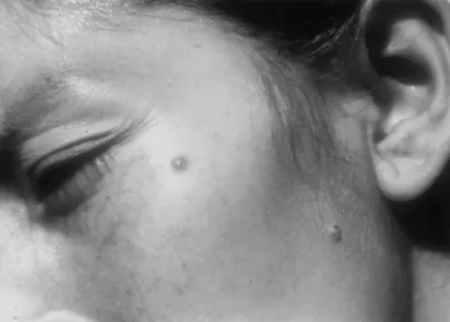
Verruga peruana is an eruptive angiomatous disease of the skin (Figs 3 and 4) that typically develops 2 months after an acute phase of bacteriemic disease known as Oroya fever or Carrion’s disease. In addition to the casual presentation of the angiomatous lesions,28,34,35 malignant-looking aspects have
been identified.36 There is a great histological similarity
between the disease BA in HIV-positive individuals.37
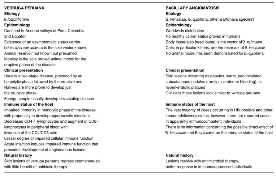
How-ever, verruga peruana differs from BA at the microscopical level, at least by the lack of aggregates of bacilli (Table 2).
Actually, verruga peruana does not seem to be confined to the recognized epidemiological villages where it was originally
documented anymore. We have indeed seen some cases of angiomatous lesions resembling verruga peruana in appar-ently immunocompetent individuals without any recognized risk factor including travel to verrucogenous areas and Oroya fever. As Bartonella spp. other than B. bacilliformis have been isolated from rodents in villages of Peru,38 some of these
angio-matous lesions with incomplete or absent epidemiological support could represent diseases caused by Bartonella spp. different from B. bacilliformis. On the other hand, there are no documented cases regarding the occurrence of diseases caused by B. bacilliformis in HIV-infected individuals. Finally, the immunodeficiency status associated with the majority of cases of BA, mainly CD4+ lymphocyte depletion and phago-cytic dysfunction, could be compared with the immunological changes produced in the Oroya fever.39 This could provide a
suitable model of the influence of the immunological status on the development of some vascular lesions of the skin.
Figure 3Verruga peruana. Two small papules on the face
Figure 4Verruga peruana, nodular type IJD_1489.fm Page 463 Wednesday, August 14, 2002 8:32 AM
Skin and the immune system in bartonelloses
The skin-restricted features of verruga peruana and the pre-dominant skin manifestations in BA together with the immuno-logical impaired status found in both diseases make it attractive to suggest a common pathobiological pathway. The skin might be a target of specific infectious agents responsible for angiomatous diseases in the setting of impaired immune function. Indeed, angiogenesis is a biological event strictly controlled and associated with wound healing, several chronic inflammatory diseases and tumor growth and meta-stasis.40 –43 The statement that Bartonella-associated vascular
lesions may represent some variants of these angiogenic responses has already been made44 and calls for further research. The
ability of Bartonella spp. to produce angiogenic factors24,44
might also be considered when analyzing the complex inter-action between bacteria, skin cells and the immune system.
The immune function of the skin was emphasized many years ago under the concept of ‘skin-associated lymphoid tissue’ constituted by keratinocytes, Langerhans cells, T-cells and endothelial cells.45 The biological interactions between
cutaneous dendritic cells, endothelial cells and skin lym-phocytes is altered in some pathological conditions. For instance, there is cumulated data related to the biology of the
skin in HIV-disease46–49 and the vascular proliferation in BA
may represent a defect in the phagocytic function and devel-opment of granulomatous reactions such as those found in CSD.50 In addition, B. bacilliformis tropism for the skin could
be caused by the better growth of the bacilli at lower temper-atures with dermal dendrocytes also playing a role.28,50
Conclusion
Bartonella spp. infections are responsible for different clinical
presentations including vascular growths in the skin. Further research must be guided in order to identify the whole spectrum of the pathogens, in particular Bartonella spp. responsible for vascular lesions. The cellular and molecular components of BA and verruga peruana lesions should be scrutinized. Some analogy among these diseases could pro-vide epro-vidence for common biological mechanisms.
References
1 Dehio C, Sander A. Bartonella as emerging pathogens.
Trends Microbiol 1999; 7: 226 – 228.
2 Maguiña C, Gotuzzo E. Bartonellosis: new and old. Infect
Dis Clin NA 2000; 14: 1 – 22.
Table 2 Similarities and differences between verruga peruana and bacillary angiomatosis
VERRUGA PERUANA BACILLARY ANGIOMATOSIS
Etiology Etiology
B. bacilliformis B. henselae, B. quintana, other Bartonella species?
Epidemiology Epidemiology
Confined to Andean valleys of Peru, Colombia and Equator
Worldwide distribution
No healthy carrier status proved in humans
Existence of an asymptomatic status carrier Body louse(also head louse) is the vector of B. quintana Lutzomyia verrucarum is the sole vector known Cats, in particular kittens, are the reservoir of B. henselae Animal reservoir not known but presumed No animal model has been demonstrated for B. quintana Monkey is the sole proved animal model for the
eruptive phase of the disease
Clinical presentation Clinical presentation
Usually a two-stage disease, preceded by an hemolytic phase followed by the eruptive one
Skin lesions occurring as papules, warts, pedunculated, subcutaneous nodules (rarely ulcerated or bleeding), or hyperkeratotic plaques
Clinically these lesions look similar to verruga peruana Natives are more prone to develop just
the eruptive phase
Foreign people usually develop devastating disease
Immune status of the host Immune status of the host
Impaired immunity in hemolytic phase of the disease The vast majority of cases occurring in HIV-positive and other with propensity to develop opportunistic infections
Decreased CD4-T lymphocytes and augment of CD8-T
immunodeficiency status, however, there are reported cases in apparently immunocompetent individuals
lymphocytes in peripheral blood with inversion of the CD4/CD8 ratio
There is no information concerning the possible direct effect of B. henselae and B. quintana on the immune status of the host Lesser degree of impaired cellular immune function
Acute infection induces impaired immune function that precedes development of angiomatous lesions
Natural history Natural history
Skin lesions of verruga peruana regress spontaneously with little benefit of antibiotic therapy
Lesions resolve with antimicrobial therapy, better response in immunosuppressed individuals
465
Chian, Arrese, and Piérard Skin manifestations of Bartonella infections Review
3 Cartihers H. Cat scratch disease. an overview based on a study of 1200 patients. Am J Dis Child 1985; 139: 1124. 4 Spach D, Koehler J. Bartonella-associated infections. Infect
Dis Clin NA 1998; 12: 137 – 155.
5 Tuli M, Cockerell C. Rickettsial and Bartonella infections. In. Farmer E, Hood A., eds. Pathology of the skin. New York, McGraw & Hill 2000, 530 – 542.
6 Anderson B, Neuman M. Bartonella spp. as emerging human pathogens. Clin Microbiol Rev 1997; 10: 203 – 219. 7 Fredericks D, Relman D. Sequence-based identification of
microbial pathogens. a reconsideration of Koch’s postulates.
Clin Microbiol Rev 1996; 9: 18 – 33.
8 Kordick D, Hilyard E, Hadfield T, et al.Bartonella
clarridgeiae, a newly recognized zoonotic pathogens causing
inoculation papules, fever, and lymphadenopathy (cat scratch disease). J Clin Microbiol 1997; 35: 1813 – 1818. 9 Margileth A, Baehren D. Chest-wall abscess due to cat
scratch disease (CSD) in an adult with antibodies to Bartonella clarridgeiae. case report and review of the thoracopulmonary manifestations of CSD. Clin Infect Dis
1998; 27: 353 – 357.
10 Regnery R, Olson J, Perkins B, Bibb W. Serologic response to ‘Rochalimacea henselae’ antigen in suspected cat scratch disease. Lancet 1992; 339: 1443 – 1445.
11 Relman D. Has trench fever returned? N Engl J Med 1995;
332: 463–464.
12 Stoler M, Bonfiglio T, Steigbrgel R, Pereira M. An atypical subcutaneous infection associated with acquired immunodefficiency syndrome. Am J Clin Pathol 1983; 80: 714 – 718.
13 Koehler J, Quin F, Berger T, et al. Isolation of Rochalimacea
species from cutaneous and osseous lesions of bacillary angiomatosis. N Engl J Med 1992; 327: 1625 – 1631. 14 Mohle-Boetani J, Koehler J, Berger T, et al. Bacillary
angi-omatosis and bacillary peliosis in patients infected with human immunodeficiency virus. Clinical characteristics of a case-control study. Clin Infect Dis 1996; 22: 794.
15 Kemper C, Lombard C, Deresinski S, et al. Visceral bacillary epithelioid angiomatosis: possible manifestations of disseminated cat scratch disease in the immunocompromised host: a report of two cases. Am J Med 1990; 89: 216. 16 Myers S, Prose N, García J, et al. Bacillary angiomatosis in a
child undergoing chemotherapy. J Pediat 1992; 121: 574. 17 Cockerell C, Bergstresser R, Myrie W, Tierno P. Bacillary
epithelioid angiomatosis occuring in a immunocompetent individual. Arch Dermatol 1990; 126: 787 – 790.
18 Smith K, Skelton H, Tuur S, et al. Bacillary angiomatosis in an immunocompetent child. Am J Dermatopathol 1996; 18: 597.
19 Plettenberg A, Rasokat H, Kalibe T, et al. Bacillary angiomatosis in HIV-infected patients. An epidemiological and clinical study. In: World AIDS Conference. Geneva 1998, 12: 824.
20 Koehler J, Sánchez M, Garrido C, et al. Molecular epidemiology of Bartonella infections in patients with bacillary angiomatosis-peliosis. N Engl J Med 1997; 337: 1876 – 1883.
21 Arvand M, Wendt C, Regnath T, et al. Characterization of
Bartonella henselae isolated from bacillary angiomatosis
lesions in human immunodeficiency virus-infected patient in Germany. Clin Infect Dis 1998; 26: 1296 – 1299.
22 Cockerell C, Le Boit P. Bacillary angiomatosis: a newly characterized pseudoneoplastic, infectous, cutaneous, vascular disorder. J Am Acad Dermatol 1990; 22: 501 – 512. 23 Conley T, Slater L, Hamilton K. Rochalimacea species
stimulate human endothelial cell proliferation and migration in vitro. J Laboratory Clin Med 1994; 124: 521 – 528. 24 Maeno N, Oda H, Yoshiie K, et al. Live Bartonella henselae
enhances endothelial cell proliferation without contact.
Microb Pathog 1999; 27: 419–427.
25 Alkan S, Orenstein J. Bacillary peliosis hepatis. N Engl J Med 1991; 324: 1513–1514.
26 Le Boit P, Berger T, Egbert B, et al. Bacillary angiomatosis: The histopathology and differential diagnosis of a pseudoneoplastic infection in patients with human immunodefficiency virus disease. Am J Surg Pathol 1989;
13: 909 – 920.
27 Schwartz R, Nychay S, Janniger C, Lambert W. Bacillary angiomatosis: presentation of six patients, some with unusual features. Br J Dermatol 1997; 136: 60 – 65. 28 Arrese J, Pierard G. Dendrocytes in verruga peruana and
bacillary angiomatosis. Dermatology 1992; 184: 22–25. 29 Robert C, Fuhlbrigge RC, Kieffer D, et al. Interaction of
dendritic cells with skin endothelium: a new perspective on immunosurveillance. J Exp Med 1999; 189: 627 – 635. 30 Moretti S, Spallanzani A, Pinzi C. Skin angiogenesis;
biologic basis for pathological processes. Clin Dermatol 1999; 17: 629 – 631.
31 Arrese J, Maguiña C, Pierard G. La verruga peruana: pasado, presente y futuro. Piel 1992; 7: 350 – 353.
32 Childs J, Rooney J, Cooper J, et al. Epidemiologic observations on infection with Rochalimacea among cats living in Baltimore. J Am Vet Med Assoc 1994; 204: 1175 – 1178. 33 Maurin M, Raoult D. Bartonella (Rochalimacea)
quintana infections. Clin Microbiol Rev 1996; 9: 273 – 292.
34 Ellis B, Rotz L, Leake J, et al. An outbreak of acute bartonelosis (Oroya fever) In Urubamba Region Peru Am
J Trop Med Hyg 1999; 61: 244–249.
35 Arias-Stella J, Lieberman P, Erlandson R, Arias-Stella J Jr. Histology, immunohistochemistry and ultrastructure of the verruga in Carrion’s disease. Am J Surg Pathol 1986;
10: 595 – 610.
36 Arias-Stella J, Lieberman P, Garcia-Cáceres U, et al. Verruga peruana mimicking malignant neoplasms. Am J
Dermat-opathol 1987; 9: 279 – 291.
37 Le Boit P, Berter T, Egbert B, et al. Epithelioid hemangioma-like vascular proliferation in AIDS. Manifestation of cat-scratch disease bacillus or infection? Lancet 1988; I: 960 – 963.
38 Birtles R, Canales J, Ventosilla R, et al. Survey of
Bartonella species infecting intradomicillary animals in the
Huayllacallan Valley, Ancash, Peru, a region endemic for human bartonellosis. Am J Trop Med Hyg 1999; 60: 799– 905.
malignant melanomas. Eur J Cancer 1997; 33: 1888 – 1892. 43 Heymans O, Blacher S, Brouers F, Pierard GE. Fractal
quantification of the microvasculature heterogeneity in cutaneous melanoma. Dermatology 1999; 198: 212 – 217. 44 Garcia F, Wojta J, Broadley K, et al. Bartonella bacilliformis
stimulates endothelial cells in vitro and is angiogenic in vivo.
Am J Pathol 1990; 136: 1125 – 1135.
AIDS example. J Laboratory Clin Med 1996; 128: 531 – 535. 49 Pope M. Mucosal dendritic cells and immunodeficiency
viruses. J Infect Dis 1999; 179: S427–S430.
50 Karem K, Paddock C, Regnery R. Bartonella henselae,
B. quintana, and B. bacilliformis: historical pathogens of
emerging significance. Microb Infect 2000; 2: 1193 – 1205.