Cytokines and chemokines in follicular fluids and potential of the
corresponding embryo: the role of granulocyte colony-stimulating factor
N. Lédée1, R. Lombroso1, L. Lombardelli2, J. Selva1, S. Dubanchet3, G. Chaouat3, F. Frankenne4, J.M. Foidart4,
E. Maggi2, S. Romagnani2, Y. Ville1 and M.-P. Piccinni2
1 Service de Gynécologie-Obstétrique et Médecine de la Reproduction et Service de cytogenétique et Biologie de la Reproduction, Centre hospitalier intercommunal Poissy-Saint Germain en Laye, Univ UVSQ, UPRES-EA 2493, Poissy F-78300, France
2
Centre of Excellence for Research, Transfer and High Education DENOTHE of the University of Florence, Department of Internal Medicine-Immunoallergology Unit, viale Morgagni 85, 50134 Florence, Italy
31NSERM U782, Univ Paris-Sud, UMR-S0782, Clamart F-92140, France
4 Département universitaire de Gynécologie et d'obstétrique, CHR la Citadelle, Liège, Belgium
BACKGROUND: The cytokine/chemokine levels of individual follicular fluids (FFs) were measured to determine whether a biomarker could be linked to the developmental potential of the derived embryo. METHODS: Fluid was collected from 132 individual FFs that were the source of oocytes subsequently fertilized and transferred. In each, a bead-based multiplex sandwich immunoassay (Luminex) was used to measure 28 cytokines and chemokines simultaneously. RESULTS: Significantly higher levels of interleukin (IL-2) and interferon (IFN-γ) were detected in FF for embryos that underwent early cleavage. IL-12 was significantly higher in FF corresponding to highly fragmented embryos and the chemokine CCL5 was
significantly higher in FF related to the best quality (Top) embryos. The level of granulocyte colony-stimulating factor (G-CSF) in individual FF samples was correlated with the implantation potential of the corresponding embryo. The area under the receiver operating characteristics curve, which distinguished the embryos that definitely led to delivery from those that did not, was 0.84 (0.75-0.90) (P = 0.0001) for FF G-CSF. FF G-CSF was significantly lower in patients older than 36 years compared with those <30-year old. When the FF G-CSF was 20 pg/ml or higher, the ratio between Top and non-Top embryos was significantly higher than for the group with FF G-CSF below 20 pg/ml (45 versus 20.45%, P = 0.007). CONCLUSIONS: Individual FF composition is related to the development of the corresponding in vitro generated embryo and its potential of implantation. Individual FF G-CSF may provide a non-invasive biomarker of implantation that needs to be evaluated together with in vitro observation to select the oocyte, and hence the embryo, to transfer.
Keywords: G-CSF ; follicular fluid ; embryo implantation ; IVF/ICSI-ET ; bead-based multiplex immunoassays
Introduction
Assessing the implantation potential of an embryo to be transferred is crucial for increasing the success rates of IVF-ICSI cycles while reducing or eliminating the risk of multiple pregnancies. The application of the 'single embryo transfer' (SET) policy has proven effective in decreasing the maternal and foetal morbidity and mortality associated with assisted reproductive technologies (De Neubourg and Gerris, 2003; De Sutter et al., 2003; Gerris
et al., 2004; Pinborg, 2005; Fiddelers et al., 2006). This policy, however, is applied only sporadically worldwide,
except when imposed by a specific legislation in a given country, mainly because the mean embryo implantation rates remain too low (15-20%). The transfer of at least two embryos significantly increases overall pregnancy rates (van Montfoort et al., 2006), and two embryos are routinely transferred into the uterine cavity in Europe, and 3-4 in the USA. The application of SET would be encouraged and the risk of multiple pregnancies minimized in IVF-ICSI cycles if methods could be found to select those embryos with the highest implantation potential so as to transfer them selectively.
The analysis of the morphology of the pre-implantation embryo before Day 2, although important, is generally not sufficiently informative. Additional data about the embryo's potential are required (Guerif et al., 2007). An alternative approach to applying SET would be to select the best oocyte and thereby avoid the ethical problems linked to the generation of surplus embryos and comply with the law in countries where all fertilized embryos must be transferred. Identifying prognostic markers of oocyte competence is difficult, but cytokines and chemokines present in individual follicular fluid (FF), which surrounds the oocyte-cumulus oophorus complex, may prove useful. Several previous studies have described factors in FF that appear to be related to a successful outcome in IVF-ET (i.e. a live birth) (Hammadeh et al., 2002; Ocal et al., 2004; Westergaard et al., 2004; Wiener-Megnazi et al., 2004; Asimakopoulos et al., 2005; Salmassi et al., 2005; Rizzo et al., 2007). However, these studies have generally been performed with pooled FF in which fluids from functional cysts, atretic, immature and mature follicles were all mixed. Studies of FF that is pooled or of limited traceability (Fanchin et
al., 2007) cannot demonstrate whether detection of these factors allows the selection of high-quality oocytes on
related to the implantation potential of the resulting embryo after a conventional ovarian stimulation protocol. We therefore decided to use multiplex analysis to explore the patterns of expression of cytokines, chemokines and growth factors of the FF of individual oocytes and then to relate these patterns to the morphology and implantation potential of the embryo derived from the oocyte from that follicle.
To maximize the potential of the multiplex assay, we limited our targets to molecules either known to participate in the early embryonic 'immunotrophic' activity or suspected of involvement in preparing local (uterine)
tolerance to the conceptus. We therefore focused on those cytokines which are most likely to be involved in switching the local Th-1/Th-2 equilibrium towards a Th2 profile (Wegmann et al., 1993). We also studied the TNF family, its related chemokines and the growth factors which are generally believed to be involved in mobilizing the surrounding immune cells. The main outcome measure of the study was the live delivery of each corresponding embryo.
We report here that G-CSF in individual FF appears to be an immune biomarker of implantation, related to ageing and ovarian stimulation and providing information complementary to that of embryo morphology. Study protocol
Patients
We recruited 142 women being treated for infertility and included in the ICSI program at the Obstetrics and Gynaecology department of Poissy-St Germain en Laye Hospital. To avoid any possibility of a bias in patient selection, the patients were randomly allocated (1/1) at inclusion in the ICSI program into either a group where individual FF were collected at oocyte retrieval (study group n = 71) or a group with standard oocyte retrieval where FF were pooled (n = 71). Each patient was included only once during the study period.
All patients were fully informed, and the Institutional Review Board (Comité Consultatif de Protection des Personnes, Poissy-St Germain en Laye) approved this investigation.
The mean age of the population was 33 years (range: 24-41), the mean number of previous IVF attempts was 2 (1-6) and the mean time since first attempting conception was 4 years (1-12 years). In 49 cases, the infertility was primary and in 22 cases, it was secondary. In 56 cases, the infertility was completely attributable to the man, in 10, it was mixed and in 5, it was unexplained, with a previous IVF failure.
Treatment
The ovarian stimulation protocols were selected by each treating physician: a standard long protocol with a daily GnRH agonist for 54 patients, a short protocol with a daily GnRH agonist for 11 patients and an antagonist protocol for six patients. The response to stimulation was monitored by serial blood tests as well as by ultrasound assessment of follicular and endometrial growth. Ovulation was triggered when at least four follicles had reached 16 mm. We classified the responses to ovarian stimulation in three categories: low response (estradiol (E2) below 1500 pg/ml the day ovulation was triggered), mild response (E2 from 1500 through 3500 pg/ml) and high
response (E2 over 3500 pg/ml).
Oocytes were retrieved by aspiration 35-36 h after the triggering of ovulation, with a general or local anaesthesia and vaginal ultrasound guidance. An individual 10-ml syringe was used for each follicle in the study group. We thus adapted the standard method of oocyte aspiration to collect the FF of each individual oocyte separately.
FF samples
Individual FF samples, each corresponding to one mature oocyte, were collected from women in the study group. The presence of an extra-technician was necessary at the time of the collection to record the presence of an oocyte in the FF sample collected. Samples without an oocyte were immediately discarded. The volume and appearance (yellow or reddish and bloody) of each FF sample were recorded. Each sample was centrifuged and divided into aliquots after anonymization of the samples in the database. This ensured that the subsequent analyses were performed on a blinded basis.
The samples were initially stored at -20°C and then at -80°C until they were assayed.
Only the FF samples corresponding to embryos actually transferred on Day 2 were analysed. Accordingly, of 530 FF samples collected, 146 embryos were transferred, and the fluid from follicles related to 132 of them was analysed. For 15 patients in a sub-cohort, FF retrieved from every follicle aspirated during the cycle was analysed (76 samples) to study the individual variations.
Oocyte fertilization and embryo culture until Day 2
Oocytes were collected and the cumulus and corona cells removed with hyaluronidase 80 IU (Fertipro, JCD, France). The oocytes were injected with single sperm in a 5-µl droplet of Ferticult Hepes (JCD, France), with
viscous PVP medium (Fertipro, JCD, France) to slow the sperm. The injected oocytes were individually cultured in a 40-µl micro-droplet of ISM1 (Medicult, France) under oil at 37°C in a 5% CO2 humid atmosphere. The
number of pronuclei and their appearance were assessed by Gianaroli's criteria after 20 h (Gianaroli et al., 2003). At 25 h, we assessed the early cleavage rate. On Day 2, we assessed and recorded the number, fragmentation and regularity of each individual blastomere. The decision to transfer an embryo was taken by the embryologist in charge, taking in account the morphology at 18 h, 25 h and Day 2 and the rate of development during the first 2 days. Embryo transfer was scheduled on Day 2. The transferred embryos were observed. (i) At the zygote stage (18-20 h), the pronuclear and nuclear morphology and polar body alignment were recorded for 76 embryos according to the classification described previously (Gianaroli, 2003). (ii) At 25 h, early cleavage was recorded for 92 embryos. (iii) On Day 2, the embryos were analysed in relation to:
(a) Their fragmentation (Grade 1: <10%, Grade 2: 10-30%, Grade 3: 30-50%, Grade 4: over 50% (the corresponding embryo was downgraded when blastomeres were unequal in size)
(b) Their embryo score: number of blastomeres × (5 - embryo grade) (Steer et al., 1992). We created three categories of analysis: score 16-30, 15-12 and <12.
(c) According to the 'Top' versus 'not Top' analysis: 'Top' embryos were defined as those with 4-5 cells on Day 2, <10% fragmentation and regular cells. All other embryos were classified as 'not Top'.
Determination of cytokine and chemokine concentrations in individual FF samples with bead-based multiplex sandwich immunoassays and ELISA measurements
We used a bead-based multiplex sandwich immunoassay, read with a Luminex system (Luminex Map
Technology) to measure the concentrations in individual FF samples of the following cytokines and chemokines: IL-1α, IL-1Ra, IL-2, IL-4, IL-5, IL-6, IL-8, IL-9, IL-10, IL-12, IL-13, IL-15, IL-17, IFN-α, TNF-α, G-CSF, GM-CSF, VEGF, PDGF, FGF, IP-10, MCP-1, CCL5, eotaxin, MIP-1-α and MIP-1-β. In brief, 50 µl of each
individual FF sample was added to 50 µl of antibody-conjugated beads directed against the cytokines listed above (Bio-Rad Laboratories, Hercules, CA, USA) in a 96-well filter plate (Bio-Rad). After a 30-min incubation, the plate was washed and 25 µl of biotinylated anti-cytokine antibody solution was added to each well before another 30-min incubation. The plate was then washed and 50 µl of streptavidin-conjugated PE was added to each well. After a final wash, each well was resuspended with 125 µl of assay buffer (Bio-Rad) and analysed by the Luminex array system. The cytokine concentrations were calculated and a standard curve was derived from various concentrations of a cytokine standard in the assay.
The limits of detection for G-CSF for the Bio-Rad kit are 5.33 ± 9.12 pg/ml (mean ± 3 × SD of the blanks, where G-CSF was not present). The intratest variation was assessed by measuring the same FF samples 10 times and the intratest SD for G-CSF ranged from 0.18 to 0.79 pg/ml.
As reagents for measuring LIF are not available for the Luminex system, it was analysed with the non-commercial ELISA previously described (Taupin et al., 1997), which combines two monoclonal antibodies known to not interfere with ligand receptors.
Evaluation of implantation and delivery potential
The probability of implantation, described here as the clinical implantation rate (IR), was defined for each sample tested as the ratio of the number of intrauterine gestational sacs to the number of transferred embryos. Clinical implantation was defined at 8 weeks of amenorrhea when a gestational sac was viewed by ultrasound. The delivery rate was defined as the ratio of the number of babies born to the number of embryos transferred. The 'certain implantation and delivery' category for receiver operating characteristics (ROC) curve analysis was defined when the number of embryos replaced was equal to the number of gestational sacs or babies born (i.e. one embryo replaced and a singleton pregnancy, two embryos replaced and a twin pregnancy).
Statistical analysis
In order to explore the significance of the concentrations of cytokines and growth factors in each FF with regard to the clinical profile and response of the stimulation of each patient, we created categories of analysis and used the ANOVA test to highlight significant differences.
The following demographic data were analysed:
(i) Age (younger than 30 years old, from 30 to 36, 37 years or older) (ii) Type of infertility (primary or secondary)
(iii) Aetiology
(iv) Number of previous attempts (1 or 2, 3 or 4, over 4)
The basal FSH was defined as elevated over 9 IU/ml and normal below 9 IU/ml. (i) Protocol type (long or short protocols or antagonist)
(ii) Hormonal response to stimulation measured by the E2 level on the day of the HCG injection (1500 pg/ml or
less, from 1500 through 3000, more than 3000).
For the embryo morphology analysis, we took into account at 20 h the pronuclear and nuclear morphology and the polar body alignment at 20 h, the occurrence of early cleavage at 25 h, grading according to the
fragmentation, embryo score, and classification (Top versus not Top at 48 h).
To construct the ROC curves, we took into account only two categories: 'no delivery' and 'definite delivery' (when the number of embryos replaced equalled the number of babies born). The discrimination between no and definite delivery as a function of the concentration of each cytokine and chemokine in each sample was
evaluated with ROC curve analysis (MedCalc for Windows, version 9.2.0.0 software, MedCalc Software, Mariakerke, Belgium). In a ROC curve the true positive rate (sensitivity) is plotted as a function of the false positive rate (100-specificity) for different cut-off points (Zweig and Campbell, 1993). The calculation of the area under the ROC (AUC-ROC) curve measures the accuracy, i.e. the ability of the cytokines and chemokines tested to discriminate between a successful and a failed implantation. The ROC curve analysis thus allowed us to define the lower limit implantation probability, defined by a negative predictive value of 100% from the AUC-ROC curve, and its upper limit, defined by the highest positive predictive value for implantation.
We were therefore able to compare the implantation and delivery potentials of each embryo as a function of the G-CSF concentration in a predefined category. A value of P < 0.05 was considered significant.
Results
ICSI results and pregnancy/delivery rates in the observed cohort
The mean number of oocytes collected was 8, the mean number of embryos obtained 4.5, and the mean number of embryos transferred 2 (1-3). The mean implantation rate per embryo was 21% (31 gestational sacs for 146 transferred embryos), the mean clinical pregnancy rate 30.9% (22/71) and the mean live birth rate 29.6% (21/71). The multiple pregnancy rate was 36% (8/22).
Detection of cytokines and chemokines
LIF, IL-1Ra, IL-4, IL-6, IL-8, IL-10, IL-12, IL-13, G-CSF, VEGF, IP-10, MCP-1, eotaxin and MIP-β were detected in all FF samples, while IL-1-β, IL-5, IL-7, IL-17, TNF-α and MIP-α were detected in none. IL-15, GM-CSF, CCL5, PDGF, IFN-7, IL-9, IL-2 and FGF were detected, respectively, in 95, 88, 81, 76, 65, 60, 48 and 22% of the FF samples (Table I).
Variations of the cytokine and chemokine content of FF in relation to demographic data
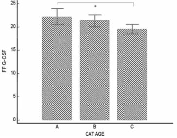
FF samples of 38 follicles were collected from patients younger than 30 years old, 57 from patients between 31 and 36, 57 over 37 years old. We observed a decrease in FF G-CSF with ageing, which was, respectively, 22.2, 21.3 and 19.6 pg/ml (Fig. 1). FF from patient younger than 30 years had a significant higher G-CSF content than those from patients 37-year old or more (P = 0.03).
We did not observe any significant differences according to the range of the ICSI attempt for the cytokines and growth factors tested.
FF G-CSF and IL-13 levels were significantly higher in patients with primary compared to those with secondary infertility (21.6 and 4.6 versus 19.9 and 4.2 pg/ml, respectively, P = 0.04 and 0.003), while CCL5 levels were significantly lower (321.56 versus 733 pg/ml, P = 0.01).
FF IL-15 was significantly higher in patients with unexplained infertility compared to those with male or mixed infertility (5 versus 1.3 and 1.6 pg/ml for IL-15, P = 0.01 and 0.03, respectively).
Hormonal response to ovarian stimulation and cytokine/ chemokine concentrations in individual FF samples
FF samples were classified according to hormonal response to stimulation: low (n = 23), normal (n = 90) and high respon-ders (n = 9) were identified. The level of G-CSF was significantly lower in high compared with low responders (18.5 versus 22.4 pg/ml, P = 0.02). We observed significant differences for many FF cytokines (G-CSF, IL-8, IL-15, IL-17, IFN-7, VEGF, IP-10, CCL5 and MIP-β), but always in the same direction, that is, the FF cytokine levels decreased when E2 levels increased.
Table I. Mean values, standard error and standard variation of the cytokines and chemokines detected in
individual FF samples with the Luminex technology (bead-based multiplex sandwich immunoassays).
Cytokines/chemokines (pg/ml) Mean Standard deviation Standard error
IL-1Ra 225 530 46 IL-2 8 5.8 0.5 IL-4 1.8 0.7 0.06 IL-6 21.2 79 6.8 IL-8 399 2785 241 IL-9 9.9 13.4 1.16 IL-10 4.6 4.6 0.4 IL-12 15.3 6.2 0.53 IL-13 4.5 0.73 0.064 IL-15 1.77 3.76 0.32 IFN-γ 32.9 43.1 3.7 G-CSF 21.06 4.64 0.40 GM-CSF 25.4 11.1 0.96 VEGF 12 616 13 565 1176 PDGF 248.8 1388 120 FGF 19 47.6 4.1 IP-10 2083 1948 168.9 MCP-1 256.6 1560 135 CCL5 449 1087 94 Eotaxin 138 103 8.9 MIP-1 beta 266 1989 172 LIF 954 1150 103
Those shown in bold have acceptable standard deviations and errors for the means.
Figure 1: Effect on ageing on individual FF G-CSF concentration (pg/ml). Three groups are shown: (A)
<30-year old (n = 38), (B) between 30 and 36-<30-year old (n = 57), (C) older than 36 <30-years (n = 57). *FF G-CSF is significantly lower in Group A versus C (22.2 versus 19.6 pg/ml, P = 0.03).
Cytokine and chemokine concentrations and the appearance of FF
FFs were classified according to the appearance and volume of each sample at the time of the collection in order to evaluate the impact of blood contamination on the cytokines/chemokines content or follicle volume. In all, 33 were yellow, 58 reddish and 48 bloody, 31 had a volume of 2 ml, 92 had a volume between 3 and 6 ml and 10 had a volume over 7 ml. The cytokine/chemokine content did not vary according to the appearance or volume of the fluid collected.
Cytokine and chemokine concentrations in individual FFs and subsequent morphology of the transferred embryos
Among the 78 embryos observed at the zygote stage, cytokine or chemokine content did not differ according to categories of pronuclear and nuclear embryo staging. There were 33 embryos with a longitudinal polar body
alignment (alpha group), 28 with a perpendicular alignment (beta group) and 19 other patterns (gamma group). IL-1Ra and TNF were significantly elevated in the gamma group compared with the alpha and beta groups (485 versus 154 and 129 pg/ml for IL-1Ra, P = 0.03 and 25 versus 9 and 8.8 pg/ml for TNF-α, P = 0.045).
Of 96 embryos observed at the early cleavage stage (25 h), 31 cleaved at 25 h and 65 did not (remaining at either the 2 or 0 pronuclei stage). FF IL-2 and IFN-γ were significantly higher in early cleavage embryo compared with the others (10 versus 7.3 pg/ml for IL-2 and 55 versus 24.4 pg/ml for interferon gamma, P = 0.03 and 0.003, respectively). On Day 2, application of the embryo score rated 68 embryos at 16 or higher, 28 from 15 through 12 and 36 below 12. IL-12 was higher in the FF from embryos scored below 12 (17 versus 14 pg/ml in the two other groups, P = 0.05).
Applying the embryo grading, 81 embryos were Grade I, 29 were Grade II, 17 were Grade III and 5 were Grade IV. IL-12 was significantly higher in FF from Grade IV embryos when compared with Grade I, II and III embryos (23.8 versus 14.6, 15.4, 16.23 pg/ml, respectively, P = 0.013).
There were 49 embryos classified as 'Top' embryos and 83 as 'non-Top' embryos. The FF CCL5 levels were significantly higher for those corresponding to high quality 'Top' embryos than for the others (756 versus 271 pg/ml, P = 0.01).
When we compare the group of embryos derived from follicles with an FF G-CSF concentration <20 and ≥20 pg/ml, the ratio of Top to not Top embryos differed significantly (20.45 versus 45%, P = 0007) (Fig. 2).
Figure 2: 'Top' versus 'non-Top' repartition in function of FF G-CSF content (pg/ml). In FF with a
concentration of G-CSF of 20 pg/ml or higher, 45% (40/ 88) of the embryos were of 'Top' quality. For the ones with a concentration below 20 pg/ml, only 20.45% (9/44) were 'Top' embryos (P = 0.007).
Cytokines, chemokines and implantation/delivery rates
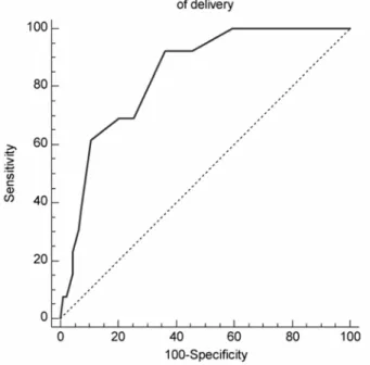
The level of only one of the substances tested—granulocyte-colony stimulating factor (G-CSF)—was correlated with the implantation potential of the corresponding embryos (r = 0.40, P < 0.001). The embryos were classified according to their implantation and delivery rates. Only the embryos with a clear outcome—that is, those which did not implant (n = 89) and those which that definitely implanted and were delivered (n = 13)—were used to construct the AUC-ROC for G-CSF. The area under the ROC curve for ongoing implantation was 0.82 (0.73-0.89) and highly significant (P = 0.0001). The area under the ROC curve for successful delivery was 0.83 (0.75-0.90) and was also highly significant (P = 0.0001) (Fig. 3). We used the AUC-ROC to define an upper and lower limit for G-CSF to determine whether the G-CSF concentration could be used to evaluate the implantation potential for each oocyte and thus for each corresponding embryo, in order to help select which and how many embryos should be transferred. The lower limit was defined according to the AUC-ROC by the highest negative predictive value of implantation (G-CSF < 20 pg/ml). When FF G-CSF was lower than 20 pg/ml, the negative predictive value was 100%. The upper limit was defined by the highest positive predictive value of implantation. According to the AUC-ROC, when the FF G-CSF exceeded 24 pg/ml, the positive predictive value reached 40%.
When we evaluated the 132 transferred embryos according to their FF G-CSF levels, we observed very significant differences in implantation rate between embryos with low and intermediate G-CSF levels (implantation and delivery rates were 9 and 6% compared with 18 and 15.8%, respectively, P = 0.003 and < 0.001) and especially between the embryos with low and high G-CSF levels (9 and 6 versus 44% for both implantation and delivery, P < 0.0001) (Table II).
Figure 3: ROC curve for measurements of G-CSF in individual FF samples from patients with 'No' and 'Certain
delivery' groups. The true positive rate (sensitivity) is plotted as a function of the false-positive rate (100-specificity) for different cut-off points of FF-GCSF concentration. Each point on the ROC plot represents a sensitivity/ specificity pair corresponding to a particular decision threshold. AUC-ROC curve is a measure of how well FF G-CSF can distinguish between two main diagnostic groups (certain delivery/no delivery). (Continuous line) The area under curve is 0.84, indicating that a randomly selected individual from the positive group has a test value larger than that for a randomly chosen individual from the negative group 84% of the time. (Dotted lines) The area under the ROC curve is 0.5, representing the null hypothesis.
Table II. Implantation and delivery rates as a function of the FF G-CSF concentrations in the individual FF
samples corresponding to the fertilised oocytes.
G-CSF concentrations
(Luminex Bio-Rad) Number of embryos
concerned Mean implantation rate (%) Mean delivery rate (%) Low G-CSF ( <20 pg/ml) 45 9 6 Intermediate G-CSF (Between 20 to 24 pg/ml) 62 18* 15.6* High G-CSF (>24 pg/ml) 25 44** 44**
The lower threshold of implantation was defined by a negative predictive value of 100% from the AUC-ROC curve (<20 pg/ml). The higher threshold of implantation was defined by the highest positive predictive value for implantation (over 24 pg/ml). *P = 0.0005 and 0.001 between intermediate and low G-CSF for implantation and delivery rates. **P < 0.0001 between high and low G-CSF for both implantation and delivery rates.
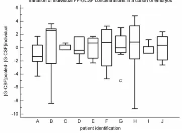
Variations of [FF]G-CSF within a single ovarian cohort
To determine whether useful information could be obtained by measuring the G-CSF concentration in pooled FF or whether it was essential to measure it for each individual follicle and oocyte, we evaluated the samples from every oocyte producing an embryo among 15 patients, independently of the outcome (embryo transfer, freezing or destruction). This allowed us to compare the variations between samples within a single ovarian cohort, that is, the oocytes produced by a single woman in a single cycle. We evaluated 76 samples with the Luminex kit manufactured by Bio-Rad. The mean value of G-CSF in the pooled FF (evaluated by adding together the separate FF G-CSF measurements for each woman and taking the mean) did not reflect the variations observed
in individual FF samples.
To assess the variations from the mean in a given ovarian cohort, we determined for each sample the following ratio: mean [FF] G-CSF in the cohort/individual [FF] G-CSF concentration. Fig. 4 shows this variation among 10 patients. Each box shows the variation of individual FF samples from the mean of all the embryos generated by these patients. The results strongly suggest that the embryos generated are not equally correlated with FF G-CSF and differ in their individual implantation potential. It is therefore the individual G-G-CSF value of the FF sample that reflects its implantation potential. Even in apparently good responders, there are embryos with a low implantation potential and vice versa.
Figure 4: The variation in concentration of individual FF in the same cohort of embryos obtained from 10
patients. Each box shows the variation of individual FF samples from the mean in the same cohort of generated embryos.
Discussion
The measurement of cytokines and chemokines of fluid from individual follicles revealed that the levels of several key immune factors were related to either the morphology of the related embryo or its implantation potential.
We observed that the development of the embryo during the first two days of in vitro culture differed
significantly in terms of the early cleavage step. Early cleavage at 25 h has been described as a good prognostic factor, when morphology remains good on Day 2 (Lundin et al., 2001; Giorgetti et al., 2007; Sundstrom and Saldeen, 2007; Terriou et al., 2007). Interferon gamma and IL-2 are well-known Th-1 cytokines and are found at significantly higher levels in FF of embryos that cleave early. This suggests that some Th-1 cytokines play a positive role in the process of embryo cleavage. In contrast, the significant elevation of IL-12, known to be cytotoxic, in highly fragmented embryos on Day 2, suggests that it has a deleterious role. This supports the previous finding that elevated IL-12 levels in pooled FFs is associated with a poor pregnancy rate (Gazvani et
al., 2002; Bedaiwy et al., 2007). CCL5 was significantly higher in FF from 'Top' embryos. CCL5, which is
thought to be produced by the surrounding T cells present in granulosa cells, acts as a chemo-attractant and may play a role. Even though preliminary, these results suggest that the assessment of the levels of some selected cytokines in individual FFs may help embryologists in their assessment of pre-implantation development. The main finding of this study, however, is that G-CSF in individual FFs appears to be an immune biomarker predictive of successful implantation and delivery. It can be measured easily before embryo transfer and even before fertilization itself, and may therefore be helpful in a strategy of SET. G-CSF is a member of the colony-stimulating-factor family, initially described as a haematopoietic growth factor (Clark and Kamen, 1987). Its main role is to act on the proliferation, differentiation and activation of haematopoietic cells of the neutrophilic lineage (Visani and Manfroi, 1995; Mielcarek et al., 1996). Although produced primarily by the latter, G-CSF is also secreted by other cells, including some in the reproductive tract: human luteinized follicular granulosa cells (Salmassi et al., 2004), endometrial cells (Giacomini et al., 1995), and cells from decidual, placental (Duan, 1990; Miyama et al., 1998) and various foetal (Calhoun et al., 1999) tissues.
Studies using western blot and immunohistochemistry techniques have located the G-CSF protein and its receptor in the ovary—mainly in the granulosa cells of the follicle and in luteal cells (Salmassi et al., 2004). At
ovulation, G-CSF concentrations are much higher in FF than in serum (Salmassi et al., 2005). This observation is quite common for the cytokines, chemokines, hormones and growth factors involved in the complex process of ovulation.
Previous studies of G-CSF in serum provided some evidence of its involvement in implantation: some authors reported that its serum levels rise at implantation in the case of successful natural cycles (Yanagi et al., 2002) and after successful IVF/ ICSI attempts (Salmassi et al., 2005). The source of this secretion seems to be the decidual and endometrial cells in the uterus, which indicates that it occurs about 10 days after oocyte retrieval (Duan, 1990; Giacomini et al., 1995).
Low levels of FF G-CSF were observed in ageing patients and in high responders to stimulation, both characteristics known to be associated with decreased pregnancy rates and to affect oocyte quality. Thus indirectly, we may postulate that the FF G-CSF is linked to oocyte quality and could represent a new tool for the evaluation of individual oocyte competence. The mean FF G-CSF concentration in women older than 36 was below the threshold of 20 pg/ml, the category associated with the highest negative predictive value for
implantation. In such patients, non-invasive tools able to identify the oocyte with the best potential would be of capital importance.
Significantly more 'Top' embryos were obtained when the FF G-CSF was 20 pg/ml or higher, even though FF-G CSF levels were not different between 'Top' and 'not Top' embryos. In terms of a strategy to choose which embryo to transfer, we may postulate that FF G-CSF and morphological development of the pre-implantation embryo are complementary tools that would not lead to discrepant evaluations. Assessment of other cytokines, such as CCL5 (for a positive selection) or IL-12 (for a negative selective) may easily complement the analysis if necessary. We also should not forget that new non-invasive tools focusing on the embryo itself are developing and could be a useful complement (Sakkas and Gardner, 2005; Seli et al., 2007; Scott et al., 2008). The assessment of G-CSF in individual FFs 2 days before embryo transfer may be very useful in cohorts in which all the embryos appear equal according to morphological criteria. This information would also be valuable in selecting which embryo(s) to freeze.
How [FF]G-CSF influences future embryo implantation is still unknown. However, there are several possibilities.
First, the FF itself may be involved in the complex dialogue that leads to implantation and could transmit messages directly to the fallopian tube and to the uterus in fertile patients (Leese et al., 2008). If we consider pregnancy as a semi-allograft, the question of maternal immune tolerance is essential. It has been reported that pretreatment of mice with G-CSF before an allograft promotes T cell tolerance towards these grafts through the induction of a switch towards a Th-2 dominated response and tolerogenic dendritic cell differentiation towards DC2 (Pan et al., 1995). Furthermore, G-CSF promotes the generation of IL-10-producing T-regulatory (T-reg) cells and promotes transplantation tolerance (Morris et al., 2004), a feature obviously relevant to the
immunoregulatory events occurring in the pre-, peri- and early post-implantation uterus. Thus, we may speculate that the [FF]G-CSF level reflects the early oocyte-uterus crosstalk that leads to the preparation of a receptive uterus, even before the embryo is generated (Herrler et al., 2003). One criterion for each oocyte's quality might therefore be its intrinsic capacity to prepare the uterus and to engage it in an immune tolerance-promoting pathway.
A second hypothesis is that [FF]G-CSF influences the mRNA content of the oocyte itself. In particular, it may induce adhesion molecules (including L selectin) at the oocyte cell surface to promote the adhesion of the future embryo to the endometrial cells. Alternatively, G-CSF could induce the cumulus granulosa cells to produce the cytokines or growth factors which are necessary for the development and implantation of the resulting embryo. It has been reported that both macrophages and CD4+T cells are normally detectable in the cumulus oophorus. More importantly, these T cells produce higher levels of IL-4 and LIF than the T cells of peripheral blood or ovarian cells isolated from the same women (Piccinni et al., 2001). Thus, [FF]G-CSF, which can induce a Th2 switch, (Rutella et al., 2005) could be responsible, at least in part, for IL-4 production by the cumulus oophorus T cells, which in turn induces the production of LIF by T cells (Piccinni et al., 1998). Amongst its many properties related to implantation, LIF has positive effects on the growth and differentiation of preimplantation human blastocysts and significantly increases the quality and number of human blastocysts in vitro (Dunglison et
al., 1996).
Another hypothesis is that [FF]G-CSF provides the embryo with crucial information on how to repair itself. Widespread use of pre-implantation diagnosis has shown not only the high incidence of mosaicism and aneuploidy in embryos in culture (Baart et al., 2006), but also their self-repair (Munne et al., 2005). Self-repair has been described in haematopoietic stem cells and appears to be affected by ageing (Rossi et al., 2007), as also observed in embryos. G-CSF has been described in various models (heart, liver) as an agent promoting
endogenous repair (Yannaki et al., 2005) either through the mobilization of haematopoietic multipotent progenitor cells (Imamura et al., 2005) or the enhancement of endogenous stem cells per se (Piscaglia et al., 2007).
All these hypotheses need further detailed investigations with animal models before their eventual validation in humans.
To conclude, although a prospective randomized study is essential to validate our hypothesis, this preliminary report strongly suggests that assessing the concentration of G-CSF in individual FF may be a valuable, non-invasive method to select those oocytes (and hence the embryos subsequently generated from them) with the highest implantation potential. It may also be a valuable biomarker to evaluate implantation potential among patients with a low ovarian reserve, which is still the major limiting step in our daily practice. In a large cohort, it would help the embryologist to choose which embryo to transfer or to freeze. This should make it possible to increase implantation rates significantly and facilitate the universal adoption of a 'SET' policy, without decreasing the subsequent pregnancy rates. It would therefore make it possible to reduce multiple pregnancy rates in IVF/ICSI-ET cycles and their related morbidity and mortality.
Acknowledgement
For the collection of the individual FF samples, we thank Robert Wainer, Marc Bailly, Audoin Delanete and Veronique Madinier. For the handling of the individual FF samples, we thank Ibrahim Hammoud for the detailed analysis of each embryo, and Marianne Bergère, Francois Vialard, Nabil Louafi, Pierre Oger and the technicians (Patrick Cavelot, Vincent Delabroye, Virginie Le Bail) for the extra work they kindly performed. We thank Prof. Ian Sargent for his help in editing the manuscript and for valuable advice throughout the research. Finally, we thank all the researchers of the European EMBIC networks, who helped in these achievements and in the construction of future directions and understanding.
Author Roles
N.L. designed the clinical setting, organized the individual FF collection and analysed the results. MP Piccinni designed the multiplex cytokine analysis, chose the cytokines to be tested, performed the Luminex analysis, with the help of L.L. R.L. and J.S. were the head of the clinical and biological Reproductive Department. Y.V. was the head of Obstetrics and Gynaecology department at Poissy Hospital. S.D. organized the shipment of samples and ensured their traceability. G.C., F.F. and J.M.F helped to decide on the cytokines and chemokines to be assessed and helped to interpret the results. Prof. S.R. was the director of the DENOTHE Centre of Excellence for Research, Transfer and Higher Education at the University of Florence and of the Department of Internal Medicine (Florence, Italy). Prof. E.M. was the director of the Immunoallergology unit (Florence, Italy). We applied for a provisional patent July, 2006 and a PCT patent of application July, 2007 (WO-2008/009705, accepted 24.01.08).
Funding
This work was supported by the European network of excellence EMBryo Implantation control EMBIC (contract 512040).
References
Asimakopoulos B, Nikolettos N, Papachristou DN, Simopoulou M, Al-Hasani S, Diedrich K. Follicular fluid levels of vascular endothelial growth factor and leptin are associated with pregnancy outcome of normal women participating in intracytoplasmic sperm injection cycles.
Physiol Res 2005;54:263-270.
Baart EB, Martini E, van den Berg I, Macklon NS, Galjaard RJ, Fauser BC, Van Opstal D. Preimplantation genetic screening reveals a high incidence of aneuploidy and mosaicism in embryos from young women undergoing IVF. Hum Reprod 2006;21:223-233.
Bedaiwy M, Shahin AY, AbulHassan AM, Goldberg JM, Sharma RK, Agarwal A, Falcone T. Differential expression of follicular fluid cytokines: relationship to subsequent pregnancy in IVF cycles. Reprod Biomed Online 2007;15:321-325.
Calhoun DA, Donnelly WH, Jr., Du Y, Dame JB, Li Y, Christensen RD. Distribution of granulocyte colony-stimulating factor (G-CSF) and G-CSF-receptor mRNA and protein in the human fetus. Pediatr Res 1999;46: 333-338.
Clark SC, Kamen R The human hematopoietic colony-stimulating factors. Science 1987;236:1229-1237. De Neubourg D, Gerris J Single embryo transfer - state of the art. Reprod Biomed Online 2003;7:615-622.
De Sutter P, Van der Elst J, Coetsier T, Dhont M Single embryo transfer and multiple pregnancy rate reduction in IVF/ICSI: a 5-year appraisal. Reprod Biomed Online 2003;6:464-469.
Duan JS. Production of granulocyte colony stimulating factor in decidual tissue and its significance in pregnancy. Osaka City Med J 1990;36:81-97.
Dunglison GF, Barlow DH, Sargent IL. Leukaemia inhibitory factor significantly enhances the blastocyst formation rates of human embryos cultured in serum-free medium. Hum Reprod 1996;11:191-196.
Fanchin R, Mendez Lozano DH, Frydman N, Gougeon A, di Clemente N, Frydman R, Taieb J. Anti-Mullerian hormone concentrations in the follicular fluid of the preovulatory follicle are predictive of the implantation potential of the ensuing embryo obtained by in vitro fertilization. J Clin Endocrinol Metab 2007;92:1796-1802.
Fiddelers AA, van Montfoort AP, Dirksen CD, Dumoulin JC, Land JA, Dunselman GA, Janssen JM, Severens JL, Evers JL. Single versus double embryo transfer: cost-effectiveness analysis alongside a randomized clinical trial. Hum Reprod 2006;21:2090-2097.
Gazvani R, Smith L, Fowler PA. Effect of interleukin-8 (IL-8), anti-IL-8, and IL-12 on endometrial cell survival in combined endometrial gland and stromal cell cultures derived from women with and without endometriosis. Fertil Steril 2002;77:62-67.
Gerris J, De Sutter P, De Neubourg D, Van Royen E, Vander Elst J, Mangelschots K, Vercruyssen M, Kok P, Elseviers M, Annemans L et
al. A real-life prospective health economic study of elective single embryo transfer versus two-embryo transfer in first IVF/ICSI cycles. Hum Reprod 2004;19:917-923.
Giacomini G, Tabibzadeh SS, Satyaswaroop PG, Bonsi L, Vitale L, Bagnara GP, Strippoli P, Jasonni VM. Epithelial cells are the major source of biologically active granulocyte macrophage colony-stimulating factor in human endometrium. Hum Reprod 1995;10:3259-3263. Gianaroli L, Magli MC, Ferraretti AP, Fortini D, Grieco N Pronuclear morphology and chromosomal abnormalities as scoring criteria for embryo selection. Fertil Steril 2003;80:341-349.
Giorgetti C, Hans E, Terriou P, Salzmann J, Barry B, Chabert-Orsini V, Chinchole JM, Franquebalme JP, Glowaczower E, Sitri MC et al. Early cleavage: an additional predictor of high implantation rate following elective single embryo transfer. Reprod Biomed Online 2007;14:85-91.
Guerif F, Le Gouge A, Giraudeau B, Poindron J, Bidault R, Gasnier O, Royere D. Limited value of morphological assessment at days 1 and 2 to predict blastocyst development potential: A prospective study based on 4042 embryos. Hum Reprod 2007;22:1973-1981.
Hammadeh ME, Ertan AK, Zeppezauer M, Baltes S, Georg T, Rosenbaum P, Schmidt W. Immunoglobulins and cytokines level in follicular fluid in relation to etiology of infertility and their relevance to IVF outcome. Am J Reprod Immunol 2002;47:82-90.
Herrler A, von Rango U, Beier HM. Embryo-maternal signalling: how the embryo starts talking to its mother to accomplish implantation.
Reprod Biomed Online 2003;6:244-256.
Imamura R, Miyamoto T, Yoshimoto G, Kamezaki K, Ishikawa F, Henzan H, Kato K, Takase K, Numata A, Nagafuji K et al. Mobilization of human lymphoid progenitors after treatment with granulocyte colony-stimulating factor. J Immunol 2005;175:2647-2654.
Leese HJ, Hugentobler SA, Gray SM, Morris DG, Sturmey RG, Whitear SL, Sreenan JM. Female reproductive tract fluids: composition, mechanism of formation and potential role in the developmental origins of health and disease. Reprod Fertil Dev 2008;20:1-8.
Lundin K, Bergh C, Hardarson T. Early embryo cleavage is a strong indicator of embryo quality in human IVF. Hum Reprod 2001;16:2652-2657.
Mielcarek M, Roecklein BA, Torok-Storb B. CD14+ cells in granulocyte colony-stimulating factor (G-CSF)-mobilized peripheral blood mononuclear cells induce secretion of interleukin-6 and G-CSF by marrow stroma. Blood 1996;87:574-580.
Miyama M, Umesaki N, Kawabata M. Identification of the granulocyte colony-stimulating factor (G-CSF) producing cell population in human decidua and its biological action on trophoblast cell. Osaka City Med J 1998;44:85-96.
Morris ES, MacDonald KP, Rowe V, Johnson DH, Banovic T, Clouston AD, Hill GR. Donor treatment with pegylated G-CSF augments the generation of IL-10-producing regulatory T cells and promotes transplantation tolerance. Blood 2004;103:3573-3581.
Munne S, Velilla E, Colls P, Garcia Bermudez M, Vemuri MC, Steuerwald N, Garrisi J, Cohen J. Self-correction of chromosomally abnormal embryos in culture and implications for stem cell production. Fertil Steril 2005: 84:1328-1334.
Ocal P, Aydin S, Cepni I, Idil S, Idil M, Uzun H, Benian A. Follicular fluid concentrations of vascular endothelial growth factor, inhibin A and inhibin B in IVF cycles: are they markers for ovarian response and pregnancy outcome? Eur J Obstet Gynecol Reprod Biol
2004;115:194-199.
Pan L, Delmonte J, Jr, Jalonen CK, Ferrara JL. Pretreatment of donor mice with granulocyte colony-stimulating factor polarizes donor T lymphocytes toward type-2 cytokine production and reduces severity of experimental graft-versus-host disease. Blood 1995;86:4422-4429. Piccinni MP, Beloni L, Livi C, Maggi E, Scarselli G, Romagnani S. Defective production of both leukemia inhibitory factor and type 2 T-helper cytokines by decidual T cells in unexplained recurrent abortions. Nat Med 1998;4:1020-1024.
Piccinni MP, Scaletti C, Mavilia C, Lazzeri E, Romagnani P, Natali I, Pellegrini S, Livi C, Romagnani S, Maggi E. Production of IL-4 and leukemia inhibitory factor by T cells of the cumulus oophorus: a favorable microenvironment for pre-implantation embryo development. Eur
J Immunol 2001;31:2431-2437.
Pinborg A. IVF/ICSI twin pregnancies: risks and prevention. Hum Reprod Update 2005;11:575-593.
Piscaglia AC, Shupe TD, Oh SH, Gasbarrini A, Petersen BE. Granulocyte-colony stimulating factor promotes liver repair and induces oval cell migration and proliferation in rats. Gastroenterology 2007;133:619-631.
Rizzo R, Fuzzi B, Stignani M, Criscuoli L, Melchiorri L, Dabizzi S, Campioni D, Lanza F, Marzola A, Branconi F et al. Soluble HLA-G molecules in follicular fluid: a tool for oocyte selection in IVF? J Reprod Immunol 2007;74:133-142.
Rossi DJ, Bryder D, Seita J, Nussenzweig A, Hoeijmakers J, Weissman IL. Deficiencies in DNA damage repair limit the function of haematopoietic stem cells with age. Nature 2007;447:725-729.
Rutella S, Zavala F, Danese S, Kared H, Leone G. Granulocyte colony-stimulating factor: a novel mediator of T cell tolerance. J Immunol 2005;175:7085-7091.
Sakkas D, Gardner DK. Noninvasive methods to assess embryo quality. Curr Opin Obstet Gynecol 2005;17:283-288.
Salmassi A, Schmutzler AG, Huang L, Hedderich J, Jonat W, Mettler L. Detection of granulocyte colony-stimulating factor and its receptor in human follicular luteinized granulosa cells. Fertil Steril 2004;81(Suppl 1):786-791.
Salmassi A, Schmutzler AG, Schaefer S, Koch K, Hedderich J, Jonat W, Mettler L. Is granulocyte colony-stimulating factor level predictive for human IVF outcome? Hum Reprod 2005;20:2434-2440.
Seli E, Sakkas D, Scott R, Kwok SC, Rosendahl SM, Burns DH. Noninvasive metabolomic profiling of embryo culture media using Raman and near-infrared spectroscopy correlates with reproductive potential of embryos in women undergoing in vitro fertilization. Fertil Steril 2007; 88:1350-1357.
Steer CV, Mills CL, Tan SL, Campbell S, Edwards RG. The cumulative embryo score: a predictive embryo scoring technique to select the optimal number of embryos to transfer in an in-vitro fertilization and embryo transfer programme. Hum Reprod 1992;7:117-119. Sundstrom P, Saldeen P. Early embryo cleavage and day 2 mononucleation after intracytoplasmatic sperm injection for predicting embryo implantation potential in single embryo transfer cycles. Fertil Steril 2008.
Taupin JL, Gualde N, Moreau JF. A monoclonal antibody based elisa for quantitation of human leukaemia inhibitory factor. Cytokine 1997;9: 112-118.
Terriou P, Giorgetti C, Hans E, Salzmann J, Charles O, Cignetti L, Avon C, Roulier R. Relationship between even early cleavage and day 2 embryo score and assessment of their predictive value for pregnancy. Reprod Biomed Online 2007;14:294-299.
van Montfoort AP, Fiddelers AA, Janssen JM, Derhaag JG, Dirksen CD, Dunselman GA, Land JA, Geraedts JP, Evers JL, Dumoulin JC. In unselected patients, elective single embryo transfer prevents all multiples, but results in significantly lower pregnancy rates compared with double embryo transfer: a randomized controlled trial. Hum Reprod 2006;21: 338-343.
Visani G, Manfroi S. G-CSF in the biology and treatment of acute myeloid leukemias. LeukLymphoma 1995;18:423-428. Wegmann TG, Lin H, Guilbert L, Mosmann TR. Bidirectional cytokine interactions in the maternal-fetal relationship: is successful pregnancy a TH2 phenomenon? Immunol Today 1993;14:353-356.
Westergaard LG, Erb K, Laursen SB, Rasmussen PE, Rex S, Westergaard CG, Andersen CY. Concentrations of gonadotrophins and steroids in pre-ovulatory follicular fluid and serum in relation to stimulation protocol and outcome of assisted reproduction treatment. Reprod Biomed
Online 2004;8:516-523.
Wiener-Megnazi Z, Vardi L, Lissak A, Shnizer S, Reznick AZ, Ishai D, Lahav-Baratz S, Shiloh H, Koifman M, Dirnfeld M. Oxidative stress indices in follicular fluid as measured by the thermochemiluminescence assay correlate with outcome parameters in in vitro fertilization, 82 Suppl. Fertil Steril 2004;3:1171-1176.
Yanagi K, Makinoda S, Fujii R, Miyazaki S, Fujita S, Tomizawa H, Yoshida K, Iura T, Takegami T, Nojima T. Cyclic changes of granulocyte colony-stimulating factor (G-CSF) mRNA in the human follicle during the normal menstrual cycle and immunolocalization of G-CSF protein. Hum Reprod 2002;17:3046-3052.
Yannaki E, Athanasiou E, Xagorari A, Constantinou V, Batsis I, Kaloyannidis P, Proya E, Anagnostopoulos A, Fassas A. G-CSF-primed hematopoietic stem cells or G-CSF per se accelerate recovery and improve survival after liver injury, predominantly by promoting endogenous repair programs. Exp Hematol 2005;33:108-119.
Zweig MH, Campbell G. Receiver-operating characteristic (ROC) plots: a fundamental evaluation tool in clinical medicine. Clin Chem 1993;39: 561-577.