HAL Id: tel-01270533
https://tel.archives-ouvertes.fr/tel-01270533v2
Submitted on 7 Mar 2016HAL is a multi-disciplinary open access archive for the deposit and dissemination of sci-entific research documents, whether they are pub-lished or not. The documents may come from teaching and research institutions in France or abroad, or from public or private research centers.
L’archive ouverte pluridisciplinaire HAL, est destinée au dépôt et à la diffusion de documents scientifiques de niveau recherche, publiés ou non, émanant des établissements d’enseignement et de recherche français ou étrangers, des laboratoires publics ou privés.
Enhanced representation & learning of magnetic
resonance signatures in multiple sclerosis
Yogesh Karpate
To cite this version:
Yogesh Karpate. Enhanced representation & learning of magnetic resonance signatures in multiple sclerosis. Medical Imaging. Université Rennes 1, 2015. English. �NNT : 2015REN1S068�. �tel-01270533v2�
THÈSE / UNIVERSITÉ DE RENNES 1
sous le sceau de l’Université Européenne de Bretagne
pour le grade de
DOCTEUR DE L’UNIVERSITÉ DE RENNES 1
Mention : Traitement du Signal et Télécommunications
Ecole doctorale MATISSE
présentée par
Yogesh Karpate
préparée à l’unité de recherche UMR CNRS 6074/CRIRBA
Nom développé de l’unité : VisAGes-INSERM U746
Composante universitaire : IFSIC
Enhanced Representation
& Learning of Magnetic
Resonance Image
Signatures in Multiple
Sclerosis
Thèse soutenue à Rennes le 14/09/2015
devant le jury composé de :
Olivier COLLIOT
RapporteurJean-Philippe RANJEVA
RapporteurKoen VAN LEEMPUT
ExaminateurFrançois ROUSSEAU
ExaminateurPatrick BOUTHEMY
ExaminateurGilles EDAN
ExaminateurChristian BARILLOT
Directeur de thèseOlivier COMMOWICK
Co-directeur de thèsei
Acknowledgments
I am profoundly grateful to my supervisors, Olivier Commowick and Christian Barillot. Their tireless pursuit of excellence in research, teaching, advising, and every other aspect of their academic work is truly inspirational. I am indebted to my supervisors for priceless advice about selecting interesting problems, making progress on difficult ones, pushing ideas to their full development, writing and presenting results in an engaging manner.
Many thanks to my friends who have had nothing to do with work in this thesis, but worked hard to keep my relative sanity throughout. I will not list all of you here, but my gratitude to you is immense.
My parents have given me unbending support and constant encouragement. I thank them for all the sacrifices they have made to ensure that their children would have better education they never had at their time. To my sister, Monali, my brother Rahul I am grateful for bringing me so much joy and love.
Contents
1 Résumé en Français 1 2 Introduction 9 3 Background 13 3.1 Introduction . . . 13 3.1.1 Causes . . . 143.1.2 Disease Course and Clinical Subtypes . . . 16
3.2 Role of MRI in MS . . . 17 3.2.1 MRI Sequences . . . 18 3.2.2 Diagnosis . . . 19 3.2.3 MS Lesions (MSL) . . . 20 3.3 Conclusion . . . 21 4 MS Lesions Segmentation 23 4.1 Introduction . . . 23 4.2 Manual Segmentation . . . 25 4.3 Semi-Automatic Segmentation . . . 25 4.4 Automatic Segmentation . . . 26 4.4.1 Multi-Sequence Information . . . 27
4.4.2 Unsupervised approaches for MS lesions segmentation . 28 4.4.3 Supervised Approaches . . . 29
4.4.4 Gd-Enhancing Lesion Detection . . . 30
4.4.5 Miscellaneous Approaches . . . 31
4.5 Performance Metrics . . . 32
4.5.1 Publicly Available Resources . . . 35
4.6 Challenges . . . 35
5 Longitudinal Intensity Normalization in Multiple Sclerosis Pa-tients 39 5.1 Introduction . . . 40
5.2 Methodology . . . 44
5.2.1 γ-Divergence . . . 44
5.2.2 γ-Loss Function . . . 45
5.2.3 Brain Tissue Intensity Modeling . . . 45
5.2.4 γ-loss Function for the Normal Distribution . . . 46
5.2.6 Intensity Correction . . . 48
5.2.7 MR Serial Change Detection . . . 48
5.2.8 Detection of active lesions in T1-w Gadolinium (Gd) Images . . . 51
5.3 Experiments and Results . . . 52
5.3.1 Dataset and Preprocessing . . . 52
5.3.2 Intialization of GMM . . . 53
5.3.3 Intensity Correction Evaluation . . . 54
5.3.4 Longitudinal Lesion Detection . . . 56
5.3.5 Active Gd-Enhanced Lesions Detection . . . 60
5.3.6 Computational Complexity. . . 61
5.4 Discussion and Conclusion . . . 62
6 Robust Detection of Multiple Sclerosis Lesions from Intensity-Normalized Multi-Channel MRI 63 6.1 Introduction . . . 63
6.2 Methodology . . . 64
6.2.1 Multiple Sclerosis Lesions (MSL) Detection . . . 64
6.2.2 Multiple Comparisons Correction . . . 65
6.3 Data Processing . . . 65
6.4 Results . . . 66
6.4.1 Quantitative Results . . . 66
6.4.2 Qualitative Results . . . 71
6.5 Conclusion . . . 71
7 Probabilistic One Class Learning for Multiple Sclerosis Le-sions Detection 77 7.1 Introduction . . . 78
7.2 State-of-the-art Methods for One Class Learning . . . 79
7.2.1 Review of Binary SVM . . . 79
7.2.2 Kernel Function . . . 80
7.2.3 One class SVM . . . 81
7.2.4 Minimum Covariance Determinant (MCD) . . . 81
7.3 Methodology . . . 82
7.3.1 Framework . . . 82
7.3.2 Probabilistic Classification . . . 82
7.4 Lesion Detection Model . . . 83
7.4.1 Aggregate Probability Score . . . 84
7.4.2 Thresholding Guided Detection . . . 84
7.5 Experiments . . . 84
Contents v
7.5.2 Data . . . 88
7.5.3 Experimental Setup. . . 89
7.6 Results and Discussion . . . 89
7.7 Conclusion . . . 96
8 Summary and Perspective 97 8.1 Longitudinal Intensity Normalization . . . 97
8.2 Robust Detection of Multiple Sclerosis Lesions from Intensity-Normalized Multi-Cha-nnel MRI . . . 98
8.3 Probabilistic One Class Learner for MS Lesion Detection . . . 99
8.4 Conclusion . . . 99
Chapter 1
Résumé en Français
Introduction
La sclérose en plaques (SEP) est une maladie neuro-dégénérative caractérisée par une évolution hétérogéne entre les patients. L’origine et l’évolution de la maladie sont encore mal comprises, et de nombreuses études ont ainsi été conduites afin d’ évaluer cette évolution et l’influence de la SEP sur les tissus du cerveau environnants. La caractéristique principale de la SEP est la démyél inisation, c’est-à-dire la destruction progressive de la myéline entourant les axones et participant au transport de l’influx nerveux. Dans la SEP, de nombreuses lésions sont présentes en supplément des tissus sains. L’imagerie par résonance magnétique (IRM) joue un rôle crucial dans la clinique de la SEP, grâce â la possibilité de caractériser la progression spatiale des lésions et un éventuel dommage microstructurel. Une meilleure compréhension de la maladie â travers la découverte de ses mécanismes permettra de mieux adapter les thérapies afin de mieux soulager le patient.
Bien que la segmentation manuelle des lésions de SEP par des experts soit considérée comme la vérité terrain (gold standard), l’évaluation objective de celles-ci devient difficile pour le radiologiste lorsque le nombre de modalités d’IRM disponibles grandit. De plus, il s’agit d’une tâche peu reproductible et son processus est fastidieux. Ainsi, de nombreuses études ont considéré la possibilité d’une segmentation automatique ou semi-automatique des lésions de SEP utilisant pour cela plusieurs modalités IRM. Le besoin de telles nouvelles méthodes d’analyse d’image est très important afin de pouvoir fournir des mesures quantitatives des lésions de SEP aux radiologistes.
Défis
De nombreux radiologistes plaident pour l’évaluation objective de la charge lésionnelle comme critère diagnostic et de suivi, ce qui reste à accomplir via les méthodes de la littérature. Bien que les techniques de segmentations visent à produire des masques exacts des lésions, elles manquent parfois totalement certaines lésions. Dans de telles situations, la détection de lésions SEP et leur localisation plutôt que leur segmentation précise peut aider à détecter toutes
les lésions. Bien que de nombreux algorithmes aient étudié la segmentation de lésions SEP, ils ne prennent pas en compte tous les aspects de l’anatomie du cerveau en présence de lésions. Voici certains de ces défis, classés selon leur ordre de considération dans la littérature.
• Segmentation en présence de volumes partiels
Lors de l’acquisition de l’IRM, un voxel comprend une résolution finie, de sorte qu’il peut contenir un ensemble de différents tissus. Ce phénomène est connu sous le nom de volumes partiels. La bordure d’une lésion peut ainsi être floue à cause de ce problème inhérent à l’acquisition. Ce phénomène a été modélisé dans le contexte de la SEP [Dugas-Phocion 2004]. La segmentation de lésions peut devenir difficile en présence de volumes partiels, spécialement pour les méthodes utilisant un modèle de distribu-tion d’intensités de chaque tissu.
• Méthodes reposant sur le recalage
Des atlas anatomiques peuvent être utilisés pour certaines méthodes de segmentation. Ils sont construits à partir d’IRM de volontaires sains. Ces atlas sont ensuite comparés aux images du patient et il est ainsi possible de traiter les lésions comme des points aberrants. Cependant, construire de tels atlas est une tâche complexe. De plus, ce type de méthodes introduit également le problème de recalage dans la segmentation de lésions SEP. Cette étape de recalage est d’autant plus difficile en présence d’une atrophie sévère du cerveau, d’un large nombre de lésions.
• Déséquilibre de classes (méthodes supervisées)
Les méthodes de segmentation basées sur un apprentissage supervisé demandent de larges bases de données afin d’être efficaces. Dans le cas de la SEP, les deux classes sont inégales: la classe de tissus normaux est toujours largement plus nombreuse que la classe de lésions ce qui cause des problèmes à la plupart des algorithmes produisant des résultats biaisés vers la classe majoritaire (tissus sains). Ainsi, ceux-ci ont une performance faible sur la classe d’intérêt (lésions SEP) [Chawla 2005]. Ce problème de déséquilibre est souvent associé à des couts asymétriques dans la mauvaise classification des éléments dans les différentes classes. De plus, la distribution des données test peut différer de celle de la base de données d’entrainement et les coûts d’une mauvaise classification être inconnus au moment de l’apprentissage. Bien que connu, ce problème clé est toujours un problème ouvert et est souvent rencontré, particulière-ment pour des bases de données massives. Une solution potentielle est la considération d’approches mono-classe, réalisant leur apprentissage
3 uniquement sur les tissus sains. Ces approches proposent une alternative intéressante aux approches discriminatives traditionnelles, dans lesquelles l’apprentissage est réalisé uniquement à partir des lésions [Tax 2004]. • Données multicentriques
Lors de larges études IRM, des données de différents scanners sont utilisées. Celles-ci peuvent comporter différents contrastes et intensités et ce même si le même protocole d’acquisition est utilisé. Des méthodes doivent alors être développées afin de prendre en compte cette variation, et ce sans biaiser les études cliniques.
• Atteinte diffuse
La majorité de la littérature se concentre sur l’analyse des lésions focales en SEP. Cependant, dans certains cas, il est impossible de trouver une frontiére claire entre les lésions et la matière blanche avoisinante. Aucune méthode n’a à ce jour considéré explicitement ces problèmes d’atteinte diffuse de la matière blanche.
• Segmentation longitudinale de lésions SEP
Pour le moment, un nombre restreint de méthodes est défini spécifique-ment dans le but de détecter les nouvelles lésions apparaissant dans le temps. Un challenge de segmentation de lésions SEP orienté spécifique-ment sur cet aspect longitudinal s’est tenu à la conférence ISBI 2015 (International Symposium on Biomedical Imaging). 1
Les techniques et algorithmes issus du domaine de l’apprentissage (ma-chine learning) sont un outil puissant permettant de résoudre des tâches de classification. Ces techniques permettent également de définir des outils automatiques de prédiction d’un phénomène reposant sur des observations connues. De plus, l’objectif du machine learning est non seulement de fournir des prédictions les plus exactes possible mais également de fournir une com-préhension de la structure des données. Ces méthodes sont particulièrement adaptées au domaine de la vision par ordinateur. L’état de l’art dans ce domaine considère de larges volumes de données pour la détection d’objets
[Dalal 2005, Felzenszwalb 2010, Uijlings 2013] et les techniques de machine
learning y sont donc particulièrement adaptées. Ces méthodes emploient des techniques de recherche de données avancées afin de trouver parmi des millions d’occurrences négatives les occurrences importantes qui sont ensuite utilisées pour entrainer un classificateur. Une approche de fenêtre glissante traite la détection d’objet comme un problème de classification, explorant plusieurs
échelles à chaque position de l’image afin de détecter si l’objet est présent. Un tel détecteur aura ainsi souvent plusieurs détections proches à différentes échelles pour le même objet.
Inspiré de ces méthodes, nous proposons un cadre permettant l’apprentissage discriminatif basé sur des patchs extraits des images, permettant de propager des annotations riches et nombreuses issues de modalités multiples afin d’entrainer un détecteur de lésions SEP. Ce cadre est proche des méthodes de segmentation dans sa philosophie de recherche voxel à voxel. Cependant, des différences existent: la segmentation a pour objectif l’obtention de contours précis; nous sommes plutôt intéressés ici par le problème de détection et de localisation de ces lésion en accordant moins d’importance à leur définition précise. Une telle détection ne conduit alors pas à une segmentation précise mais plutôt un masque d’intérêt au sein duquel les lésions sont localisées. Cette localisation des lésions SEP est cruciale, permettant de fournir au clinicien un chiffre im-portant pour le diagnostique (nombre de lésions). Pour cette localisation, nous reposons sur une approche de fenêtre glissante se reposant sur l’information du patch pour discriminer l’existence d’une lésion. Ce type d’approche fonctionne mais est très couteuse car chacun des millions de pixels de l’image du patient doit être testé.
L’état de l’art des méthodes de segmentation fait également face à des problèmes d’extension à de larges bases de données en termes de temps de calcul, de robustesse et de complexité. Des algorithmes efficaces sont ainsi nécessaires afin de prendre en compte des représentations d’images de haute dimension et de permettre la recherche dans de grandes masses d’images. De plus, l’obtention des données d’entrainement peut être très consommatrice de temps, celles-ci requérant d’être annotées par un radiologiste. Ainsi, les méthodes de l’état de l’art doivent également faire un compromis entre les données d’entrainement labellisées requises et la flexibilité de l’apprentissage. Dans ce contexte, l’objectif de cette thèse est également de fournir un cadre basé sur un ensemble bien analysé et limité d’algorithmes.
Organisation de la thèse
Cette thèse est organisée en deux parties. La première partie consiste en trois chapitres et présente le contexte et le raisonnement derrière les travaux présentés. En particulier, le chapitre 2 présente l’architecture et l’organisation générale de la thèse. Le chapitre 3 aborde le contexte clinique de la sclérose en plaques et le rôle de l’imagerie médicale (IRM) dans la prise en charge des patients et leur diagnostic. Le chapitre 4 se focalise quant à lui sur une étude large de la littérature en segmentation de lésions de SEP. Ce chapitre fournit
5 également un préambule aux expérimentations réalisées dans la thèse.
Dans une seconde partie, quatre chapitres couvrent les contributions réal-isées dans cette thèse ainsi que les expérimentations réalréal-isées et leurs résultats. Le chapitre 5 présente une technique de normalisation d’intensité en IRM permettant d’aider dans le suivi de l’évolution de la SEP pour un patient à différents temps successifs. Se reposant sur cette technique de normalisa-tion, le chapitre 6 présente une technique de détection de lésions SEP via un cadre statistique de comparaison entre le patient et une population de sujets contrôles. Ensuite, le chapitre 7 considère la détection comme un problème d’apprentissage à une classe (celle des tissus sains), considérant les lésions comme des éléments aberrants de cette classe. Enfin, le chapitre 8 conclue la thèse par des perspectives sur les travaux accomplis.
Contributions
Chapitre 3 (Contexte). Ce chapitre présente une introduction au contexte clinique et la physiopathologie de la sclérose en plaques. Nous y décrivons aussi l’importance qu’a prise l’imagerie par résonance magnétique (IRM) dans son étude et son diagnostic. Ainsi, les différentes séquences IRM utilisées en routine clinique sont étudiées, et les différents aspects et types de lésions visibles dans ces images sont investigués.
Chapitre 4 (Revue des techniques de segmentation de lésions). Ce chapitre présente une revue de l’état de l’art en segmentation de lésions. Cette revue catégorise les différentes techniques en plusieurs sous groupes et présente leurs avantages et inconvénients. Ainsi, une large variété de méthodes manuelles, semi-automatiques et automatiques est étudiée. Dans tous les cas, l’étape automatique de segmentation est très dépendante de la qualité des im-ages d’entrée. Dans la mesure où elles requièrent un degré divers d’interaction humaine, les méthodes manuelles / semi-automatiques ne sont en général pas applicables à de larges bases de patients du fait du temps nécessaire et de la fatigue de l’utilisateur. Le paradigme de la segmentation totalement automatique a donc pris une large part des recherches effectuées. Les avancées récentes dans ce domaine ont montré la faisabilité de l’apprentissage de modèles précis pour la détection de lésions. D’un point de bue général, la segmentation automatique peut être classifiée en deux catégories:(1) supervisée et (2) non supervisée. Les cadres de segmentation supervisée sont principalement basés sur un apprentissage de patchs d’images. Ces méthodes comprennent les support vector machine (SVM), les arbres et forêts de décisions ou aléatoires, et des modèles graphiques probabilistes comme les conditional random fields (CRF). De récentes avancées dans le domaine de la parcimonie ont conduit au
développement de détection de lésion reposant sur un apprentissage de diction-naire. A l’inverse, la segmentation non supervisée considère principalement des modèles génératifs des intensités des tissus sains du cerveau en IRM (mixture finie de modèles) et décrivent les lésions comme des points aberrants de ces modèles. D’autres méthodes non supervisées incluent des étapes ou éléments supplémentaires comme les MRF ou les Graph cuts. Une autre approche populaire utilise des atlas ou des méthodes de classification floues. La majorité de toutes ces approches considèrent l’information de multiples modalités (T1-w, T2-w et FLAIR) pour effectuer la détection ou la segmentation des lésions. Un aperçu rapide de la détection de lésion prenant le produit de contraste Gadolinium est enfin présentée, ces méthodes reposant pour la plupart sur des CRF. Afin de valider ces approches, un large nombre de métriques ont été envisagées, et incluent le score de Dice, la sensibilité, la valeur prédictive positive, la distance de Hausdorff. Malgré cette large littérature, de nombreux défis restent à aborder et ceux-ci sont présentés afin d’exposer le raisonnement du reste de la thèse.
Chapitre 5 (Normalisation longitudinale d’intensités). L’IRM con-ventionnelle ne fournit pas de valeurs quantitatives telles que les unités Hounsfield en tomographie. Ceci peut causer des problèmes à des appli-cations de suivi des intensités dans les images acquises au cours du temps, notamment en détection ou segmentation, dans la mesure où ces opérations de post-processing dépendent des différences relatives d’intensité. Dans ce chapitre, nous proposons une technique de normalisation d’intensité pour des données multimodales d’IRM, et ce en étant robuste à la présence de lésions variables. Les intensités des IRM de multiples modalités sont modélisées via une transformation paramétrique et une estimation robuste d’un modèle de mixture de Gaussiennes, en utilisant la γ-divergence, non affectée par la présence de lésions. Ainsi, les intensités des lésions ne seront pas affectées par la normalisation. L’évolution de lésions peut alors être suivie par une simple soustraction d’images et un seuillage automatique de Otsu. Cette méthode est comparée à diverses techniques de l’état de l’art [Nyul 2000, Hellier 2003] sur deux jeux de données comportant respectivement 18 et 40 patients, chacun avec au minimum 3 points temporels. La distance du χ2 pour le matching d’histogrammes et des courbes ROC sont utilisées pour l’évaluation. L’aire sous la courbe ROC est bien meilleure pour la méthode proposée comparée aux autres approches évaluées.La même méthode est appliquée à la détection de lésions prenant le contraste Gadolinium (lésions actives) en considérant la normalisation d’images pré- et post-contraste.
Chapitre 6 (Détection robuste de lésions de sclérose en plaques sur des images multimodales normalisées). Nous présentons ici une nou-velle méthode de détection de lésions de SEP basée sur une analyse voxel à
7 voxel comparant les images d’un patient par rapport à un ensemble de sujets contrôle. Ce chapitre a pour objectif d’étudier les bénéfices de l’imagerie multimodale afin de détecter des différences significatives entre un patient et la base de contrôles. Cet algorithme comprend deux parties. Tout d’abord, une normalisation d’intensité telle que décrite dans le chapitre précédent est effectuée afin de minimiser les différences inter-sujet liées à la variabilité de l’acquisition et aux différentes machines IRM. La seconde partie repose sur la définition d’un cadre statistique pour la comparaison multimodale du patient et d’un atlas construits à partir des images des contrôles. La méthode proposée est évaluée sur deux jeux de données constitués respectivement de 16 et 40 patients. Les détections obtenues ont été comparées pour diverses combi-naisons de modalités, par exemple T1-w, T2-w ou FLAIR individuellement, et une combinaison des trois modalités. Les détections ont été également comparées avec et sans normalisation. L’aire sous la courbe ROC est employée comme élément d’évaluation et a démontré une meilleure performance avec normalisation et en utilisant la séquence T2.
Chapitre 7 (Détection de lésions par apprentissage d’une classe). L’apprentissage pour la détection n’utilisant qu’une classe peut Ãłtre vu comme un sous ensemble spécifique des problèmes de classification à deux classes, mais où seule une des deux classes est disponible pour la phase d’entrainement. Dans le cas de l’imagerie médicale, l’obtention de ces données d’apprentissage est une tâche particulièrement longue et fastidieuse et de telles méthodes à une classe sont donc d’un intérêt certain. Il est relativement simple d’obtenir dans notre cas les données de la classe de tissus sains via l’utilisation de données de volontaires sains. Ainsi, ce chapitre présente un algorithme de détection de lésions SEP reposant sur cette approche. Les contributions sont ici doubles: (1) la construction d’un algorithme automatique et probabiliste permettant de discriminer les lésions des tissus sains, se basant sur une représentation simple des intensités des images (utilisées pour l’entrainement d’un classificateur probabiliste au niveau du voxel); (2) la génération d’une carte de probabilité à partir du classificateur pour déterminer pour un patient donné la probabilité d’occurrence d’une lésion, carte ensuite seuillée par un seuillage automatique d’Otsu afin d’obtenir les détections. Ce cadre a été évalué sur deux jeux de données de 16 et 40 patients respectivement. Notre analyse des résultats basée sur l’aire sous la courbe ROC révèle une bonne capacité de détection des lésions, et notamment meilleure que d’autres approches telles que la méthode du minimum du déterminant de la covariance ou encore les SVM à une classe.
Chapitre 8 (Conclusion et perspectives). Ce dernier chapitre résume les contributions de cette th‘ese et ouvre des perspectives et travaux futurs reliés à chaque chapitre.
Conclusion
Cette thèse adresse principalement le problème de représentation de l’image appliqué à la détection de lésions de SEP. Les études longitudinales en SEP deviennent de plus en plus prépondérantes et l’IRM y joue un rôle crucial dans le diagnostic et l’adaptation des thérapeutiques au patient et à son évolution. Dans un futur proche, il sera crucial de définir des représentations et des algorithmes de détection adaptés afin d’analyser précisément ces images. Les méthodes statistiques et de machine learning y jouent un rôle prépondérant. L’analyse de données d’IRM longitudinales sera également de plus en plus importante pour suivre l’évolution de la maladie. Dans ces objectifs, nous avons proposé de nouvelles méthodes de représentation d’images et de machine learning étendant les méthodes de l’état de l’art afin de mieux exploiter la distribution spatiale des intensités de l’image ou en fournissant un test statistique de voisinages très proches des pixels. Ces méthodes ont été éprouvées via des expériences sur plusieurs bases de données et ont démontré une meilleure performance que les méthodes issues de l’état de l’art.
Chapter 2
Introduction
Multiple Sclerosis (MS) is a disease with heterogeneous evolution among the patients. In MS, White Matter (WM) lesions are also present in addition to healthy brain tissues. The origin and evolution of this disease are still not well understood, and numerous studies have been conducted to evaluate its evolution and its influence on neighboring brain structures. Quantitative analysis of longitudinal Magnetic Resonance Images (MRI) provides a spatial analysis of the brain tissues which may lead to the discovery of putative biomarkers of disease evolution. Better understanding of the disease will lead to a better discovery of pathogenic mechanisms, allowing suitable therapies to alleviate patient’s sufferings.
Although manual lesion detection by experts is the Gold Standard, the objective evaluation of lesions becomes difficult for the radiologist when the number of MR sequences grows dramatically. Consequently, several studies investigated the automatic/semi-automatic segmentation of MS lesions using multi-channel MR images. Therefore, there is a strong demand for automated MS lesions detection algorithms to assist radiologists.
Over the past decades, scientists have addressed problems of prediction by deriving theoretical frameworks from empirical studies or have learned prior knowledge in order to model, analyze and understand the phenomenon under study. For example, medical practitioners know from past experience that persons with high blood sugar are generally at high risk of diabetes. For an increasing number of problems however, standard techniques limit the success of the study under consideration. For example, identification and correlation of the genetic risk factors for cancers, where knowledge is still very sparse, is nearly impractical for the cognitive abilities of human beings. This is essentially because of the very high complexity and intricacies of interactions that exist among DNA. Similarly, for very fine-grained near future financial market forecasts, a large number of variables need to be taken into account, which quickly goes beyond the capabilities of experts to put them all into a mathematical model. To improve the reasoning and knowledge behind such problems and further advance science, machines of high speed and capacity have been built and designed since the twentieth century to assist humans in their calculations. With the advent of technology
in terms of hardware, developments in theoretical computer science, artificial intelligence and statistics have made machines to become more than calculators. Recent advances have made them experts of their own kind, capable to learn from data and to unleash by themselves the predictive structure of problems. Methodologies and algorithms that have stemmed from the field of statistics, machine learning have indeed now become a powerful tool for the analysis of complex and large data, successfully assisting practitioners and scientists in numerous breakthroughs in various fields of computer vision. The MS lesions and other tissues are often complex, and difficult to analyze. In this thesis, the complexity of the MS lesions and other brain tissues are brought forth with analysis, illustration and visualization. The objective is to simplify the processing of large patient databases and assist the radiologist in decision making, which would increase the statistical power of clinical trials. This thesis has endeavored to develop MS lesion detection techniques within a principled framework, based on the development of appropriate medical vision analysis and machine learning.
The thesis is organized as follows: Chapter 3: Background
MS is one of the main causes for developing physical and cognitive disabili-ties in young adults both in developed and developing world. MRI has emerged as a non-invasive imaging technique that offers the possibility of visualizing the brain. Recently, it ha been identified as a biomarker tool for MS and used extensively in diagnosis, follow-up and prognosis. In this chapter, we introduce the role of MRI in MS. Formally, MS and its main characteristics are studied.
Chapter 4: Multiple Sclerosis Lesions Segmentation
MS lesion segmentation suffers from many practical and theoretical prob-lems. Despite these problems, MS lesion segmentation research has made notable strides within the last decades. Recent advances have shown the feasibility of learning accurate models for detecting MS lesions. Within the last few years, thanks in part to work developing standardized benchmark databases and MS lesion detection challenges, researchers have set their sights on more complex problems that involve detecting MS lesions within realistic settings. This chapter presents a comprehensive review of the current state-of-the art methods as well as state-of-their strong and weak points. Furstate-of-ther, it builds state-of-the rationale for the thesis.
Chapter 5: Longitudinal Intensity Normalization in Multiple Sclerosis
MRI lacks a standard MR-sequence dependent intensity scale like the Hounsfield units in computed tomography. This may hamper the subsequent applications of the acquired images like detection, segmentation and registra-tion. Since these post processing operations may depend on intensity space of
11 the acquired images. Numerous approaches to cope with this problem were proposed recently. In this chapter we propose a new intensity normalization framework for longitudinal MR images and compare with two state-of-the-art standardization methods regarding applicability and accuracy. As a part of performance metrics, the χ2 distance for histogram and longitudinal lesion
detection are employed.
Chapter 6: Robust Detection of Multiple Sclerosis Lesions from Intensity-Normalized Multi-Channel MRI
In this chapter, to characterize MS lesions, we propose a novel paradigm to detect white matter lesions based on a statistical framework. It aims at studying the benefits of using multi-channel MRI to detect statistically significant differences between each individual MS patient and a database of control subjects. This framework consists in two components. First, intensity standardization is conducted based on the technique developed in chapter 4. The second part studies the comparison of multi-channel MRI of MS patients with respect to an atlas built from the control subjects, thereby allowing us to look for differences in normal appearing white matter, in and around the lesions of each patient.
Chapter 7: Probabilistic One Class Learning for Multiple Sclero-sis Lesion Detection
This chapter presents an automatic algorithm for the detection of multiple Sclerosis lesions (MS Lesions) from multi-sequence magnetic resonance imaging (MRI). We build a probabilistic classifier that can recognize MS Lesions as a novel class, trained only on Normal Appearing Brain Tissues (NABT). Patch based intensity information of MRI images is used to train a classifier at the voxel level. The classifier is in turn used to compute a probability characterizing the likelihood of each voxel to be a lesion.
Chapter 8: Summary and Perspective This chapter deals with per-spectives on the problems addressed in thesis and possible future directions.
Chapter 3
Background
Contents
3.1 Introduction . . . 13
3.1.1 Causes . . . 14
3.1.2 Disease Course and Clinical Subtypes . . . 16
3.2 Role of MRI in MS . . . 17
3.2.1 MRI Sequences . . . 18
3.2.2 Diagnosis . . . 19
3.2.3 MS Lesions (MSL) . . . 20
3.3 Conclusion. . . 21
Multiple sclerosis (MS) is a chronic progressive disabling auto-immune disorder of the central nervous system with considerable social impact and economic consequences. It is a major cause of non-traumatic disability in young adults [Sadovnick 1993]. The socio-economic costs of MS are high. They are higher than those for stroke and Alzheimer’s disease because of the long disease course, its higher prevalence and incidence among young adults, the subsequent early loss of productivity because of physical disability, fatigue and co-morbidity. Managing MS is an ongoing process, beginning with the very first symptoms and continuing throughout the disease course.
We first introduce the role of Magnetic Resonance Imaging (MRI) in MS. The first part of thsi chapter describes MS. Next, we describe how and why MRI has emerged as a para-clinical tool in MS.
3.1
Introduction
MS involves an immune mediated process in which an abnormal response of the body immune system is directed against the central nervous system (CNS), which is made up of the brain, spinal cord and optic nerves. The exact antigen or target that the immune cells are sensitized to attack are still unknown,
which is why MS is considered by many experts to be immune-mediated rather than autoimmune [Weiner 2004].
Myelin is a material that forms a layer, the myelin sheath, usually around only the axon of a neuron. The formation of the myelin sheath is called myelination. Within the CNS, the immune system attacks myelin the fatty substance that surrounds and insulates the nerve fibers as well as the nerve fibers themselves. It is essential for the proper functioning of the nervous system. It is an outgrowth of a type of glial cell.
CNS myelin is produced by special cells called oligodendrocytes. It plays an instrumental role in proper functioning of CNS. The main purpose of a myelin layer (or sheath) is to increase the speed at which electrical impulses propagate along the myelinated fiber. Myelin decreases capacitance and increases electrical resistance across the cell membrane. Thus, myelination helps prevent the electrical current from leaving the axon. It has been suggested that myelin permits larger body size by allowing fast communication among distant body parts. Demyelination is the loss of the myelin sheath insulating the nerves, and is the hallmark of MS. When any part of the myelin sheath or nerve fiber is damaged or destroyed, nerve impulses traveling to and from the brain and spinal cord are distorted or interrupted, producing a wide variety of symptoms. Damaged myelin forms scar tissue (sclerosis), which gives the disease its name. The expression of pathological feature in the form of MS lesion is essentially demyelination phenomenon. Both the myelin sheath and the oligodendrocyte itself are impaired within lesions. Consequently, it leads to attacks by cells of the immune system that react with myelin-related sites, such as myelin basic protein. Immune attack consists of two types of attacks upon: (1) T cells directed at myelin and oligodendrocytes in cellular immunity thus leading to phagocytosis by macrophages; (2) humoral immunity, with the secretion of anti-myelin antibodies from B cells and subsequent fixation of complement of the myelin sheath along with possibility of making it vulnerable to phagocytosis and the oligodendrocyte by macrophages [Brück 2005]. Figure
3.1 depicts the demyelinated neuron to be the primary consequence of MS. The disease is thought to be triggered in a genetically susceptible individual by a combination of one or more environmental factors [Ebers 2008].
3.1.1
Causes
The cause of MS is not fully understood; however, it is believed to occur as a result of some combination of environmental factors such as infectious agents and genetics [DH 2005]. MS affects more than 2.3 million people worldwide. While the disease is not contagious or directly inherited, epidemi-ologists have identified factors in the distribution of MS around the world
3.1. Introduction 15
Figure 3.1: Image showing the demyelinated axons. Courtesy:http://www. nationalmssociety.org/
that may eventually help determine what causes the disease. These factors include gender, genetics, age, geography and ethnic background. MS is not considered a hereditary disease; however, a number of genetic variations have been shown to increase the risk [Dyment 2004]. The probability is higher in relatives of an affected person, with a greater risk among those more closely related. Specific genes that have been linked with MS include differences in the human leukocyte antigen (HLA) system a group of genes on chromosome 6 that serves as the major histocompatibility complex (MHC) [Compston 2008]. The abnormal deviation of gene expression in the MHC and HLA region are related to has been known identified as one of the main factor for susceptibility of the disease. Females are more often affected than males by a ratio of 2:1 [Mumford 1992], and age at clinical onset is typically between 20 and 40 years of age. The risk factors include the alleles associated with major histocompati-bility complex molecules, infectious agents such as the EpsteinBarr virus and Chlamydia pneumoniae, lack of exposure to sunlight and vitamin D, and smok-ing [Levin 2005, Lincoln 2005, Pekmezovic 2006]. Different populations and ethnic groups have a different prevalence of MS. In France alone, there exists 94.7 MS patient per 100000 people [Fromont 2010]. The spatial distribution of MS patients density around the world is shown in Figure 3.2. MS is more common in people who live farther from the equator. MS is more common in regions with northern European populations and the geographic variation may simply reflect the global distribution of these high-risk populations.
Figure 3.2: High regional prevalence of MS across the world. Courtesy : [Pietrangelo 2015]
3.1.2
Disease Course and Clinical Subtypes
A survey of international MS experts proposed the guidelines to describe types of the disease [Lublin 1996]. Patients usually suffer a first neurological event suggestive of MS known as Clinically Isolated Syndrome (CIS). It lasts for at least a day, with symptoms and signs indicating either a single lesion (monofocal) or more than one lesion (multifocal) within the central nervous system. Four types of MS have been established: relapsing-remitting MS (RRMS), primary-progressive MS (PPMS), secondary-progressive MS (SPMS),
and progressive-relapsing MS (PRMS). Each of these disease courses might be mild, moderate or severe. Around 85% of patients initially suffer from a relapsing remitting disease type. RRMS consists of clearly defined disease relapses with full or partial recovery and no further progression of disease between relapses. Inflammation and lesion formation are likely to be the precursor to relapses. Of those patients with RRMS, most will go on to develop a progressive form of the disease within an average of 20 years [Vukusic 2003]. This is called SPMS and these patients may have occasional superimposed relapses, minor remissions and plateaus during the progressive phase. PPMS describes the 15% of people who have a progressive form of the disease from onset with gradual but almost continuous worsening of disability and only occasional plateaus and temporary minor improvements in function. New inflammatory lesions are seen less in the progressive stages of the disease. A progressive relapsing disease course has also been described and is seen in a
3.2. Role of MRI in MS 17 minority of patients. It is characterized by progressive disease from onset, but with clear acute relapses and continued progression between relapses. The progression of MS subtypes is shown in Figure 3.3.
Figure 3.3: The progression of MS subtypes. Courtesy: http://en.wikipedia. org/wiki/Multiple_sclerosis
3.2
Role of MRI in MS
In order to acquire MRI, the patient is positioned inside a MRI machine which uses a strong magnetic field around the area to be imaged. In most medical applications, protons (hydrogen atoms) in tissues containing water molecules are used to create a signal that is processed to form an image of the tissues.
With the advent of technology, sophisticated techniques added to MRI made it possible to detect MS lesions earlier and with more precision than ever. It has emerged as a key principal tool in the diagnosis of MS and is increasingly used in studies seeking to monitor disease progression. It is a non-invasive technique and does not utilize ionizing radiation like computerized tomography, making it more practical for repeated examination of people with a condition that may persist for long years. Technological advances of MR in recent years have dramatically improved our understanding of MS disease. MRI scanning gave researchers faster and more sophisticated ways of testing drugs to treat MS. The benefits of a new drug can be seen on MRI scans before they can be seen in patients themselves [Ge 2006].
3.2.1
MRI Sequences
Different MRI sequences provide different information on the anatomy. The following sequences are complementary and routinely used in clinical practice: 1. A T1-w brain MRI scan, enhanced with gadolinium (injected intra-venously for enhancement of scan), provides prognosis of disease activity by highlighting areas of active inflammation. Gadolinium (Gd) is a contrast agent that marks blood activities. Due to the large size of the Gd molecule, it cannot normally penetrate the blood-brain barrier. The blood-brain barrier is a highly selective permeability barrier that separates the circulating blood from the brain extracellular fluid in the central nervous system. However, in case of active inflammation, the blood brain barrier is broken and Gd can penetrate and highlight the inflamed areas. These areas of inflammation appear as active lesions, meaning that they are new or getting bigger or active. T1-weighted images without contrast agent depict dark areas (black-holes) that are suggestive areas of permanent nerve damage.
2. Fluid-attenuated inversion-recovery (FLAIR) images, in which the white matter lesions appear as bright spots, reflecting different levels of myelin loss and inflammatory activity. The sensitivity of the sequence is high but its specificity is low, as other lesions (e.g. vascular lesions) can mimic MS Lesions. Besides these, some part of healthy brain tissues like also exhibit the similar intesnity profile as MS lesions.
3. Dual-echo T2- and proton density-weighted images, in which the white matter lesions appear hyper-intense similarly to FLAIR images. They can be particularly useful in the posterior fossa, where FLAIR images have limited sensitivity.
3.2. Role of MRI in MS 19 4. Magnetization transfer (MT-MRI) is a scan which provides information about tissue damage associated to the disease. The sequences mentioned above are sensitive for detecting lesions and track their evolution over time. Lesions do not adhere to single-phenomenon of deviation from normal brain tissue. They are indicators of various areas of inflamma-tion, demyelinainflamma-tion, ischemia, edema, cell loss and gliosis. Sequences decscribed as above are unable to differentiate among these different pathologies. Conventional imaging also poorly characterizes the degree of injury in demyelinated lesions. In addition, conventional imaging does not identify all of the pathology in MS: there are widespread abnormali-ties in the white matter which appears normal on T2- and T1-weighted images. This problem is alleviated by MT because of its higher specificity than conventional T2-weighted scans [Filippi 2007, Fox 2011].
5. Diffusion-weighted imaging (DWI) MR scans provide a contrast that enables the diffusional motion of water molecules to be quantitatively measured. DWI provides information about of the brain micro structures. It is helpful to quantify apparent mean diffusion coefficient (MD) and Fractional Anisotropy (FA). The values for MD and FA are larger and smaller for lesion areas than for the normal appearing white matter [Filippi 2001].
3.2.2
Diagnosis
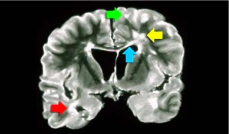
The diagnostic criteria for MS in conjunction with MRI observations with clinical and other para-clinical tools were introduced in 2001. The diagnosis of MS requires elimination of more likely diagnoses and demonstration of dissemination of lesions in space and time. The McDonald criteria shown in Figure 3.4 for MS were recommended in 2001 [McDonald 2001] by an international panel and revised in 2005 [Polman 2005] and 2010 [Polman 2011]. The McDonald criteria take into account the clinical presentation and MRI. When a patient experiences two or more episodes with clinical evidence of two or more neurological deficits, there is no need for additional requirements to make the diagnosis of MS, because there is dissemination in space and time. In all other cases, which are less than two episodes or less than two clinical significant lesions, there arises a need for MRI to fulfill the diagnostic criteria by demonstrating dissemination in space, in time or both. The McDonald criteria are quite specific pertaining to the fact that use of MRI to diagnose MS is only utilized when patient is screened for MS. Typical types of lesions which are suggestive of MS are shown in Figure 3.5. An involvement of the temporal lobe is shown by the red arrow, the green arrow indicates juxtacortical lesions
Figure 3.4: McDonald criteria for MS. Courtesy:http://www. radiologyassistant.nl/
touching the cortex, involvement of the corpus callosum is depicted as blue arrow and periventricular lesions touching the ventricles. The lesions in the deep white matter which are non specific to MS are shown by yellow arrow.
Figure 3.5: Coronal PD image of a brain specimen with MS involvement. Courtesy: http://www.radiologyassistant.nl/
3.2.3
MS Lesions (MSL)
Several MR sequences are necessary in order to detect the MS lesions. These lesions are classified into three subtypes of MS lesions depending upon the peculiar intensity characteristics they possess on respective sequences. They are Active/Gd-Enhancing lesions, black-holes and T2-w as shown in Figure
3.3. Conclusion 21 • T2w lesions: These lesions exhibit a hyper intensity profile compared to normal-appearing white matter on T2w, PDw and FLAIR sequences. They may be iso- or hypo-intense in T1w images. T2w lesions are not clinically specific and can result from inflammation, edema, demyelina-tion, or axonal loss. New contrast-enhanced lesions with hyper intensity also corroborate for the same location with a hyperintense lesion on T2-weighted images. These new T2 hyperintense lesions tend to reduce in size over time and their intensity decreases because of tissue repair [Meier 2007b, Meier 2007a].
• Gd Enhancing lesions: Longitudinal and cross-sectional MRI studies have demonstrated that formation of new MSL can strongly be linked with a focal area of contrast enhancement on T1-weighted images obtained after Gd injected intravenously. Typically, it can be observed in patients with RR or SP MS [Lassmann 2008]. This enhancement correlates with altered blood brain barrier permeability in the setting of acute perivascular inflammation and enables differentiation between acute, active lesions and chronic, inactive ones. In Figure 3.6, FLAIR image shows multiple focal demyelinating lesions that are hyperintense relative to the normal appearing brain tissue. After contrast administration, some of the lesions are hyperintense on T1-weighted images, indicating increased permeability of the bloodbrain barrier, a feature that distinguishes acute from chronic demyelinating lesions. The Gd enhancement varies in size and shape, and usually lasts from a few days to weeks, with an average duration of 3 weeks (97 % of lesions enhance during less than 2 months)
[Cotton 2003]. Lesions which are new and increase in size are classified
as active lesions.
• Black Holes: A T1-weighted MRI scan shows black holes which are suggestive areas of permanent axonal damage. These are hypo-intense lesions because of their dark intensity profile. To be a candidate for black hole, a T1-w lesion should not enhance with gadolinium and should generally be persistent for at least several months.
3.3
Conclusion
The origin and evolution of MS are still not well understood, and numerous studies have been conducted to evaluate its evolution and its influence on neighboring brain structures. Nowadays, a strong emphasis is put on early detection to slow down the disability and disease.
Figure 3.6: Conventional magnetic resonance imaging in multiple sclerosis. FLAIR (left) and gadolinium-enhanced T1-weighted (right) sequences. Cour-tesy: [Rovira 2013]
Figure 3.7: Example of MS lesions on MRI. From left to right: FLAIR, PDw, Gd-enhanced T1w, T1w, and T2w images. Several types of MS le-sions can be observed: Enhancing lele-sions (blue), lele-sions visible only on T2w (green), black holes (red). In Gd-enhanced T1w and FLAIR images, multi-ple bright regions are observed that may be mislabeled as lesions. courtesy: [Garcia-Lorenzo 2013]
of the brain tissues, which may lead to the discovery of putative biomark-ers of disease evolution. Nowadays, clinical trials use the total lesion load in conventional images. In the next chapter, we will focus on the state of the art methods of MS lesion segmentation and detection on conventional MRI.
Chapter 4
MS Lesions Segmentation
Contents
4.1 Introduction . . . 23 4.2 Manual Segmentation . . . 25 4.3 Semi-Automatic Segmentation. . . 25 4.4 Automatic Segmentation . . . 26 4.4.1 Multi-Sequence Information . . . 274.4.2 Unsupervised approaches for MS lesions segmentation . 28
4.4.3 Supervised Approaches. . . 29
4.4.4 Gd-Enhancing Lesion Detection . . . 30
4.4.5 Miscellaneous Approaches . . . 31
4.5 Performance Metrics . . . 32
4.5.1 Publicly Available Resources . . . 35
4.6 Challenges . . . 35
4.1
Introduction
MRI is playing an increasing role in the scientific investigation and clinical management of MS. Conventional MRI sequences are highly sensitive for detecting brain pathologies, like MS lesions and can provide quantitative assessment of inflammatory activity and lesion load. Quantitative MRI provides assistance for a large variety of applications, e.g. to predict brain lesion load and monitoring, longitudinal studies of cognitive aging [Bakshi 2008], or even the analysis of the fetal brain development [Rousseau 2013]. MRI achieves a great tissue contrast enabling the distinction between brain tissues; namely gray matter (GM), white matter (WM), and the cerebrospinal fluid (CSF).
Detecting and localizing MS lesions in MRI is a hard task and generally requires an expert neurologist or radiologist. The detection process is also time consuming and includes some subjectivity in interpreting the images. It
requires multi-sequence intensity fusion, deep anatomical knowledge and solid spatial awareness. MS lesions do not exhibit peculiar shapes and geometries. They possess nodular and oval like, ring to hole like shapes. Consequently, the MS Lesions detections performed by different experts can vary in the number and size of MS Lesions identified. Consequently, the MS Lesions detection performed by different experts can vary in the number and size of MS Lesions identified. As statistics reveal that eye fatigue is commonly encountered problem with radiologists as they visually need to inspect copious imaging data.1 This becomes more prevalent when the imaging modalities grow
dramatically. Due to the volume overload and constrained clinical information available as part of imaging studies, there may be room for diagnosis errors. Radiologists are a scarce resource in many countries. Therefore, it is of paramount importance to reduce the burden of data to be investigated by radiologists. To alleviate this problem, many computer assisted methods have been proposed for diagnostic interpretation of medical imaging datasets guided by clinical knowledge. These methods have the advantage to be consistent and repeatable, although they do not always achieve results as good as manual expert annotations.
In spite of these clear challenges, MS lesion segmentation research has made notable strides within the last decades. The techniques take different approaches to the problem of MS Lesions segmentation and consist of compre-hensive frameworks made of several steps, including pre- and post-processing. Recent advances have shown the feasibility of learning accurate models for detecting MS lesions. Adapting various methodologies from different streams of science, researchers are making efforts for detecting MS lesions within realistic settings. As a part of the effort, various standardized benchmark databases and MS lesion detection challenges have been developed. While a numerous studies have been done, they all must make a few common choices: how will the MR images be represented? using that representation, how is a model learned? Given a new MR image, how is detection carried out? This chapter reviews state-of-the-art of strategies for MS lesions detection/segmentation methods with the aim of pointing out their strengths and weaknesses which in turn explains the rationale behind the proposed methods for this thesis. Further, it concludes with a discussion of the techniques as well as the perspectives on future improvements.
There are three main types of segmentation approaches depending on the user intervention: manual, semi-automatic, and automatic.
4.2. Manual Segmentation 25
4.2
Manual Segmentation
The first method to delineate MSL is manual segmentation. An expert rater examines different MR sequences to identify the lesion voxels. Unfortunately, the manual process is time consuming and somewhat subjective. Different experts (inter-rater variability) and even the same expert (intra-rater variabil-ity) may therefore provide different segmentations for the same data. Even so, manual annotations are considered the best results available and usually serve as the baseline for evaluating other methods. The expert segmentations can be considered as a silver standard since they are not perfect representations of the ground truth but provide the best estimates available. Automatic meth-ods provide some assistance to MSL segmentation. Where experts can have difficulty in infusing multi modal MRI information, well designed frameworks can efficiently blend this data. As a result, it is interesting to pursue the development of semi-automatic and automatic lesion segmentation methods.
4.3
Semi-Automatic Segmentation
In order to reduce the inter- and intra- rater variability in segmentation of MSL, several semi-automatic methods have been developed. Semi automatic techniques need some human input as the prior knowledge for additional automatic processing steps. This knowledge could be an input in the form of focused region of interests (fROI) or a coarse-grained selection of object of interest. Though semi-automatic methods can relieve some of the work from radiologists, they do require some human interaction. A method based on prior knowledge with fuzzy logic is presented in [Horsfield 2007]. The prior knowledge resides in the form of probabilistic feature distribution and feature size maps, in a standard anatomical space. The fuzzy affinity between pixels is modified to capture this information. Here, the user is required to identify each lesion with a mouse click, to provide a set of seed pixels. The algorithm then grows the features from the seeds to define the lesions as a set of objects with fuzzy connectedness above a preset threshold. Ruben et al. [Cárdenes 2003] proposed the technique for interactive segmentation. It consists of three steps. First, a KNN classifier is applied to classify brain tissues in CSF, GM, WM and MS lesions on template based on user input. The second step concerns the detection of MS lesion which is done by computing a fast distance transformation in conjunction with intensity information on template. Last, a connected component technique is used to refine the voxels detected as MS lesions. Another approach proposed by [Derraz 2010] was segmentation based upon Active Contour Model and statistic prior knowledge of MS lesions
in fROI within MRI. In particular, the user selects coarse fROI that encloses potential MS lesions and a sufficient background of the healthy White Matter tissues (WM). Texture features corresponding to Normal Appearing Brain Tissues and MS lesions were incorporated to achieve final segmentation. Graph Cuts (GC) algorithm is a method for finding the maximum a posteriori (MAP) estimate of a binary image [Boykov 2006]. The method treats the image like a flow graph with two nodes, the source and the sink. The source represents the object class in the image, in this case the lesions. The sink represents the background: the NABT. The other nodes of the graph are the image voxels. A network of weighted and directed edges connects the nodes in the graph. The GC makes use of regional and voxel-neighborhood information to differentiate between the two classes. The MAP estimate corresponds to the maximum flow through the node network. The result is two sets of strongly connected nodes that correspond to the MSL and NABT [Biediger 2014]. Some authors
[Lecoeur 2009] proposed to use GC with spectral gradient and multi-sequence
MRI for lesions segmentation. All these methods need seed points defined by the user.
In any case, the automated step of the framework is highly dependent on the quality of the input. Since they require some level of user intervention, the semi-automatic methods may not cater the needs of large patient studies because they are still time consuming and tedious for the user.
4.4
Automatic Segmentation
Automatic methods require no user intervention. The comprehensive surveys
of [Lladó 2012b, Garcia-Lorenzo 2013] provide the different types of
segmen-tation frameworks. In general, there are three main types of fully automated segmentation schemes: data guided methods, learning based methods, and statistical methods. The data dependent methods use thresholding and region growing to segment the lesions in an image, like the watershed and graph cut methods. The learning based methods require a training set and some feature extraction. These methods learn the characteristics of lesions and then classify based on discriminative learning approaches. The statistical meth-ods involve estimations of probability density functions of intensity of voxels. These methods are based on inference methods with some neighborhood or classification examples and include probabilistic graphical models and support vector machines. All have pros and cons in their use and the results they provide. Figure 4.1 shows the broad range of methods based on supervised and unsupervised techniques. It helps to take a glimpse on the huge literature of automatic MSL segmentation.
4.4. Automatic Segmentation 27
Dictionary Learning/Sparsity Conditional Random Fields
Decision Forest,SVM Manifold Learning Generative Models based on EM like Framework Modeling MSL as an extra clus-ter,outliers Graphical Models:MRF, graph cut Atlas based frameworks Fuzzy Logic Approaches Super vised Framew orks Unsupervised Frameworks MSL Segmentation MSL Segmentation
Figure 4.1: The various approaches for MSL detection based on their charac-teristics
4.4.1
Multi-Sequence Information
By and large, a typical MRI under consideration can be a representation in one or combination of the four possible sequences, namely, PD-w, T1-w, T2-w, and FLAIR. The objective is to find the various MS lesions through the use of these sequences. Prior works in the context of MS lesion detection deal with either single or multi-sequence approaches i.e. the use of a single MRI sequence or combination of several MRI sequences respectively. Single-sequence approaches are mainly used to segment the brain tissues.
For instance, T1-w sequences are widely used for this purpose, since they show the best contrast between the three main brain tissues: WM, GM and CSF. Another example of the single-sequence approach is the segmentation of MS lesions using just the FLAIR sequence [Khayati 2008]. The multi-sequence approaches, on the other hand, use at least two sequences. One of the benefits of using more than one of the different MRI sequences is that it increases the intensity feature space, producing a better discrimination between brain tissues. Garcia et al. [Garcia-Lorenzo 2011] propose a typical example of this approach which uses T1-w, T2-w, and FLAIR to detect MS lesions. There is also a method to make use of the initial single sequence approach to be used as a basis for further analysis in multi-sequence context to obtain the final lesion map. For example, the T1-w sequence could be used to influence a multi-sequence approach using T2-w and PD-w.
4.4.2
Unsupervised approaches for MS lesions
segmenta-tion
Generative methods remain the popular choice for MS lesions segmentation. It consists of tissue classification with an expectation maximization (EM)
[McLachlan 2008] algorithm. The approaches based on EM typically modify
the EM algorithm to factor it into their classification methodologies.
These EM derived algorithms are typically modified to be robust against lesion affected regions. The outcome is then parsed in order to detect outliers which, in this case, coincide with MS lesions. In their seminal work, Van Leemput et al. [Leemput 2001] developed a framework for segmentation of MS lesions based on multi-sequence information. It relies on classification based upon the intensity information of tissues using a stochastic model for normal brain sequences. Furthermore, it models MS lesions as outliers. It removes MR field inhomogeneities and incorporates contextual information in the clas-sification of lesions using a Markov random field (MRF). Another similar work proposed by Garcia et al. [AïtAli 2005, Garcia-Lorenzo 2011] incorporates a tissue classification methodology based on a model of intensities of the normal appearing brain tissues. In order to estimate the model parameters, a trimmed likelihood estimator [Neykov 2007] is initialized with a hierarchical random approach in order to be robust to MS lesions and other outliers present in MR sequences. An iterative scheme of recursive EM was then used to compute this estimator. It is a robust algorithm using 3D+t MR data to segment MS lesions over time in a standardized clinical protocol. In the last step, refinement of the segmentation was done using both the Mahalanobis distance of intensity of WM voxels and prior information coming from clinical knowledge on lesion appearance across sequences. Another approach considers the EM with a partial volume model among tissue classes in conjunction with a Mahalanobis distance thresholding which detects MS lesions [Dugas-Phocion 2004]. A post-processing morphological operation was performed to refine the segmentation from regions of interest in order to improve the classification performance [Souplet 2008]. Freifeld et al. [Freifeld 2009] proposed a Constrained Gaussian Mixture Model (CGMM) technique based on a mixture of multiple spatially oriented Gaussians per tissue. The intensity of a tissue remains unchanged over the entire set of Gaussians for that tissue. MS lesions are modeled explicitly as an extra class by GMM in addition to healthy tissue classes. MS lesions were detected by estimation of parameters for outlier class followed by the refinement of lesion contours based upon the probability-based curve evolution technique. Rather than estimating the distribution of lesions, [Harmouche 2006] proposed to cast lesions as a separate class. It is based on unsupervised Bayesian frame-work. It models the different intensity distributions for different tissues of
4.4. Automatic Segmentation 29 the brain. MS lesion detection was performed using posterior probabilities along with entropy based features. Khayati et al. [Khayati 2008] classified MS lesions voxels based upon adaptive mixtures method (AMM) and a MRF model from a FLAIR sequence. The intensity of each lesion voxel is modeled as a linear combination of intensities related to the normal and pathological tissues. Applying an optimal threshold, the voxels with new intensities are primarily classified into two stages: chronic and acute MS lesions. Finally, the acute lesions are classified into two new stages based upon their activities, early- and recent acute. Schmidt et al. [Schmidt 2012] developed MS lesion detection using three-dimensional (3D) gradient echo (GRE) T1-w and FLAIR sequences. It initially classifies the three tissue classes of CSF, WM and GM from the T1-weighted image. In the subsequent stage, the FLAIR intensity distribution of each tissue class is taken into account to detect outliers, which are called lesion beliefs. The neighboring voxels in lesion belief maps are analyzed and assigned to lesions. This is done for all voxels that are associ-ated with the MS lesions. A fuzzy C-Means algorithm was also investigassoci-ated
[Aymerich 2010a,Aymerich 2010b]. These approaches pursue the grouping of
voxels into a number of clusters, which maximize inter-cluster variability while minimizing intra-cluster variability. Rather than a crisp or hard classification, the fuzzy approach establishes the degree to which a pixel belongs to a given cluster. In this way, a voxel can belong to more than one cluster to varying degrees.
4.4.3
Supervised Approaches
Machine learning plays an essential role in the medical imaging field, including computer-aided diagnosis, image segmentation, image registration, image fusion, image-guided therapy, image annotation, and image database retrieval. The objects such as lesions and organs may not be represented accurately by a simple models; thus, medical computer vision requires learning from training examples. One of the most popular uses of machine learning is classification of objects such as tissues into certain classes (e.g., pathological or non-pathological) based on input features obtained from segmented object candidates.
Support vector machines (SVM) [Vapnik 1995] is a popular and widely used supervised learning algorithm. It has also been used in the context of MS lesion detection [Fiot 2008, Abdullah 2011]. The method extracts intensity values as features from examples of lesion and non-lesion voxels. It then attempts to divide the two classes by dividing the hyper-plane of features in a discriminating fashion. While there are many possible dividing planes, the method seeks the plane with the widest margin between classes. SVM in conjunction with
various kernels facilitates the non-linear classification by projecting the features into Reproducible Kernel Hilbert Space (RKHS) [Shawe-Taylor 2004]. One problem with the SVM approach in MS lesion detection is the imbalance between class representations. In general, the number of voxels that represent normal brain tissue far exceeds the number of voxels that represent MS lesions. This can lead to the over fitting of the classifier.
Ensemble learning is the process by which multiple models, such as classifiers or experts, are strategically generated and combined to solve a classification problem. Two ensemble learning approaches were studied in the context of MS lesions detection. In a first study, Geremia et al. [Geremia 2010] proposed to build a discriminative random decision forest framework to provide a voxel-wise probabilistic classification of the image. The method uses multi-sequence data (T1, T2, FLAIR), a prior knowledge of tissue classes and long-range spatial context to distinguish lesions from healthy tissues. The authors also utilize a symmetry feature, which takes into account the fact that MS lesions tend to develop in an asymmetric way compared to healthy brain which remains approximately symmetric with respect to the mid-sagittal plane. In a second study, [Wels 2008] developed a framework based on the probabilistic boosting trees technique. It incorporates the context of a voxel under consideration and its transformation into feature space of an over-complete set of Haar-like features. This information establishes the class specific characteristics. A discriminative model for voxel classification was developed based upon boosting within a tree structure. It consists of selection and combination of most discriminative features which are established recursively in cascade. Consequently it yields posterior probabilities for voxels in learning phase. The final segmentation was obtained after refining the preliminary result by stochastic relaxation and a standard level set approach.
Further, a multi-scale segmentation can be combined with discriminative classification to take into account regional properties [Akselrod-Ballin 2006]. It relies on a combination of segmentation by Soft Weighted Aggregation (SWA), a rich feature vocabulary describing the segments, and a decision tree-based classification of the segments. Then, successively selecting and combining the most discriminative features during ensemble, the overall procedure was able to learn in terms of posterior probabilities. Beyond the information introduced via the spatial prior atlases, these methods are limited in their ability to take advantage of long-range spatial context in the classification task.
4.4.4
Gd-Enhancing Lesion Detection
All approaches mentioned above work on the conventional MRI sequences without contrast agent. However, it must be noted that in the context of active
![Figure 3.2: High regional prevalence of MS across the world. Courtesy : [Pietrangelo 2015]](https://thumb-eu.123doks.com/thumbv2/123doknet/11344485.284322/24.892.213.654.169.474/figure-high-regional-prevalence-ms-world-courtesy-pietrangelo.webp)