Science Arts & Métiers (SAM)
is an open access repository that collects the work of Arts et Métiers Institute of Technology researchers and makes it freely available over the web where possible.
This is an author-deposited version published in: https://sam.ensam.eu Handle ID: .http://hdl.handle.net/10985/15789
To cite this version :
Christine MUTH-SENG, Laure-Lise GRAS, ANTHONY ROUX, Sébastien LAPORTE - Modelling of fascia lata rupture during tensile tests via the discrete element method - Computer Methods in Biomechanics and Biomedical Engineering - Vol. 20, n°sup1, p.147-148 - 2017
Any correspondence concerning this service should be sent to the repository Administrator : archiveouverte@ensam.eu
Modelling of fascia lata rupture during tensile tests via the discrete element
method
C. Muth-senga, L. L. Grasb, A. Rouxa and S. Laportea
aarts et métiers paristech, institut de Biomécanique humaine george Charpak, paris; biFsttar, lBmC umr_t9406univ lyon, université Claude
Bernard lyon 1, lyon, France
KEYWORDS Fascia lata; discrete element method; rupture
1. Introduction
Musculoskeletal models are often used to better under-stand the behaviour of the many components of the body, to predict injuries and design safety devices. These models become increasingly more detailed by including connec-tive tissues. However, their implementation is often chal-lenging due to the small amount of data regarding their mechanical properties.
Fascia lata is a connective tissue wrapped around the muscles of the thigh. Histological studies on goat fascia lata (Pancheri et al. 2014) have shown its heterogeneous structure composed of two layers of collagen fibres con-nected by a proteoglycan matrix. Both layers have a spe-cific fibre orientation, and are not orthogonal.
The discrete element method (DEM) makes it possible to model complex structures by discretizing them into simple elements, associated to a mass, and cohesive bonds, such as springs. This allows for a better understanding of the relationship between micro and macroscale prop-erties of materials. While this method is usually used to model composite materials, it has been adapted to model biological materials, such as the muscle-tendon complex (Roux et al. 2016).
The aim of this study is thus to reproduce numerically the macroscopic behaviour of the fascia lata submitted to a tensile test using DEM.
2. Methods
2.1. Geometrical construction of the model
GranOO (Granular object oriented, www.granoo.org) software was used to build the fascia lata model using microstructural dimensions extracted from the micro-scopic view and histological sections made by Pancheri
et al. 2014. The microstructure of fascia lata was discre-tized using spherical elements linked with spring bonds arranged in two layers. Spherical discrete elements were put at each crossing of fibres, with an interval of 143 μm along the longitudinal orientation and an interval of 132 μm along the transverse orientation. As the longitudi-nal fibres are larger and arranged on a different layer than the transverse ones, elements on the longitudinal layer have a diameter of 162 μm, while those on the transverse layer a diameter of 75 μm.
The collagen fibres and the proteoglycan matrix were modelled as follows. Each element is linked by a spring to its 17 closest neighbours: the eight neighbouring ele-ments on the same layer and the nine eleele-ments facing them on the opposite layer. Among these 17 spring bonds, two of them model a portion of collagen fibre, linking the element to its neighbours along the corresponding fibre orientation, while the 15 other bonds model the proteo-glycan matrix (Figure 1(a)).
2.2. Mechanical properties
Each spring stiffness was computed from the Young mod-ulus, cross-sectional area and initial length of links. The Young modulus of the matrix was the same as that used by Roux et al. to model the extracellular matrix in muscle, 0.1 MPa. A Young modulus of 44 MPa for collagen fibres was computed using the apparent Young modulus of fascia lata assessed by Henderson et al. (2015) and the previously mentioned modulus for the matrix via an estimation of fibre-matrix ratio in a section. Longitudinal springs are given a broader cross-section than transverse springs and are thus stiffer.
The strain limit causing the failure of the springs for collagen fibres was set at 0.4, corresponding to some of
The biggest advantage of using DEM is to assess the rupture phenomenon at a microstructural scale. Failure occurred following the delamination between the fibres once the shear stress was sufficient to tear them from one another.
4. Conclusions
While the results of these simulations are still prelimi-nary, they are encouraging as they seem to show a similar behaviour as the one found experimentally. Use of DEM is promising and our next step is to analyse the influence of various microstructural parameters, such as fibre orienta-tion or mechanical properties distribuorienta-tion, on the global mechanical properties of fascia lata. The model should be able to be adapted to other fasciae as well, but it still needs to take into account slack length of the fibres and more accurate material properties.
References
Henderson ER, Friend EJ, Toscano MJ, Parsons KJ, Tarlton JF. 2015. Biomechanical comparison of canine fascia lata and thoracolumbar fascia: an in vitro evaluation of replacement tissues for body wall reconstruction. Veterinary Surgery. 44:126–134.
Pancheri FQ, Eng CM, Lieberman DE, Biewener AA, Dorfmann L. 2014. A constitutive description of the anisotropic response of the fascia lata. J Mech Behav Biomedl Mater. 30:306–323.
Roux A, Laporte S, Lecompte J, Gras L-L, Iordanoff I. 2016. Influence of muscle-tendon complex geometrical parameters on modeling passive stretch behavior with the discrete element method. J Biomech. 49:252–258.
Shen ZL, Dodge MR, Kahn H, Ballarini R, Eppell SJ. 2008. Stress-strain experiments on individual collagen fibrils. Biophys J. 95:3956–3963.
the lowest strain limit to rupture in literature (Shen et al.
2008). Moreover, due to the lack of data on the strain limit of the matrix, it was once again set at the same value used by Roux et al. (2016), i.e. 0.3.
2.3. Numerical tensile test simulation
Numerical simulations were carried out to replicate the tensile tests up to rupture performed by Henderson et al.
2015; The model dimensions were thus taken according to their samples, meaning 3 × 1 cm rectangular samples. One extremity of the model was fixed, allowing no dis-placement, while a linear displacement was imposed to the other one at a quasi-static strain rate (Figure 1(b)). The force induced in the springs was measured at each step. 3. Results and discussion
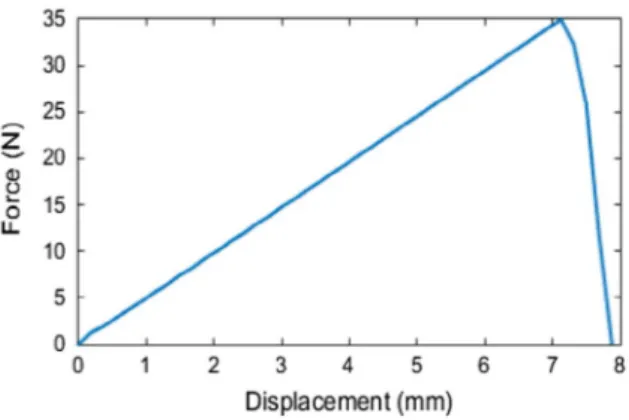
Evolution of the force induced in the springs with regards to the imposed displacement is provided on Figure 2. This curve shows that fascia lata is a rather brittle material, hardly showing any yield portion, much like the results obtained by Henderson et al. in their study of canine fascia lata. However, as the current model does not take into account any slack for the fibres and considers them ready to be stretched, the curves do not show any toe/heel region, which is characteristic of most biological materi-als. Furthermore, while the force at rupture of 35 N was about 2/3 lower than the one found experimentally by Henderson et al., the maximum displacement of 7 mm is of the same order of magnitude. The lack of data on the mechanical properties of the proteoglycan matrix led to using the properties of similar tissues and may explain this underestimation of the force.
Longitudinal fibre Transverse fibre Matrix (a)
(b)
Figure 1. (a) illustration of all bonds for the yellow element and the neighbouring fibres. (b) model of fascia lata sample and boundary conditions. the arrows indicate fibre orientation.
Figure 2. Force-displacement curve from uniaxial tensile test simulation along longitudinal fibres.