The Role of NHL-2 in Regulating C. elegans P granule
Function
par Rana Amini
Département de Pathologie et Biologie Cellulaire
Faculté de Médecine
Mémoire présenté à la Faculté des études supérieures en vue de l’obtention du grade de Maîtrise (M. Sc.)
en Pathologie et Biologie Cellulaire Option biologie du cancer
Faculté des études supérieures et postdoctorales
Ce mémoire intitulé:
The Role of NHL-2 in Regulating C. elegans P granule Function
Présenté par : Rana Amini
a été évaluépar un jury composé des personnes suivantes :
Dr. Marc Therrien, président-rapporteur Dr. Jean-Claude Labbé directeur de recherche Dr. Katherine Borden membre du jury
Abstract
Asymmetric cell division is a process that enables stem cells to simultaneously self-renew and generate progeny committed to differentiation. For instance the totipotent and immortal germ lineage in C. elegans is a stem cell lineage that contains ribonucleoprotein organelles called P granules. P granules are present specifically in germ cells and have been proposed to function as germ line determinants. Interestingly, various RNP granules, such as P bodies that regulate post-transcriptional control have been also observed in all organisms. Nevertheless, the precise function of C. elegans P granules remains elusive. Recently our lab showed that NHL-2, a C. elegans homologue of Drosophila Mei-P26, localizes to P granules in early embryos and plays a role in asymmetric cell division and cell polarity. All P granules contain NHL-2, raising the possibility that NHL-2 plays a role in regulating P granule biogenesis and function. We investigated this possibility by imaging and quantifying the intensity of PGL-1, a core component of P granules, in fixed embryos. This analysis revealed that there is a reduction in: 1) the number of P granules and 2) the mean fluorescence intensity (MFI) and total fluorescence intensity (TFI) of PGL-1 staining in nhl-2 null mutant embryos compared to wild type. Our further analysis of nhl-2 loss of function demonstrates that NHL-2, similar to P granule core components such as DEPS-1, GLH-1 and PGL-1, is required for proper germ cell proliferation and fertility at elevated temperature. One aspect of the nhl-2 loss of function phenotype is that nhl-2 mutants are distributed in different groups based on both their P granules number and PGL-1 intensity: one population has wild type PGL-1 intensity while the second group has severely reduced amounts of PGL-1. Strikingly, such variability is observed even in the sterility phenotype of nhl-2 mutants at elevated temperatures. Taken together, our results suggests that NHL-2 loss of function disrupts germ cell proliferation as well as P granule formation or stability during early stages of germ cell precursor development and that NHL-2 function may be partially redundant with other regulators of P granule stability.
Résumé
La divison cellulaire asymétrique est un processus essentiel qui permet aux cellules souches de s’auto-renouveller et de produire une cellule fille destinée à la différenciation. La lignée germinale de C. elegans, totipotente et immortelle, est une lignée de cellules souches qui contient des organites ribonucléoprotéiques appelés granules P. Au cours du développement ces derniers sont toujours localisés spécifiquement dans les cellules précurseurs de la lignée germinale, suggérant qu’ils sont des déterminants de la lignée germinale. De façon intéressante, des granules ribonucléoprotéiques, comme les P bodies impliqués dans le contrôle post-transcriptionnel, ont été observés chez tous les organismes. Néanmoins, la fonction précise des granules P de C. elegans est inconnue. Récemment, notre laboratoire a montré que NHL-2, un homologue de Mei-P26 de Drosophile, colocalise avec les granules P dans des embryons précoces et joue un rôle dans la division cellulaire asymétrique et dans la polarité cellulaire. Tous les granules P contiennent NHL- 2, ce qui nous a mené à poser l’hypothèse que NHL-2 régule la biogenèse et la fonction des granules P. Nous avons testé cette hypothèse par imagerie et quantification de l'intensité de PGL-1, un composant essentiel des granules P, dans des embryons fixés. Nos résultats montrent que dans des embryons mutants pour nhl-2 il y a une réduction du nombre de granules P, de l'intensité de fluorescence moyenne (IFM) et de l'intensité de fluorescence total (IFT) de PGL-1. Une analyse plus poussée a montré qu'il existe deux populations distinctes d’embryons mutants pour nhl-2 : l’une présente une intensité de PGL-1 comparable à celle d’une population sauvage alors que le second groupe présente une forte réduction des quantités de PGL-1 et est comparable à des mutants pour pgl-1. Cette variabilité est aussi observée dans le phénotype de stérilité de nhl-2 mutant à des températures élevées. Globalement, nos résultats suggèrent que la perte de fonction de NHL-2 perturbe la prolifération des cellules germinales ainsi que la formation et/ou la stabilité des granules P au cours des étapes précoces du développement des précurseurs de la lignée germinals. D’autre part, ils suggèrent que la fonction de NHL-2 pourrait être partiellement redondants avec les autres régulateurs de la stabilité des granules P.
Table of Contents
Abstract... i
Résumé ... ii
List of the Figures………vi
Acknowledgments………ix
1. Introduction ... 1
1.1. Asymmetric cell division: ... 2
1.2. Polarity is required for asymmetric cell division: ... 3
1.3. Asymmetric Cell Division and Cell Polarity in Development and Disease: ... 4
1.4. Germline development in C. elegans ... 7
1.4.1. Germ line specification occurs during embryogenesis ... 7
1.4.2. Postembryonic development of the germline... 9
1.5. Characteristics of the Germline... 11
1.5.1. Germ cells behave like stem cells: ... 11
1.5.2. Germ Cells are in a Transcriptionally Repressed State:... 11
1.5.3. Germ Granules: ... 12
1.6. C. elegans P granules ... 15
1.6.1. P granule Localization and Physical Nature ... 15
1.6.2. P granule composition... 18
1.6.3. P granule assembly pathway: ... 20
1.6.4. What are the functional consequences of loss of P granule Components? ... 21
1.7. P granules and Translational Control... 23
1.7.1. P granule localization and composition: ... 23
1.7.2. Links between different RNP granules in C. elegans: ... 25
1.8. NHL-2 and Translational Control ... 28
2. Materials and Methods ... 33
2.1. Strains and Alleles... 34
2.2. C. elegans techniques- Dissection, Immunofluorescence and Microscopy... 35
2.2.1. Antibodies and Markers ... 35
2.2.2. Immunofluorescence of C. elegans embryos ... 36
2.3.3. Imaging ... 38
2.3. Sterility Assays: ... 41
3. Results ... 42
3.1. The number of P granules is decreased in nhl-2 mutant embryos ... 43
3.2. PGL-1 Fluorescence Intensity is significantly reduced in nhl-2 mutant embryos .... 45
3.3.The underlying mechanisms of P granule reduction... 50
3.3.1. NHL-2 loss of function phenotype is not due to P granule fusion... 51
3.3.2. nhl-2 loss of function causes a general decrease in PGL-1 intensity levels at 2-cell stage embryos ... 53
3.4. NHL-2 is required for fertility at elevated temperatures... 55
4. Discussion... 58
4.1. NHL-2 loss of function and P granules Defects... 59
4.2. NHL-2 might function redundantly with other components of P granule assembly pathway ... 60
List of the Figures
1. IntroductionFigure 1.1 : Polarity establishment and asymmetric cell division in C.
elegans………....4
Figure 1.2 : Germ cells are set apart from somatic cells during early embryogenesis in C. elegans………8
Figure 1.3 : The C. elegans gonad………...10
Figure 1.4 : Germ granules are perinuclear in most organisms………...………14
Figure 1.5 : P granule segregation in C. elegans……….16
Figure 1.6 : P granules are localized by dissolution/condensation mechanisms………….17
Figure 1.7 : NHL-2 is a member of the conserved TRIM-NHL- family of proteins ……..30
Figure 1.8 : NHL-2, PAB-1 and TIA-1 granules have similar distribution patterns……...32
2. Materials and Methods Figure 2.1 : C. elegans dissection for immunofluorescence………36
Figure 2.2 : P granule quantification………39
3. Results
Figure 3.1 : NHL-2 is important for regulating the P granule stability in early embryos..44
Figure 3.2 : Different populations of nhl-2 mutants……….. 45
Figure 3.3 : nhl-2 mutants have less PGL-1 intensity……….48
Figure 3.4 : Different populations of nhl-2 mutants………...49
Figure 3.5 : Two models for P granule reduction in nhl-2 mutants………51
Figure 3.6 : P granules do not fuse in the absence of NHL-2……….52
Figure 3.7 : A general reduction of PGL-1 intensity levels in nhl-2 mutants……….54
Figure 3.8 : Removing NHL-2 and three other CeBrats affects fertility at 26°C…………56 Figure 3.9 : Normaski images of wild type and mutant hermaphrodites grown at 26°C…57
To my mother for her endless love and support
Acknowledgments
I would like to deeply thank my research director, Dr. Jean-Claude Labbé, for accepting me in his laboratory, for his presence, support, exceptional understanding and of course for his knowledge and his advices.
Thanks to all current and previous members of Labbé laboratory, especially Dr. Vincent Hyenne and Dr. Nicolas Chartier for their helpful advice.
I would also like to thank Dr. Paul Maddox for providing us with CENP-A antibody and access to Delta Vision Microscope.
I am really grateful to Christian Charbonneau, the adviser of the bio-imaging platform in IRIC, for his assistance in microscopy and his patience in teaching me how to work with illustrator.
1.1. Asymmetric cell division:
In contrast to normal cell division, which gives rise to equivalent daughter cells, asymmetric cell division (ACD) is a process by which a mother cell divides to generate two daughter cells that adopt distinct cell fates. In principle, asymmetric cell division occurs through two different mechanisms: extrinsic (induced) or intrinsic (Horvitz and Herskowitz, 1992). In the case of extrinsic or induced ACD, the daughter cells are initially equivalent but signaling between the cells induces a difference, whereas in the intrinsic ACD the difference in two daughter cells is generated by the asymmetrical segregation of intracellular proteins before division. These proteins are subsequently partitioned differentially into the two daughter cells so that they are inherited to only one of the daughter cells, causing that cell to be different from its sibling. Because these proteins determine what becomes of a cell they are called cell fate determinants.
Asymmetric cell division is a conserved mechanism used by different organisms to generate cell diversity during animal development (Horvitz and Herskowitz, 1992). For instance, intrinsic asymmetric cell division is a fascinating property of stem cells that enables them to undergo self-renewal and differentiation at the same time (Morrison and Kimble, 2006). In addition, during Drosophila neurogenesis, neuroblasts divide asymmetrically giving rise to two daughter cells, with different sizes, different cell cycle times and distinct fates. Later the larger cell keeps dividing asymmetrically, whereas the smaller cell called GMC (Ganglion Mother Cell) undergoes differentiation to generate two neurons or glial cells. Another classic example of intrinsic asymmetric cell division is the C. elegans zygote, which divides along its anterior–posterior (a–p) axis to produce a large anterior blastomere (AB) and a small posterior blastomere (P1).
1.2. Polarity is required for asymmetric cell division:
Cell Polarity is a fundamental property of many cells and refers to the asymmetry either in shape or in the content of a cell created through unequal distribution of its molecular components. Cell polarity is essential for various cellular processes such as cell migration. Strikingly, cell polarity plays a critical role in governing asymmetric cell division and therefore for the development of multicellular organisms.
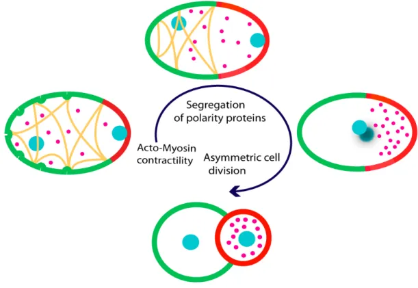
In C. elegans, cell polarity is established by the asymmetric distribution of a group of proteins called PAR proteins (for ‘partitioning defective’). The seven par genes (par-1 to par-6 and pkc-3) in C. elegans encode proteins that are highly conserved across species. PAR-1, PAR-4 and PKC-3 are serine-threonine protein kinases; PAR-3 and PAR-6 are PDZ domain proteins, PAR-2 contains a RING finger domain and PAR-5 is a 14-3-3 protein (Kemphues et al., 1988). In C. elegans, polarization is initiated at fertilization with sperm entry into the oocyte. The sperm defines the posterior end of the zygote and also brings the microtubule-organizing center (MTOC) to the zygote. After a couple of maternal meiotic cycles, the uniformly distributed PAR proteins start to rearrange so that the PAR-1 and PAR-2 are restricted to the posterior cell cortex of the embryo whereas PAR-3, PAR-6 and PKC-3 are enriched at the anterior cell cortex. Polarity along the anterior-posterior axis then influences spindle positioning, leading to the first asymmetric segregation and division of cell fate determinants, including P granules and MEX-5 protein into two different daughter cells (Figure 1.1). The AB cell will mainly form ectoderm, whereas P1 will give rise to the germline, as well as endoderm and some mesoderm. Removing any of the PAR proteins disrupts the establishment of polarity, leading to an abnormal cell division and ultimately to embryonic lethality.
Figure 1.1: Polarity establishment and asymmetric cell division in C. elegans. Acto-myosin network: yellow lines; pronuclei and nuclei: blue, cortical PAR-3/PAR-6/PKC-3: green; cortical PAR-2 and PAR-1: red, P granules: pink. In C. elegans, the cortex contractions that occurs shortly after fertilization, determines polarity by the unequal distribution of a group of conserved polarity proteins or PARs. This anterior-posterior polarity in turn directs the differential segregation of cell fate determinants to opposite ends of the embryo, leading to an asymmetric cell division, after which two unequal cells are formed, each possessing a particular set of molecules and thus a distinct identity.
1.3. Asymmetric Cell Division and Cell Polarity in Development and Disease:
Asymmetric cell division enables stem cells to self-renew and differentiate with a single division; however symmetric stem cell divisions have been also observed during both vertebrate and invertebrate development. For instance, germline stem cells divide
symmetrically in the distal tip of the gonad in C. elegans larvae, whereas they divide asymmetrically during embryogenesis. Indeed symmetric cell division is the strategy that stem cells use in order to increase their population (expansion). However, this unique feature of stem cells, which is essential for the development of organisms, can be deleterious if the balance between asymmetric and symmetric cell divisions becomes defective. While an excess of stem cell self-renewal may lead to tumorogenesis, an excess of differentiation may lead to tissue degeneration and / or tissue aging (Morrison and Kimble, 2006). But what causes the disruption of this balance and deregulation of asymmetric cell divisions?
Cell polarity mechanisms are responsible for regulating the asymmetric cell divisions of stem cells (Wodarz, 2005; Wodarz and Nathke, 2007). Several lines of evidence indicate that disruption of cell polarity is a hallmark of cancer: several cell polarity proteins function either as proto-oncogenes or tumor suppressors (Lee and Vasioukhin, 2008). For instance, the Drosophila polarityproteins Lgl, Dlg and Scrib are potent tumor suppressors. It has been shown that removing any of these proteins results in the growth of tumor-like structures in larval imaginal discs and brains (Bilder, 2004). Moreover, there is clear evidence that aPKC is also a potentproto-oncogene in mammalian cells and is involved in the regulation of cell proliferation as well as tumor development (Regala et al., 2005; Stallings-Mann et al., 2006). In addition, it has been shown that aPKC is involved in the squamous-cellcarcinoma of the head and neck (Cohen etal., 2006) as well as the proliferation of humanglioblastoma cell lines (Donson et al., 2000). It was also shown that overexpression of PKCι, a human homologue of aPKC results in ovarian, lung and colon cancers. Interestingly, tumors with elevated levels of PKCι had lost their polarity demonstrating that PKC could contribute to the development, establishment and maintenance of cell polarity (Eder et al., 2005, Wodarz and Nathke, 2007). Moreover, mutations in LKB1, a human homologue of PAR-4 cause the Peutz-Jeghers syndrome (PJS) by impairing LKB1’s ability to induce cell polarity (Wodarz and Nathke, 2007).
Taken together, these studiesdirectly connect cell polarity pathways to the developmentof cancer in vertebrates and invertebrates.
1.4. Germline development in C. elegans
In sexually reproducing animals, germ lineage is responsible for the development of an organism by transmitting genetic information from one generation to the next.
1.4.1. Germ line specification occurs during embryogenesis
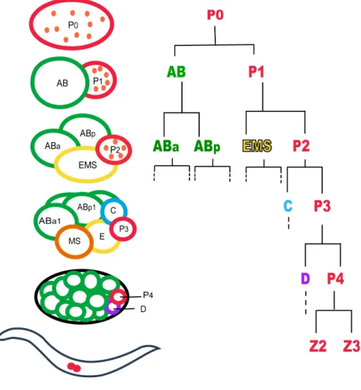
In C. elegans, the germ line is set apart from the soma early in embryogenesis through a series of asymmetric divisions. The zygote (P0) undergoes an asymmetric cleavage in order to give rise to the AB and P1 blastomeres. AB and P1 are unequal in size and fate and are also different in their cell cycle lengths with AB dividing prior to P1. AB then divides to produce somatic descendents while P1 divides asymmetrically to give rise to germ line blastomeres (P2, P3 and P4). P4 is a germline founder cell that divides only once (at the 100-cell stage) during embryogenesis in order to generate the two primordial daughter cells Z2 and Z3. During larval development, Z2 and Z3 begin to divide symmetrically to give rise to the entire germ cells of the adult gonad (Figure 1.2).
Figure 1.2: Germ Cells are set apart from somatic cells during early embryogenesis in C. elegans. The germline precursor cells or P cells (shown in red). Embryos divide asymmetrically to give rise to AB and P1. The AB lineage then loses the germline determinants, P granules (small dots shown in orange) and therefore all her daughters are restricted to form the somatic tisuues of the animal such as hypodermis and neurons: EMS produces the somatic gonad, muscle, the majority of the pharynx, neurons, gland cells and intestine; C produces muscle, hypodermis and neurons; D produces muscle. On the contrary, the germline determinants are transferred to the second daughter cell, P1 that gives rise to all germline blastomeres (P2, P3, and P4). The asymmetric cell division continues so that at L1 two primordial germ cells, Z2 and Z3 are present (shown in red).
1.4.2. Postembryonic development of the germline
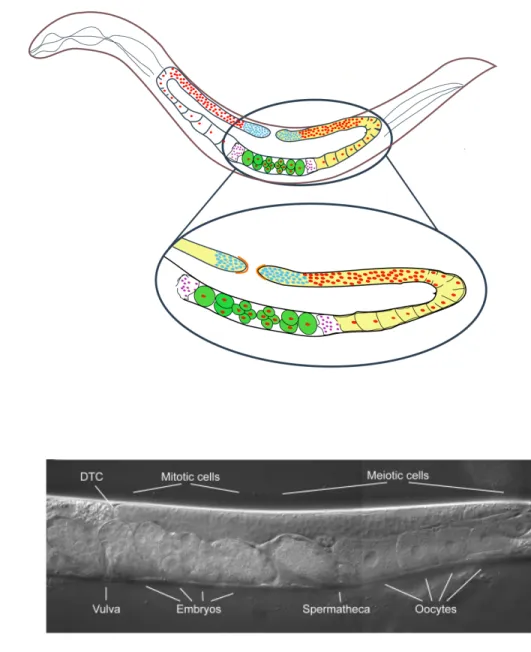
At hatching, the gonad contains four cells: the two germline precursor cells, Z2 and Z3, and the somatic gonad founder cells, Z1 and Z4, which are derived from the MSt lineage. In the middle of L1 larval stage, Z1 and Z4 descendants form the Distal Tip Cells (DTCs) while Z2 and Z3 undergo extensive proliferation to give rise to all the germ cells of the developing gonad. DTCs are located at the distal tips of the two U-shaped gonad arms throughout development in hermaphrodites and at the distal tip of the single J-shaped gonad in males (Kimble and Hirsh, 1979). The adult gonad of hermaphrodite C. elegans has two symmetrical U-shaped tubes consisting of an ovary in a distal arm and an oviduct in the proximal arm, connected through a spermetheca to the common uterus (Figure1.3). The gonad in C. elegans exhibits distal-proximal polarity that corresponds to the development of the germ line from mitosis through meiosis and gametogenesis. The distal tip (mitotic region) of the adult gonad contains a population of stem cells that are mitotically dividing to form a syncitium in the distal gonad arm (Hirsh et al., 1976). The DTC functions as a niche providing the necessary signals that regulate the balance between self-renewal and differentiation that is needed for proper progression through gametogenesis.
Later, during the third larval stage, as germ cells move away from the mitotic region, they enter a transition zone where they progress into different stages of meiotic prophase from prophase I to diakinesis. As germ cells move in the transition zone they exit pachytene in the loop region of the gonad to form oocytes then undergo cellularization. Oocyte maturation occurs in the most proximal end of the gonad arm upon receiving a signal from sperm. Oocytes are then fertilized by stored sperm as they move through the spermatheca into the uterus. After fertilization embryogenesis starts in the uterus and the embryos are then eventually extruded from the uterus. In hermaphrodites when sperm is abundant, oocytes undergo maturation through diakinesis; however, in the absence of
sperm, oocytes are arrested in diakinesis (Dernburg et al., 1998; Francis et al., 1995).
A
B
Figure 1.3: The C. elegans Gonad. (A) Schematic and (B) DIC image of the gonad in wild type hermaphrodite. The gonad has two identical arms, each of which produces first sperm, and later oocytes. The oocytes are fertilized as they move towards the uterus, which is common to both gonad arms. The most distal region of the gonad contains a pool of germline stem cells that continuously undergo mitosis. These cells are capped by the somatic distal tip cell (DTC). As the cells move away from DTC, they enter into meiotic phase; they go into meiosis (transition zone) where they undergo an extended period of pachytene. Germ cells specified to become oocytes exit pachytene in the loop region of the gonad, undergo cellularization, and begin swelling from increased cytoplasmic volume.
1.5. Characteristics of the Germline
Germ cells are the founder cells of all sexually reproducing organisms and must be established in early embryogenesis and then maintained throughout development. Due to their unique role, germ cells possess special properties that are essential for reproduction and species survival. Several defining features that distinguish germ cells from somatic cells are conserved among several species including Drosophila melanogaster, Caenorhabditis elegans and Xenopus laevis.
1.5.1. Germ cells behave like stem cells:
Because the germline is the only lineage to contribute its genetic information to the next generation, it is often referred to as an immortal and totipotent lineage. Totipotency is defined as the ability of a single cell or a cell lineage to generate all the cell types of an organism, including the germline. One unique feature of germ cells in most animals is their ability to divide and give rise to all the differentiated tissues of future generations of multi-cellular organisms. Unlike somatic cells, germ cells undergo meiosis to reduce their ploidy in order to generate gametes. The germ lineage is often referred to as an immortal and totipotent lineage, since at fertilization the haploid gametes unite and give rise to a fully differentiated organism, more germ cells and thus the endless germ line cycle continues from one generation to the next. In contrast to somatic cells that undergo only mitosis and ultimately die with each generation, the germline escapes mortality (Cinalli et al., 2008, Kawasaki et al., 2004).
1.5.2. Germ Cells are in a Transcriptionally Repressed State:
Germ cells are considered to function as totipotent stem cells that give rise to highly differentiated cells: sperms and oocytes. However, during development, the totipotency of germ cells is suppressed, and it is not until fertilization that this totipotency is unleashed (reactivated) and transferred to the zygote to form a new organism. In other
words, although germ cells contain somatic factors, including those required for the patterning of the early embryo, germ cells are prevented from responding to these factors that determine (promote) somatic differentiation in their somatic neighboring cells.
But what protects germline cells from following a somatic fate? In C. elegans, after the initial determination of the germ line, germ cells are kept separated from somatic cells throughout development. During asymmetric cell divisions, proteins important for germline development are segregatedpreferentially to the germline blastomeres while being excluded from somatic daughter cells. One of these proteins is a maternally encoded CCCH zinc finger proteins named PIE-1. PIE-1 is a global transcriptional repressor that accumulates in the developing germline of C. elegans. PIE-1 interferes with phosphorylation of the carboxy terminal domain (CTD) repeats of RNA polymerase II to repress the RNA polymerase ΙΙ-dependant gene expression through blocking transcriptional elongation. Due to PIE-1 function, germline blastomeres lack many embryonically transcribed RNAs that somatic cells contain (Seydoux et al., 1996). It has been shown that loss of pie-1 function affects the patterns of transcription; embryonic transcribed RNAs are observed in both somatic and germline cells resulting in transforming of the germ cells into somatic cells. In addition, ectopic expression of PIE-1 in somatic blastomeres inhibits transcription in these cells, suggesting that PIE-1 has a key role in protecting early germ cells from expressing somatic differentiation and therefore promoting germline fate (Seydoux et al., 1996).
1.5.3. Germ Granules:
One fascinating feature of germline cells is their continuity from one generation to the next. This property is due to the segregation of a special cytoplasm called germ plasm, passed on from parent germ cells to the germ cells of the succeeding individual (Seydoux and Braun, 2006). In most, if not all animals germ plasm contains large,
non-membrane bound, ribonucleoprotein (RNP) organelles referred to as germ granules, on which germ lineage differentiation depends. Germ granules are inherited maternally with the germ plasm and segregated preferentially to the primordial germ cells (PGCs) during germ line development and they have been called by various names in different organisms; polar granules in flies, P granules in worms, mitochondrial clouds in frogs and chromatid body in mammalian spermatocytes (Seydoux and Braun, 2006).
It has been shown that mRNAs coding for important germline factors are present in germ granules. For instance, Vasa a conserved germ-granule component is required for germ plasm assembly in the posterior pole of Drosophila; vasa mutants lack localized polar granules, fail to form the germ cell lineage and develop into sterile adults (Lasko and Ashburner, 1988; Hay et al., 1990; Lehmann, 1992; Lang et al., 1994). The presence of germ granules in germline cytoplasm across species and the germline defects after depleting germ granule components, suggests that germ granules are essential for germ cell formation and development. However, one question remains: How do germ granules play their role?
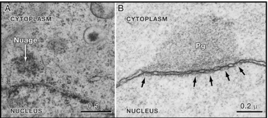
Germ granules in all species contain various RNAs and different RNA-binding proteins, many of which are associated with RNA metabolism. It has been revealed that during most of the germline development, germ granules are perinuclear and are associated with nuclear pores (Pitt et al., 2000) (Figure 1.4). Furthermore, germ granules in Drosophila associate with ribosomes (Mahowald, 1968). Altogether this suggests that germ granules among species might be involved in mRNA processing and/or mRNA trafficking from the nucleus to the cytoplasm (Seydoux and Braun, 2006).
Figure 1.4: Germ Granules are Perinuclear in most organisms (Seydoux and Braun, 2006). Electron micrographs of P granules on germ nuclei. Anti-nuclear pore antibody (mAb414) and anti-P granule antibody (K76) were used. Germ granules in (A) the primordial germ cell of a rat at embryonic day 10 (nuage) and (B) in an adult C. elegans germ cell (Pg). P granules (Pg) appear as electron-dense zones around each nucleus. Arrows show nuclear pores. Five pores are visible beneath the P granules.
1.6. C. elegans P granules
In C. elegans, the germ granules are called P granules and are thought to have determinative roles in germline development. P granules are cytoplasmic particles present throughout life in the germline of C. elegans (with the exception of mature sperm), are exclusively localized to germ cells and can be visualized by immunofluorescence (Strome and Wood, 1982) or by electron microscopy (Wolf et al., 1983).
1.6.1. P granule Localization and Physical Nature
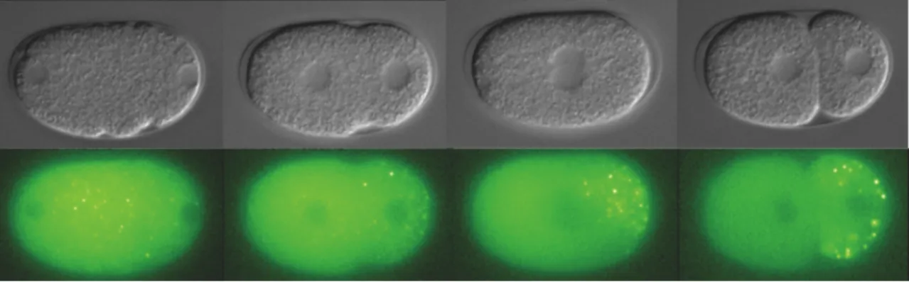
In 1996, Hird et al., injected fluorescently labeled OIC1D4 monoclonal antibody, which presumably recognize the P granule specific protein PGL-1, into the germline syncitium of adult hermaphrodites in order to monitor the germline-specific P granule movements in the early C. elegans embryo. Their studies showed that P granules are maternally contributed to the embryo and are initially distributed equally throughout the cytoplasm of the unpolarized 1-cell embryo. During pseudo-cleavage and before the oocyte pronucleus migration toward the sperm pronucleus, P granules start to move posteriorly so that by the end of pseudo-cleavage, almost all P granules are located at the posterior end of the embryo. At the first asymmetric division, all the P granules are segregated preferentially to the posterior germline blastomere (P1), while the anterior P granules disappear progressively during mitosis, so that none are detectable in the AB cell. The asymmetric segregation of P granules continues at each asymmetric cleavage resulting in the inheritance of P granules by the germline blastomeres (P2, P3 and P4). P4 then divides and passes the P granules to the two primordial germ cells, Z2 and Z3 (Figure 1.5).
Figure 1.5: P granule segregation in C. elegans. The images are taken with an Axio-Imager microscope, using a PGL-1:GFP strain. The P granules are initially distributed throughout the cytoplasm, but begin to move posteriorly before the oocyte pronucleus starts to migrate toward the sperm pronucleus. After the two pronuclei meeting, most P granules are localized to the posterior cell so that after the first division, all the P granules are inherited by P1; no P granules are seen in the anterior cell AB.
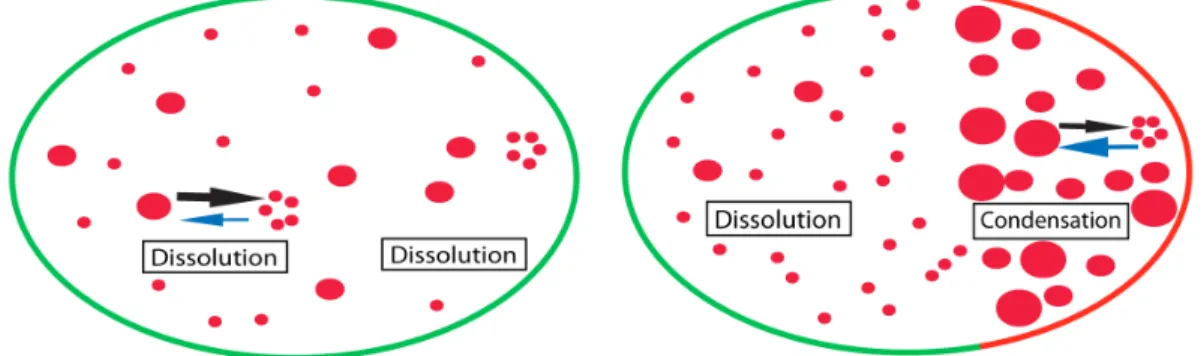
How are P granules targeted specifically to germline blastomeres during C. elegans divisions? For a long time it was thought that P granule posterior partitioning is either controlled by their migration with the cytoplasmic flow toward the posterior pole (Hird and White, 1993, Cheeks et al., 2004 and Strome and Lehmann, 2007), or by the degradation or disassembly of the P granules remaining in the anterior (Hird et al., 1996 and Spike and Strome, 2003). However, it has recently been shown that a dissolution/condensation process controls P granule localization. Using a 3D particle tracking approach, Brangwynne et al. showed that before symmetry breakage, there is an overall P granule dissolution throughout the cytoplasm. Breaking of the symmetry however, results in the condensation of the P granules at the posterior while dissolution continues in the anterior pole of the zygote. Thus the rapid transition between P granules dissolution and condensation causes the migration of P granule components to the posterior, which eventually leads to the disappearance of the anterior components (Figure
1.6). Interestingly, they also reported that P granules behave like liquid droplets. During the condensation phase, P granules are spherical but since their transition from a soluble phase to a condensed structure is required for their proper segregation, they become nonspherical as they associate with the nucleus at the four-cell stage (Brangwynne et al., 2009). In addition, there is also evidence that some polarity proteins such as PAR-1 and MEX-5 function in P granule localization mainly by regulating dissolution/condensation of P granules (Brangwynne et al., 2009).
P granules are known to have a dynamic morphology. During the early embryogenesis, P granules get progressively larger; as they increase in size, their number decreases, suggesting that P granules aggregate in order to form larger granules. In addition, during most of the germline development, P granules have a perinuclear positioning; however during oogenesis; they dissociate from the nuclear surface and become localized to the cytoplasm. It has been shown that P granules reattach to the nucleus during embryogenesis, in 4-cell stage germline blastomeres (Strome and Wood, 1982, Pitt et al., 2000).
Figure 1.6: P granules are localized by dissolution/condensation mechanisms. (A) Before symmetry breakage the high concentration of P granule components leads to the dissolution of P granules into diffusing components (small red circles) in both anterior and posterior cells. (B) However, symmetry breaking causes the P granule condensation in the posterior while P granules at the anterior pole undergo dissolution.
1.6.2. P granule composition
P granules are large non-membrane bound ribonucleoprotein (RNP) organelles and their function is essential for germline development but they have not been characterized biochemically. However it has been shown that P granules are composed of some RNAs and proteins important for proper germline specification and development.
• RNAs
P granules like their mammalian counterparts contain RNA molecules. After the onset of asymmetric cell divisions, one group of maternal RNAs known as the class II maternal RNAs, show unequal distribution between somatic and germline blastomeres. Although these RNAs are distributed to both daughter cells after each cleavage, they are maintained in germline cells while being selectively degraded in somatic blastomeres. In addition, several clusters of poly(A)+ RNAs co-localize with the germline-specific P granules (Seydoux and Fire, 1994). nos-2 (Subramaniam and Seydoux, 1999) and pos-1 (Tabara et al., 1999, Schisa et al., 2001), are two mRNAs which associate with P granules during early embryogenesis. For instance, the nos-2 mRNA is present throughout the cytoplasm of early embryonic blastomeres, but appears to be concentrated and enriched in P granules of germ cells (Subramaniam and Seydoux, 1999). nos-2 encodes a protein similar to the Drosophila Nanos and functions redundantly with nos-1 to regulate germ-cell proliferation and viability (Subramaniam and Seydoux, 1999). pos-1 encodes a CCCH-type zinc-finger protein which is also essential for the proper germline development. In addition to nos-2 and pos-1, some other class II maternal mRNAs shown to localize to P granules in adult gonads are also degraded rapidly in somatic cells while they are protected in germline blastomeres (Schisa et al., 2001).
• Proteins
P granules contain different proteins most of which serve an essential role in C. elegans germline development. One group of proteins is transiently associated with P granules; they are detected in P granules only during early embryogenesis while they disappear in late-stage embryos or in larval or adult germlines. These include PIE-1 (Mello et al., 1996, Tenenhouse et al., 1998), MEX-1 (Guedes and Pries, 1997), MEX-3 (Draper et al., 1996), MEX-5 (Schubert et al., 2000), POS-1 (Tabara et al., 1999b) and IFE-1, an isoform of eIF4E (Amiri et al., 2001). Almost all proteins in this group are known to function during early embryogenesis by preventing the germline blastomeres from adopting somatic fate or the opposite.
Some of the protein components of P granules, like PGL-1/3, GLH-1/4 and DEPS-1, are structurally associated with P granules throughout the life cycle of the worm and regulate assembly and function of these particles, as removing any of them affects proper germline proliferation and development in C. elegans (Kawasaki et al., 1998, 2004; Spike et al., 2007). It was also shown that removing GLH and PGL family proteins alters the proper P granule morphology (Schisa et al., 2001). DEPS-1 and PGL proteins are nematode specific components while the GLH family is homologous to Vasa; a highly conserved Dead-box RNA helicase with a role in germline development and fertility in several species, including Xenopus and mouse (Hay et al., 1988; Lasko and Ashburner, 1988). Several other proteins are also found to be localized to P granules. For instance, our lab showed that NHL-2, a homologue of Brat in Drosophila, also localizes to P granules in C. elegans early embryo (Hyenne et al., 2008).
Interestingly, all known protein components of P granules contain putative RNA-binding motifs, suggesting that they might regulate either the structure or function of the P granules through their RNA-related function.
1.6.3. P granule assembly pathway:
Several molecular epistasis analyses have revealed the mechanism of P granule assembly, stability and function. Studies have shown that loss of some components of P granules affects the structure of P granules in germlines, suggesting their role in P granule assembly pathway. In addition, removing of these components results in sterility and germ cell underproliferation (Kawasaki et al., 1998, Kawasaki et al., 2004, Spike et al., 2008b). These properties provide clues that the role of P granule components is perhaps to regulate the postembryonic germline development. For instance, DEPS-1, a P granule-associated protein is required for the proper localization of PGL-1. Moreover, RT-PCR and western blot analysis revealed that deps-1 mutant germlines display a reduction in the levels of glh-1 mRNA and protein, suggesting that DEPS-glh-1 functions upstream of GLH and PGL family of proteins in the P granule assembly pathway (Spike et al., 2008b). Another P granule component is a DEAD-box RNA helicase, GLH-1, loss of which causes a partial dispersal of PGL proteins (PGL-1, PGL-2 and PGL-3), suggesting that in addition to DEPS-1, GLH-1 participates in the recruitment of PGL proteins to P granules (Spike et al., 2008 a, Spike et al., 2008b). In contrast, it has been shown that PGL-1 depletion is not needed for the assembly of GLH-1, GLH-4 and DEPS-1 to P granules. However, IFE-1, an isoform of eIF4E that binds mRNA caps is a component of P granules that requires PGL-1 associate with P granules (Amiri et al., 2001, Kawasaki et al., 2004, Spike et al., 2008a, Spike et al., 2008b). Together these findings place DEPS-1 at the top of the P granule assembly pathway, while GLH-1 is upstream and IFE-1 is downstream of PGL-1 (Kawasaki et al., 2004, Updike and Strome, 2009).
Moreover, studies showed that mutations in other P granule components such as MEX-1 (Mello et al., 1992; Guedes and Priess, 1997), MEG-1 (Leacock and Reinke, 2008) and OMA-1 (Shimada et al., 2006) also disrupt PGL-1 in the germline. Recently, a genome-wide RNAi screen for PGL-1 accumulation and localization defects was performed and it has been shown that 173 genes are required for the normal P granule stability, localization and function in C. elegans. Interestingly many of these genes are involved in
important cellular processes such as protein degradation, translation, splicing and nuclear export (Updike and Strome, 2009). For instance, some Sm components of the splicesome have been shown to colocalize with P granules within the germ cells and it has been revealed that RNAi depletion of some Sm proteins affects the proper localization of all three PGL proteins to P granules (Barbee et al., 2002). The finding that RNAi depletion of other splicing components did not affect P granule distribution pattern supported the idea that the role of Sm proteins in P granule localization is not through their function in splicing (Barbee et al., 2002). Strikingly however, the RNAi screen performed by Updike and Strome revealed that some pre-mRNA splicing factors are also required for the proper localization of PGL-1 in germline blastomeres. As the germline cells are transcriptionally silenced (Seydoux et al., 1996), this data suggests that proper mRNA splicing might affect P granule localization, assembly and/or stability (Kawasaki et al., 2004, Updike and Strome, 2009). In addition, the proper localization of P granules to the nuclear periphery was also shown to depend on nuclear pores and nuclear transport components, suggesting that mRNA export from the nucleus is also required for the stability and proper localization of P granules to the nuclear periphery (Updike and Strome, 2009).
1.6.4. What are the functional consequences of loss of P granule components? Phenotypic analysis of mutants for P granule components provide clues that the role of P granule components is perhaps to regulate postembryonic germline development, as pgl-l, glh-1 and deps-1 mutants display temperature sensitive sterility and germ cell under-proliferation (Kawasaki et al., 1998, Kawasaki et al., 2004, Spike et al., 2008b). It is now known that absence of any of these genes does not severely compromise fertility at low temperature, while mutations in any of them lead to 100% sterility at high temperatures (26°C for pgl-1 and glh-1 mutants, 24.5°C for deps-1 mutants). pgl-1, glh-1 and deps-1 mutants have under-proliferated germlines that generally lack gametes suggesting that the primary role of these components is in regulating germ cell proliferation and gametogenesis
to ensure fertility. Furthermore, there is evidence that removing of PGL-1 affects proper oogenesis. Interestingly, this sterility phenotype has both a maternal and a non-maternal (zygotic) component indicating that both maternal and zygotic PGL-1, GLH-1 and DEPS-1 are required to ensure normal germline development in C. elegans (Kawasaki et al., 1998, Spike et al., 2008a,b).
Another P granule component is IFE-1, one of five eIF4E (Eukaryotic translation initiation factor 4E) isoforms identified in C. elegans. IFE-1 is expressed primarily in thegermline and associates with P granules. Complete loss of ife-1 function affects the translation of several maternal mRNAs(pos-1, pal-1, mex-1 and oma-1) as well as male germ cell mRNAs(Henderson et al., 2009). IFE-1 is involved in spermatogenesis since loss of IFE-1 affects the proper cytokinesis of spermatocytes (Amiri et al., 2001, Henderson et al., 2009). In addition, ife-1 mutants also show oogenesis defects and therefore ife-1 mutants have abnormal gametogenesis resulting in 100% sterility at 25°C (Henderson et al., 2009).
One fascinating feature of the sterility observed in deps-1, glh-1, pgl-1 and ife-1 mutants is its temperature sensitivity. One explanation is that there is a functional redundancy with other members of their respective gene family at lower temperatures. For instance, in glh-1 mutants, at lower temperature other GLH proteins compensate for the absence of GLH-1, whereas GLH-1 depletion leads to sterility at 26°C (Kawasaki et al., s2004). Another explanation is that the processes that are regulated by P granule components, such as germline proliferation and/or mRNA export from germ nuclei are temperature sensitive (Kawasaki et al., 2004).
1.7. P granules and Translational Control
Germline development and embryogenesis rely on gene expression and cytoplasmic mRNA control. Transcriptionis active during oogenesis allowing the cell to produce maternal messenger RNAs required for the development. Messenger RNAs are transcribed in the nucleus and exported to the cytoplasm for translation. The maternal mRNAs then are packaged into oocytes until fertilization. Upon fertilization the maternal mRNAs will be actively translated in a specific spatial and temporal manner.
In the germline, mRNAs programmed for the delayed translation are accumulated and stored in P granules (Seydoux and Braun, 2006). Thus P granules are thought to play an essential role in regulating the accurate temporal and spatial mRNA translation or degradation. In addition, the finding that IFE-1 associates with PGL-1 in P granules, raises the possibility that P granules might are involved in activation or inhibition of translation (Henderson et al., 2009). However the biochemical function of P granules remains elusive. Nevertheless, several lines of evidence support the idea that P granules are involved in the posttranscriptional regulation of gene expression:
1.7.1. P granule localization and composition:
Mahowald in 1968 observed that Drosophila germ granules associate with ribosomes. This observation led him to propose that mRNAs required for germ cell development might store into Drosophila polar granules (Mahowald, 1968). Today it is clear that P granules contain mRNAs encoding germline-specific proteins and poly(A)+ RNAs (Seydoux and Fire 1994). In 2001, Schisa et al showed that mRNAs that function during development are enriched in P granules whereas the ubiquitously expressed actin and tubulin mRNAs are distributed uniformly throughout the cytoplasm (Schisa et al., 2001). Moreover, there is evidence that SL1 sequences, which are trans-spliced onto the 5' end of most C. elegans mRNAs (Zorio et al. 1994), also colocalize with P granules (Pitt et al., 2000; Schisa et al., 2001; Updike and Strome, 2009). In addition, P granules are composed of multiple proteins that all contain RNA-binding motifs, which might enable
them to interact with RNAs exiting from the nucleus. The question is whether this association is important?
GLH-1, a core component of P granules, contains two different regions, a DEAD-box helicase domain and a Gly-rich region containing 3 CCHC zinc fingers, which are both able to bind to RNA (Sengoku et al., 2006). GLH-1 proteins lacking any of these two regions are not able to localize properly to P granules. Together these observations suggest that the interaction between GLH-1 and RNA contributes to P granule function, perhaps by regulating the assembly of GLH-1 to P granules (Spike et al., 2008b). It has also been reported that the Phe-Gly (FG) domain of GLHs mediates their association with the FG-rich nuclear pore protein (Frey et al., 2006). Furthermore, it was shown that 75% of nuclear pores in the germ cells are associated with the P granules (Pitt et al., 2000). According to a genome-wide RNAi screen that was performed recently, removal of nuclear pore proteins (NPP) leads to the detachment of P granules from the nuclear envelope and disrupts the proper localization and assembly of PGL-1 to P granules. Thus the association between NPPs and P granule components is required for proper P granule location and PGL-1 intensity levels (Updike and Strome, 2009).
Studies revealed that some P granule components such as DEPS-1 are required for proper RNAi response (Spike et al, 2008b). Moreover, csr-1, one of the C. elegans Argonauts, is also involved in a germline endo-RNAi pathway. csr-1 worms have larger P granules in germline blastomeres (Updike and Strome, 2009). Thus one possibility is that P granules are involved in the regulation of the posttranscriptional gene expression.
P granule composition together with their distribution over nuclear pores suggests that they are most likely to encounter mRNAs during their transport from nucleus to cytoplasm and therefore could potentially contribute to the majority of post-transcriptional gene regulation procedures such as mRNA trafficking, translation and stability that occur in germ cells (Schisa et al., 2001; Seydoux and Braun, 2006 and Spike et al., 2008b).
1.7.2. Links between different RNP granules in C. elegans:
Messenger RNAs that are not immediately translated after being exported from the nucleus accumulate in special discrete cytoplasmic domains referred to as RNA granules. These granules contain various ribosomal subunits, ribosomal RNAs, translation factors, decay enzymes, helicases, scaffold proteins and RNA-binding proteins in order to control the localization, stability, and translation of their RNAs. Strikingly P granules share certain components with these RNA granules of somatic cells. They have various names; P bodies, processing bodies and stress granules and are widely conserved among species.
Untranslated mRNAs localize to translationally repressed mRNP granules called Processing bodies or P bodies (also referred to as GW182 or Dcp bodies) of somatic cells in order to be degraded or reactivated (Brengues et al., 2005; Anderson and Kedersha, 2006 and Seydoux and Braun, 2006). P bodies contain factors essential for translational repression (eIF4e, Dhh1/RCK/p54, Scd6/RAP55) and components of mRNA decapping (DCP1 and DCP2), activators of decapping (LSM-1) and deadenylation (CCR4-NOT deadenylase complex) machinery and are involved in mRNA repression and degradation (Seydoux and Braun, 2006).
Non-translating mRNAs can also accumulate in other cytoplasmic structures termed stress granules. Certain P body components (e.g. Dhh1/RCK/p54, Scd6/RAP55 and XRN1) also localize to stress granules (Kedersha and Anderson, 2002). Stress granules are formed when translation initiation is compromised in response to environmental stress such as heat or UV irradiation. Under these conditions, translation of mRNAs encoding housekeeping proteins is arrested, and mRNAs accumulate in stress granules. The T-cell intracellular antigen-1 (TIA-1), an mRNA binding protein, is essential for the accumulation of stalled translational pre-initiation complexes to stress granules (Kedersha et al., 1999, Kedersha et al., 2005). Although they have some similarities, some features distinguish stress granules from P bodies. Unlike P bodies, stress granules contain ribosomal subunits. In addition, the initiation factors such as, eIF3, G3BP, eIF4G, TIA-1 and PAB-1 are found
exclusively in stress granules but not in P bodies, whereas DCP1and DCP2 are only restricted to P bodies. Another difference is that mRNA degradation does not occur in stress granules, which makes them available for rapid reinitiation after recovering from stress (Sheth and Parker, 2003; Cougot et al., 2004a). Thus mRNA degradation machinery must be somehow disabled during the stress response, suggesting that stress granules function in mRNA sorting rather than decay (Kedersha et al., 2005) whereas P bodies are sites ofmRNA degradation (Sheth and Parker, 2003).
Several lines of evidence indicate that P bodies and stress granules are distinct granules that interact with each other, suggesting that stress granules and P bodies may function together to regulate the balance of translated, repressed and degraded mRNAs in somatic cells (Kedersha et al., 2005; Gallo et al., 2008). Interestingly, P bodies and stress granules contain the same species of mRNAs. One explanation could be that some mRNAs exiting translation assemble into stress granules. Later, mRNAs destined for degradation are transported from stress granules to P bodies through the physical association between these two RNA granules (Kedersha et al., 2005).
Several lines of evidence suggest parallels between P granules, P bodies and stress granules. P granules share components with both processing bodies and stress granules of somatic cells in C. elegans. P granules contain protein involved in translation initiation, translation control and mRNA decay:
• Decapping proteins including PATR-1, DCAP-1, DCAP-2, CGH-1 and CAR-1 (Audhya et al., 2005, Boag et al., 2005, Gallo et al., 2008, Lall et al., 2005, Navarro et al., 2001, Squirrell et al., 2006)
• RNA dependent silencing machinery components (Argonauts and microRNAs) (Updike and Strome, 2009)
• Translational initiation factors and markers of stress granules: IFE-1 (Amiri et al., 2001), TIA-1 and PAB-1 (Gallo et al., 2008)
P granules and stress granules have been shown to have similarities. P granules and stress granules, in addition to their dynamic structures, both share some components of P bodies while they have some factors such as TIA-1 and PAB-1 that are not found in P bodies. Furthermore, it has been shown that nos-2 mRNA accumulates in both P granules and stress granules whereas its association to P bodies is not very stable, suggesting that P granules might associate with stress granules (Gallo et al., 2009).
An interesting issue consists of identifying how different RNP granules regulate mRNA translation. CGH-1, a highly conserved DEAD-box family protein localizes to both P granules and P bodies. cgh-1 mutants show a reduction in their mRNA level. In addition the nos-2 mRNA that should be translationally silenced during oogenesis, is prematurely translated in the oocytes of the cgh-1 worms (Boag et al., 2008, Gallo et al., 2008), suggesting that CGH-1 functions in both mRNA translational repression and protection from degradation. In addition, it has been shown that while PATR-1 has a key role in P body formation, P granules are formed independently of PATR-1 (Boag et al., 2008). Based on these observations, Rajyaguru and Parker proposed that CGH-1 has different roles in posttranscriptional gene expression, depending on its assembly to P granules or P bodies. In germline blastomeres, CGH-1 colocalization to P granules leads to the recruitment of CAR-1 and PAB-CAR-1 to P granules. This complex protects the germline mRNA from being degraded by blocking the association of decapping and degrading enzymes to P granules. In contrast, CGH-1 associates with PATR-1 and other decapping and degrading factors of the somatic P bodies in order to degrade the mRNA in somatic cells (Rajyagur and Parker, 2009). Altogether, these observations support the idea that different RNA granules use distinct mechanisms to control maternal mRNA translation and stability.
1.8. NHL-2 and Translational Control
NHL-2, a homologue of the Drosophila Brain Tumor (Brat), is enriched in P granules during early embryogenesis in C. elegans (Hyenne et al., 2008). Brat is a member of the NHL family of proteins found in Drosophila (Arama et al., 2000; Sonoda, 2001). The NHL family name derives from three of the founding members: NCL-1, HT2A, and LIN-41, which have been shown to be involved in RNA metabolism: NCL-1 is required for ribosomal RNA synthesis (Frank et al., 1998) and LIN-41 is involved in the temporal regulation of the expression of the LIN-29 transcription factor (Slack, 2000). The Brat protein contains conserved motifs: N-terminal B-box zinc-finger domains and several NHL repeats in their C-terminus. All these motifs are reported to be involved in protein-protein interactions. In addition to its role as a cell growth inhibitor in Drosophila, Brat is also known as a tumor suppressor functioning in the posttranscriptional gene regulation. Brat forms a complex with Pumilio (homolog of C. elegans FBF-1/2) and Nanos (homolog of C. elegans NOS-1/2 and 3) and interacts with the 3’-UTR of the Hunchback (HB) mRNA through its NHL domains in order to inhibit the translation of HB in the posterior pole of the Drosophila embryo, suggesting a role for other NHL family of proteins in controlling the posttranscriptional gene expression (Sonoda et al., 2001).
Drosophila Brat has five homologues in C. elegans: NCL-1, NHL-1, NHL-2, NHL-3 and LIN-41, which are collectively referred to as CeBrats (for C. elegans Brats) (Hyenne et al., 2008). In addition to the conserved domains present in Brat, all CeBrats except for NCL-1 also contain an N-terminal RING domain (Figure 1.7). In C. elegans, the ortholog of Brat, NCL-1, was shown to function as a cell growth inhibitor, since worms lacking NCL-1 have enlarged nucleoli, increased amounts of ribosomal RNA and a higher rate of protein synthesis (Frank and Roth, 1998), whereas NHL-1, NHL-2 and NHL-3 are not likely to play the same role (Hyenne et al., 2008). Interestingly, it has been reported that while LIN-41 cannot affect par-2 lethality, 4 of the CeBrats; NCL-1, NHL-1, NHL-2 and NHL-3 are able to suppress par-2 lethality and therefore are involved in regulating embryonic polarity (Hyenne et al., 2008).
Recently it has been shown in Drosophila that Brat and another member of the TRIM-NHL family of proteins, Mei-P26, bind to Argonaute-1, a core miRISC (miRNA-induced silencing complex) component. Mei-P26 inhibits the microRNA biogenesis in Drosophila ovaries, since its removal leads to a general upregulation of most microRNAs. In addition, both Mei-P26 and Brat, bind to AGO-1 (Argonaute-1), a core component of RISC machinery, through their NHL domains. Mei-P26 lacking its NHL domains is not able to bind to AGO-1, leading to the possibility that NHL domains in other proteins might be also involved in regulating microRNAs (Neumuller et al. 2008). In support of this idea, it was recently shown that NHL-2 controls microRNA activity in C. elegans larvae. It has been also reported that NHL-2 similar to its ortholog Mei-P26, has a physical interaction with key factors of miRISC pathway: ALG-1 and AIN-1 (Hammel et al., 2009). Similarly, TRIM32, a mouse homolog of Drosophila Brat and Mei-P26 also binds Argonaute-1 and is involved in the regulation of microRNAs (Schwamborn et al., 2009).
It was shown that NHL-2 localizes to somatic P bodies together with CGH-1, another key component of P bodies that binds to ALG-1 and AIN-1. NHL-2 and CGH-1 have been shown to interact physically and both are involved in regulating the posttranscriptional repression of several microRNA targets in somatic cells of developing worm larvae (Hammell et al., 2009). In addition, there is evidence that NHL-2 functions in posttranscriptional gene regulation mainly via modulating the efficacy of the interactions between miRNAs and their targets in C. elegans larvae (Hammell et al., 2009), however it is not known whether NHL-2 has the same role during embryogenesis.
Figure 1.7: NHL-2 is a member of the conserved TRIM-NHL- family of proteins (Hyenne et al., 2008). (A) Schematic representation of D. melanogaster Brat and its homologs in C. elegans. All of them contain N-terminal B-box zinc-finger domains, a BBC or coiled-coil domain and several NHL repeats in their C-terminus. All of these but NCL-1 and Brat contain an N-terminal RING domain. Protein amino acid length is given in brackets. (B) Phylogenetic reconstruction of Brat homologues in C. elegans, D. melanogaster and H. sapiens. C. elegans NCL-1 and NHL-2 together with D. melanogaster Brat and Mei-P26, are separated from their other homologs. The length of the tree branches are proportional to the relative evolutionary distance between Brat homologs which is numbered above.
1.9. Objectives of the Master’s Project
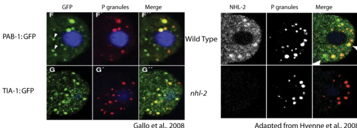
Recently, our lab has shown that NHL-2 localizes to P granules in the embryo (Figure 1.8.A). In addition, studies in our lab revealed that NHL-2 functions in embryonic polarity in C. elegans (Hyenne et al., 2008). NHL-2 is also known to be involved in the posttranscriptional repression in C. elegans larvae (Hammell et al., 2009). Nevertheless the precise function of NHL-2 at the embryonic level is not known. Interestingly, it was shown that two markers of stress granules; PAB-1 and TIA-1 have the same distribution pattern as NHL-2 as they both localize to P granules (1.8.B) (Gallo et al., 2008). In addition, little is known about the regulation and exact function of P granules in C. elegans. In order to investigate the underlying mechanisms of P granule function and stability, we postulated that NHL-2 plays its role in early embryogenesis by regulating P granule function, perhaps through association with PAB-1 and/or TIA-1. Furthermore, the presence of NHL-2 on P granules makes it also possible that, similar to P granule core components, NHL-2 also functions in germline development and proliferation. Thus we decided to explore the molecular function of NHL-2 in P granule biogenesis, in order to gain a better understanding of P granules function.
The aim of this project is to understand the possible role of NHL-2 in controlling P granule function and germline development. Thus, we developed an image analysis approach to determine whether NHL-2 functions in P granule stability. Furthermore, we are also interested in investigating how P granules are regulated in C. elegans during development and to identify proteins that mediate this control. Studying how NHL-2 and P granules function in C. elegans could elucidate and provide key insights into the roles and relationships of various RNA granules in different species.
Figure 1.8: NHL-2, PAB-1 and TIA-1 granules have similar distribution patterns. They all colocalize partially to P granules. (A) Confocal images of embryos expressing the indicated GFP fusion and double-stained with anti-GFP and anti-P granules antibodies (B) Immunofluorescence of 4-cell embryos stained with anti-P granules and anti-NHL-2 antibodies. Note that some NHL-2, PAB-1 and TIA-1 positive granules colocalize with P granules, while others are devoid of P granule components.
2.1. Strains and Alleles
All strains were maintained as described by Brenner (1974) and were grown at 15° C unless otherwise stated. The wild-type strain was the N2 (Bristol) strain. The following mutants have been used:
1- nhl-2(ok818) III
Deletion allele considered as a null allele (Hyenne et al., 2008). 2- pgl-1(bn101) IV
Point mutation considered as a null allele (Kawasaki et al., 2004). 3- nhl-3(tm2516) II; nhl-2(ok818) ncl-1(e1942) nhl-1(gk15) III
nhl-1(gk15), nhl-3(tm2516) are deletion alleles and ncl-1(e1942) is a point mutation allele and it can be considered as a null allele since it induces a strong decrease in its respective protein expression levels (Frank et al., 2002).
4- nhl-2(ok818) III; pgl-1(bn101) IV same as above
Mutant strains were generated by genetic crosses. nhl-3(tm2516); nhl-2 (ok818) ncl-1(e1942) nhl-1(gk15) strain was generated by Vincent Hyenne (Hyenne et al., 2008).
I generated 2(ok818); pgl-1(bn101) double mutants. The presence of nhl-2(ok818) was confirmed by PCR. The presence of pgl-1(bn101) in this mutant was confirmed by examining the sterility of mutants at the elevated temperature of 26°C under the dissecting microscope.
2.2. C. elegans techniques- Dissection, Immunofluorescence and Microscopy
2.2.1. Antibodies and Markers
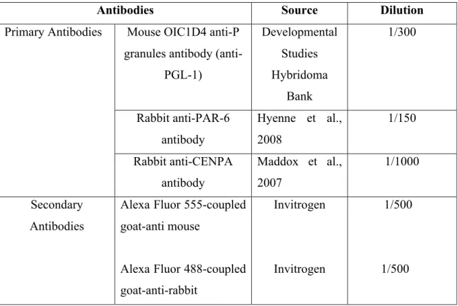
Table I: List of the markers and antibodies used for embryo staining.
Antibodies Source Dilution
Mouse OIC1D4 anti-P granules antibody
(anti-PGL-1) Developmental Studies Hybridoma Bank 1/300 Rabbit anti-PAR-6 antibody Hyenne et al., 2008 1/150 Primary Antibodies Rabbit anti-CENPA antibody Maddox et al., 2007 1/1000 Secondary Antibodies
Alexa Fluor 555-coupled goat-anti mouse
Alexa Fluor 488-coupled goat-anti-rabbit
Invitrogen
Invitrogen
1/500
2.2.2. Immunofluorescence of C. elegans embryos
L4s were kept for 24h at 25°C and were then fixed and stained using the following protocol:
1. 20-30 worms were placed in 6µl water on a polylysine-coated slide and were sliced twice, once near each spermatheca, using two 25-gauge needles. This releases embryos (Fig 2.1).
Figure 2.1: C.elegans dissection for immunofluorescence (Prasad, Jang et al. 2007).
2. The slides were immediately frozen on dry ice. The coverslips were then flicked off the slides with a razor blade (to crack the egg shell) and the slides were put into cold methanol for fixation for 20 minutes at -20°C.
3. The slides were taken out of methanol and were dried using a Kim wipe (to remove all the liquid surrounding the embryos). To remove the methanol, the slides were washed (3-4 times) with PBS.
4. The PBS was aspirated off and then 100µl of PBT (PBS + 0.05% Tween 20) was added on top of the slides.
5. The PBT was aspirated off and 25 µl of goat serum was then added on embryos. The slides were then left in a humid chamber at room temperature for 30 minutes.
6. The goat serum was aspirated and 20 µl of OICD4 (1:300 dilutions in PBT) was added on each slide.
7. The slides were incubated at 4° C overnight in a sealed humidity chamber.
8. The next day the slides were washed with PBT 3 times and then 25 µl of secondary antibody was put on them. The slides were incubated at room temperature for 1 hour and they were washed with PBT 3 times.
9. Six µl of mounting medium containing DAPI was added on each slide, the coverslips were then put gently on each slide and later the slides were sealed with nail polish.
2.3.3. Imaging
2.3.3.1. P granule Quantification: • Image Acquisition
For P granule numbers quantification, images were acquired with a Zeiss LSM 510 Meta laser scanning confocal microscope using a Plan Apochromat 63x/1.4 NA objective. Images from wild type and mutant embryos were acquired using identical parameters. The principle of confocal microscopy is based on the fact that only a portion of the emitted light passes through a "pinhole" for the high-resolution observation of very thin optical sections of cells. This allows for a higher resolution image by preventing out of focus light from reaching the detector. Then a 3D reconstruction can be performed by superposition of these sections.
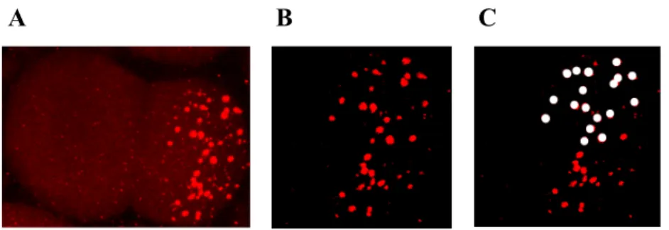
• Image Analysis
The images acquired in confocal fluorescence were analyzed with the LSM program, and then processed using Adobe Photoshop. Since P granules are only present in the posterior pole of the embryo, a threshold was applied in order to eliminate the background puncta in the anterior and then we counted the total number of P granules in the posterior of each embryo (Figure 2.2). The number of P granules in each embryo is the average of three times quantification.