Université de Montréal
L’IMPORTANCE DE LA VOIE DE SIGNALISATION AU NIVEAU DE LA BARRIÈRE HÉMATO-ENCÉPHALIQUE
THE IMPORTANCE OF THE HEDGEHOG SIGNALING PATHWAY AT THE LEVEL OF THE BLOOD-BRAIN BARRIER
par
Aurore Dodelet-Devillers
Département de Physiologie Faculté de Médecine
Mémoire présenté à la Faculté des Études Supérieurs en vue de l’obtention du grade de Maîtrise
en Sciences Neurologiques
Septembre, 2009
Université de Montréal Faculté des études supérieures
Ce mémoire intitulé :
L’IMPORTANCE DE LA VOIE DE SIGNALISATION AU NIVEAU DE LA BARRIÈRE HÉMATO-ENCÉPHALIQUE
THE IMPORTANCE OF THE HEDGEHOG SIGNALING PATHWAY AT THE LEVEL OF THE BLOOD-BRAIN BARRIER
Présenté par : Aurore Dodelet-Devillers
A été évalué par un jury composé des personnes suivantes
Dr. Adriana Di Polo Président-rapporteur
Dr. Alexandre Prat Directeur de recherche
Dr. Christine Vande Velde Membre du jury
contrôlant le passage des substances sanguines et des cellules immunitaires. La BHE est formée de cellules endothéliales liées ensemble par des jonctions serrées et ses fonctions sont maintenues par des astrocytes, celles ci sécrétant un nombre de facteurs essentiels. Une analyse protéomique de radeaux lipidiques de cellules endothéliales de la BHE humaine a identifié la présence de la voie de signalisation Hedgehog (Hh), une voie souvent liées à des processus de développement embryologique ainsi qu’au niveau des tissus adultes. Suite à nos expériences, j’ai déterminé que les astrocytes produisent et secrètent le ligand Sonic Hh (Shh) et que les cellules endothéliales humaines en cultures primaires expriment le récepteur Patched (Ptch)-1, le co-récepteur Smoothened (Smo) et le facteur de transcription Gli-1. De plus, l’activation de la voie Hh augmente l’étanchéité des cellules endothéliales de la BHE in vitro. Le blocage de l’activation de la voie Hh en utilisant l’antagoniste cyclopamine ainsi qu’en utilisant des souris Shh déficientes (-/-) diminue l’expression des protéines de jonctions serrées, claudin-5, occcludin, et ZO-1. La voie de signalisation s’est aussi montrée comme étant immunomodulatoire, puisque l’activation de la voie dans les cellules endothéliales de la BHE diminue l’expression de surface des molécules d’adhésion ICAM-1 et VCAM-1, ainsi que la sécrétion des chimiokines pro-inflammatoires IL-8/CXCL8 et MCP-1/CCL2, créant une diminution de la migration des lymphocytes CD4+ à travers une monocouche de cellules endothéliales de la BHE. Des traitements avec des cytokines pro-inflammatoires TNF-α and IFN-γ in
vitro, augmente la production de Shh par les astrocytes ainsi que l’expression de surface
de Ptch-1 et de Smo. Dans des lésions actives de la sclérose en plaques (SEP), où la BHE est plus perméable, les astrocytes hypertrophiques augmentent leur expression de Shh. Par contre, les cellules endothéliales de la BHE n’augmentent pas leur expression de Ptch-1 ou Smo, suggérant une dysfonction dans la voie de signalisation Hh. Ces résultats montrent que la voie de signalisation Hh promeut les propriétés de la BHE, et qu’un environnement d’inflammation pourrait potentiellement dérégler la BHE en affectant la voie de signalisation Hh des cellules endothéliales.
Mots-clés : radeaux lipidiques, microdomaine, barrière hémato-encéphalique, jonctions serrées, astrocyte, cellule endothéliale, endothélium, perméabilité, voie de signalisation hedgehog, sonic hedgehog, sclérose en plaques, neuroimmunologie, neuroinflammation
regulates the entry of blood-borne molecules and immune cells into the CNS. Recent studies indicate that the Hedgehog (Hh) signaling pathway in adult tissues plays an important role in vascular proliferation, differentiation and tissue repair. Using a lipid membrane raft-based proteomic approach, I have identified the Hedgehog (Hh) pathway as a signaling cascade involved in preserving and upkeeping BBB functions. My study shows that human astrocytes express and secrete Sonic Hh (Shh) and conversely, that human BBB-ECs bear the Hh receptor Patched-1 (Ptch-1), the signal transducer Smoothened (Smo) as well as transcription factors of the Gli family. Furthermore, activation of the Hh pathway in BBB-ECs restricts the passage of soluble tracers in vitro. By blocking the Hh signaling in vitro and by using Shh knock-out (-/-) embryonic mice, I demonstrate a reduced expression of TJ molecules claudin-5, occludin and ZO-1. Hh activation also decreases the surface expression of cell adhesion molecules ICAM-1 and VCAM-1, and decreases BBB-ECs secretion of pro-inflammatory chemokines IL-8/CXCL8 and monocytes chemoattractant protein 1 MCP-1/CCL2, resulting in a reduction of migrating CD4+ lymphocytes across human BBB-EC monolayers. In vitro treatment with inflammatory cytokines TNF-α and IFN-γ, upregulates the production of astrocytic Shh and the BBB-EC surface expression of Ptch-1 and Smo. In active Multiple Sclerosis (MS) lesions, in which the BBB is disrupted, Shh expression is drastically upregulated in hypertrophic astrocytes, while Ptch-1 and Smo expression is down-regulated or left unchanged, suggesting that a deregulation in the Hh signaling pathway may prevent the barrier stabilizing properties of Hh. Our data demonstrate an anti-inflammatory and BBB-promoting effect of astrocyte-secreted Hh and suggest that a pro-inflammatory environment disrupt the BBB by impacting, at least in part, on Hh signaling in brain ECs.
Key words: lipid membrane raft, DRM, microdomain, blood-brain barrier, tight junction, astrocyte, endothelial cell, endothelium, permeability, hedgehog pathway, sonic hedgehog, Multiple Sclerosis neuroimmunology, neuroinflammation
TABLE OF CONTENTS
TABLE OF CONTENTS...1
LIST OF ABBREVIATIONS...5
DEDICATION AND ACKOWLEDGEMENTS ...8
LITERARY REVIEW ...10
1.0 THE BLOOD-BRAIN BARRIER (BBB) ...11
1.1 Unique BBB properties... 11
1.1 Tight and adherens junctions ... 13
2.0 CUES IN THE VASCULATURE ... 17
2.1 Glial cues control of the vasculature and blood-brain barrier function ... 17
2.2 Neural cues control of the vasculature... 20
2.3 Neural cues control of blood-brain barrier function ... 22
3.0 THE HEDGEHOG (Hh) SIGNALING PATHWAY ...24
3.1 The Hh signaling pathway in vasculogenesis ... 28
3.2 The Hh signaling pathway in angiogenesis ... 29
3.3 Evidence of the Hh signaling pathway in barrier function ... 31
4.0 THE BASAL LAMINA... 32
5.0 IMMUNE INTERACTIONS AT THE BBB... 33
5.1 Multiple sclerosis (MS) and experimental autoimmune encephalomyelitis (EAE) ... 34
5.2 The Hh signaling pathway in injury and MS ... 37
6.0 LIPID MEMBRANE MICRODOMAINS ...40
PART I:...43
HYPOTHESIS AND OBJECTIVES...43
MATERIALS AND METHODS... 44
1.0 Detergent-resistant membrane (DRM) isolation and analysis ... 44
1.1 Western blot of lipid membrane rafts ... 44
1.2 BBB-ECs lipid membrane raft proteomic analysis... 45
RESULTS ... 46
1.1 BBB-EC lipid membrane rafts contain junctional and integrin proteins... 48
1.2 BBB-EC lipid membrane rafts contain adhesion molecules... 48
1.3 BBB-EC lipid membrane rafts contain transporter proteins... 51
1.4 BBB-EC lipid membrane rafts contain proteins typically associated with the nervous system ... 53
PART II:...55
HYPOTHESIS AND OBJECTIVES...55
ORIGINAL ARTICLE...57
SONIC HEDGEHOG PROMOTES...57
BLOOD-BRAIN BARRIER INTEGRITY AND IMMUNE-QUIESCENCE...57
Abstract... 58
Introduction... 59
Results... 61
Astrocyte and blood-brain barrier endothelial cell expression of hedgehog pathway components ... 61
Astrocyte-secreted sonic hedgehog decreases permeability in vitro ... 62
Cyclopamine induces BBB disruption in vivo... 63
Sonic hedgehog promote and maintain tight junction protein expression... 63
Hedgehog activation influences cell adhesion molecule expression, cytokine secretion and lymphocyte migration ... 65
Transcription factor upregulation upon hedgehog activation ... 66
Pro-inflammatory cytokines modulates the hedgehog signaling pathway in vitro... 67
Modulation of the hedgehog signaling pathway in situ in Multiple sclerosis ... 67
Discussion... 68
Materials and methods ... 73
Primary cell isolation & culture... 73
Immunocytofluorescence... 73
Tissue and immunohistochemistry ... 74
Reverse-transcription and real-time quantitative polymerase reaction... 75
Western blots ... 76
In vivo permeability ... 78
Flow cytometry and enzyme-linked immunosorbent assay... 78
Statistical analysis... 79
Figure legends... 80
Supplemental Figure Legend ... 85
Acknowledgements... 86
DISCUSSION ...96
1.0 LIPID MEMBRANE RAFTS IN BBB...97
1.1 Role of membrane lipid rafts in junctional protein complexes... 97
1.2 Role of lipid membrane rafts in adhesion molecules... 99
1.3 Proteomic analysis of BBB-EC lipid membrane rafts reveal proteins typically associated with the nervous system ... 100
2.0 THE Hh SIGNALING PATHWAY ...102
2.1 The Hh signaling pathway in lipid membrane rafts... 102
2.1 The Hh signaling pathway in the BBB ... 104
2.2 The Hh signaling pathway in MS ... 107
LIST OF FIGURES AND TABLES FIGURES
Figure i: The Hedgehog signaling pathway ... 16 Figure ii: Different detergents isolate cholesterol-enriched DRMs from
human primary cultures of BBB-ECs ... 27 Figure iii: Isolated lipid membrane rafts contain TJ proteins, integrins and
CAMs... 47 Figure iv: Hypothetical model representing different lipid membrane rafts
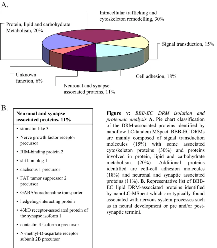
found in BBB-ECs ... 52 Figure v: BBB-EC DRM isolation and proteomic analysis reveals neuronal
and synaptic associated proteins ... 54 Figure 1: Human astrocytes express and secrete Shh and human BBB-ECs
express receptor Ptch-1 and signal transducer Smo... .87 Figure 2: Astrocyte secreted Shh decreases BBB-EC permeability via Smo
signaling... 88 Figure 3: Shh is important in TJ maintenance ... 89 Figure 4: Modulating Hh activity influences BBB-EC expression of CAMs,
chemokine secretion and leukocyte ability to transmigrate... 90 Figure 5: In vitro expression pattern of transcription factors in BBB-ECs. ... 91 Figure 6: Pro-inflammatory cytokines upregulate Hh components in
astrocytes and BBB-ECs in vitro. ... 92 Figure 7: Expression of Shh in MS tissue... 93 Figure 8: Expression of Hh components in MS tissue... 94 Supplemental Figure 1: Activation of Hh pathway is dose-dependent and
affects permeability of small molecule tracer 14C-sucrose, without
affecting proliferation ... 95
TABLES
Table i: Lipid membrane raft-associated transporters identified by proteomic
LIST OF ABBREVIATIONS
Ab: Antibody
ACM: Astrocyte conditioned media AJ: Adherens junction
AKAP12: A-kinase anchoring protein 12
ALCAM: Activated leukocyte cell adhesion molecule
Ang: Angiopoitein
AngII: Angiotensin II
APC: Allophycocyanin (fluorochrome) BBB: Blood-brain barrier
Boc: Brother of Cdon or Biregional cell adhesion molecule-related/downregulated by oncongenes binding proteins BDNF: Brain-derived neurotrophic factor
BMP: Bone morphogenic protein BSA: Bovine serum albumin C-terminal: Carboxyl-terminal
CAM: Cell adhesion molecule CCL: CC chemokine ligands CD: Cluster of differentiation
Cdon: Cell adhesion molecule-related/down-regulated by oncogenes
CNS: Central nervous system CSF: Cerebrospinal fluid CXCL: CXC chemokine ligand
Cy3: Cyanine (fluorochrome) DAB: 3,3-diamino-benzidine
Dhh: Desert Hedgehog
DMEM: Dulbecco's Modified Eagle's Medium DRM: Detergent resistant membrane
EAE: Experimental autoimmune (allergic) encephalomyelitis EC: Endothelial Cell
ECM: Extracellular matrix EGF: Epidermal growth factor ELISA: Enzyme linked immunosorbent assay EPC: endothelial precursor cell Eph: Ephrin receptor
FBS: Fetal bovine serum FGF: Fibroblast growth factor FITC: Fluorescein (fluorochrome) GDNF: Glial-derived neurotrophic factor GFAP: Glial fibrillary acidic protein
Gli-1: Glioblastoma-associated oncogene homolog 1 Glut-1: Glucose transporter-1
HDL: High density lipoprotein HEK: Human epithelial kidney
Hh: Hedgehog
Hip: Hedgehog interacting protein
HS: Human serum
HUVEC: Human umbilical vein endothelial cells HRP: horseradish peroxidase
hrShh: human recombinant sonic hedgehog ICAM: Intercellular Adhesion Molecule ICS: Intracellular stain
IFN-β: Interferon beta IFN-γ: Interferon gamma IgG: Immunoglobulin G Ihh: Indian Hedgehog
IL: Interleukin
IP-10: Interferon protein-10 JAM : Junctional adhesion molecule
-/-: Knock-out
LC-MSpect: Liquid chromatography-mass spectrometry LDL: low density lipoprotein
MAGI: Membrane-associated guanylate kinase inverted MAGUK: Membrane-associated guanylate kinase
MBP: myelin basic protein
MCAM: melanoma cell adhesion molecule MCP-1: Monocyte chemoattractant protein-1 MHC I: Major histocompatibility complex, class I MIP-1α: Macrophage inflammatory protein-1alpha MOG: Myelin oligodendrocyte glycoprotein MS: Multiple Sclerosis
mRNA: Messenger ribonucleic acid MRP-1: Multiple drug resistance protein-1 MMLV: Murine moloney leukemia virus N-cadherin: Neural cadherin
N-terminal: Amino-terminal
NAWM: Normal appearing white matter NSC: Neural stem cell
OPC: Oligodendrocyte precursor cell
qPCR: Quantitative real-time polymerase chain reaction PBS: Phosphate buffered saline
P-glycoprotein Permeability glycoprotein PDGF: Platelet-derived growth factor PDZ: PSD-95/discs-large/ZO-1 domain
PFA: Paraformaldehyde
PECAM-1: Platelet/endothelial cell adhesion molecule-1 PLP: Proteolipid protein
PNS: Peripheral nervous system Ptch-1: Patched-1
PTX: Pertussis toxin
PVDF: polyvinylidene fluoride
RANTES: Regulated upon activation, normal T cell expressed and secreted
Robo: Roundabout
RT: Room temperature
SANT-1: Smoothened Antagonist-1 SEM: Standard error of mean SH3: src homology domain 3 Shh: Sonic Hedgehog
Smo: Smoothened
SSeCKS: src-suppressed C-kinase substrate SVZ: Subventricle zone
T cell: thymus-matured lymphocyte CD4+ T cell: T helper cell expressing CD4 TEER: Transendothelial electric resistance TfR: Transferrin receptor
TGF-β: Transforming growth factor- beta
TJ: Tight junction
TNF-α: Tumor necrosis factor-alpha
TSP: Thrombospondin
UEA-I: Ulex europaeus agglutinin I UNC: Uncoordinated genes
VCAM-1: Vascular Cell Adhesion Molecule-1 VE-cadherin: Vascular endothelial-cadherin VEGF: Vascular endothelial growth factor vWF: von Willebrand factor
WB: Western blot
Wnt: Combination of Wingless (Drosophila) and int-1 (mouse, now called Wnt)
The vascular system is composed of blood vessels that pervade through the entire body, transporting blood and providing nutrients, oxygen, and hormones to the tissues, while removing carbon dioxide and waste metabolites. Vascular development is typically divided into two separate stages. The first stage, vasculogenesis, is defined as the de novo formation of endothelial tubes from newly differentiated endothelial precursor cells (EPC), or angioblasts. Aggregations of two or more angioblasts initiate the process of vascular tube formation, also known as tubulogenesis1. Lumens are formed by fusion of intracellular vacuoles with cell membranes, enlarging the enclosed space between the cells and generating the appearance of clear slit-like spaces between angioblasts. Establishment of apical-basal cell polarity and interactions between angioblasts and surrounding extracellular matrix (ECM) are also important steps in establishing early vasculature2-4 The later stages of vasculogenesis include the formation of vascular channels and capillary plexus, which are then remodeled into a circulatory network via angiogenesis5. This second separate stage of vascular development is thus the subsequent growth, elaboration and remodeling of existing blood vessels to form a mature vasculature and is also, in general, the process of new blood vessel formation in the adult organism4,6,7. Once angiogenesis has ceased, vessel maturation begins.
1.0 THE BLOOD-BRAIN BARRIER (BBB) 1.1 Unique BBB properties
Maturing endothelial cells (ECs) of the central nervous system (CNS) attain
unique properties compared with those present in other organs, such as high resistance intercellular complexes, low pinocytosis and specialized transport
systems, leading to the formation of the blood-brain barrier (BBB), or barriergenesis 8,9. During this final developmental step of the brain vascular system, cerebral and spinal cord capillaries form a continuous cellular barrier, restricting the movement of electrolytes, xenobiotics and circulating immune cells between the systemic circulation and the CNS parenchyma in order to maintain an optimal milieu for neuronal functions. Low transcellular passage is a result of the loss of cell fenestration, and tightly adhering
junctions between BBB-ECs permit additional low paracellular diffusion10. These optimal BBB properties are achieved through intricate cellular interactions between BBB-ECs and perivascular glial cells, creating a dynamic neurovascular unit.
In order to promote an optimal milieu for neuronal functioning, BBB-ECs also express highly active metabolic enzymes and numerous polarized transporters which regulate oxygen and nutrient transport to the CNS8,11. In fact, BBB-ECs contain abundant mitochondria, as compared to non-CNS ECs, reflecting the high energy demand required to achieve barrier functions8. These active transport systems include nutrient transporters (specific for glucose, amino acids, nucleoside, fatty acid, minerals and vitamins), peptides and protein transport systems (oligopeptide transporters, absorptive and receptor mediated endocytosis) as well as various ion transporters8,11,12. Thus, expression of BBB-specific transporters, such as glucose transporter-1 (Glut-1), sodium/glucose co-transporters and transferrin receptor (TfR), are necessary for proper nutrient transport from the blood to the CNS11. Many drug efflux pumps, such as P-glycoprotein, and Multiple drug resistance protein-1 (MRP-1), carry toxic substrates from the interstitial space of the CNS back to the blood and thus by restricting toxin accumulation within the CNS, provide an extremely important mechanism by which the BBB prevents damage to the brain13.
Other molecular markers for in vivo and in vitro BBB-ECs exist, but few are specific to the brain vasculature, as they also reside on peripheral ECs, epithelial cells, or other CNS cells. To name a few, von Willebrand factor (vWF), a large multimeric glycoprotein, is selectively produced in ECs and is involved in coagulation14. The plant-derived lectins Ulex europaeus agglutinin I and Lycopersicon esculentum are known to bind ECs and their uninterrupted staining are commonly used as a marker of EC and BBB integrity15,16. Other BBB-EC markers include tissue transglutaminase, Neurothelin/HT7/EMMPRIN, and γ-glutamyl transpeptidase (γGT), an enzyme involved in the catalytic transport of amino acids8,17-20. Antibodies directed against caveolin-1 also bind to ECs in vitro and in situ and can be used as a reliable marker of human CNS vessels21. Indeed, caveolin-1 is expressed throughout the CNS vasculature, is not vessel size-dependant nor substantially affected by inflammation and is only weakly expressed by perivascular astrocytes22,23.
1.1 Tight and adherens junctions
Intercellular tight junctions (TJs) and adherens junctions (AJs) between BBB-ECs gradually restrict the passage of blood-borne molecules and cells from the blood to the CNS as the junctions mature24. TJ complexes are the main protein structures responsible for the barrier properties and are located at the apical, or luminal, plasma membrane between adjacent BBB-ECs. They are viewed to polarize the cell into its apical and basolateral domains, in addition to providing high transendothelial electrical resistance (TEER) and restrictive permeability25,26. TJs consist of at least three known types of integral/transmembrane proteins including occludin, claudins and the junctional adhesion molecules (JAMs)8,11,25-27. Occludin does not seem to be functionally required for the structural integrity or formation of the TJs as the occludin knock-out (-/-) animals developed normal TJs and functional BBB. However, other developmental abnormalities during development in the occludin -/- mice, such as aberrations in brain function and inflammation of the gastric epithelium, suggest that occludin may modulate TJs by other means28. Claudins, on the other hand, are members of a large family of proteins (over 20 claudins) that share structural homologies with occludin and are reported to be essential TJ components29. Claudins create an impermeable seal between the cells through homophilic and heterophilic binding of their extracellular loops between adjacent cells. Claudin-1, -3, -5 and -12 are expressed in BBB-ECs and contribute to cerebral vessel impermeability30,31. In particular, claudin-5 is an essential TJ protein as in vivo permeability experiments in claudin-5 deficient mice demonstrated selective loss of BBB integrity to small molecules. Although the general morphology ofblood vessels was not altered in these claudin-5 -/- animals, these mice died within hours of birth31. Claudin-3 has also been observed to be selectively lost from BBB TJs under pathological conditions where BBB integrity is compromised, such as in experimental autoimmune encephalomyelitis (EAE) and in glioblastoma multiforme30,32. This suggests that claudin expression and function might be regulated by pro-inflammatory cytokines and in pathological conditions. The third protein reported to be part of TJ complex is JAM-1,
also known as JAM-A. JAM-A localizes within TJs and is involved in the recruitment and organization of the TJs, as its expression enhances the accumulation of zona occludens (ZO)-1 and occludin by linking to the actin cytoskeleton33. Recent reports described reduced JAM-A expression during the early phase of BBB breakdown, suggesting that JAM-A contributes to BBB integrity34. JAM-2 and -3 are also present in BBB-ECs11,35,36, but their contribution to TJ complexes and BBB integrity remains unclear.
Occludin, claudins and JAMs interact with a variety of intracellular proteins and may therefore regulate a wide array of signaling pathways essential for TJ assembly, maintenance and regulation. In fact, the cytoplasmic region of transmembrane TJ complexes interacts with a vast number of adaptor proteins that allow binding of actin microfilaments to the TJ complexes while also acting as signaling molecules. ZO -1, -2 and -3 are members of the membrane-associated guanylate kinase (MAGUK) protein family of adaptor proteins that contain many protein-protein interaction domains (such as SH3 and PDZ domains), which can interact with the cytoplasmic tail of TJ proteins and bind to other adaptor proteins, leading to the formation of intricate signaling scaffolding platforms27. ZO-1 is important for TJ platform stability as disruption of ZO-1 correlates with loss of BBB properties15. In fact, the suppression of ZO-1 and ZO-2 in cultured epithelial cells lead to the cytoplasmic accumulation of claudins and absence of TJ formation37. Additional MAGUK proteins expressed by BBB-ECs include MAGUK inverted proteins (MAGI-1 and -3) and multi-PDZ protein (MUPP1)27. Well characterized signaling molecules, such as the small GTPases RhoA, Rac and Cdc42 control actin cytoskeleton organization and are also involved in the maintenance of TJs 38-40. Overall, the expression levels of TJ proteins have been found to decrease in many neurological disorders that are characterized by the loss of BBB permeability integrity, such as in brain tumors, ischemia, and multiple sclerosis (MS)15,41-46. Additional putative TJ and intracellular adaptor proteins are still being identified and found to be associated with the junctional complexes, such as EMP-1 (epithelial membrane protein)47, LYRIC (LYsine-RIch CEACAM1)48, coxsackie adenovirus receptor (CAR)49, AF-650, cingulin27,51, CASK52 and 7H6 antigen53. The exact contributions of these molecules are yet to be established.
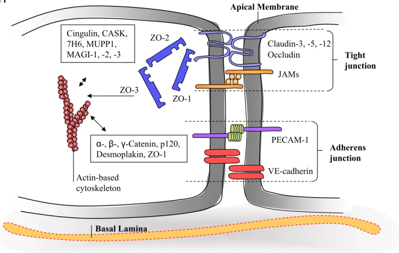
Another type of junction that contributes to BBB permeability and intermingles with the TJs in ECs is the adherens junction (AJ). AJs of BBB-ECs are primarily composed of platelet-endothelial cell adhesion molecule (PECAM)-1 and vascular endothelial cadherin (VE-cadherin) which interact with intracellular proteins, including the different catenins (isoform α, β, γ), ZO-1, p120 and desmoplankin, therefore creating an association with the actin cytoskeleton54-63. In addition, catenins are also crucial signaling molecules which can enter the nucleus and induce gene transcription upon AJ disruption64. AJ expression temporally precede the expression of TJs at intercellular contacts, and may be necessary for proper TJ formation as recent reports demonstrated that VE-cadherin expression and clustering modulates the activity of various transcription factors leading to an upregulation of claudin-5 gene transcription in ECs65. To summarize, the various proteins involved in the junctional complexes are illustrated in Figure i.
Claudin-3, -5, -12 Occludin JAMs Tight junction Adherens junction PECAM-1 VE-cadherin ZO-2 ZO-3 ZO-1 Actin-based cytoskeleton α-, β-, γ-Catenin, p120, Desmoplakin, ZO-1 Cingulin, CASK, 7H6, MUPP1, MAGI-1, -2, -3 Basal Lamina
Figure i: Schematic illustration of the Tight and Adherens Junction of BBB-ECs. TJs are compromised of the transmembrane proteins occlduin and the claudins, which are similar in homolgy and JAMs. BBB-EC AJ consists mainly of PECAM-1 and VE-cadherin. The cytoplasmic region contains adaptor proetins which link TJ and AJ proteins to the actin cytoskeleton. ZO-1, through its protein-protein domain, can bind the cytosolic tails of transmembrane proteins. With the help of other cytosolic proteins, such as Cingulin, CASK, MAGIs, TJs can be associated to the cytoskeleton. Catenins are the main adaptor protein for AJs, interacting with additional proteins such as desmoplankin, p120 and ZO-1, to stabilize the junctions.
2.0 CUES IN THE VASCULATURE
The cues and receptors most known to be involved in vasculogenesis and angiogenesis are largely specific to the cardiovascular system, such as vascular endothelial growth factor (VEGF) and components of the transforming growth factor (TGF)-β signaling pathway, angiopoietins (Ang)-1 and -266. VEGF and Ang-1 are potent mitogens and survival factors for the vasculature as they stimulate the initially scattered EPCs to proliferate, fuse together and form a primitive plexus of vessels that consequently enlarges and remodels, in the end, to develop into highly conserved organ-specific vascular patterns7,66. For precise and functional vascular networks to be reproduced, additional signals assist in the temporal and spatial guidance and proliferation of cells within the developing vasculature, permitting pervasive navigation of blood vessels to their appropriate target sites and to acquire a mature vasculature that is functionally and elaborately adjusted to its organ-specific environment, such as the CNS.
2.1 Glial cues control of the vasculature and blood-brain barrier function
Barriergenesis and the unique EC properties in the CNS are not predetermined by brain-specific EPCs but are induced by the local neural environment during the development of the vascular system. Persistence of a functional BBB throughout adulthood is thus similarly maintained and regulated by numerous brain-derived factors11,67. The complex communication and diverse signaling events between BBB-ECs and surrounding astrocytes, pericytes and microglia, allow for regulated BBB functions and promote TJ maintenance, metabolic and specialized transporter expression as well as immune quiescence of the brain ECs68. These proximate signals also promote rapid regulation and remodeling of the BBB, a phenomena crucial for maintaining CNS homeostasis in response to physiological and pathological stimuli8,11,15,21,24,45,69-72. One relatively important cellular component of the neurovascular unit comprises of pericytes which are believed to play a role in the maturation and maintenance of the BBB by
secreting growth factors and producing ECM constituents73. Pericytes contact ECs via N-cadherin-dependent binding74, and are reported to secrete TGF-β75, Ang-1 and -276,77, and platelet-derived growth factor (PDGF)78, which promote vascular maturation, integrity and BBB impermeability76. Although pericytic abnormalities and dysfunctions have been associated with CNS pathologies such as hypertension, Alzheimer’s disease and stroke, their exact nature and function in regards to the BBB remain under investigation73,76,77.
Other cellular constituents of the neurovascular unit which are crucial inducers of BBB properties are the astrocytes. Astrocyte-BBB-EC interactions are known to regulate vascular proliferation, angiogenesis, transporter protein expression, TJ protein expression and morphology and finally, inflammatory responses in the brain8,11,32,43,68,71,79-83. Astrocytic endfeet, separated only by a thin, but compact basal lamina, contact and ensheathe the entire surface of brain and spinal cord blood vessels84,85. The importance of astrocytes on BBB properties becomes clear when BBB-ECs are isolated and cultured in
vitro in the absence of astrocytes. BBB-derived ECs rapidly lose some of their barrier
characteristics, such as P-glycoprotein and TfR expression86,87. BBB function can be reinstated by co-culturing BBB-derived ECs in the presence of astrocytes or supplementation of astrocyte-conditioned media (ACM)43,88,89. Conversely, culturing non-neural ECs in the presence of astrocytes, or astrocyte-secreted factors, induces BBB-specific properties, such as P-glycoprotein and TJ expression11,68,81,90. These observations underscore the importance of signals provided by astrocytes and provide evidence that factors needed for reliable BBB function are at least partly soluble and secreted91. A number of studies have identified a variety of soluble and contact-dependent factors provided by astrocytes. Taken together, the microenvironment of the healthy brain and cellular interactions within the neurovascular unit provide inductive and maintenance signals for the BBB.
Astrocytes can be induced to secrete classic angiogenic factors that are important in vascular growth. VEGF, primarily acting through Flk1 receptor, is a potent mitogen for endothelial cells92 and is required for the formation, remodeling and survival of embryonic blood vessels as VEGF deficient embryos develop few or no angioblasts and die prematurely93. Later in development and adulthood, astrocyte-derived VEGF can be induced under hypoxic conditions94 to stimulate angiogenesis and, in consequence,
provokes BBB disruption95-97. On the other hand, Ang-1 and -2 bind to the receptor tyrosine kinase Tie-2 on ECs. Whilst Ang-2 activates angiogenesis and is expressed in early phases of BBB breakdown98, Ang-1 is involved in vascular maturation and quiescence, events that are characteristic of BBB differentiation99,100. In fact, Ang-1 causes a time- and dose-dependent decrease in endothelial permeability by upregulating TJ expression100. Astrocyte-secreted thrombospondin (TSP)-1 and -2 also have anti-angiogenic properties, inhibiting EC proliferation and has been suggested to promote vascular maturation101. Thus, a balance between angiogenic and BBB promoting agents occurs at the level of brain ECs.
Additional potential cues that promote BBB maintenance include members of the fibroblast growth factor (FGF) family which have the ability to decrease EC permeability102,103. In fact, FGF-2 and -5 -/- mice have decreased occludin and ZO-1 expression and show defects in barrier functions103. Astrocytic secretion of glia-derived neurotropic factor (GDNF) has also been seen to increase BBB integrity via its ligation to EC-expressed GDNF receptor α1104. Src suppressed C kinase substrate, also known as SSeCKS, produced by astrocytes, regulates angiogenesis, by decreasing the expression of VEGF and stimulates expression of Ang-1, augments ZO-1 and claudin-1 protein expression and decreases cerebral EC monolayers permeability 81. Additional novel astrocytic factors, recently identified by Dr. Kim and colleagues, include meteorin and A-kinase anchor protein-12 (AKAP12), both which are capable of regulating barrier development and formation via induction of TSP-1 and-2 expression and reduction of angiogenic factor secretion101,105. TGF-β, a well known lymphocyte-produced cytokine with multiple and complex physiological functions, is also known to be secreted by astrocytes and ECs and to induce a downregulation of the level of leukocyte migration across ECs in vitro and in vivo in a dose-dependent manner106-108. Finally, work by Wosik and colleagues have shown that astrocytes secrete angiotensinogen and angiotensin II (AngII) which decreases BBB permeability. Angiotensinogen -/- mice have a diminished expression of occludin and disorganized TJ strands in brain capillaries, supporting the notion that AngII is involved in the formation of BBB-EC TJs43. Therefore, the current view is that astrocytes are key regulators of BBB development, maintenance and regulation and that understanding the complex astrocyte-BBB-EC interactions under
physiological and pathological conditions may lead to the development of novel therapeutic strategies.
2.2 Neural cues control of the vasculature
The search for additional cues that could potentially promote barrier formation can benefit from drawing on similar processes which utilizes signaling molecules. Prior to barriergenesis, vascular development requires directional guidance to establish a precise, conserved branching pattern in the vertebrate body, including those in the brain9. Due to the nutrient and oxygen demand of neurons, the development of the vascular system is interconnected to the development of the nervous system. Recent evidence suggests that blood vessels and nerves share common mechanisms to expand, navigate and mature during development, as well as post-natally109-111. In fact, some neurons follow the path of blood vessels and inversely, the differentiation and branching of certain blood vessel depends on the emergence of new neural circuits112,113. It is now clear, that many guidance cues, first found and characterized for their function in neural development, are recently also recognized to function during vascular development. The vascular network shares much of the extensively studied neuronal navigational methods to guide its blood vessels and many parallels have been drawn between the two systems. In neurons, the growth cone at the tip of the axon is a highly motile structure that explores the microenvironment by extending finger-like filopodia and veil-like lamellipodia. These extensions contain guidance cue receptors that have the potential to activate various signaling cascades affecting the arrangement and composition of the cytoskeleton and therefore impact on the directional movement and elaboration of the growing axon114. Recent studies have shown that in nascent capillary sprouts, endothelial tip cells can be seen as the vascular equivalent of the growth cone as they act as sensors, signal transducers and motility devices than can regulate extension of capillary sprouts109. Although tip cells proliferate minimally, endothelial stalk cells further down the sprout, divide to generate new vessels along the path chosen by the tip cell115. Like the axonal growth cone, the tip cells can extend filopodia and sense guidance cues in ECM or on
neighboring cells that dictate the direction of growth of sprouting capillary. Fusion of tip cells and lumen formation allows the generation of new vessels. Interestingly, both vessels and axons respond not only express but to common cues109-112.
The long search for morphogens and guidance cues culminated in the identification of several highly conserved, distinct but multifunctional protein families. Members of the Wnt, Hedgehog (Hh), FGF and bone morphogenic protein (BMP) families were shown to act as classical morphogens in a number of different contexts. On the other hand, proteins of the Netrin, Slit, Ephrin and Semaphorin families were found to act as guidance cues for migrating cells and axons. Morphogens are expressed early in development in spatially very distinct patterns. Through the formation of gradients (for example along a developing body- or organ-axis), morphogens can directly induce different cell fates in a concentration-dependent manner. Thus, morphogens govern the pattern of tissue development and the position of various specialized cell types within a tissue116,117. Diffusible or cell surface-bound guidance cues can attract, repel or induce turning of growing axons and thus aid in the pathfinding process of neurons118-120. With recent studies, axon guidance functions have now been documented for members of each of the three major families of classical morphogens: Hhs, BMPs and Wnts, clearly demonstrating the complexity of these developmental cues121-123. In addition to these groups of major signaling molecules being active in early developmental processes, they are utilised repeatedly in a variety of different organs and systems.
Most of the morphogenic/guidance pathways have been studied in other angiogenic processes109,124-129. EC differentiation and vascular morphogenesis are dependent on morphogenic factors such as Hh, Wnt and BMP127,130,131. Furthermore, Hh has been associated with several stages of vascular development, which will be reviewed later on (section 3.0). In analogy to the growth cone, the extending filopodia of the endothelial tip cell express Netrin-receptor UNC5, Slit-receptor Robo4, and Ephrin-receptors EphA and B, interpreting signals as attractive or repulsive when blood vessels navigate in through environment129,132,133. For example, loss of UNC5H2/B or Netrin-1 in mice and zebrafish lead to abnormal vessel guidance and excessive vessel branching, suggesting they provide critical repulsive guidance for navigating blood vessels124,134. Netrin-4, however, seemed to be involved in angiogenesis after ischemic injury as it
upregulates in astrocytic endfeet and blood vessels 135. Similarly, Slit-2 inhibits the migration of Robo4-expressing ECs, while attracting ECs expressing the receptor Robo1125,133. EphrinB2 seems to provide a repulsive cue for intersomitic vessels136. In addition, Sema4D was seen to actually induce blood vessel formation, tubologenesis on top of EC migration. In fact, Semaphorin receptor Neuropilin-1 has now been recognized as a co-receptor for VEGF isoforms, suggesting complex interactions between vascular and axonal networking137,138. In fact, capillary sprouts radially invade the developing neuroectoderm along concentration gradients of VEGF113. While angiogenic factors are seen to act as chemoattractant cues, guidance and morphogenic cues can, on the other hand, act as angiogenic factors. Netrin-4 can increase blood-vessel density by enhancing blood vessel proliferation135 and BMP2 can both play a chemotactic role on angioblasts and microvascular ECs as well as stimulating their proliferation and inducing tube formation139. While EphrinB2/EphB4 deletion resulted in a general failure in angiogenic remodeling of vascular plexus, EphrinA1 may regulate post-natal angiogenesis129. However, much controversy remains in this new area of research, concerning the expression and function of axonal guidance cues in angiogenesis and EC maintenance.
This recycling of molecules during development suggests that these multifunctional proteins act globally in providing positional information in a variety of developmental processes such as embryonic patterning, CNS induction, and angiogenesis. In addition, many of these morphogens and axon guidance cues are also important in post-natal and adult processes that utilize and recapitulate embryonic processes such as maintenance of organotypic tissue, remodeling after repair and barriergenesis.
2.3 Neural cues control of blood-brain barrier function
Due to the close interaction of BBB-ECs with CNS processes, it is perhaps not surprising that cues typically associated with neural processes have thus been shown to impact on BBB-ECs. As mentioned previously, FGF-2 and -5 -/- mice have decreased occludin and ZO-1 expression and show defects in barrier functions103. BMP signaling
has also been seen to be important in astrocyte-endothelial interactions, where the loss of the receptor BMPR1a leads to an increase in leakage of Evans Blue and immunoglobulins (class G; IgG) through cortical vessels in vivo140. Stimulation of Robo4 on ECs with its ligand Slit-2 induced barrier function in vitro. In vivo, Slit-2 prevented VEGF-induced hyperpermeability and Evans Blue leakage into tissues, implying their role in vascular bed maturation133. Recent studies have shown that the canonical Wnt pathway is important for vascular development and for the formation of a mature BBB 141-143. The Wnts are secreted glycoproteins that accumulate in the ECM that trigger activation of the Frizzled receptor on adjacent cells. Frizzled activation promotes β-catenin stabilization and entrance into the nucleus, where it can modulate target gene transcription. The Wnt pathway is activated in brain ECs during embryogenesis, where it is required for CNS angiogenesis and capillary bed formation141. Together, Wnt 7a and 7b, together, are essential for CNS vascular development as Wnt7a/b double -/- mice display severe CNS-specific hemorrhaging and enhanced vascular fragility through destabilization of inter-endothelial contacts144. In vitro, Wnt3a induces claudin-3 upregulation in mice brain vascular ECs142 and Wnt7a elicits strong migration of mouse brain ECs across a Boyden chamber filter and induces the expression of BBB-specific transporter Glut-1141. Post-natally, conditional loss-of-function of B-catenin in mouse ECs has decreases claudin-3 expression and Evans Blue extravasation in the CNS-parenchyma142. Additional studies on CNS-related signaling molecules could potentially lead to identifying other BBB promoting factors.
3.0 THE HEDGEHOG (Hh) SIGNALING PATHWAY
The Hh pathway is a conserved signaling cascade involved in embryonic morphogenesis, pathfinding, and as recent studies show, angiogenesis. There are three vertebrae homologues of the originally identified Hh in Drosophila melanogaster: Desert Hh (Dhh), Indian Hh (Ihh) and Sonic Hh (Shh). All three activate the same signaling pathway and the presence of divergent regulatory elements cause spatial and temporal differences in their expression patterns during embryogenesis145-149. While Ihh and Dhh are best known for their importance in chondrocyte and gonadal differentiation respectively146, Shh has been widely associated with morphogenic events such as posterior identity of the limb bud150, ventral cell identity of the neural tube, and motor neuron development151,152. As other morphogens, Shh has also been characterized as an axonal guidance cue, capable of both stimulating commissural axon migration towards the floorplate 121,122 and repelling them after the axons have crossed the floor plate and turned rostrally153. Recent studies suggest that the Hh pathway may also play an important role in adult tissue homeostasis, neural progenitor proliferation and wound healing, which all draws parallels to the embryonic developmental processes 130,154.
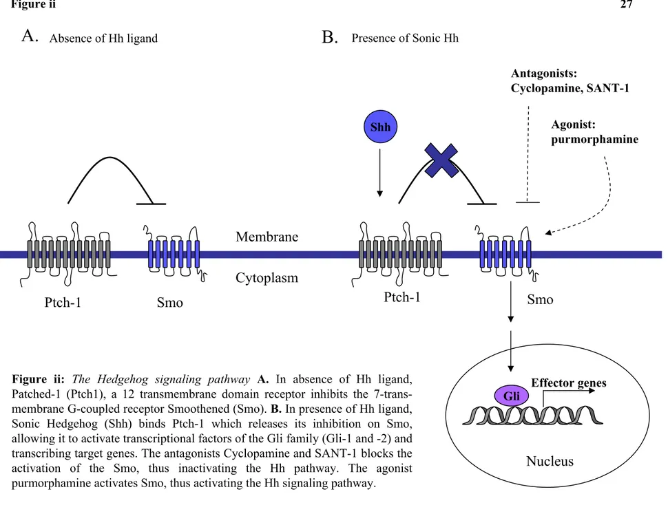
Shh is initially translated as a ~45kDa precursor protein. Essential to Hh signaling is the internal autocatalytic cleavage of the 45kDa protein mediated by the carboxy (C)-terminal of Hh, yielding two products, the ~25kDa C-terminal fragment and the ~20kDa amino (N)-terminal signaling domain. During autoproteolysis of the precursor protein, the C-(N)-terminal fragment becomes a functionally active cholesterol transferase that covalently adds a cholesterol molecule to the C-terminus of the N-terminal fragment. A palmitoyl moiety is also added to the N-terminal of the processed signaling molecule by Skinny Hh 145,155,156. These two lipid modifications control, in part, the ability of the Hh molecule to bind to its receptor and to signal at short- or long range. Following the post-translational addition of cholesterol and palmitoyl moieties, Shh is released from the producing cell with the help of the 12-transmembrane protein Dispatched130. Tethered to the cell surface or secreted, Shh binds with high affinity to the cell surface receptor Patched (Ptch-1)-1, a 12-transmembrane domain receptor. In mammals, Ptch-2 has been isolated but not much is known about its exact role in
the signaling pathway157. Binding of Shh to Ptch-1 alleviates repression of the signal transducer Smoothened (Smo), a serpentine transmembrane protein with a topology reminiscent of Frizzled family of Wnt receptors and other G-protein coupled receptors158. Although the exact mechanism in which Ptch-1 inhibits or activates Smo is unknown, it is speculated that Smo is regulated by an undiscovered small molecule, such as oxysterols or vitamin D3 that would induce conformational changes within Smo159-161. The interaction of Hh with Ptch-1 can be promoted by its binding to two other transmembrane proteins Boc and Cdon. Several other surface proteins can bind, mobilize and limit the range of Shh movement such as Hip (Hh interacting protein), a membrane bound glycoprotein which sequesters Hh ligands, but has no effect on Smo activity and thus attenuates the signal162. Once Smo is in its active conformation, activation of zinc-finger transcription factors of the Gli family (Gli-1 through -3) act at the last step of the Shh-signal-transduction pathway145,147 (Figure ii). Activation of cytoplasmic-bound Gli components is achieved through their sequential phosphorylation by various kinases, allowing them to translocate into the nucleus and induce or repress transcription of target genes. Elegant genetic studies have exposed both the redundant and unique functions of the three Gli transcription factors. Gli-1 and Gli-2 act primarily as activators, while Gli-3 can undergo autoproteolysis to function as a transcriptional repressor. Thus, the ratio of the Gli activator to Gli repressor forms of the proteins is critical in final transcription processes163. In addition to the induction of specific cell fate determinants in response to the Hh signal received, proteins of the Hh cascade itself, such as Ptch-1, Hip and Gli-1 are upregulated. It is thought that this positive feedback upregulation of expression results in increased levels of Ptch-1 and Hip protein at the cell membrane, further sequestering Hh and limiting spreading of the signal 146,147,164. Therefore, Ptch-1, Hip and more often, Gli-1 mRNA or protein upregulation assays are used as indicators of Hh pathway activation.
Given that Hh signaling is a critical developmental pathway during embryogenesis and organogenesis, mutations in pathway components result in congenital defects. The forebrain of the fetal Shh mutant fails to develop into two hemispheres, or holoprosencephaly, resulting in defects in the development of proper brain structures and functions as well as facial properties 150,165. In human foetuses, loss of one Shh allele is enough to cause varying penetrance up to and including cyclopia (fusion of developing eyes at the midline), whereas both alleles need to be lost in the Shh -/- mouse to produce the same phenotype166-169. In mouse embryos, deletion
of Shh leads to cyclopia and defects in ventral neural tube, somite and foregut patterning. At later developmental stages, defects include severe distal limb malformations and failure of lung branching150,170,171.
In addition to the vital role Hh plays in embryonic development, Hh signaling also functions as a regulator of cell proliferation, differentiation and survival in adult tissue. The importance of Hh functions, in the adult, is reflected in the loss of growth control when the pathway is damaged. Studies have shown that inappropriate reactivation of this pathway later in life can lead to the development of malignancies as Hh signaling is a molecular hallmark of several subsets of familial and sporadic tumors, including basal cell carcinoma syndrome172, medullablastoma146,173, pancreatic174, breast175, prostate176, gastrointestinal177 and lung cancers178,179. In addition, the family of Gli genes are named for their role in glioblastoma formations180.
The highly teratogenic steroidal alkaloid toxins, cyclopamine and jervine, are extracted from corn lilies. Early gestating sheep grazing on wild corn lilies resulted in severe nervous system defects in their offspring that exhibited strong resemblances to holoprosencephaly with associated cyclopia. The toxins were later characterized to act as antagonistic compounds binding Smo and inhibiting Hh signaling181. Pertussis toxin (PTX) has also been seen to inhibit Smo activity through its ability to prevent G proteins from interacting with their G protein-coupled receptors, even though no known G protein has been shown to bind to G-protein-coupled receptor-like Smo182. High-throughput screens of chemical libraries for Hh pathway modulators have identified four compounds that potentially inhibit Shh (Smo antagonist, SANT-1 through SANT-4) and two that activate Hh signaling (Smo agonist, SAG and purmorphamine), both by binding directly to Smo (Figure ii) 183,184. Of the antagonists, SANT-1 was found to exhibit the highest affinity for Smo and attenuated Shh stimulation to a much greater extent than the other antagonists183.
A.
Shh
Gli Effector genes
Ptch-1
Smo
Membrane
Cytoplasm
Nucleus
Antagonists: Cyclopamine, SANT-1 Agonist: purmorphamine Presence of Sonic HhFigure ii: The Hedgehog signaling pathway A. In absence of Hh ligand, Patched-1 (Ptch1), a 12 transmembrane domain receptor inhibits the 7-trans-membrane G-coupled receptor Smoothened (Smo). B. In presence of Hh ligand, Sonic Hedgehog (Shh) binds Ptch-1 which releases its inhibition on Smo, allowing it to activate transcriptional factors of the Gli family (Gli-1 and -2) and transcribing target genes. The antagonists Cyclopamine and SANT-1 blocks the activation of the Smo, thus inactivating the Hh pathway. The agonist purmorphamine activates Smo, thus activating the Hh signaling pathway.
Absence of Hh ligand
B.
3.1 The Hh signaling pathway in vasculogenesis
As other morphogens and axonal guidance cues, the Hh pathway has been associated with the vascular system. In the developing embryo, the visceral yolk sac is the first site of blood vessel formation in the murine embryo. Ihh was shown to regulate murine yolk sac angiogenesis as the Ihh -/- mice, as well as Smo -/- or cyclopamine-treated murine yolk sacs formed smaller, flattened, disorganized vessels that easily collapsed7,130,185. Smo deficient embryos display a more severely abnormal yolk sac phenotype than do Ihh -/-, suggesting that Shh may compensate for the loss of Ihh, but that it is not completely redundant185. In zebrafish models, Shh signaling is crucial for the organization of the angioblasts into major vascular channels such as the aorta and axial vein186,187. Similarly, in avian and mouse embryos, Shh and Ihh act together to mediate embryonic formation of aortic and coronary vessels4,187,188. Vokes and colleagues4 remarked that the first blood vessels within the embryo always formed in the mesoderm tissue in close proximity to the endoderm, and that an endodermic signal was essential for the assembly of angioblasts into tubular vessels. While VEGF treatment was not sufficient in inducing tubulogenesis in embryos lacking an endoderm, Shh was both found to be produced by the endoderm and to promote the assembly of angioblasts into vessel tubes. This elegant study showed that while VEGF was important in EPC differentiation, Shh signaling was crucial for the structural morphological formation of the vessels. Although at first, no obvious vascular defects were observed in Shh deficient mouse embryos150, other groups have now described decreased and abnormal vascularization in tissues such as the lung, heart, pharynx, and pulmonary tissue171,189. Cyclopamine-treated embryos exhibited a variety of vascular abnormalities such as interrupted tubes, unassembled clusters of angioblasts and fewer vessel tubes, compared to control embryos4. In embryos treated with cyclopamine at embryonic stage E8.5-10.5, neural tube angiogenesis was also impaired, as perineurial vascular plexus vessel sprouting does not occur190. On the other hand, hypervascularization of neuroectoderm occurred in response to Shh overexpression in the dorsal neural tube of zebra fish191 and overexpression of Shh by injection of Shh mRNA causes the formation of lumenized ectopic vessels187.
3.2 The Hh signaling pathway in angiogenesis
During angiogenesis, remodelling of the vasculature is a process that depends on migration and reassembly of ECs. Through Smo activation, Shh can provoke cell morphology alterations together with formation of lamellipodia, suggesting a migratory response192. Furthermore, embryonic mouse yolk sac ECs responded to Shh treatment by activation of gene transcription involved in cytoskeletal organization, migration, and angiogenesis, as detected by GeneChip assays193. Wound healing is often seen as a recapitulation of angiogenesis as it requires recruitment of EPCs and endothelial tip cell chemoattraction to the target area154,193. Added exogenously, Shh significantly enhances wound healing and migration of embryonic ECs in wounded areas in a scratch assay, a process mediated through migration rather than cellular proliferation193. In an in vivo study of wound healing in mice, Shh treatment resulted in smaller wounds after 5 days, compared to control, a possible result of bone-marrow derived EPC recruitment, increased cellular infiltration, collagen deposition and enhanced overall vascularity in the wounded area154. As a morphogen, it is perhaps expected that several in vitro studies have shown that in addition to migration, Shh binding to Ptch-1 mediates capillary formation and morphogenesis, in mature murine brain capillary ECs, HUVECs and putative angioblasts from the bone marrow4,154,182. In these cells, Shh treatment increased nuclear Gli-1 localization and mediates gene transcription and protein synthesis as actinomycin D and cycloheximide, respectively, inhibit capillary morphogenesis. Cyclopamine and PTX also suppress Shh-inducing capillary morphogenesis182. By augmenting cytoplasmic extensions and increasing cellular contacts between cells, Shh promotes cell type specific cellular adhesion which is important in structure formation. Similar models in drosophila proposed Hh-mediated cell adhesion pathways as a mechanism of wing imaginal disc formation194.
Shh signaling stimulates vascular remodelling and angiogenesis in various tissues at later developmental stages195. Decreased levels of Shh signaling as a result of treatment of mice with a Shh neutralizing antibody or with cyclopamine leads to angiogenic malformation and loss of ability of existing vessels to remodel, fuse and form branches, causing malformations and haemorrhages. In particular, Shh-supported processes such as the development of branchial region vessels and pulmonary arteries are defective in Shh null mice embryos196. Postnatal and adult angiogenesis can also be enhanced by Hh signaling. Ptch-1 was
also found to be normally expressed in cardiovascular tissues of juvenile and adult mice and activation of Hh signaling in the adult heart is sufficient to promote coronary neovascularization and protect from ischemia, implying an important role for Shh in maintaining cardiac homeostasis and function. Temporal and tissue-specific deletion of Smo lead to reductions in proangiogenic gene expression and loss of coronary vasculature, which resulted in tissue hypoxia, cardiomyocyte apoptosis, ventricular failure and subsequent lethality197,198. When Shh is administered to aged mice, new vessel growth can be observed in adult corneas and ischemic hind limbs. Indeed, Shh treatment can promote an increase in capillary density, blood flow and vessel diameter, suggesting neovascularization199. Similarly, implantation of Shh-containing pellets promotes neovascularization and formation of large, well-organized branched vessels in corneal angiogenesis assays in mice199,200. In perspective, the Hh pathway possibly aids in tumor angiogenesis. The fact that the usually high expression of Hip on ECs is diminished in cancers supports the hypothesis that Hh is enhanced in tumor tissues and contributes to tumor angiogenesis and growth201.
Shh not only can be viewed as a potent angiogenic agent, but activation of the Hh pathway can also have an indirect role in angiogenesis, by acting upstream of angiogenic factors. One of the first publications on the Hh pathway in angiogenic processes describes the ability of Shh to upregulate angiogenic factors such as VEGF (all three isoforms of VEGF1, -189, -165, -121) and both Ang-1 and Ang-2 in mesenchymal fibroblasts, possibly placing Hh upstream of these vascular-specific growth factors and vessel stabilization potentials199. However, Shh treatment did not seem to have an effect of EC migration and proliferation, implying that EC response may be dependent on their differentiation status, developmental age and organ origin193,199. Since, others have observed VEGF and Ang-1 upregulation in other contexts, such as in Shh-treatment of human EC lines202, and coronary vasculature198. Blocking Shh with neutralising antibodies or cyclopamine inhibits VEGF upregulation and thus angiogenesis, as seen in mouse models of hind limb and cornea ischemia199,203. Ang-1 and its receptor Tie-2 are even downregulated in the lungs of Shh -/- embryos204. These studies suggest that the Hh signaling pathway is important in vascular proliferation and differentiation as well as in vitro capillary morphogenesis.
3.3 Evidence of the Hh signaling pathway in barrier function
Little is known for the Hh signaling pathway during the last step of vasculature maturation, barriergenesis. In the developing mouse submandibular gland, lumen formation occurs in the epithelial cell lining, leading to the establishment of apical-basal polarity and TJ formation, processes reminiscent of EC barrier formation. Shh null mice exhibit developmentally arrested submandibular gland epithelium, whereas treatment with Shh enhances epithelial lumen formation, full cell polarization and ZO-1, claudin-3 and occludin TJ protein distribution in these deficient explants205. In addition, laminin-5 deposition at the basal lamina region of terminal buds was accelerated in Shh-treated glands205, suggesting a role of Shh in both junctional and extracellular cell attachment. Importantly also, in the peripheral nervous system (PNS), Dhh is expressed in myelinating Schwann cells. This Dhh signal is needed for the formation and maintenance of the perineurium, a component of the blood-nerve barrier that acts as a barrier to protect against serum molecules and cellular infiltration206. Comparable to the endothelial lining of blood vessels, perineurial cells are sealed by tight and gap junctions and are supported by a prominent basal lamina, thus forming an effective diffusion barrier. In Dhh -/- animals, the perineurium is looser, and the basal lamina encloses less collagen and is overall discontinuous. In addition, perineurial cells of Dhh null mice lack the gap junction protein connexion43, while expression of TJ proteins occludin and ZO-1 is disorganized. Freeze fracture electron microscopy further revealed abnormal and interrupted TJ arrays in Dhh null mice when compared to normal control perineurium TJ strands, leading to a defective nerve-tissue barrier, shown by extravasation of Evans Blue dye and immune cells invasion into the endoneurium206. Similarly, after a crush injury, more macrophage infiltrations is visible in both injured and non-injured nerves in Dhh -/- mice207. Therefore, the Hh pathway is likely to be involved in the formation of an ordered and functionally competent perineurium, seen as a later stage of perineurial sheath maturation208. By analogy, these studies raise the question of whether the Hh pathway plays a role in other barrier contexts, such as the BBB.
4.0 THE BASAL LAMINA
The basal lamina, found between the BBB-ECs and perivascular cells, is composed of several ECM components, including fibronectin, laminins, collagen type IV and heparin sulfate proteoglycans209-211. The basal lamina functions as a tissue boundary on which cells are attached to and a substrate for cellular differentiation and induction of gene expression. Astrocyte, and to a lesser degree, pericyte-derived ECMs enhanced BBB integrity, demonstrating the importance of additional glial matrix constituents209,211,212. For example, astrocytes can express agrin which could serve as a basal membrane factor maintaining barrier functions, as loss of agrin in human glioma accompanied a loss in TJ8,213. In addition, soluble factors secreted by glial cells can be captured by ECM proteins, and thus increase their local concentrations. BBB-ECs, in return, constitutively express a number of integrins that are important for EC adhesion to the basal lamina and control many signaling events critical for cell survival, growth and gene expression210,214,215. Integrins are heterodimers of α- and β- integrin and pairs such as α1β1, α3β1 and α6β1 are commonly expressed on the cerebrovasculature and recognize the basal lamina. Alterations in expression of integrins have been shown to correlate with angiogenesis, BBB breakdown and increased barrier permeability during focal cerebral ischemia and/or MS. For instance, appearance of αvβ3 on BBB-ECs was found to be significantly upregulated after the onset of focal ischemia 210,214. This suggests that integrin receptor function is needed for the maintenance of the BBB.
5.0 IMMUNE INTERACTIONS AT THE BBB
Under physiological conditions, a low number of immune cells, or leukocytes, continuously cross the BBB and monitor the CNS in a process called immune surveillance. The BBB-ECs are, thus, the first cells leukocytes must contact before entering the CNS and the molecular mechanisms by which leukocytes accomplish transendothelial migration is complex and involves a series of sequential and tightly controlled steps216. Initially, leukocyte rolling consists of a weak interaction between EC selectins, or cell adhesion molecules (CAMs), and their ligands on leukocytes. Since the CAM-ligand interaction is originally one of low-affinity, leukocytes can roll along the blood-vessel wall and sense activation factors deposited on the EC surface, such as various chemokines217.
During inflammatory conditions, BBB-ECs can actively influence and participate in neuroinflammatory reactions by regulating cytokine/chemokine and CAM expression, both directly affecting immune cell migration into the CNS218-221. In addition to the ECs, CNS parenchymal cells can become activated by inflammatory cytokines to secrete chemokines in their vicinity. With over 50 identified members, chemokines constitute a large group of small cytokines (chemotactic cytokines) which are classified, according to their cystein configuration, into two main families, the CC and CXC chemokines. In response to inflammatory challenge, BBB-ECs can produce and secrete pro-inflammatory among others, monocyte chemoattractant protein (MCP)-1, also known as CC ligand (CCL)-2, MIP-1α/CCL3, RANTES/CCL5, interleukin (IL)-8/CXCL8, and IP-10/CXCL10217,222-224. However, MCP-1/CCL2 and IL-8/CXCL8 were seen as the major chemokines produced by BBB-ECs grown under culture conditions and upregulated upon inflammatory conditions225. All these chemokines bind specific G-protein coupled receptors on the leukocytes, rapidly inducing integrin conformational changes through an outside-in signaling mechanism that promote their high-affinity interactions to the cell CAMs expressed on BBB-ECs.217
In addition to the tight adherence, EC expression of CAMs and leukocyte integrins also mediate the ensuing diapedesis of immune cell across vascular beds. Under
normal conditions, CNS-ECs express low levels of all the appropriate CAMs, such as intercellular CAM-1 (ICAM-1) and vascular CAM-1 (VCAM-1)226-228. However, in the presence of inflammation, these molecules are strongly upregulated, further promoting leukocyte arrest and transmigration. Recent studies have identified additional CAMs that regulate the transmigration of lymphocytes EC barriers by the means of ALCAM (activated leukocyte cell adhesion molecule or CD166)23 and Ninjurin-1 229 which, for example, play important roles in invasion of particular subsets of leukocytes such as CD4+ T cells and monocytes, respectively, from the blood to the CNS, especially during inflammatory contexts.
The final step of immune cell transmigration across the BBB consists of arrested leukocytes crawling along the surface of inflamed endothelium toward inter-endothelial vascular junctions. Homophilic interactions with junctional molecules such as PECAM-1 and JAMs results in junctional loosening, leukocyte extravasation and eventual basal lamina breach through gaps of low laminin expression217,230,231. Further proteolysis of the compact basal membrane surrounding the BBB by matrix metalloproteinases occurs to permit leukocyte entry into the parenchyma.
5.1 Multiple sclerosis (MS) and experimental autoimmune encephalomyelitis (EAE)
In non-pathological conditions, BBB-ECs control the CNS microenvironment and limit the entrance of immune cells by expressing low levels of integrin ligands and activation markers, to achieve a precisely regulated biochemical and immunological homeostasis essential for reliable CNS function. Immune cell infiltration and BBB breakdown is associated with many CNS diseases, including Alzheimer’s disease232, HIV-1-associated encephalomyelitis233, stroke234, amyotrophic lateral sclerosis235 and especially, MS79,223. MS is an inflammatory and demyelinating disease of the CNS characterized by multifocal perivascular immune infiltration, leading to demyelination and astrogliosis (astroglia activation and proliferation)236.
CD4+ T cells, also called T helper (Th) cells, are a part of the adaptive immune system which generally recognizes foreign peptides, or antigens, and subsets of Th cells
are generally distinguished by their cytokine secretion profile. Th1 associated soluble mediators, interferon (IFN)-γ and tumor necrosis factor (TNF)-α, are pro-inflammatory cytokines and MS has been considered to be initiated by auto-reactive CD4+ Th1 cells that migrate across the BBB to access the CNS237,238. In fact, MS is generally considered a CD4+ T cell-mediated inflammatory disease, based on the cellular composition and number of immune cells that infiltrate the brain and cerebrospinal fluid (CSF) and on data from the mouse model of MS, experimental autoimmune (allergic) encephalomyelitis (EAE). Th17 cells are another subset of Th cells which secrete IL-17 and IFN-γ and has often been implicated in autoimmune diseases as these cells are found to be in MS lesions and IL-17 deficient mice significantly suppress EAE disease scores239,240. Finally, Th2 cells secrete anti-inflammatory cytokines such as IL-4 and interferon (IFN)-β, thereby downregulating and suppressing the effects of pro-inflammatory Th1 and 17 effects. In fact, IFN-β is currently used as a therapy for MS due to immunomodulatory abilities241.
Oligodendrocytes are CNS-derived glial cells responsible for the myelination of neurons. In MS, oligodendrocytes are a central target of the attacks by the host immune system242,243. Autoreactive CD4+ T cells have been found to recognize protein components of the myelin sheathe, such as myelin oligodendrocyte glycoprotein (MOG), proteolipid protein (PLP) and myelin basic protein (MBP), and initiate thus local inflammation, axonal demyelination lesions, and plaque formation236. Leukocytes accumulate in these lesions, most notably in the perivascular space surrounding small blood vessels. Continuous chronic inflammation can lead to recruitment of additional immune cells, furthering demyelination, axonal injury and glial scaring. In EAE, the injection of defined myelin components, together with an immune stimulating adjuvant, into naïve susceptible animals leads to a predominantly CD4+ T cell-mediated autoimmune diseases that shares similarities with MS. Whilst EAE cannot be transferred by antibodies, passively transferring encephalitogenic myelin-specific T cells in susceptible rodent and strands also leads to the diseases244. This demonstrates that EAE, and possibly MS, are T cell mediated autoimmune diseases. However, in certain EAE models used, the infiltration is characterized by a predominant infiltration of mostly CD4+ T cells into the spinal cord and brain stem, which are significant differences between the rodent model and the human pathology245. However, to induce EAE, PTX is
given, which opens the BBB246,247. This suggests that EAE is a combination of at least two processes, autoreactive T cells, and BBB disruption.
In MS lesioned areas, the BBB becomes compromised and leaky, allowing entry of serum proteins and cells into the CNS. Clinically, BBB breakdown can be visualized by magnetic resonance imaging using gadolinium as a tracer molecule for lesions. Gadolinium leakage into the CNS parenchyma serves as a reliable marker for disease activity as it indicates inflammatory processes, making it an essential detection and diagnosis tool248,249. Gadolinium leakage may even precede the appearance of new lesions, suggesting potential BBB deregulation pathologies prior to inflammatory processes250. Lesion development is characterized by BBB activation and disruption in TJs organization, ensuing in changes in EC permeability42,64. Activated ECs, containing increased number of vesicles, decreased density of mitochondria, and expressing higher levels of CAMs, are also present in MS plaques251,252, which correlated with the accumulation of serum proteins in these areas. Nonetheless, active cell recruitment across BBB-ECs seems to be an essential step in contributing to CNS inflammation and subsequent tissue injury and BBB breakdown. Activated, myelin-specific leukocytes are primed through signaling via high levels of integrins and chemokine receptors, promoting their entry into CNS parenchyma. In fact, neutralizing antibodies (Natalizumab) against a leukocyte integrin suppress leukocyte entry into the CNS through inhibition of integrin-binding to its ligand on the BBB-EC253.
Upon entry into the CNS, leukocytes induce molecular changes in the nervous tissue and initiate transient or even chronic inflammatory reactions. Inflammatory cytokines such as IL-1, TNF-α, and IFN-γ, increase CAM expression and chemokine secretion by BBB-ECs and further modulate their TJs expression and function, aiding in the recruitment and entrance of more leukocytes to the site. BBB-ECs thus become an important source of pro-inflammatory chemokines, cytokines239,254 and CAMs23,72. This is the basis for the inflammatory cascade that leads to the focal inflammation of BBB-ECs and the recruitment and infiltration of additional immune cells. The infiltrated immune cells will subsequently secrete additional cytokines and chemokines that may further enhance CNS inflammation or promote a gradual resolution of the inflammation. CD4+ T cell-associated soluble mediators such as IFN-γ and TNF-α are found in elevated