HAL Id: dumas-02483860
https://dumas.ccsd.cnrs.fr/dumas-02483860
Submitted on 18 Feb 2020HAL is a multi-disciplinary open access
archive for the deposit and dissemination of sci-entific research documents, whether they are pub-lished or not. The documents may come from teaching and research institutions in France or abroad, or from public or private research centers.
L’archive ouverte pluridisciplinaire HAL, est destinée au dépôt et à la diffusion de documents scientifiques de niveau recherche, publiés ou non, émanant des établissements d’enseignement et de recherche français ou étrangers, des laboratoires publics ou privés.
and emergence of multi-resistant bacteria
Coralie Hardy Raad
To cite this version:
Coralie Hardy Raad. Bloodstream infections in immunocompromised children and emergence of multi-resistant bacteria. Pédiatrie. 2019. �dumas-02483860�
BLOODSTREAM INFECTIONS IN IMMUNOCOMPROMISED
CHILDREN AND EMERGENCE OF MULTI-RESISTANT
BACTERIA
THESE
Présentée à la Faculté de Médecine Hyacinthe BASTARAUD
des Antilles et soutenue publiquement à l’Université Claude Bernard à LYON et examinée par les Enseignants des deux Facultés pour le compte de la Faculté des Antilles
Le 15/03/2019
Pour obtenir le grade de
DOCTEUR EN MEDECINE
Par
RAAD Coralie née HARDY
Jury: M. ELENGA Professeur, Président du jury
M. BERTRAND Professeur
M. GILLET Professeur
Mme HALFON-DOMENECH Docteur en Médecine, Directrice de thèse
2019
HYACINTHE BASTARAUD
Liste des Enseignants
Le Président de l'Université des Antilles : Eustase JANKY Doyen de la Faculté de Médecine : Raymond CESAIRE
Vice-Doyen de la Faculté de Médecine : Suzy DUFLO
Professeurs des Universités - Praticiens Hospitaliers
Pascal BLANCHET Chirurgie Urologique
CHU de POINTE- À -PITRE/ABYMES Tel : 05 90 89 13 95
André-Pierre UZEL Chirurgie Orthopédique et Traumatologie
CHU de POINTE-A-PITRE/ABYMES Tel : 05 90 89 14 66
Pierre COUPPIE Dermatologie et vénéréologie
CH de CAYENNE Tel : 05 94 39 53 39
Thierry DAVID Ophtalmologie
CHU de POINTE-A-PITRE/ABYMES Tel : 05 90 89 14 55
Suzy DUFLO ORL – Chirurgie Cervico-Faciale
CHU de POINTE-A-PITRE/ABYMES Tel : 05 90 93 46 16
Eustase JANKY Gynécologie-Obstétrique
CHU de POINTE-A-PITRE/ABYMES Tel 05 90 89 13 89
François ROQUES Chirurgie Thoracique et Cardiovasculaire
CHU de FORT- DE - FRANCE Tel : 05 96 55 22 71
Jean ROUDIE Chirurgie Digestive
CHU de FORT- DE - FRANCE Tel : 05 96 55 21 01 - Tel : 05 96 55 22 71
Jean-Louis ROUVILLAIN Chirurgie Orthopédique
CHU de FORT- DE - FRANCE Tel : 05 96 55 22 28
André CABIE Maladies Infectieuses
CHU de FORT- DE - FRANCE Tel : 05 96 55 23 01
Philippe CABRE Neurologie
CHU de FORT- DE - FRANCE Tel : 05 96 55 22 61
Professeurs des Universités - Praticiens Hospitaliers (Suite)
Raymond CESAIRE Bactériologie-Virologie-Hygiène option virologie
CHU de FORT- DE - FRANCE Tel : 05 96 55 24 11
Maryvonne DUEYMES-BODENES Immunologie
CHU de FORT- DE - FRANCE Tel : 05 96 55 24 24
Annie LANNUZEL Neurologie
CHU de POINTE- À -PITRE/ABYMES Tel : 05 90 89 14 13
Louis JEHEL Psychiatrie Adulte
CHU de FORT- DE - FRANCE Tel : 05 96 55 20 44
Mathieu NACHER Epidémiologie, Economie de la Santé et Prévention
CH de CAYENNE Tel : 05 94 93 50 24
Magalie DEMAR - PIERRE Parasitologie et Infectiologue
CH de CAYENNE Tel : 05 94 39 53 09
Vincent MOLINIE Anatomie Cytologie Pathologique
CHU de FORT DE FRANCE Tel : 05 96 55 20 85/55 23 50
Philippe KADHEL Gynécologie-Obstétrique
CHU de POINTE-A-PITRE/ABYMES Tel : 05 90 89 13 20
Michel DE BANDT Rhumatologie
CHU de FORT- DE - FRANCE Tel : 05 96 55 23 52
Karim FARID Médecine Nucléaire
CHU de FORT- DE - FRANCE Tel : 05 96 55 21 67
Mehdi MEJDOUBI Radiodiagnostic et imagerie Médicale
CHU de FORT- DE - FRANCE Tel : 05 96 55 21 84
Rémi NEVIERE Physiologie
CHU de FORT- DE - FRANCE Tel : 05 96 55 20 00
Christian SAINTE-ROSE Radiodiagnostic et imagerie Médicale
CHU de FORT- DE - FRANCE Tel : 05 96 55 20 00
Sébastien BREUREC
Bactériologie & Vénérologie
CHU de POINTE- À -PITRE/ABYMES Tel : 05 90 89 12 80
Professeurs des Universités - Praticiens Hospitaliers (Suite)
Félix DJOSSOU Maladies infectieuses et tropicales
CH de CAYENNE Tel : 05 94 39 50 50
Nicolas VENISSAC Chirurgie thoracique et cardiovasculaire
CHU de FORT- DE - FRANCE Tel : 05 96 55 20 00
Moustapha DRAMÉ Épidémiologie, Économie de la Santé
CHU de FORT- DE - FRANCE Tel : 05 96 55 20 00
Christophe DELIGNY Médecine Interne
CHU de FORT- DE - FRANCE Tel : 05 96 55 22 55
Narcisse ELENGA Pédiatrie
CH de CAYENNE Tel : 05 94 39 77 37
Michel CARLES Anesthésie Réanimation
CHU de POINTE- À -PITRE/ABYMES Tel : 05 90 89
Professeur de Médecine Générale
Jeannie HELENE-PELAGE Médecine Générale
CHU de Pointe-à-Pitre / Cabinet libéral Tel : 05 90 84 44 40
Professeur Associé de Médecine Générale
Franciane GANE-TROPLENT Médecine générale
Cabinet libéral les Abymes Tel : 05 90 20 39 37
Maîtres de Conférences des Universités - Praticiens Hospitaliers
Jocelyn INAMO
Cardiologie
CHU de FORT- DE - FRANCE
Tel : 05 96 55 23 72 - Fax : 05 96 75 84 38
Fritz-Line VELAYOUDOM épse CEPHISE
Endocrinologie
CHU de POINTE- À -PITRE/ABYMES Tel : 05 90 89 13 03
Marie-Laure LALANNE-MISTRIH Nutrition
CHU de POINTE- À -PITRE/ABYMES Tel : 05 90 89 13 00
Moana GELU-SIMEON Gastroentérologie hépatologie
CHU de POINTE-A-PITRE/ABYMES Tel : 05 90 89 10 10
Maturin TABUE TEGUO
Médecine interne : Gériatrie et Biologie du vieillissement
CHU de POINTE-A-PITRE/ABYMES Tel : 05 90 89 10 10
Véronique BACCINI Hématologie
CHU de POINTE-A-PITRE/ABYMES Tel : 05 90 89 10 10
Maître de Conférence des Université de Médecine Générale
Philippe CARRERE
Médecine générale
Ruelle de la colline Section Dupré 97141 VIEUX FORT
Tel : 05 90 80 84 05
Maître de Conférence Associé de Médecine Générale
Franck MASSE Médecine générale
Maison de Santé de Ducos
Chefs de Clinique des Universités - Assistants des Hôpitaux
BLAIZOT Romain Dermatologie
CH de CAYENNE Tel : 05 94 39 53 39
BROUZENG-LACOUSTILLE Charlotte Endocrinologie
CHU de POINTE- À -PITRE/ABYMES Tel : 05 90 89 13 03
BUTORI Pauline Ophtalmologie
CHU de Pointe-à-Pitre Tél. : 0590 89 14 50 / 0690 00 93 95
CHAUMONT Hugo Neurologie
CHU de POINTE- À -PITRE/ABYMES Tel : 05 90 89 14 13
CHEVALLIER Ludivine Chirurgie Digestive et Viscérale
CHU de Martinique Tél. : 0596 55 20 00
DUDOUIT Sylvain Chirurgie Orthopédique
CHU de Martinique Tél. : 0596 55 20 00
DURTETTE Charlotte Médecine interne
CHU de Martinique Tél. : 0596 55 22 55
HENNO Florent Anesthésie-Réanimation
CHU de Pointe-à-Pitre Tél. : 0590 89 10 10
HUYGHUES DES ETAGES Gunther ORL/Chirurgie maxillo faciale
CHU de Pointe-à-Pitre Tél. : 0590 89 14 60
JEREMIE Jean-Marc Psychiatrie
CHU de Martinique Tél. : 0596 55 20 44
LEFEVRE Benjamin Maladies infectieuses
CHU de Pointe-à-Pitre Tel : 05 90 89 10 10
MONFORT Astrid Cardiologie
CHU de Martinique Tél. : 0596 55 23 72
PARSEMAIN Aurélie ORL/Chirurgie maxillo faciale
CHU de Pointe-à-Pitre Tél. : 0590 89 14 60
PASQUIER Jérémie Maladies Infectieuses
CHU de Martinique Tél. : 0596 55 20 00
Chefs de Clinique des Universités - Assistants des Hôpitaux (Suite)
PERROT Emmanuel Urologie
CHU de Pointe-à-Pitre Tél. : 0590 89 13 95
POUY Sébastien Cardiologie
CHU de Martinique Tél. : 0596 55 23 72
RENARD Guillaume Chirurgie Orthopédique
CHU de Martinique Tél. : 0596 55 20 00
ROLLE Amélie Réanimation
CHU de Pointe-à-Pitre Tél. : 0590 89 10 10
SAINTE-ROSE Vincent CH “Andrée ROSEMON” de Cayenne Parasitologie et Mycologie
Tél. : 0594 39 53 59
SYLVESTRE Emmanuelle Maladies Infectieuses
CHU de Martinique Tél. : 0596 55 20 00
TRAMIER Ambre Gynécologie-Obstétrique
CHU de Pointe-à-Pitre Tél. : 0590 89 19 89
Chefs de Clinique des Universités – Médecine Générale
BONIFAY Timothée Médecine Générale
CHU de Cayenne, Croix Rouge Tél. : 0594 39 50 50
CARPIN Jamila Médecine Générale
Cabinet du Dr GANE-TROPLENT Tél. : 0590 20 39 37
NIEMETZKI Florence Médecine Générale
CH « Andrée Rosemon » de Cayenne/Cabinet Tél. : 05 94 39 50 50 poste 59 28
PLACIDE Axiane Médecine Générale
CHU de Martinique, Cabinet Tél. : 05 90 72 12 04
Professeurs ÉMÉRITES (au 31/08/2019)
Serge ARFI Médecine interne
CHU de FORT- DE – France Tel : 05 96 55 22 55 - Fax : 05 96 75 84 45
Georges JEAN-BAPTISTE
Rhumatologie
CHU de FORT- DE - FRANCE Tel : 05 96 55 23 52 - Fax : 05 96 75 84 44
Remerciements
J’aimerai remercier les membres de mon jury de thèse d’avoir pris le temps de lire et évaluer mon travail.
Je souhaite tout particulièrement remercier le Dr Carine Halfon-Domenech, ma directrice de thèse, qui m’a accompagnée depuis le début de mon travail. Présente, positive, organisée et toujours à l’écoute, merci. En semaine, le week-end, en France et à l’étranger, et même en arrêt maladie, le Dr Halfon-Domenech m’a soutenue. Merci.
Merci à toute l’équipe - infirmiers, médecins, biologistes - de l’institut d’hématologie et d’oncologie pédiatrique de Lyon. Merci au Professeur Yves Bertrand, aux Drs Faure-Conter, Cuzzubo et Fuhrmann d’avoir relu avec attention mon manuscrit. Merci à l’équipe du service de rhumatologie et néphrologie pédiatrique de l’HFME qui fut bienveillante pendant tout l’été 2018.
Je suis très reconnaissante à ma famille pour son soutien inconditionnel. Merci à mes parents, votre éducation et votre amour m’ont aidé à grandir et m’ont construit. Je vous en suis très reconnaissante. Un immense merci à Aude et Alex, les Hardy Sisters. Kader, le meilleur beau-frère, sans toi je n’aurai pas de si belles tables et figures. Merci pour le soutien de ma belle-famille, Huguette et Aamo’brahim, Vincent et Nico.
Merci à mon mari, Mathieu qui a su m’épauler et me motiver tout au long de mon travail. Que de chemin parcouru, voyages au bout du monde, vie associative, internat sous les tropiques, mariage, une magnifique petite fille, et en parallèle nos études de médecine. Merci pour l’amour que tu me donnes au quotidien. Un très grand merci Adélie d’avoir été si sage pendant que je finalisai mon manuscrit.
Sans oublier les amis. Merci à Chloé, Jeanne et Oriane la sous colle du cha, mais surtout de vraies amies. Clotilde, une oreille attentive à l’autre bout du monde. Anne, Benjamin, Camille, Camcam, Valou, Horti et toute la bande scoute (de France). La bonne humeur et la joie de vivre des carabins rouges et de leurs ainés (Dib et Barès).
Sommaire
1. Résumé en français ... - 11 -
2. Résumé en anglais ... - 12 -
3. Revue de la littérature ... - 13 -
4. Contributions des auteurs du travail ... - 18 -
5. Statut de l’article ... - 18 -
6. Manuscrit ... - 19 -
-1. Résumé en français
Objectif : Les bactériémies sont responsables d’une morbidité élevée chez les enfants
immunodéprimés. La documentation d’une bactériémie demeure difficile en cas de survenue d’un épisode fébrile chez ces patients fragiles, c’est pourquoi une antibiothérapie probabiliste à large spectre est rapidement initiée. L’objectif de cette étude est double. Il consiste à la fois à caractériser l’épidémiologie des bactéries responsables de bactériémie chez des enfants immunodéprimés, mais également d’étudier l’émergence de bactéries résistantes aux antibiotiques chez ces mêmes patients
Méthodes : Une étude descriptive et rétrospective fut menée de janvier 2014 à décembre 2017
à l’institut d’hématologie et d’oncologie pédiatrique de Lyon, un centre unique représentatif de tous les niveaux d’immunodépression de l’enfant. Notre cohorte comprenait des patients atteints de maladies hématologiques et oncologiques, des enfants ayant reçu une greffe de cellules souches hématopoïétiques, et de patients souffrant de déficit immunitaire combiné sévère. Une bactériémie était définie par une hémoculture positive associée à de la fièvre. L’antibiothérapie probabiliste était adaptée au niveau d’immunodépression de l’enfant et à une éventuelle colonisation bactérienne antérieure, et comprenait de la vancomycine, de l’amikacine et une pénicilline à large spectre.
Résultats : 310 (4.2%) bactériémies furent identifiées chez 186 patients sur les 7301
hospitalisations recensées en 4 ans. Les Cocci Gram-Positifs étaient majoritaires et représentaient 72,9 % des bactériémies documentées avec 49.7 % de staphylocoques à coagulase négatifs et 6.5% de Staphylococcus aureus. Les Bacilles Gram-Négatifs représentaient 21.6% des pathogènes identifiés avec 4.8% d’infections à Pseudomonas. L’incidence annuelle des bactériémies diminua de 0.75% (p =0.002) par an. Seules 11 des 310 bactériémies (3.5%) furent causées par des bactéries multirésistantes.
Conclusion :
Les bactériémies chez les patients immunodéprimés sont majoritairement dues à des Cocci-Gram-Positifs. Dans notre étude, l’utilisation raisonnée d’antibiotiques à large spectre n’était pas associée à l’émergence de bactéries multirésistantes.
2. Résumé en anglais
Objective: Bloodstream infections (BSI) remain a major cause of morbidity and mortality in immunocompromised children. Documenting bacteremia at the onset of fever in immunocompromised patients is problematic: this leads to an early administration of broad-spectrum antibiotics. This study was performed to investigate the epidemiology of BSI: the bacterial spectrum and antibiotic resistance profiles.
Methods: A retrospective, descriptive study was conducted from January 2014 to December 2017, in a tertiary care center in France, Lyon. Our cohort included a large scale of immunocompromised children: hematological malignancies, oncology patients, children undergoing hematopoietic stem cell transplantation, as well as patients with severe combined immunodeficiency syndromes. A BSI was defined by a positive blood culture sample, associated with fever. Empirical antibacterial therapy was adjusted to the immunosuppression risk group, and prior bacterial colonisation. All patients received vancomycin, amikacin and a broad-spectrum penicillin.
Results: We identified 310 (4.2%) BSI episodes among 186 patients, for a total of 7301 hospitalisations. Gram-positive cocci were the most common isolates, 226/310 (72.9%). Coagulase negative staphylococci were identified in 49.7 % (154/310) BSI and Staphylococcus aureus caused 6.5% (20/310) infections. None of the latter were methicillin resistant. Gram-negative-bacilli accounted for 21.6% (67/310) isolated bacteria and Pseudomonas for 4.8% (15/310). The incidence of BSI annually decreased of 0.75% (p =0.002). Only 11/310 (3.5%) BSIs were caused by multidrug-resistant bacteria (MDRB), none were vancomycin resistant.
Conclusion: BSI in our immunocompromised patients was mainly caused by Gram-positive
bacteria. The controlled use of broad-spectrum antibiotics was not associated with the
3. Revue de la littérature
La médecine est une science qui ne cesse d’évoluer et les avancées thérapeutiques des dernières décennies soulèvent de nouveaux enjeux médicaux. Aux bénéfices d’un meilleur contrôle de la maladie sous immunosuppresseurs, d’une guérison au prix de chimiothérapies intensives ou de thérapies ciblées, s’ajoutent les risques infectieux liés à l’immunodépression induite, ou innée. Les polynucléaires neutrophiles jouent un rôle majeur dans l’immunité innée en attaquant les agents bactériens et fongiques, déclenchant ainsi une cascade pro-inflammatoire au site de l’infection. Une neutropénie, qualitative ou quantitative, expose ainsi les patients immunodéprimés à un risque d’infection bactérienne et/ou fongique grave.
L’altération de la fonction immunitaire peut être congénitale, comme cela se retrouve chez les enfants présentant un déficit immunitaire combiné sévère ou bien une granulomatose septique. Elle peut également être secondaire aux corticothérapies prolongées, aux cancers (hématologiques en particulier), aux chimiothérapies, à l’utilisation d’immunodépresseurs au long cours, et aux greffes (de moelle ou d’organe). Cependant, les niveaux d’immunodépression ne sont pas les mêmes pour tous ces patients. Dans cette population hétérogène, une augmentation en nombre et en durée des épisodes de neutropénie fébrile est constatée1.
En cas de neutropénie fébrile, les bactériémies sont les infections les plus fréquemment documentées (jusqu’à 22%)2. Les complications infectieuses demeurent la cause majeure de
morbidité chez ces enfants immunodéprimés2,3. Ces épisodes infectieux allongent la durée moyenne d’hospitalisation, impactant la qualité de vie des patients et de leurs familles. Les soins engendrés ne sont pas dénués de risques : infections nosocomiales, effets secondaires et toxicité des antibiotiques, retard dans les protocoles de chimiothérapie… Ces épisodes infectieux entraînent un surcoût médico-économique4.
Dans les pays développés, la mortalité liée à ces infections bactériennes demeure basse. Dans son étude prospective multicentrique, réalisée dans 8 instituts d’oncologie pédiatrique allemands et suisses, de 2007 à 2010, Ammann et al. rapportent 3 décès (1,8% ; 3/179) attribuables aux septicémies. Un des pathogènes identifiés était un Enterobacter cloacae
sécréteur de bêtalactamase à spectre élargi résistant au traitement de première ligne par pipéracilline - tazobactam et gentamicine4. Une étude plus ancienne de L’European Organisation for Research and Treatment of Cancer (EORTC) comparant les bactériémies chez
les enfants et les adultes immunodéprimés rapporte une mortalité liée aux infections de 1% chez les enfants contre 4% chez les adultes (p=0,001)5.
Pour mémoire, les infections fongiques opportunistes (Candida, Aspergillus, Mucorales) peuvent également être à l’origine de tableaux sévères chez ces enfants immunodéprimés, notamment pour ceux ayant une neutropénie profonde et prolongée. En cas d’infection invasive à Aspergillus, le taux de survie était de 58% (étude de 2001-2010) dans une étude française 6. Ce taux chutait drastiquement en cas d’infection mucorale. Une origine infectieuse virale ou encore parasitaire ne doit cependant pas être oubliée en fonction du contexte clinique.
De plus en plus de pédiatres sont amenés à prendre en charge ces épisodes fébriles, voire infectieux, dans cette population hétérogène d’enfants immunodéprimés. L’objectif étant de soigner, tout en minimisant l’échec clinique, la toxicité thérapeutique et la sélection de souches bactériennes résistantes.
DEFINITIONS :
Il faut dans un premier temps identifier et définir ces situations à risque. A l’heure actuelle, il n’existe pas en France ou à l’international de consensus exact sur la définition de Neutropénie Fébrile (NF) chez ces enfants immunodéprimés1. Selon l’Infectious Diseases Society of America (IDSA), la neutropénie fébrile (NF) est définie par une température orale ≥ 38,3°C une fois, ou bien ≥ 38°C pendant une durée > 1 heure. Le chiffre de polynucléaires neutrophiles (PNN) doit être inférieur à 500/mm3 ou pouvant le devenir dans les prochaines 48
heures. Pour la Société Européenne d’Oncologie Médicale (ESMO), la NF associe une fièvre > 38,5°C en une seule prise ou > 38°C en deux prises à un intervalle de deux heures, associée à un compte de PNN inférieur à 500/mm3 ou pouvant le devenir1. Le risque d’infection devient majeur si les PNN sont < 100/mm3.
EPIDEMIOLOGIE :
Seules 40 à 50% des NF sont d’origine infectieuse, et 10 à 30 % seront documentées par une bactériémie 7. Toutefois, l’introduction d’une antibiothérapie probabiliste à large spectre demeure une urgence. Il n’existe pas à ce jour de test clinico-biologique sensible et spécifique permettant de différencier les NF bactériémiques des autres. Le choix d’un traitement empirique exige d’une part une connaissance fine et actualisée du spectre microbiologique en cause dans
les septicémies des patients immunodéprimés. D’autre part, il faut identifier les facteurs de risque propres à l’hôte : portes d’entrée infectieuses (muqueuses ORL et digestives, voie veineuse centrale), antécédents de portages ou de bactériémies à germes multi-résistants, compétences immunologiques de l’hôte (pathologie sous-jacente, phase de chimiothérapie, profondeur et durée de la neutropénie, existence d’une réaction du greffon contre l’hôte en cas de greffe). Chez cette population, les bactéries responsables de septicémies sont souvent issues de leur propre flore (nasopharynx, intestin, peau) constituant un réservoir endogène et exogène7.
L’épidémiologie des agents pathogènes responsables des bactériémies est en constante évolution géographique et temporelle. Depuis les années 1970, on observe en Europe, un glissement du spectre bactérien des septicémies chez les patients atteints de neutropénie fébrile, des Gram-Négatifs (GN) aux Gram-Positifs (GP)8. Les staphylocoques à coagulase négative (CoNS) sont majoritaires, dont plus de la moitié sont résistants à la méthicilline. Les causes de cette modification écologique sont plurifactorielles : l’utilisation d’antibiothérapie prophylactique, la décontamination digestive, la présence de voies veineuses centrales au long cours, l’altération de la barrière cutanéomuqueuse (mucites) induite par les chimiothérapies de plus en plus agressives, l’utilisation d’antibiotiques à large spectre sélectionnant les Cocci Gram-Positifs (CGP). Concernant les bactéries à Gram Négatifs (BGN) , Escherichia coli est la plus fréquemment identifiée dans les bactériémies symptomatiques, devant les entérobactéries et Pseudomonas aeruginosa (6-7% selon les études)2,3. De récentes études
menées en Allemagne, aux Pays-Bas et en Suisse sur les bactériémies lors de NF, ont montré des taux bas de bactéries multi-résistantes aux antibiotiques utilisés en routine (Staphylococcus aureus résistant à la méthicilline, entérocoques résistants à la vancomycine, BLSE) 3,9.
Toutefois les bactéries circulent facilement à l’ère de la mondialisation et des transports aériens : il faut donc s’intéresser également à l’épidémiologie bactérienne des autres continents. Avec 70% de la population mondiale vivant en Asie/Pacifique, cette région est devenue un épicentre des résistances bactériennes. Une étude sur les bactériémies, menée en Inde en 2013 dans une unité d’oncologie pédiatrique, rapporte des taux alarmants de bactéries multirésistantes. Soixante-et-onze pour cent des pathogènes identifiés étaient des GN avec 24% de bactéries sécrétrices de bêtalactamases à spectre élargi (BLSE) et 26,9% de résistances aux carbapénèmes. La moitié des Klebsielle était résistante aux carbapénèmes (probablement NDM-1) ; 41,6% des staphylocoques dorés étaient résistants à la méthicilline 8.
une étude rétrospective multicentrique (2000-2008) dans 12 centres d’oncologie pédiatrique. Cent-vingt-sept enfants avaient une bactériémie à Pseudomonas aeruginosa. : 31% (39/127) des souches étaient multi-résistantes (résistance à la pipéracilline, aux céphalosporines, aux carbapénèmes et aux fluorochinolones). La mortalité était de 36% (14/39) en cas de résistance bactérienne aux antibiotiques contre 13% (11/88) en l’absence de résistance.
ANTIBIOTHERAPIE :
L’antibiothérapie probabiliste initiale doit donc être adaptée au niveau d’immunodépression ainsi qu’à cette nouvelle écologie : elle doit couvrir les pathogènes majoritaires : les GP, tels que les staphylocoques CoNS et les streptocoques du groupe viridans. Cependant, à cause de la morbidité et de la mortalité élevée associée aux bactériémies à GN, l’antibiothérapie probabiliste doit également couvrir ces pathogènes. En effet en cas de NF et bactériémie à GN, la mortalité peut atteindre 70%7.
Il existe peu de recommandations mais de nombreux protocoles locaux concernant le choix de la stratégie antibiotique en cas de survenue d’aplasie fébrile en pédiatrie. En 2012 et 2017, un panel international d’experts en oncologie pédiatrique et en maladies infectieuses ont rédigé des recommandations concernant la prise en charge des neutropénies fébriles chez des enfants atteints de cancer et/ou bénéficiant d’une greffe de cellules souches hématopoïétiques10.
Ce travail avait pour objectif d’établir des recommandations solides pour la prise en charge pédiatrique des neutropénies fébriles. Il existe en effet une hétérogénéité dans les pratiques françaises concernant la prise en charge initiale des neutropénies fébriles.1. Ces
recommandations proposaient chez les patients à haut risque de NF l’usage d’une monothérapie à large spectre (bêtalactamine active sur le Pseudomonas : carbapénème ou céphalosporine de 4ème génération) en première intention en l’absence de sepsis (recommandation forte). Un second antibiotique actif sur les agents GN ou un glycopeptide doit-être administré dans les formes septicémiques/instables ou bien dans les unités à haut taux de résistances bactériennes. En effet, une revue systématique des études cliniques randomisées en cas de NF (3 études avaient des patients à haut risque de NF) comparant une monothérapie vs une combinaison avec adjonction d’un aminoglycoside ne montrait pas de différence significative en terme d’échec thérapeutique et de mortalité10. Lorsque le risque de NF est faible, ils recommandent (recommandation faible) une antibiothérapie orale. En France, en 2016, seuls 9 centres d’hématologie pédiatrique de la Société Française de Lutte contre les Cancers et Leucémies de
l’Enfant et de l’Adolescent sur 30 (30%) mettaient en pratique cette approche pour les patients neutropéniques fébriles sans sepsis1.
Dans ce travail de thèse, notre objectif principal a donc été d’étudier l’épidémiologie des bactériémies chez des enfants ayant des niveaux d’immunodépressions différents, allant de patients traités pour des tumeurs solides (faible niveau d’immunodépression) à des patients bénéficiant d’une allogreffe de moelle osseuse (fort niveau d’immunodépression). Notre second objectif a été d’étudier l’existence de résistances bactériennes aux antibiotiques dans notre centre, de 2014 à 2017 inclus.
1. Delebarre M, Tiphaine A, Martinot A, Dubos F. Risk-stratification management of febrile neutropenia in pediatric hematology-oncology patients: Results of a French nationwide survey: D ELEBARRE ET AL. Pediatr Blood Cancer. 2016;63(12):2167-2172.
2. Haeusler GM, Mechinaud F, Daley AJ, et al. Antibiotic-resistant Gram-negative bacteremia in pediatric oncology patients--risk factors and outcomes. Pediatr Infect Dis J. 2013;32(7):723-726.
3. Simon A, Furtwängler R, Graf N, et al. Surveillance of bloodstream infections in pediatric cancer centers - what have we learned and how do we move on? GMS Hyg Infect Control. 2016;11:Doc11.
4. Ammann RA, Laws HJ, Schrey D, et al. Bloodstream infection in paediatric cancer centres--leukaemia and relapsed malignancies are independent risk factors. Eur J Pediatr. 2015;174(5):675-686.
5. Hann I, Viscoli C, Paesmans M, Gaya H, Glauser M. A comparison of outcome from febrile neutropenic episodes in children compared with adults: results from four EORTC studies. International Antimicrobial Therapy Cooperative Group (IATCG) of the European Organization for Research and Treatment of Cancer (EORTC). Br J Haematol. 1997;99(3):580-588.
6. Domenech C, Leick-Courtois C, Bienvenu A-L, et al. Improvement in the Outcome of Invasive Aspergillosis in a Pediatric Hematology Department: A 10-Year Review. J Pediatr Hematol Oncol. 2015;37(7):560-565.
7. Zimmer AJ, Freifeld AG. Optimal Management of Neutropenic Fever in Patients With Cancer. J Oncol Pract. 2019;15(1):19-24. doi:10.1200/JOP.18.00269
8. Thacker N, Pereira N, Banavali SD, et al. Epidemiology of blood stream infections in pediatric patients at a Tertiary Care Cancer Centre. Indian J Cancer. 2014;51(4):438-441. 9. Miedema KGE, Winter RHLJ, Ammann RA, et al. Bacteria causing bacteremia in pediatric cancer patients presenting with febrile neutropenia--species distribution and susceptibility patterns. Support Care Cancer Off J Multinatl Assoc Support Care Cancer. 2013;21(9):2417-2426.
10. Lehrnbecher T, Robinson P, Fisher B, et al. Guideline for the Management of Fever and Neutropenia in Children With Cancer and Hematopoietic Stem-Cell Transplantation Recipients: 2017 Update. J Clin Oncol Off J Am Soc Clin Oncol. 2017;35(18):2082-2094.
4. Contributions des auteurs du travail
Mme Raad et Dr Domenech ont conçu et mené l’étude, collecté les données, analysé les résultats, rédigé, révisé et édité le manuscrit.
Drs Fuhrmann, Grando, Rasigade et Mme Bruchon ont collecté les données, analysé les résultats, et révisé le manuscrit.
Drs Cuzzubbo, Faure-Conter, Garnier, Bergeron, Renard et Bertrand ont collecté et interprété les données et révisé le manuscrit.
Dr Behdenna a réalisé et interprété les analyses statistiques et révisé le manuscrit.
5. Statut de l’article
6. Manuscrit
Bloodstream infections in immunocompromised children and emergence of
multi-resistant bacteria, Lyon, France, from 2014 to 2017
Coralie Raad1,2, Abdelkader Behdenna3, Christine Fuhrmann4,Jacqueline Grando5, Christine Bruchon5, Jean-Philippe Rasigade4,6, Cécile Faure-Conter1,7, Daniela Cuzzubbo1,2, Nathalie
Garnier1,2, Christophe Bergeron1,7, Cécile Renard1,2, Yves Bertrand1,2, Carine Domenech1,2
Affiliations:
1 Institute of Pediatric Hematology and Oncology, Lyon
2 Hospices Civils de Lyon, Claude Bernard University-Lyon1, Lyon
3 Institute of Biodiversity, Animal Health and Comparative Medicine, University of Glasgow 4 Institute of infectious disease Hospices Civils de Lyon, Lyon
5 Department of Epidemiology, GH EST, Hospices Civils de Lyon, Lyon
6 CIRI – Centre International de Recherche en Infectiologie, Inserm, U1111, Université Claude
Bernard Lyon 1, CNRS, UMR5308, Ecole Normale Supérieure de Lyon, Univ Lyon, F-69007, Lyon
7 Léon Berard Center, Lyon
INTRODUCTION:
Thanks to the many therapeutic advances made in recent decades, the outcome of immunocompromised children has greatly improved. An alteration in the child’s immune system can be caused by either a congenital disease, such as severe combined
immunodeficiency syndrome, or can be acquired: hematological malignancies, solid tumors,
chemotherapy, and radiotherapy. For all these immunocompromised children, bacterial
bloodstream infections (BSI) remain a leading cause of morbidity and mortality (1,2). These
BSIs account for nearly half of all nosocomial identified infections (3).
The epidemiology of bacteria causing BSIs has shifted from Gram-Negative organisms
in the 1970s to Gram-Positive organisms over the last years (4). BSIs also increase the length
of hospitalisation, affect the quality of life of both children and their families and generate
significant health costs (5). Thus, many measures are implemented to prevent such events,
including antibacterial prophylaxis and early administration of broad-spectrum antibiotic
therapy adapted to risk stratification in case of fever. Indeed, the difficulty of documenting
of broad-spectrum antibiotics, without waiting for further clinical or microbiological evidence
of infection. The introduction of an empirical antibiotic therapy in febrile neutropenia has
reduced the morbidity of infectious complications but differs among pediatric care units (1,2).
Antimicrobial resistance occurs naturally over time, and the rapid development of multidrug
resistant bacteria (MDRB) has become a major health care issue worldwide. Does our practice
trigger the emergence of MDRB?
In this study, we report four years of experience in our tertiary center. Our
pluridisciplinary center takes care of patients with congenital immunodeficiency, as well as
immunocompromised children induced by chemotherapy for cancer (solid tumor, or
hemopathy) and/or HSCT. Our first objective was to analyse the incidence and microbiological
spectrum of bacteria-related BSIs. The second objective was to assess if our current large
METHODS:
Study design and setting
We conducted a monocentric, retrospective and descriptive study over a 4-year period
from January 2014 to December 2017. The patients were all treated at theInstitute of Pediatric
Hematology and Oncology in Lyon, France. This institute is unique in France: it treats children
and young adults for hematological diseases (malignancies, severe combined
immunodeficiency), solid tumors and has a bone marrow transplantation unit.
All hospitalised patients with a documented BSI were included. Patients with acute
lymphoblastic leukemia (ALL) and acute myeloid leukemia (AML) including first line therapy
as well as relapse, were treated according to the national and european trials (3,5–7). Patients
suffering from malignant lymphomas and solid tumors were treated according to the current
ongoing protocols (8–14).
HSCT (allogeneic and autologous) were performed according to the French Society of
Bone Marrow Transplantation recommendations. Most patients received a myeloablative
conditioning regimen with either total body irradiation or busulfan. Allogeneic HSCT was
performed in high-risk acute leukemias in first complete remission, after leukemia relapses,
in haemoglobinopathies, in metabolic disorders, in severe combined immunodeficiency
syndromes and in aplastic anaemias. Autologous HSCT was performed in high risk solid
tumors.
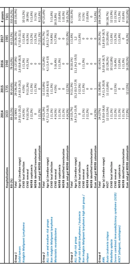
Risk stratification of immunocompromised children
The children were divided into 4 infectious risk groups regarding their
immunosuppression level. The lower risk group, group 1, included patients with solid tumors,
medium risk ALL, localised Burkitt lymphoma, lymphoblastic lymphoma, and high-risk
neuroblastoma. In group 3 were included the patients with high risk ALL, high-risk Burkitt
leukemia/lymphoma. Group 4 representing the highest risk group, included patients treated for
AML, infants under 1-year old with ALL, ALL relapses, allogeneic and autologous HSCT,
aplastic anemias, severe combined immunodeficiency syndrome.
Supportive care
Our center has 2 different units (protected unit and conventional unit) with dedicated
protective isolation methods. The highest isolation measures were taken for the most
immunocompromised patients, group 4. Our protected unit was already described (15). Briefly,
this unit consisted of positive air pressure single rooms equipped with HEPA filters. Patients
were cared for in a clean non-sterile outfit (gowns and mask). Non-sterile gloves were only
required with contagious patients. Food restrictions were extended to a low microbial diet.
Upon entering the unit, hands were washed with soap and water followed by an alcohol-based
rub. Thereafter, only alcohol-based hand rub was used before being in contact with the patient.
Children hospitalised in the conventional unit were isolated in single rooms only if they
had contagious disease. The other patients were free to wander in the unit or in collective areas
of the hospital: the school, the playground, the main lobby. Alcohol-based hand rub was used
by caregivers before and after any contact with patients.
Children in group 3 and 4 received non-absorbable antibiotics for gut and gum
decontamination. Gut and gum decontamination were performed with a Gram-negative bacilli
(GNB) targeting aminoglycoside (gentamycin). Gum decontamination targeted
alpha-hemolytic streptococcus through vancomycin mouthwash (16). Antifungal decontamination
group 2, 3 and 4 and at arrival for group 1 patient previously treated in a foreign hospital. This
monitoring was conducted to assess microbial flora characteristic, inquire colonisation with
MDRB, and guide empiric antibiotic therapy in the occurrence of fever. No primary antifungal
prophylaxis was used for our patients, including group 3 and 4.
All patients had a long-term central venous access device (CVAD) implanted at the
diagnosis. Tunneled catheters and peripherally inserted central catheters (PICCs) were cared
accordingly to protocol. Clinically infected (local inflammatory signs, pain, redness, swelling,
purulent drainage) central venous access devices were removed. Positive blood cultures despite
an adapted antibiotic therapy was an indication of CVAD removal. In the occurrence of a
Staphylococcus aureus related BSIs, CVAD removal was strongly discussed. The presence of Pseudomonas or Stenotrophomonas related BSIs was an absolute indication for CVAD removal.
Data collection and definitions
Microbiological records were reviewed to identify patients with BSIs. Their medical
records were analysed. The patient’s legal guardian signed an informed consent before
treatment, which included authorisation for the report of their data.
A bacteremia was defined by a positive blood culture sample obtained through a CVAD,
associated with fever. For coagulase-negative staphylococci (CoNS) two distinct positive blood
cultures within 48 h were mandatory. A polymicrobial infection was defined by a positive blood
culture with different pathogens. Fever was defined as a single body temperature > 38.5°C or
at least two measures > 38°C in an interval of 1 hour. Neutropenia was defined as an absolute
Multidrug resistant bacteria were defined as having acquired non-susceptibility to at
least one antimicrobial agent in three or more antimicrobial categories. Highly resistant
bacteria were defined as non-susceptibility to at least one agent in all but two or fewer
antimicrobial categories. (17)
Empirical intravenous antibiotic therapy protocols
All patients with a CVAD and febrile neutropenia were empirically treated with a
glycopeptid (vancomycin), an aminoglycoside (amikacin) and a broad-spectrum penicillin
adjusted to the risk group. Group 1 received ceftriaxone, groups 2, 3 and 4 received a
broad-spectrum penicillin. We also adapted our empirical therapy to prior identified gum or gut
bacterial colonisation and history of MDRB. If an infection was found, antibiotic therapy was
secondarily tailored to the identified germ. Vancomycin was stopped after 48 hours if the
multiple blood cultures did not identify a Gram-positive-cocci. The aminoglycoside was
stopped after 3 injections in the absence of bacterial documentation, and single kidney patients
received a single shot.
Statistical analysis
Descriptive statistics were reported in terms of absolute frequencies and percentages.
Distribution of data was described in terms of a median or mean value. Simple linear regression and Pearson’s correlation tests were used to compare the relationship between different variables. P values <0.05 were considered significant.
RESULTS:
Patients’ characteristics
There was a total of 7301 stays in our 2 units from January 2014 to December 2017,
with an annual increase. During these 7301 hospitalisations (> to 1 day), we identified 310 BSI
among 186 patients; corresponding to a ratio of 4.2% of stays. BSIs accounted for 37.5% to 44% of the center’s hospitalisations for fever and febrile neutropenia over these years. Patients with BSIs were aged from 3 days old to 22 years old; 134/310 (43,1%) were females. Patients’
characteristics are described in table 1 and suppl. tables 1 and 2.
Bacteria spectrum and risk group distribution (Fig. 1, suppl. fig. 1)
BSIs were mostly observed in groups 1 and 4, with a stable percentage of infections per
year in these groups over time. An average of 33,9 % (27,8-36,5%) BSIs in 4 years were
recorded in group 1, and 34,1% (27-41%) in group 4. The percentage of BSIs in groups 2, and
3 were respectively 21,6% (13,8-31,7%) and 10,3 % (1,6-15,7%), they were more labile over
time (Table 1).
Over the 4 years, among 310 infections, Gram-positive cocci accounted for 72. 9 % of
all BSIs: 49.7 % were documented with CoNS with 37.1% of Staphylococcus epidermidis,
6.5% with Staphylococcus aureus and 3.2% with group D Streptococcus. None of the
Staphylococcus aureus were methicillin resistant. Gram-negative-bacilli accounted for 21.6% of all BSIs, including 14.5% enterobacteria (Escherichia coli 14/45 (31.1%); Enterobacter
cloacae 11/45 (24.4%); Klebsielle pneumoniae 8/45 (17.8%)) and 4.5% Pseudomonas BSIs. The analysis of the bacterial spectrum according to the groups was not statistically significant.
of BSIs due to enterobacteria was also the highest in these two groups, respectively 15.6% and
23%. (Fig. 2).
Incidence
The incidence of BSIs dropped over time: 5.5% of annual stay in 2014 and 3.2% in
2017, thus an annual decrease of 0.75% (p=0.002) (Table 1). This statistically significant effect
is the resultant of the annual decrease of staphylococcal infections with a drop of 4.4 (p=0.001)
BSIs per year. Streptococcal and enterobacteria infections also declined over time, with
respectively an annual decrease of 1.8 (p = 0.077) and 3.2 (p = 0.09). Incidence of Pseudomonas
related BSIs were stable over time.
Reinfections
Our patients had different types of CVADs: 78 central ports, 20 PICCs and 212 tunneled
catheters. CVAD were considered as clinical infected in 30 cases (9.7%), and 60 CVAD
(19.4%) were removed.
Fifty-eight patients (31%) suffered from a second or more BSI. Thirty-nine patients
underwent 2 BSIs, 19 patients underwent 3 BSIs or more BSIs. Fifteen (26.3%) patients were
in group 1, and 25 (40.9%) in group 4. There was no statistical difference between the groups
(Fig. 3) Among these 58 re-infected patients, 25 had their CVAD removed once and 3 had their
CVAD removed twice. Among the patients infected twice, 15/39 patients were re-infected by
Staphylococcus (CoNS and Staphylococcus aureus). Twenty-four patients were re-infected by a different pathogen.
Polymicrobial infections:
We identified 26 episodes of polybacterial infections: 2, or more, identified bacteria in
separate blood cultures during the same infectious episode. They occurred in 23 (12.4%)
patients, 10 patients were in group 1, 6 in group 2, and 7 in group 4. Three patients suffered
from 2 polybacterial BSIs (group 1 and 4). We did not observe a statistical significance between
the risk group and the occurrence of polybacterial infections.
Multiresistance, gum and gut colonisation:
Gum and/or gut MDRB colonisation was identified in 65 patients, mostly in group 4
(39/65). Six out of these 65 MDRB were identified as extensively drug resistant bacteria
(non-susceptibility to at least one agent in all but two or fewer antimicrobial categories) (17). One
child in group 1 had been previously treated in Romania and was colonised with a Klebsiella
pneumoniae producing carbapenemase (OXA 48) and an Enterococcus faecium resistant to
glycopeptides. Three other children, treated in our center only were colonised with 2
Enterobacter cloacae, 2 Klebsielle pneumoniae.
Eleven BSIs (3.5%) were identified as MDRB: 5 E.coli, 3 Klebsiella pneumoniae, 1
Enterobacter cloacae, 1 Klebsielle oxytoca, 1 Hafnia alvei. Eight of these MDRB infections were primo-infections. One patient had originally been treated in eastern Europe and was
colonised with MDRB at his arrival. Three MDRB infections were caused by reinfections. Six
of them were associated with a gum or gut bacteria translocation, 3 in group 1, 1 in group 2 and
2 in group 4. All gram-positive bacteria were susceptible to vancomycin. We report no
DISCUSSION:
Immunocompromised children, due to malignancies and their treatments or caused by
primary immunodeficiency, encounter a high risk of BSIs. BSIs are the most frequent
complications and causes of hospitalisation in this specific population. The epidemiology of
bacteria causing BSIs has shifted from Gram-Negative organisms in the 1970s to Gram-Positive
organisms over the last years (4). Furthermore, the rapid development and the global spread of
multidrug resistant bacteria is a major health care issue worldwide. Knowledge of pathogens
distribution and resistance patterns is paramount for an optimal empiric antibiotic therapy and
outpatient management.
In this study, we examined the spectrum and resistance profile of bloodstream infections
in pediatric immunocompromised patients in relation to their immunosuppression level. In our
center the incidence of BSI dropped over time 4.3 % (3.2-5.5%). The majority of BSIs (72.9
%) were caused by Gram-positive cocci, particularly CoNS. Among the Gram-negative
bacteria, enterobacteriae were the most common ones. These results are coherent with
worldwide experience of BSI in pediatric hematology and oncology (18). Opportunistic
infections caused by Gram-positive bacteria among our population may be the product of
several cofactors including neutropenia, intensive chemotherapy, presence of invasive devices
such as CVADs, and mucositis. In accordance to Miedema et al (1) we did not identify a single
case of BSIs due to methicillin resistant Staphylococcus aureus or vancomycin-resistant
enterococci. We report no extensively drug resistant enterobacteria. These results are also
consistent with European studies performed in pediatric oncology treatment centers in
Germany, Switzerland and the Netherlands (19) where the proportion of MDRB-related BSIs
remained consistently low (1,5% of all BSIs). Bacterial spectrum in the current study was
comparable to the one observed in non-hemato-oncological pediatric centers such as intensive
vancomycin targeted CoNS whereas Gram-negative bacteria were controlled by a
broad-spectrum penicillin. An aminoglycoside was added for the synergic effect.
Our protocol diverges from the 2012 guidelines for management of febrile neutropenia
in children with cancer and/or undergoing HSCT (21). Indeed, these guidelines strongly
recommend monotherapy as first line treatment, with an antipseudomonal -lactam or carbapenem as empiric therapy in the high-risk group. A second anti Gram-negative agent or a
glycopeptide was added only for clinically unstable patients. We agree with the overall goal:
minimising exposure to avoidable antibiotics while providing coverage for virulent organisms.
An excessive use of broad-spectrum antibiotics may accelerate resistance rates. However, with
our step-down strategy, we were able to rapidly stop unnecessary antibiotics at 48h. Moreover,
initial broad-spectrum multiple therapy enabled a rapid control of BSIs: the early use of
glycopeptides allowed a rapid control of more than 50% of our BSI and does not appear to
trigger vancomycin resistant CoNS. Pseudomonas is responsible for a minority of our BSI
whereas enterobacteria represents the most common Gram-negative bacteria. We then prefer to
reserve carbapenem for documented penicillin resistant germs and MDRB, and thus avoid
selection of new resistant bacteria. The lack of resistance of our Gram-negative isolates is
reassuring and reflects the tight regulation of broad-spectrum antibiotics in our unit. There are
major limits to our study which is a retrospective evaluation over a short period of time, in a
single center. We could consider, in order to diminish antibiotic exposure, to restrain the use of
aminoglycosides. They would be used in case of severe sepsis or in patients with a high risk of
BSI caused by digestive pathogens. This would then be individually discussed and re-evaluated
over time.
Thanks to our antibiotic protocol, the early administration of intravenous antibiotics
biological features. The patients with a controlled CoNS BSIs (apyrexia, benign clinical
features, sterilisation of blood cultures, vancomycin adapted concentration levels (22)) could
be discharged within 72 hours and the treatment was pursued at home with the outpatient
hospitalisation. This early outpatient management strategy considerably increased the children’s quality of life. With a rapid control of the infection we were also able to sterilise 81% of CVAD without the use of an antibiotic lock. For thirty-three patients we controlled the BSIs
without adding the burden of another intervention. The 25 remaining patients had a second
surgery to replace the CVAD. These results are meaningful: changing a CVAD is a
non-negligible intervention in terms of morbidity and the health costs. This outcome highlights the
importance of medical and parental education: a key point in the primary prevention of BSIs.
The nurses as well as the parents followed strict aseptic rules regarding the use and
manipulation of the CVAD. Furthermore, our unit has set up a training program for the nurses
over the last years.
To our knowledge this is the first study that analyses the microbiological spectrum of
bacteria related BSIs in immunocompromised children, with risk stratification on the level of
immunosuppression. A strength of this study is that we are unique: we treat in the same institute
blood cancers, solid tumors, benign platelet and red blood cell pathologies, congenital
immunodeficiency and we perform HSCT. The diversity of the diseases we treat gives
legitimacy to the study, even if monocentric.
Our study reveals the same number of documented BSIs in the highest immunosuppression
group (group 4) than in the less immunocompromised group (group 1). This last group could
be compared to children treated by immunosuppressive treatments on the long term. In adult
with oral antibiotics. The 2012 guidelines for the management of pediatric febrile
neutropenia(21) also recommend (weakly, 2B) oral antibiotic administration in children with
low risk febrile neutropenia. Our present study demonstrates a high proportion of BSIs caused
by Staphylococcus (50%) and enterobacteria (15%) in this low risk group. Such pathogens, in
association with a CVAD, cannot be treated with oral medication only (21). Additionally, oral
antibiotics encounter major difficulties in children compared to parenteral antibiotics: drug
form, palatability, cooperation of younger children, impaired gastrointestinal absorption,
mucositis. These results stress the importance of a close follow up during the entire treatment
in all risk groups.
Reinfections were a major issue in our study: 31% patients suffered from a reinfection.
Interestingly their immunocompromised status was not correlated to a higher risk of reinfection
as we could have supposed. The patients were predominantly reinfected by different pathogens
and were not more likely to have MDRB. We did not identify the sources of all reinfections. It
could be interesting to study the different risk factors of reinfections. They are a true burden for
the families and the patients who are re-hospitalised in most cases. Survival rates now exceed 70% in child cancer however reducing the morbidity is tomorrow’s challenge.
A decontamination was performed in our center in the highest immunocompromised
groups 3 and 4. However, prophylactic administration of oral antibiotics stays a debated issue. By modulating the composition of the hosts’ intestinal microbiota to prevent infections, decontamination damage the gut epithelial barrier. Murine studies suggest that metabolites
generated by the gut microbiota not only regulate intestinal immune homeostasis, but also
influence circulating immune cells (23). A recent study revealed the occurrence of a higher rate
The authors suggested that microbiota alteration could participate in a systemic
pro-inflammatory response and alter intestinal immune homeostasis (22). One must also consider
that the gut microbiota can be altered by other cofactors during these infectious episodes such
as fasting, mucositis, parenteral nutrition. The major limit to gut and gum decontamination is
non-observance, especially in teenage patients (24). Indeed, an incomplete decontamination
will inevitably select resistant bacteria. Interestingly, in our study, we observed only 6 BSI
(9.2% of the 65-positive gut/gum samples) caused by a bacterial agent identified in both blood
culture and gut/gum sample. However, this number is probably underestimated, the sensitivity
of bacterial identification through gum and gut sample remains low. Prospective studies that
evaluate the contribution of bacterial decontamination to the emergence of MDRB are needed.
CONCLUSION:
BSI in immunocompromised children are mainly caused by Gram-positive bacteria, this
result is consistent with worldwide experience. In our center, the incidence of BSIs declined
over the 4 years due to a decrease of staphylococcal infections. This result stresses the
importance of medical and parental education concerning the management of CVADs. The
low and constant number of MDRB related BSIs is encouraging. The short use of vancomycin
does not trigger the emergence of resistant bacteria. Regarding the immunosuppression levels
we must stay alert, BSIs remain a major cause of morbidity even in the less immunosuppressed
patients. A prospective multicenter study involving pediatric oncology and hematology units is
REFRENCES :
1. Miedema KGE, Winter RHLJ, Ammann RA, Droz S, Spanjaard L, de Bont ESJM, et al. Bacteria causing bacteremia in pediatric cancer patients presenting with febrile neutropenia--species distribution and susceptibility patterns. Support Care Cancer Off J Multinatl Assoc Support Care Cancer. 2013 Sep;21(9):2417–26.
2. Delebarre M, Tiphaine A, Martinot A, Dubos F. Risk-stratification management of febrile neutropenia in pediatric hematology-oncology patients: Results of a French nationwide survey: D ELEBARRE ET AL. Pediatr Blood Cancer. 2016 Dec;63(12):2167–72.
3. Entz-Werle N, Suciu S, van der Werff ten Bosch J, Vilmer E, Bertrand Y, Benoit Y, et al. Results of 58872 and 58921 trials in acute myeloblastic leukemia and relative value of chemotherapy vs allogeneic bone marrow transplantation in first complete remission: the EORTC Children Leukemia Group report. Leukemia. 2005 Dec;19(12):2072–81.
4. Thacker N, Pereira N, Banavali SD, Narula G, Vora T, Chinnaswamy G, et al. Epidemiology of blood stream infections in pediatric patients at a Tertiary Care Cancer Centre. Indian J Cancer. 2014 Dec;51(4):438–41.
5. Felix A, Leblanc T, Petit A, Nelkem B, Bertrand Y, Gandemer V, et al. Acute Myeloid Leukemia With Central Nervous System Involvement in Children: Experience From the French Protocol Analysis ELAM02. J Pediatr Hematol Oncol. 2018 Jan;40(1):43–7. 6. Biondi A, Schrappe M, De Lorenzo P, Castor A, Lucchini G, Gandemer V, et al. Imatinib
after induction for treatment of children and adolescents with Philadelphia-chromosome-positive acute lymphoblastic leukaemia (EsPhALL): a randomised, open-label, intergroup study. Lancet Oncol. 2012 Sep;13(9):936–45.
7. Domenech C, Mercier M, Plouvier E, Puraveau M, Bordigoni P, Michel G, et al. First isolated extramedullary relapse in children with B-cell precursor acute lymphoblastic leukaemia: results of the Cooprall-97 study. Eur J Cancer Oxf Engl 1990. 2008 Nov;44(16):2461–9.
8. Landmann E, Burkhardt B, Zimmermann M, Meyer U, Woessmann W, Klapper W, et al. Results and conclusions of the European Intergroup EURO-LB02 trial in children and adolescents with lymphoblastic lymphoma. Haematologica. 2017 Dec;102(12):2086–96. 9. Cairo MS, Sposto R, Gerrard M, Auperin A, Goldman SC, Harrison L, et al. Advanced stage, increased lactate dehydrogenase, and primary site, but not adolescent age (≥ 15 years), are associated with an increased risk of treatment failure in children and adolescents with mature B-cell non-Hodgkin’s lymphoma: results of the FAB LMB 96 study. J Clin Oncol Off J Am Soc Clin Oncol. 2012 Feb 1;30(4):387–93.
10. Wrobel G, Mauguen A, Rosolen A, Reiter A, Williams D, Horibe K, et al. Safety assessment of intensive induction therapy in childhood anaplastic large cell lymphoma: report of the ALCL99 randomised trial. Pediatr Blood Cancer. 2011 Jul 1;56(7):1071–7. 11. Worch J, Ranft A, DuBois SG, Paulussen M, Juergens H, Dirksen U. Age dependency of primary tumor sites and metastases in patients with Ewing sarcoma. Pediatr Blood Cancer. 2018 Jun 1;e27251.
12. Wilde JCH, Aronson DC, Sznajder B, Van Tinteren H, Powis M, Okoye B, et al. Nephron sparing surgery (NSS) for unilateral wilms tumor (UWT): the SIOP 2001 experience. Pediatr Blood Cancer. 2014 Dec;61(12):2175–9.
13. Gaspar N, Occean B-V, Pacquement H, Bompas E, Bouvier C, Brisse HJ, et al. Results of methotrexate-etoposide-ifosfamide based regimen (M-EI) in osteosarcoma patients included in the French OS2006/sarcome-09 study. Eur J Cancer Oxf Engl 1990. 2018;88:57–66.
14. Morgenstern DA, Pötschger U, Moreno L, Papadakis V, Owens C, Ash S, et al. Risk stratification of high-risk metastatic neuroblastoma: A report from the HR-NBL-1/SIOPEN study. Pediatr Blood Cancer. 2018 Jul 17;e27363.
15. Domenech C, Leick-Courtois C, Bienvenu A-L, Pracros J-P, Picot S, Bleyzac N, et al. Improvement in the Outcome of Invasive Aspergillosis in a Pediatric Hematology Department: A 10-Year Review. J Pediatr Hematol Oncol. 2015 Oct;37(7):560–5. 16. Brunet AS, Ploton C, Galambrun C, Pondarré C, Pages MP, Bleyzac N, et al. Low
incidence of sepsis due to viridans streptococci in a ten-year retrospective study of pediatric acute myeloid leukemia. Pediatr Blood Cancer. 2006 Nov;47(6):765–72. 17. Magiorakos A-P, Srinivasan A, Carey RB, Carmeli Y, Falagas ME, Giske CG, et al.
Multidrug-resistant, extensively drug-resistant and pandrug-resistant bacteria: an international expert proposal for interim standard definitions for acquired resistance. Clin Microbiol Infect Off Publ Eur Soc Clin Microbiol Infect Dis. 2012 Mar;18(3):268–81. 18. Mvalo T, Eley B, Bamford C, Stanley C, Chagomerana M, Hendricks M, et al.
Bloodstream infections in oncology patients at Red Cross War Memorial Children’s Hospital, Cape Town, from 2012 to 2014. Int J Infect Dis IJID Off Publ Int Soc Infect Dis. 2018 Dec;77:40–7.
19. Simon A, Furtwängler R, Graf N, Laws HJ, Voigt S, Piening B, et al. Surveillance of bloodstream infections in pediatric cancer centers - what have we learned and how do we move on? GMS Hyg Infect Control. 2016;11:Doc11.
20. Hooven TA, Polin RA. Healthcare-associated infections in the hospitalized neonate: a review. Early Hum Dev. 2014 Mar 1;90:S4–6.
21. Lehrnbecher T, Phillips R, Alexander S, Alvaro F, Carlesse F, Fisher B, et al. Guideline for the management of fever and neutropenia in children with cancer and/or undergoing hematopoietic stem-cell transplantation. J Clin Oncol Off J Am Soc Clin Oncol. 2012 Dec 10;30(35):4427–38.
22. Hoegy D, Goutelle S, Garnier N, Rénard C, Faure‐Conter C, Bergeron C, et al. Continuous intravenous vancomycin in children with normal renal function hospitalized in hematology–oncology: prospective validation of a dosing regimen optimizing steady-state concentration. Fundam Clin Pharmacol. 2018 Jun 1;32(3):323–9.
23. Whangbo J, Ritz J, Bhatt A. Antibiotic-mediated modification of the intestinal microbiome in allogeneic hematopoietic stem cell transplantation. Bone Marrow Transplant. 2017 Feb;52(2):183–90.
24. Lehrnbecher T, Laws H-J, Boehm A, Dworzak M, Janssen G, Simon A, et al. Compliance with anti-infective preventive measures: A multicentre survey among paediatric oncology patients. Eur J Cancer Oxf Engl 1990. 2008 Sep;44(13):1861–5.