Interfering with Activating Protein-1
and Estrogen Stimulations
Flavia Pernasetti*†, Laure Caccavelli, Ce´cile Van de Weerdt,
Joseph A. Martial, and Marc Muller
Laboratoire de Biologie Mole´culaire et de Ge´nie Ge´ne´tique
Universite´ de Lie`ge
Institut de Chimie B6
B-4000 Sart Tilman, Belgium
Transcription of the human PRL (hPRL) gene in the pituitary is subject to tissue-specific and multihor-monal regulation involving two main regulatory re-gions, a proximal promoter and a distal enhancer. In this report we show that thyroid hormone in-hibits the expression of the hPRL gene in rat pitu-itary cells. Transient expression experiments show that thyroid hormone regulation involves a strong inhibitory element, located in the proximal (2164/
235) promoter, which is modulated by a more
dis-tal stimulatory response control region. Gel retar-dation experiments reveal that the thyroid hormone receptor does not bind to the proximal negative element.
We show the existence of an activating protein-1 (AP-1) response element located at positions261 to254 of the proximal promoter, conferring AP-1 stimulation to the hPRL promoter. This AP-1 induc-tion is abolished when hormone-bound thyroid hormone receptor is present, indicating that there is an interference between the thyroid hormone receptor and AP-1 regulatory pathways.
Furthermore, using the complete hPRL upstream region, we show that estrogen induction is abol-ished by simultaneous thyroid hormone treatment. (Molecular Endocrinology 11: 986–996, 1997)
INTRODUCTION
In vertebrates, thyroid hormone (T3) regulates a vast array of biological processes, including metamorpsis, development, growth, metabolism, and ho-meostasis, mainly through specific regulation of gene transcription (1). The effects of T3 are mediated by thyroid hormone receptors (TRs) belonging to the large nuclear hormone receptor superfamily (2, 3). Some members of this superfamily, such as the estrogen and
glucocorticoid receptors, bind to conserved hormone response elements (AGGTCA and AGAACA, respec-tively) in the promoter regions of target genes. The binding of TR to thyroid hormone response elements (TREs) is apparently more complex. TREs vary con-siderably in nucleotide sequence and with regard to the number, spacing, and arrangement of binding sites (4–7). Most TREs described to date are composed of two or more copies of the motif (A/G)GG(A/T)CA (or a related sequence) arranged as palindromes or direct repeats. The spacing of these half-sites is critical for TR homodimer formation or heterodimerization with other members of the nuclear receptor superfamily, such as the retinoic acid (RAR and RXR) and vitamin D receptors (8). Most well known TREs mediate positive regulation by thyroid hormone. Negative T3response elements (nTREs) have been less well characterized, being sometimes described as half-site sequences to which TR would bind as a monomer (9). Functional interactions between various nuclear receptors and between such receptors and other transcription fac-tors are well documented (10–14).
Recently, thyroid hormone receptors have been shown to inhibit gene expression by interfering with the activity of the activating protein-1 (AP-1) transcrip-tion factor (15–22). This factor is a dimer whose com-ponents are respectively encoded by the jun/fos pro-tooncogene family (23). It binds to a specific DNA sequence (ATGAGTCA) and stimulates gene tran-scription in response to activation of the protein kinase C pathway (24). TRs exert their inhibitory effect through direct interaction with the AP-1 complex in solution, leading to the formation of a transcriptionally inactive TR/AP-1 complex. TRs do not compete with AP-1 for DNA binding sites (15).
PRL is a hormone essentially secreted by the ante-rior pituitary. Transcription of the human PRL (hPRL) gene in the pituitary is subject to tissue-specific and multihormonal regulation involving the POU domain transcription factor Pit-1 (25–28). In the 59-flanking sequence of the hPRL gene, two regulatory regions 0888-8809/97/$3.00/0
Molecular Endocrinology
Copyright © 1997 by The Endocrine Society
have been identified, a proximal promoter (2250/235) containing three Pit-1-binding sites and a distal en-hancer (22000/21200) with eight additional Pit-1-binding sites (29, 30). Regulation of hPRL gene tran-scription by phorbol esters, epidermal growth factor, TRH, Ca21, and cAMP depends on elements located
within the first 250 bp of the promoter (31–34). Estro-gens are reported to stimulate hPRL gene expression through a response element next to the distal en-hancer (34). Nothing is known about T3regulation of hPRL, whereas contradictory results have been ob-tained in GH pituitary tumor cell lines for rat PRL (rPRL) T3regulation. T3was found to inhibit rPRL promoter-driven transcription (35–37) in GH1cells but to stimu-late it in GH4C1cells (38) through the same response element, located in the proximal promoter (coordi-nates2176/211). From studies in GH3cells, two TREs were identified in the rPRL gene: a positive response element in the distal enhancer next to the estrogen receptor response element and a negative response element in the 292-bp span of the proximal promoter (39).
This report focuses for the first time on the regula-tion of the hPRL gene by thyroid hormone. We show that the hPRL gene promoter contains both a positive and a negative TRE. The negative effect is stronger than the positive one and involves cross-talk between TR and AP-1.
RESULTS
The hPRL Promoter Is Inhibited by Thyroid Hormone
To identify possible T3response elements in the 5 9-flanking region of the hPRL gene, we used various constructs containing either the complete 5000-bp pituitary hPRL promoter or a 59-deleted portion of it fused to the chloramphenicol acetyltransferase (CAT) reporter gene (29). The constructs were introduced by electroporation into GH3B6 cells (a PRL-producing pituitary cell line). Figure 1 shows the CAT activity produced by each construct in the absence and pres-ence of T3as well as the inhibition factor in the pres-ence of T3.
A construct containing the 500-bp promoter region of the human placental lactogen B gene (hCS-B, struct p500CSBCAT) (14) was used as a positive con-trol. In this construct, T3causes 4-fold stimulation of reporter gene expression (0.25-fold repression). As expected, constitutive thymidine kinase (TK)-driven CAT gene expression is unaffected by T3treatment. In contrast, expression from all hPRL constructs is inhib-ited in the presence of T3. The shorter constructs (p250PRLCAT and p740PRLCAT) are inhibited ap-proximately 4-fold, whereas the longer ones (p1330-PRLCAT, p1750PRLCAT, p2627PRLCAT, p3500-PRLCAT, and p5000PRLCAT) are inhibited only 2-fold. Similar results were obtained in GC cells (data not
shown). These results suggest the presence of two distinct T3response elements in the hPRL promoter
whatever pituitary cell line used. One response ele-ment, located in the 250-bp proximal promoter, would mediate a strong inhibitory effect; the second, located between coordinates21330 and 2740, would have a weaker positive effect, attenuating the inhibitory effect of the proximal region.
The Inhibitory Effect Is not Pit-1 Dependent
The first 250 bp of the hPRL promoter (proximal pro-moter) include three well known Pit-1-binding sites (30). They also include the A site involved in the re-sponses to cAMP, TRH, and Ca21(32, 33) (Fig. 2a). To study T3regulation in this proximal region, we tested two constructs: one bearing the entire proximal region without the TATA box (bp2250/235) and one bearing the same fragment lacking the P3 Pit-1-binding site (bp 2164/235). Both proximal promoter segments were cloned in front of the TK promoter fused with the CAT reporter gene. The constructs were introduced by electroporation into GH3B6cells (Fig. 2b). As a positive control we used a construct containing three palin-dromic TRE sequences cloned in front of the TK-CAT fusion. As expected, T3 stimulates CAT expression Fig. 1. Influence of T3on hPRL Promoter Activity
GH3B6cells were transfected by electroporation with 30
mg of the indicated fusion construct. The various PRLCAT constructs carry 59-deletions of the hPRL promoter (decreas-ing in length from p5000PRLCAT to p250PRLCAT). Construct p500CSBCAT carries the human placental lactogen promoter fused to the CAT reporter gene. Plasmid pTKCAT (or pBLCAT2) carries the CAT reporter gene under the control of the TK promoter. Electroporated cells were incubated for 48 h in medium with or without 10 nMT3. Cells were
har-vested, and CAT activity was assayed in cell lysates. The percentage of CAT conversion in the absence (hatched
boxes) or presence (gray boxes) of T3is shown, and the ratio
between these values is indicated to the left as fold repres-sion for each construct. Values are the mean6SEMof five independent transfections, each performed in duplicate.
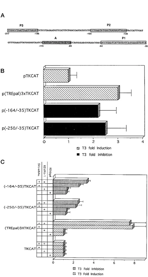
Fig. 2. T3Regulation of a Heterologous Promoter in Pituitary and Nonpituitary Cells
A, Sequence of the sense strand of the 217-bp hPRL promoter (57). The positions of the three proximal, high affinity Pit-1-binding sites and of the A sequence are indicated, respectively, by boxes P1, P2, P3, and A. B, GH3B6cells were transfected
with 15 mg of the indicated plasmid. Plasmid pTKCAT carries the TK promoter fused to the CAT reporter gene. Plasmids p(2164/235)TKCAT and p(2250/235)TKCAT additionally carry the indicated base pairs of the hPRL proximal promoter in front of the TK promoter; p(TREpal)3XTKCAT carries instead three copies of the TREpal sequence in front of the TK promoter. Cells were grown for 48 h in the absence or presence of 10 nMT3. CAT activity was measured, and the data are presented as described
from this control plasmid approximately 3-fold while having no effect on the negative control (pTKCAT), indicating that T3effects are specific under our exper-imental conditions. CAT expression from both p(2250/235)TKCAT and p(2164/235)TKCAT is inhib-ited about 3-fold by T3. This shows that the T3 re-sponse element can mediate inhibition of the heterol-ogous TK promoter. Our experiments further show that the nTRE is located between bp2164 and 235 of the hPRL proximal promoter.
To detect a possible involvement of Pit-1 in T3 inhi-bition of the hPRL proximal promoter, we performed cotransfection experiments in which nonpituitary HeLa cells received one of the above-mentioned constructs in combination with a vector expressing either hTRa1 complementary DNA (cDNA; pSV2c-erbAa), hPit-1 cDNA (pRSVPit-1), or both vectors (Fig. 2c). Expres-sion from pTKCAT is unaffected by the presence of one or both of these expression vectors. As expected, T3stimulates expression from p(TREpal)3XTCAT in the presence of pSV2c-erbAa; this effect is unaltered by the additional presence of pRSVPit-1. In cells receiv-ing either p(2250/235)TKCAT or p(2164/235)TKCAT constructs, the presence of pSV2c-erbAa is sufficient to promote T3inhibition of reporter gene expression, so clearly Pit-1 synthesis is not required for this effect. Furthermore, the inhibition factor is the same regard-less of whether the Pit-1 expression vector is present. That Pit-1 is indeed synthesized in HeLa cells trans-fected with pRSVPit-1 is shown by the increased basal level of CAT expression recorded with p(2250/ 235)TKCAT (4-fold stimulation) and p(2164/235)TK-CAT (3-fold stimulation). These results suggest that Pit-1 is not involved in the inhibitory effect exerted by T3through the proximal promoter region. We obtained similar results in other Pit-1-deficient cell lines, such as CV1 and JEG-3 (data not shown), reinforcing the con-clusion that the effect of T3 on the hPRL proximal promoter is not pituitary specific.
Involvement of Receptor-DNA Binding in the T3 Effects
The2164/235 region contains two Pit-1-binding sites (P1 and P2) and the cAMP response element A (Fig. 2a). A search for potential TR-binding sites in this region revealed only one imperfect half-site, GGGTaA (2113/2108). To test the affinity of TR for this putative binding site, we performed gel retardation assays with bacterially expressed TR (Fig. 3). The probes used
were an oligonucleotide containing the 2115/287 PRL proximal promoter sequence and, as a positive control, an oligonucleotide containing the DR4 con-sensus TR-binding sequence (two TR concon-sensus half-sites directly repeated, spaced by 4 bp). As expected, we observed a retarded band with the DR4 oligonu-cleotide, corresponding to the TR monomer (Fig. 3, lane 2); this band disappeared in the presence of a 100-fold excess of cold DR4 oligonucleotide (lane 3), but not in the presence of a 100-fold excess of Sp1 oligonucleotide (lane 5). Moreover, the retarded band was supershifted in the presence of anti-TR antibodies (lane 4). All of these results prove the specificity of the Fig. 3. TR Does not Bind to the Proximal TRE of the hPRL
Promoter
Extracts of TR-expressing bacteria (4.5mg protein) were incubated with the indicated32P-labeled DNA fragment, i.e.
either the DR4 oligonucleotide (positive control) or the (2115/ 287) sequence of the proximal hPRL promoter. The presence of competitor oligonucleotides (in 100-fold excess), anti-TR antibody (a-TR), recombinant RXR (4.5 mg), and 10 nMT3is
indicated by a1.
in Fig. 1B. Black bars, Inhibition factors; hatched bars, induction factors. Values are the mean6 SEMof four independent
transfections, each performed in duplicate. C, HeLa cells were cotransfected by calcium phosphate precipitation with 5mg of the indicated reporter construct and with 10mg of each expression vector indicated. The pSV2c-erbAa expresses human TRa1; pRSVPit-1 expresses human Pit-1; pRSVbgal, expressing b-galactosidase, was used as a control and to maintain a constant amount of expression vector (20mg). Cells were incubated for 48 h in the absence or presence of 10 nMT3, and CAT activity was
measured. Inhibition factors (gray bars) and induction factors (hatched bars) were calculated as described in Fig. 1B. The data are the mean6SEMof five independent experiments, each performed in duplicate.
retarded band. Addition of bacterially expressed RXR led to the formation of TR/RXR heterodimers (lane 6), as expected.
In contrast, TR does not specifically bind to the proximal PRL promoter sequence (2115/287), even in the presence of RXR or T3 (lanes 7–13). The results were the same when the probe used was the entire 2164/235 PRL proximal sequence (data not shown). This strongly suggests that the T3effect in this region occurs via an indirect mechanism that does not re-quire TR binding to the DNA.
T3Inhibition of the hPRL Proximal Promoter Is Independent of the Forskolin Pathway, but Involves an AP-1-Binding Site
To identify the indirect T3repression mechanism, two alternative pathways were investigated. TR has been shown to interfere with stimulation of the PIT1 gene promoter by forskolin (40). As the proximal hPRL pro-moter contains cAMP response elements (2164/235) (32, 33), we examined whether T3 treatment might affect the response to forskolin mediated by this re-gion. GH3B6 cells were transfected with a construct containing the first 164 bp of the proximal promoter cloned in front of the luciferase (LUC) reporter gene (p164PRL-LUC). The cells were treated with forskolin and/or T3. Gene expression increased significantly (15-fold) in response to forskolin. T3 treatment re-duced the basal promoter activity approximately 2-fold. When both treatments were applied, forskolin stimulation was reduced by a factor of 2, clearly indi-cating that forskolin and thyroid hormone act through distinct pathways (data not shown).
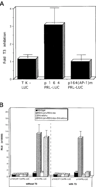
In search of an alternative pathway that might be modulated by T3, sequence analysis of the hPRL prox-imal promoter was performed. This analysis revealed the presence of a highly conserved AP-1 consensus motif (261 TGAaTCAT 254) containing only one mis-match (see also Caccavelli, L., I. Manfroid, J. A. Mar-tial, and M. Muller, in preparation). This led us to in-vestigate whether this putative AP-1 site is functionally active and how destruction of this site affects the response of the hPRL proximal promoter to T3. To this end, we produced a mutation in the AP-1 consensus motif of p164PRL-LUC, yielding plasmid p164(AP-1)mPRL-LUC. We then transfected GH3B6cells with either p164(AP-1)mPRL-LUC, its wild-type parent, or the control plasmid pTKLUC bearing the LUC gene under the control of the TK promoter. The cells were treated or not with T3. The results are shown in Fig. 4a. T3treatment did not affect expression from pTK-LUC, but it inhibited expression from p164PRL-LUC (3-fold inhibition). The mutant construct p164(AP-1)mPRL-LUC displayed 2-fold lower basal expression and did not respond to T3.
These results show for the first time that an AP-1 consensus sequence located close to the proximal Pit-1-binding site P1 contributes to the basal activity of the hPRL promoter in pituitary cells. In addition, this
Fig. 4. T3Inhibits the Response of the hPRL Proximal
Pro-moter to the AP-1 Transcription Factor
a, GH3B6 cells were electroporated with 10 mg
p164PRL-LUC or p164(AP-1)mPRL-LUC (containing a mu-tated AP-1 response element). As a control we used pTK-LUC, a plasmid carrying the luciferase gene under the control of the TK promoter. The cells were incubated for 48 h with or without 10 nMT3, and luciferase activity was
measured. Inhibition factors were calculated by dividing the activity recorded for treated cells by that obtained with the corresponding untreated cells. The basal LUC activity obtained with p164(AP-1)mPRL-LUC plasmid is 2 times less than that obtained with p164PRL-LUC. The data are the mean6SEMof three independent transfections, each
performed in duplicate. b, HeLa cells were cotransfected by calcium phosphate precipitation with 10mg p164PRL-LUC or p164(AP-1)mPRL-p164PRL-LUC and either 20 mg of the control expression vector pRSVbgal or 5 mg of each other expression vector indicated plus enough pRSVbgal to reach a total of 20mg. The cells were incubated for 48 h with or without 10 nMT3, and luciferase activity was
mea-sured. The data are the mean6SEMof three independent
sequence is directly involved in T3repression of the hPRL promoter.
T3Abolishes AP-1 Activation of the hPRL Promoter in Heterologous Cells
Since T3 inhibition appears to involve a putative AP-1-binding site, we next analyzed the action of AP-1 on the hPRL proximal promoter in nonpituitary cells. We cotransfected HeLa cells with p164PRL-LUC in combination with vectors expressing c-jun and c-fos (pRSVc-jun and pRSVc-fos) and/or with pSV2c-erbAa. It should be mentioned that none of these vectors has any effect on reporter gene ex-pression from control plasmid pTK-LUC (not shown). The cells were incubated with or without T3. Cells harboring a vector expressingb-galactosidase cDNA (pRSVbgal) were included as a control. The results are presented in Fig. 4b. In the presence of T3, as expected, a 3-fold inhibitory effect is ob-served using p164PRL-LUC only when the thyroid hormone receptor is coexpressed. In the absence of TR, transcription was stimulated approximately 20-fold in the presence of c-jun and c-fos regardless of whether T3was present. In contrast, stimulation by c-jun and c-fos was nearly abolished when both T3 and its receptor were present. Furthermore, muta-tion of the consensus AP-1 site in p164(AP-1)mPRL-LUC completely abolished both c-jun/c-fos activa-tion and T3inhibition of the hPRL promoter in HeLa cells.
These results strongly suggest that AP-1 is able to activate the hPRL promoter and that T3 exerts its inhibitory effect via an interaction between the hor-mone-bound receptor and the AP-1 complex.
AP-1 Binds to the hPRL Proximal Promoter
To confirm the involvement of AP-1 in hPRL expres-sion, we performed gel retardation assays. The264/ 235 fragment was used as a probe in the presence of AP-1-enriched HeLa cell extracts. The results are pre-sented in Fig. 5. One retarded complex was obtained (lane 2), which disappeared in the presence of a 100-fold excess of an oligonucleotide containing the AP-1 consensus sequence (lane 3), but not in the presence of the nonspecific DR4 oligonucleotide in a 100-fold excess (lane 4). In addition, the complex disappeared in the presence of antibodies recognizing the DNA-binding region of c-jun (lane 5). All of these data show that the observed band is a specific AP-1 DNA complex.
Taken together, our results confirm the following model for T3inhibition of hPRL gene transcription: the hormone-bound thyroid hormone receptor interacts with the AP-1 complex, thereby preventing the com-plex from activating hPRL gene transcription via the proximal promoter.
T3Abolishes Estradiol (E2) Stimulation of the hPRL Promoter
The transfection experiments using 59-deletion mu-tants of the hPRL promoter suggested the presence of a modulatory element in the distal region (21330/ 2740) that leads to a decrease in the overall T3
inhi-bition. Preliminary studies identified a complex ele-ment centered at21200 that is able to bind TR and, in addition, contains an estrogen response element (34). To further analyze the role of this distal regulatory region, we examined the effects of thyroid hormone and estrogen on a fragment of the hPRL promoter containing both the proximal promoter and the distal regulatory region (p2627PRLCAT; Fig. 6). The results Fig. 5. AP-1 Specifically Binds to the Proximal hPRL
Pro-moter
Total extracts from HeLa cells enriched in AP-1 factor (described in Materials and Methods; 5mg) were incubated with the 32P-labeled (264/235) hPRL promoter sequence
and used in a gel retardation assay. Competition with a 100-fold excess of cold oligonucleotide and addition of anti-Jun antibody (a-jun) are indicated by 1.
show, as expected, that T3exerts an approximately
3-fold inhibitory effect, and E2has a 2-fold stimulatory effect. In cells treated with both hormones, the re-corded CAT activity is surprisingly low, reaching only half the basal level. In conclusion, the impact of the distal element on T3regulation appears to be weak, even when it is stimulated by estrogens.
DISCUSSION
We have studied, for the first time, the regulatory ef-fects of thyroid hormone on the hPRL gene promoter. We present evidence that thyroid hormone down-reg-ulates transcription of the hPRL gene in pituitary cells. In the hPRL promoter we have identified two main T3-responsive regions. The first, located in the proxi-mal promoter, mediates a strong negative effect, whereas the second, located in the distal promoter, mediates a weak positive response. The overall effect of the two combined regions is repressive.
Our finding that T3inhibits the hPRL promoter is in keeping with the physiological data on the endoge-nous human gene. Hypothyroidism in human patients is frequently associated with increased serum PRL levels, whereas hyperthyroid patients present lower PRL levels than euthyroid control subjects (40). Al-though it is unclear whether this effect is due to direct regulation of the hPRL gene, recent studies on a
hu-man pituitary cell line suggest 2-fold inhibition of en-dogenous hPRL gene transcription by T3(41).
The Proximal Promoter
The proximal hPRL promoter mediates T3inhibition of a heterologous promoter (TK) in both pituitary (GH3B6) and nonpituitary cells (HeLa, CV1, and JEG-3; Fig. 2). On the other hand, we observe no binding of TR to the proximal T3response element in gel retardation as-says (Fig. 3). This suggests that T3 acts indirectly. Recently, T3treatment was shown to inhibit Pit-1 gene transcription through interference between the thyroid hormone receptor and Pit-1 (42). As T3inhibition of the hPRL promoter is observed in nonpituitary cells (Fig. 2c), we can exclude an indirect action of T3through inhibition of Pit-1 expression. Furthermore, coexpres-sion of Pit-1 and TR does not affect T3inhibition in HeLa cells (Fig. 2c), so we can rule out a mechanism involving interference between the TR and Pit-1 lead-ing to inhibition of the hPRL gene promoter, nor is the forskolin pathway involved in the response of the hPRL promoter to T3(our data not shown). This con-trasts with T3 inhibition of Pit-1 gene expression, which involves interference of T3with the cAMP in-duction pathway (42).
An AP-1-responsive element has been located in the hPRL proximal promoter between coordinates 261 and 254 (see also Caccavelli, L., I. Manfroid, J. A. Martial, and M. Muller, in preparation). It contains an AP-1-binding sequence that differs by only one mis-match from the consensus AP-1-binding site. We show that this sequence can specifically bind AP-1 in gel retardation assays (Fig. 5) and that it mediates 15-fold stimulation of reporter gene transcription in the presence of coexpressed c-jun and c-fos (Fig. 4b). This stimulation is almost completely abolished in the presence of hormone-bound TR. We conclude that thyroid hormone exerts its inhibitory effect via interac-tion of hormone-bound TR with AP-1. Furthermore, mutation of the AP-1 site completely abolishes the stimulatory effect of c-jun and c-fos and the inhibitory effect of T3 (Fig. 4). This confirms that the two re-sponses are interrelated.
The transcription-stimulating activity of AP-1 is in-creased by protein kinase C phosphorylation. Gellersen et al. (34) observed synergistic stimulation of the hPRL proximal promoter (250 bp) by TPA (12-O-tetradecanoyl-phorbol-13-acetate) treatment and Pit-1 expression in the SKUT-1B-20 human uterine cell line. Here we show strong stimulation of the hPRL promoter in HeLa cells cotransfected with a reporter construct and with c-jun and c-fos expression vectors, but this effect is completely Pit-1 independent. This discrepancy suggests that TPA stimulation in SKUT 1B-20 cells might involve another mechanism(s) in addition to the increased AP-1 activity.
Several groups have reported interaction of AP-1 with members of the nuclear receptor family, e.g. the glucocorticoid, retinoic acid, progestin, estrogen, and Fig. 6. The Inhibitory Effect of T3Overrides the Stimulatory
Effect of E2Exerted via the 2627-bp hPRL Promoter
Frag-ment
GH3B6 cells were electroporated with 30 mg
p2627PRLCAT. After electroporation, the cells were incu-bated for 48 h in the presence of the antiestrogen ICI 64384 (1mM) and/or T3(10 nM) or in the presence of E2(100 nM)
and/or T3(10 nM). CAT activity was measured, and the
in-duction factors were calculated as the ratio between the measured activity to the value obtained with ICI-treated con-trol cells. The data are the mean6SEMof five independent experiments, each performed in duplicate.
thyroid hormone receptors (15–22). Different mecha-nisms have been observed for this cross-modulatory action according to the promoter context (22). Direct interaction between TR and AP-1 has been demon-strated in coimmunoprecipitation assays (20). Further-more, in gel retardation experiments, binding of AP-1 to DNA was inhibited by TR when the latter was added before addition of the labeled binding sequence (19, 20). The hPRL proximal promoter contains one AP-1-responsive element and no TR-binding site. This sug-gests that TR blocks the action of AP-1 by hindering its binding to its response element. Whether the interac-tion between TR and AP-1 is direct or mediated by another factor(s) is unclear.
In striking contrast to the overall inhibition of hPRL transcription by T3, the combined positive and nega-tive acting elements of the rPRL promoter mediate overall activation of this promoter by T3in GH3cells (39). The rPRL promoter responds differently to T3 according to the pituitary cell line examined (36–39). Nevertheless, studies in rats have shown that treat-ment with the antiarhythmic drug amiodarone, a T3 antagonist, reduces PRL mRNA levels (43), suggesting that rPRL expression is up-regulated by T3. The rat sequence corresponding to the hPRL proximal AP-1 sequence differs by three nucleotides from the AP-1 consensus binding sequence, suggesting that rat and human PRL genes might be differently regulated by AP-1. Indeed, recently it was shown that expression of c-jun inhibits rat PRL expression in GH4pituitary cells (44). This repression involves the FPII region centered at2125 in the rat promoter and is only seen in GH4 cells. The proposed mechanism is an indirect binding of c-jun to the promoter by recruitment of a pituitary-specific FPII-binding factor. Whether T3would be able to block this repression, which would result in an over-all activation by T3, is at present unknown. These observations contrast with our results using the hPRL promoter in GH3B6 cells. However, the same re-searchers describe a synergistic activation of the rPRL promoter by c-jun and Pit-1 expression in HeLa cells. In this case, the proposed mechanism is an interaction of c-jun with Pit-1 bound to the proximal binding site, but again without direct DNA binding of c-jun. Nothing is known about T3modulation of this effect, which is suppressed in GH4cells by the c-jun repression de-scribed above. Thus, it seems very likely that the mechanism of T3inhibition of the hPRL promoter de-scribed here does not apply to the homologous rPRL promoter.
Complete hPRL Promoter
The combined action of the proximal, negative pro-moter and the distal, weakly positive region results in inhibition by T3 of the complete upstream region. In addition, this region contains a consensus estrogen-responsive element (ERE) located close to a TR-bind-ing sequence (see also Van de Weerdt, C., F. M. Per-nasetti, L. Caccavelli, J. A. Martial, and M. Muller, in
preparation). The same hPRL ERE can mediate 2-fold stimulation of a heterologous promoter in SKUT-1B-20 cells, a PRL-producing uterine carcinoma cell line (34). The fact that T3 is also able to block E2 activation of this construct (Fig. 6) stresses the impor-tance of the T3regulation mediated by the proximal AP-1 site.
Activation of rPRL gene transcription by E2involves the stabilization of a chromatin loop permitting critical interactions between proteins of the distal enhancer and proximal promoter (45). Dexamethasone-bound GR has been found to bind to the distal ERE, compet-ing in this with estrogen receptor and preventcompet-ing for-mation of the loop (46). We have no inforfor-mation as to whether a particular chromatin structure characterizes the plasmid constructs used here, but hindering of loop formation by TR is certainly a possible mecha-nism of repression, especially as factors bound to the proximal promoter are known to be important in loop formation. If the AP-1 complex plays a role in the interactions between the distal and proximal regions, then T3blocking of AP-1 binding to the DNA would inhibit loop formation, thus preventing E2stimulation. Alternatively, T3 might down-regulate the expression of other proteins necessary for the response to estro-gen mediated by the complete hPRL promoter.
In conclusion, we demonstrate that hPRL gene tran-scription is inhibited by thyroid hormone. We show that the inhibitory effect of T3 involves interference with the AP-1 transactivation mediated by an AP-1-binding site located in the proximal hPRL promoter. A weak positive TRE, located in the distal region close to an ERE, modulates this inhibition. However, T3 treat-ment reduces the E2stimulatory effect at a promoter region containing both proximal and distal response elements. Thus, our work illustrates the complexity of mechanisms involved in gene transcriptional regula-tion. Cross-talk between nuclear receptors and other transcription factors provides an intricate set of dis-tinct regulatory mechanisms permitting precise con-trol of a specific gene expression by intra- and extra-cellular factors.
MATERIALS AND METHODS
Plasmid Constructs
PRLCAT constructs with 59-deletions in the hPRL promoter (24777, 23474, 22627, 21750, 21330, 2740, and 2250 indicate the first remaining upstream nucleotide) have been described (30, 32). The chimeric hPRLTKCAT construct p(2250/235)TKCAT was constructed as follows. The DNA fragment was amplified by PCR using strategically designed 59- and 39-primers (see oligonucleotides), digested by HindIII and XbaI, gel purified, and inserted in front of the TK promoter by ligation into the HindIII-XbaI-digested pBLCAT2 (47) plasmid. Plasmid p164PRL-LUC was built by digesting p164PRLCAT (32) with BglII and HindIII, after which the hPRL promoter fragment was gel purified and cloned into the pXP2 vector (48). Plasmid p164(AP-1)mPRL-LUC was built using the Chameleon double stranded, site-directed mutagenesis
kit from Stratagene (La Jolla, CA), using the mutated AP-1 m oligonucleotide as a template. The sequence of all constructs was verified by dideoxy chain termination sequencing. The (TREpal)3XTKCAT construct has been described previously (14). The expression vector pSV2c-erbAa (containing human TRa cDNA expressed under the control of the simian virus 40 early promoter) was provided by J. Ghysdael. The pRSVc-jun and pRSVc-fos vectors, which contain coding sequences for c-jun and c-fos under the control of the Rous sarcoma virus (RSV) promoter, respectively, are gifts from P. Herrlich. The pRSVbgal and the pRSVPit-1 expression vectors have been described previously (49, 50). All plasmids were prepared using the Nucleobond kit from Macherey-Nagel Co. (Du¨ren, Germany).
Oligonucleotides
The double or single stranded oligodeoxyribonucleotides used for plasmid construction or gel mobility assays were obtained from Eurogentec (Seraing, Belgium). The AP-1 oli-gonucleotide contains the AP-1 site of the human collage-nase promoter (56). The numbers represent positions in the PRL upstream sequence, where relevant.
For each oligonucleotide the sequence of one strand is presented: 2250PRL, 59-CCCAAGCTTAGATCTCACCTT-TCAAC-39; 235PRL, 59-GCTCTAGATATCTTCATGAATATA-ATG-39; DR4, 59-TCGAAGCTTCAGGTCACAGGAGGTCAA-GCT-39; AP-1 m, 59-CTTCATGAATATAATCGAGCAGGCAT-TCGTTTCCC-39; and Sp1, 59-AGTTCCGCCCATTCTCC-GCCCCA-39.
Cell Cultures and Transient Transfection
GH3B6and HeLa cells were grown in monolayers in DMEM
supplemented with 10% FCS plus E2(53 10210M).
Twenty-four hours before transfection, the cells were grown in phenol red-free DMEM supplemented with 2% AG1X8 resin-char-coal-stripped FCS (52). T3, E2, and forskolin were obtained
from Sigma (Deisenhofen, Germany).
Electroporation GH3B6cells were harvested with
trypsin-EDTA and resuspended in phenol red-free DMEM with 2% depleted FCS (FCSt; final concentration, 23 106cells/ml).
Thirty micrograms of each plasmid were mixed with 1.63 106
cells. The cells were then exposed to a single pulse of 250 V/4 mm and 1500mF capacitance with an Easyject electroporator (EquiBio, Seraing, Belgium). Transfected cells were immedi-ately transferred to phenol red-free DMEM with 2% FCSt and incubated for 48 h. Treatment of the electroporated cells is detailed in the figure legends.
Calcium Phosphate Precipitation HeLa cells were trans-fected by the CaPO4method, as previously described (53).
The day before transfection, 53 105cells were plated in petri
dishes (9 cm in diameter) in phenol red-free DMEM with 2% FCSt. Two to 10mg reporter plasmid were mixed with 5 mg of each expression vector specified; the total amount of expres-sion vector was adjusted to 20 mg by the addition of the appropriate amount of pRSVbgal. After a 12-h incubation with CaPO4precipitate, the cells were washed with PBS and
incubated in phenol red-free DMEM with 2% FCSt for 48 h. Hormone treatments are detailed in the figure legends.
CAT Assay After a 48-h incubation, the cells were har-vested by scraping and resuspended in 100ml 250 mM Tris-HCl (pH 7.6). Cell disruption and the CAT assay were per-formed as described previously (32).
Luciferase Assay After a 48-h incubation, the cells were harvested by scraping and directly resuspended in lysis buffer. The assays were performed as described previously (54).
Protein concentrations in extracts were determined by the Bradford assay. Fifty micrograms of total extract were used in both the CAT and LUC assays.
Mobility Shift Assays
Oligonucleotides and PCR fragments were [32P]ATP labeled
using T4 polynucleotide kinase. Labeled probes were purified by elution from polyacrylamide gels. Proteins were preincu-bated in a buffer containing 20 mMHEPES (pH 7.8), 4 mM
MgCl2, 2 mMdithiothreitol (DTT), 10% glycerol, 0.2 mg/ml
BSA, and 80 mMKCl with 1mg poly(dI-dC) for 30 min at 4 C. When necessary, antibodies were preincubated with proteins for 30 min. After preincubation, 10,000 cpm probe were added, and incubation proceeded for 30 min at 4 C. The resulting protein-DNA complexes were resolved by electro-phoresis on a prerun 5% polyacrylamide gel with 0.53 TBE as the running buffer for 2 h at 4 C. The gel was dried and autoradiographed overnight. Polyclonal rabbit Jun anti-bodies were obtained from Santa Cruz Biotechnology (Santa Cruz, CA). The antigen sequence used to produce the anti-bodies corresponds to residues 247–263 within the C-termi-nal DNA-binding domain of the mouse c-Jun protein. Poly-clonal rabbit antithyroid hormone receptora1 antibody was purchased from Affinity Bioreagents (Neshanic Station, NJ). The antigen sequence used to produce the antibodies cor-responds to residues 403–410 of the extreme C-terminal region of the human TRa, which is identical to the rat se-quence. Recombinant RXR was provided by Dr. Baes, Uni-versity of Leuven (Leuven, Belgium).
Bacterial Expression and Purification of hTRa1 The bacterial strain BL21(DE3)pLYS S (55) was transformed with the vector pET-hTRa1 (provided by L. J. De Groot). A freshly transformed clone was used to inoculate 500 ml Luria Bertoni medium. The culture was grown at 37 C to an OD of 0.4, isopropyl-b-D-thiogalactoside (IPTG) was added to a 0.5-mM
final concentration, and the culture was gently rocked for 5 h at 20 C. The bacteria were harvested by centrifugation and resuspended in 20 ml lysis buffer (20 mMTris, pH 8.0; 100 mM
NaCl; 0.5 mMMgCl2; 1 mMphenylmethylsulfonylfluoride; and
1% aprotinin). The suspension was sonicated on ice to obtain a clear lysate; 2 ml of a mixture of 0.1MDTT, 0.5MEDTA, and 5% Nonidet P-40 were added; and bacterial debris were removed by centrifugation at 60,0003 g. Soluble proteins were precipitated from the supernatant by the addition of 0.33 g/ml ammonium sulfate and recovered by centrifugation at 60,0003 g. The pellet was resuspended in 1 ml HS buffer (25 mMHEPES-KOH, pH 7.6; 5 mMMgCl2; 1 mMEGTA; 1 mM
DTT; and 10% glycerol), and the solution was dialyzed against HS buffer. The fraction was loaded on a heparin-Sepharose column, and proteins were eluted with a linear KCl gradient. Human TRa1 was detected in the fractions by means of a gel retardation assay using a DR-4 probe and by SDS-PAGE followed by Coomassie staining or Western blot-ting. Eluates at 0.4–0.7MKCl contained greater than 95%
pure, soluble, bacterially expressed hTRa1. We used 4 mg purified TR extract in each gel retardation assay unless oth-erwise indicated in the figure legends.
HeLa Cell Extracts Enriched with AP-1 Complex HeLa cells were transfected with 25mg pRSVc-jun and pRSVc-fos according to the diethylaminoethyl-dextran transfection pro-tocol (56). The cells were harvested 48 h after transfection. Total protein extracts were prepared by resuspending the cells in 20 mMHEPES, pH 7.8; 400 mMKCl; 20% glycerol; and 2 mMDTT. They were disrupted by three freeze-thaw
cycles, followed by centrifugation at 12,000 rpm for 30 min. Total enriched extract was added in each gel retardation assay in an amount corresponding to 4.5 mg protein, as determined by the Bradford assay.
Acknowledgments
We are grateful to P. Herrlich for RSVc-jun and RSVc-fos expression vectors, to J. Ghysdael for the SV2c-erbAa ex-pression vector, and to M. Baes for recombinant RXR.
This work was supported in part by grants from the Ser-vices Federaux des Affaires Scientifiques, Techniques et Cul-turelles (PAI P3–042 and PAI P3–044); Fonds National de la Recherche Scientifique (3.4537.93 and 9.4569.95); and Ac-tions de Recherche Concertes (95/00–193).
Received May 20, 1996. Revision received December 30, 1996. Rerevision received March 14, 1997. Accepted March 14, 1997.
Address requests for reprints to: Dr. M. Muller, Laboratoire de Biologie Mole´culaire et de Ge´nie Ge´ne´tique, Universite´ de Lie`ge, Institut de Chimie B6, B-4000 Sart Tilman, Belgium.
* Present address: Department of Reproductive Medecine, 9500 Gilman Drive, University of California-La Jolla, San Di-ego, California 92093-0674.
† Fellow of the National Council for Scientific and Tech-nological Development, Conselho Nacional de Desenvolvi-mento Cientifico e Tecnologico, Brazil.
REFERENCES
1. De Groot LJ, Larsen PR, Refetoff S, Stambury JB 1984 The Thyroid and its Diseases, ed 5. Wiley and Sons, New York
2. Evans RM 1988 The steroid and thyroid hormone recep-tor superfamily. Science 240:889–895
3. Beato M 1989 Gene regulation by steroid hormones. Cell 56:335–344
4. Brent GA, Harney JW, Chen JW, Warne R, Moore RL, Larsen PR 1989 Mutations of the rat growth hormone promoter which increase and decrease response to thy-roid hormone define a consensus thythy-roid hormone re-sponse element. Mol Endocrinol 3:1996–2004
5. Naar AM, Boutin JM, Lipkin SM, YUVC, Holloway JM, Glass CK, Rosenfeld MG 1991 The orientation and spac-ing of core DNA-bindspac-ing motifs dictate selective tran-scriptional responses to three nuclear receptors. Cell 65:1267–1279
6. Glass CK, Holloway JM, Devary OV, Rosenfeld MG 1988 The thyroid hormone receptor binds with opposite tran-scriptional effects to a common sequence motif in thyroid hormone and estrogen response elements. Cell 54:313–323
7. Baniahmad AC, Steiner C, Kohne AC, Renkavitz R 1990 Modular structure of a chicken lysosyme silencer: in-volvement of an unusual thyroid hormone receptor bind-ing site. Cell 61:505–514
8. Umesono K, Murakami KK, Thompson CC, Evans RM 1991 Direct repeats as selective response elements for thyroid hormone, retinoic acid, and vitamin D 3 recep-tors. Cell 65:1255–1266
9. Carr FE, Wong NCW 1994 Characteristics of a negative thyroid hormone response element. J Biol Chem 269:4175–4179
10. Cato ACB, Heitlinger E, Ponta H, Klein-Hitpass L, Ryffel GU, Bailly A, Rauch C, Milgrom E 1988 Estrogen and progesterone receptor-binding sites on the chicken vitel-logenin II gene: synergism of steroid hormone action. Mol Cell Biol 8:5323–5330
11. Cato ACB, Ponta H 1989 Different regions of the estro-gen receptor are required for synergistic action with the glucocorticoid and progesterone receptors. Mol Cell Biol 9:5324–5330
12. Meyer M-E, Gronemeyer H, Turcotte B, Bocquel M-T, Tasset D, Chambon P 1989 Steroid receptors compete for factors that mediate their enhancer function. Cell 57:433–442
13. Schule R, Muller M, Kaltschmidt C, Renkawitz R 1988
Many transcription factors interact synergistically with steroid receptors. Science 242:1418–1420
14. Voz ML, Peers B, Wiedig MJ, Jacquemin P, Belayew A, Martial JA 1992 Transcriptional regulation by triiodothy-ronine requires synergistic action of the thyroid receptor with another trans-acting factor. Mol Cell Biol 12:3991–3997
15. Zhang X-K, Wills KN, Husmann M, Hermann T, Pfahl M 1991 Novel pathway for thyroid hormone receptor action through interaction with jun and fos oncogene activities. Mol Cell Biol 11:6016–6025
16. Schuele R, Rangarajan P, Kliewer S, Ransone LJ, Bolado J, Yang N, Verma IM, Evans RM 1990 Functional antag-onism between oncoprotein c-jun and the glucocorticoid receptor. Cell 62:1217–1226
17. Schuele R, Rangarajan P, Yang N, Kliewer S, Ransone LJ, Bolado J, Verma IM, Evans RM 1991 Retinoic acid is a negative regulator of AP-1-responsive genes. Bio-chemistry 88:6092–6096
18. Desbois C, Aubert D, Legrand C, Pain B, Samarut J 1991 A Novel mechanism of action for v-erbA: abrogation of the inactivation of the transcription factor AP-1 by reti-noic acid and thyroid hormone receptors. Cell 67:731–740
19. Schmidt EDL, Cramer SJ, Offringa R 1993 The thyroid hormone receptor interferes with transcriptional activa-tion via the AP-1 complex. Biochem Biophys Res Com-mun 192:151–160
20. Wondisford FE, Steinfelder HJ, Nations M, Radovick S 1993 AP-1 antagonizes thyroid hormone receptor action on the thyrotropin a subunit gene. J Biol Chem 268:2749–2754
21. Savouret J-F, Rauch M, Redeuilh G, Sar S, Chauchereau A, Woodruff K, Parker MG, Milgrom E 1994 Interplay between estrogens, progestins, retinoid acid and AP-1 on a single regulatory site in the progesterone receptor gene. J Biol Chem 269:28955–28962
22. Herrlich P, Ponta H 1994 Mutual cross-modulation of steroid/retinoid acid receptor and AP-1 transcription fac-tor activities. A novel property with practical implications. Trends Endocrinol Metab 5:341–346
23. Vogt PK, Bos TJ 1990 jun: oncogene and transcription factor. Adv Cancer Res 55:1–35
24. Karin M 1990 The AP-1 complex and its role in transcrip-tional control by protein kinase C. In: Cohen P, Foulkes G (eds) Molecular Aspects of Cellular Regulation. Elsevier, Amsterdam, vol 6:143–161
25. Ingraham HA, Chen R, Mangalam HJ, Elsholtz HP, Flynn SE, Lin CR, Simmons DM, Swanson L, Rosenfeld MG 1988 A tissue-specific transcription factor containing a homeodomain specifies a pituitary phenotype. Cell 55:519–529
26. Mangalam HJ, Albert VR, Ingraham HA, Kapiloff M, Wilson L, Nelson C, Elsholtz H, Rosenfeld MG 1989 A pituitary POU domain protein, Pit-1, activates both growth hormone and prolactin promoters transcription-ally. Genes Dev 3:946–958
27. Bodner M, Castrillo J-L, Theill LE, Deerinck T, Ellisman M, Karin M 1988 The pituitary-specific transcription fac-tor GHF-1 is a homeobox-containing protein. Cell 55:505–518
28. Nelson C, Albert VR, Elsholtz HP, Lu LI-W, Rosenfeld MG 1988 Activation of cell-specific expression of rat growth hormone and prolactin genes by common transcription factor. Science 239:1400–1405
29. Berwaer M, Monget P, Peers B, Mathy-Hartert M, Bellefroid E, Davis JRE, Belayew A, Martial JA 1991 Multihormonal regulation of the human prolactin gene expression from 5000 bp of its upstream sequence. Mol Cell Endocrinol 80:53–64
30. Peers B, Voz ML, Monget P, Mathy-Hartert M, Berwaer M, Belayew A, Martial JA 1990 Regulatory elements con-trolling pituitary-specific expression of the human
pro-lactin gene. Mol Cell Biol 10:4690–4700
31. Berwaer M, Peers B, Nalda AM, Monget P, Davis JRE, Belayew A, Martial JA 1993 Thyrotropin-releasing hor-mone and epidermial growth factor induce human pro-lactin expression via identical multiple cis elements. Mol Cell Endocrinol 10:4690–4700
32. Peers B, Monget P, Nalda MA, Voz ML, Berwaer M, Belayew A, Martial JA 1991 Transcriptional induction of the human prolactin gene by cAMP requires two cis-acting elements and at least the pituitary-specific factor Pit-1. J Biol Chem 266:18127–18134
33. Peers B, Nalda MA, Monget P, Voz ML, Belayew A, Martial JA 1992 Binding of a 100 kDa ubiquitous factor to the human prolactin promoter is required for its basal and hormone-regulated activity. Eur J Biochem 210:53–58 34. Gellersen B, Kempf R, Telgmann R, DiMatttia G 1995
Pituitary-type transcription of the human prolactin gene in the absence of Pit-1. Mol Endocrinol 9:887–901 35. Maurer RA 1982 Thyroid hormone specifically inhibts
prolactin synthesis and decreases prolactin messenger ribonucleic acid levels in cultured pituitary cells. Endo-crinology 110:1507–1514,
36. Stanley F, Samuels HH 1982 n-Butyrate affects thyroid stimulation of prolactin production and m RNA levels in GH1cells. J Biol Chem 259:9768–9775
37. Forman BM, Yang C-r, Stanley F, Casanova J, Samuels HH 1988 c-erbA protooncogenes mediate thyroid hor-mone-dependent and2independent regulation of the rat growth hormone and prolactin genes. Mol Endocrinol 2:902–911
38. Stanley F 1989 Transcriptional regulation of prolactin gene expression by thyroid hormone–alternate supres-sion and stimulation in different GH cell lines. Mol Endo-crinol 3:1627–1633
39. Day RN, Maurer RA 1989 Thyroid hormone-responsive elements of the prolactin gene: evidence for both positive and negative regulation. Mol Endocrinol 3:931–938 40. Watanabe H, Sasaki S 1995 Effect of thyroid status on
the prolactin-releasing action of vasoactive intestinal peptide in humans: comparison with the action of thyrotropin-releasing hormone. Neuroendocrinology 61:207–212
41. Chomczynski P, Soszynski PA, Frohman LA 1993 Stim-ulatory effect of thyroid hormone on growth hormone gene expression in a human pituitary cell line. J Clin Endocrinol Metab 77:281–285
42. Sanchez-Pacheco A, Palomino T, Aranda A 1995 Nega-tive regulation of expression of the pituitary specific tran-scription factor GHF-1/Pit-1 by thyroid hormone through interference with promoter enhancer elements. Mol Cell Biol 15:6322–6330
43. Franklyn JA, Gammage MD, Sheppard MC 1987 Amio-darone and thyroid hormone effects on anterior pituitary hormone gene expression. Clin Endocrinol (Oxf)
27:373–382
44. Farrow KN, Manning N, Schaufele F, Gutierrez-Hartmann A 1996 The c-jund-domain inhibits neuroendocrine pro-moter activity in a DNA sequence- and pituitary-specific manner. J Biol Chem 271:17139–17146
45. Cullen KE, Kladde MP, Seyfred MA 1993 Interaction be-tween transcription regulatory regions of prolactin chro-matin. Science 261:203–206
46. Gothard LQ, Hibbard JC, Seyfred MA 1996 estrogen-mediated induction of the rat prolactin gene transcription requires the formation of a chromatin loop between the distal enhancer and the proximal promoter regions. Mol Endocrinol 10:185–195
47. Luckow B, Schutz G 1987 CAT constructions with mul-tiple unique restriction sites for the functional analysis of eukariotic promoters and regulatory elements. Nucleic Acids Res 15:5490
48. Nordeen SK 1988 Luciferase reporter gene vectors for analysis of promoters and enhancers. BioTechniques 6:454–456
49. MacGregor GR, Mogg AE, Burke JF, Caskey CT 1987 Histochemical staining of clonal mammalian cell lines expressing E. coliMb-galactosidase indicates heteroge-neous expression of bacterial gene. Somat Cell Mol Genet 13:253–265
50. Pernasetti F, Wera S, Belayew A, Martial JA 1996 Cloning of a human GHF-1/Pit-1 cDNA variant. Nucleic Acids Res 21:3584
51. Angel P, Imagawa M, Chiu R, Stein B, Imbra RJ, Rahmsdorf HJ, Jonat C, Herrlich P, Karin M 1987 Phor-bol ester-inducible genes contain a common cis element recognised by TPA modulated trans-acting factor. Cell 49:729–739
52. Samuels HH, Stanley F, Casanova J 1979 Depletion of
L-3,5,39-triiodothyronine andL-thyroxine in euthyroid calf serum for use in cell culture studies of the action of thyroid hormone. Endocrinology 105:80–85
53. Baniahmad A, Muller M, Steiner Ch, Renkawitz R 1987 Activity of two different silencer elements of the chicken lysozyme gene can be compensated by enhancer ele-ments. EMBO J 6:2297–2303
54. Sekkali B, Belayew A, Martial JA 1994 A comparative study of reporter gene activities in fish cells and embryos. Mol Marine Biol Biotechnol 3:30–34
55. Studier FW, Rosenberg AH, Dunn JJ, Dubendorff JW 1990 Use of T 7 RNA polymerase to direct expression of cloned genes. Methods Enzymol 185:60–89
56. Martin B, Renkawitz R, Muller M 1994 Two silencing sub-domains of v-erbA synergize with each other, but not with RXR. Nucleic Acids Res 22:4898–4905 57. Truong AT, Duez C, Belayew A, Renard A, Pictet R, Bell
GI, Martial JA 1984 Isolation and characterisation of the human prolactin gene. EMBO J 3:429–437