Etude comparative des méthodes de quantification des orientations de fibres dans les renforts des composites
Texte intégral
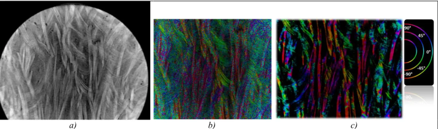
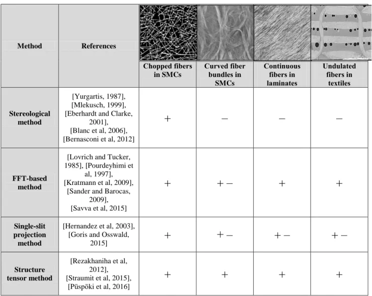
Figure
![Fig. 2. a) Scheme of the projection of image features through a slit [10]; b) initial validation image; c)projection of the validation image through a slit oriented at -60°](https://thumb-eu.123doks.com/thumbv2/123doknet/7922194.265339/5.892.75.816.790.1075/scheme-projection-features-initial-validation-projection-validation-oriented.webp)
Documents relatifs
To take the argument developed into account rigorously, we will define that the solution ψ 1 + ψ 2 for the wave function ψ 3 of the double-slit wave equation follows a
Sektion Physik, Karl-Marx-UniversitSt Leipzig, DDR 7010 Leipzig, D.R.G. bbstract - Using a two-dimensional isotropic adparticle diffusion law the cross-correlation function
We make this distinction between the superposition principle (with incoherent summing) and a Huygens’ principle (with coherent summing) to make sure that we respect what we can do
arithmetic lifting property for all groups and fields (i.e., the Black conjecture) implies the Regular Inverse Galois Problem (Proposition 1.2) (we note this (’)Note
Because the evolution of the probability density of the wave packet just after it exits the slits raises the issue of interpreting the wave/particle dualism, we also
However, concerning measurement, equation (27) shows that at a given physical-time-instant
18 Rather, in this case, a further process had to take place, most probably analogy across prepositions or, possibly, semantic levelling with a near-synonymous preposition,
The letter was from a law firm in New York, and the attorney’s name was Thomas Campbell who was writing a letter on behalf of the artist Mary Ellen Carroll and it was
