Distinct and common cerebral activation changes during mental time travel
in relapsing-remitting multiple sclerosis patients
A. Ernst1,2 · V. Noblet2,3 · E. Denkova4 · F. Blanc2,3,5 · J. De Seze2,5 · D. Gounot2,3 · L. Manning1,2,6 1 Cognitive Neuropsychology and Physiopathology of Schizophrenia (INSERM UMR 1114), 67000 Strasbourg, France
2 Psychology Department, University of Strasbourg, 12, rue Goethe, Strasbourg 6700, France
3 ICube laboratory (CNRS; UMR 7357), Fédération de Médecine translationnelle de Strasbourg (FMTS), 67000 Strasbourg, France
4 Department of Psychology, University of Miami, Miami, FI 33158, USA
5 Department of Neurology and Centre Mémoire de Ressources et de Recherche (CMRR), University Hospital of Strasbourg, 67000 Strasbourg, France
6 INSERM U1114, 1, Place de l'Hôpital, 67000 Strasbourg, France
Abstract
Mental time travel (MTT) entails the ability to mentally travel into autobiographical memory (AM) and episodic future thinking (EFT). While AM and EFT share common phenomenological and cerebral functional properties, distinctive characteristics have been documented in healthy and clinical populations. No report, to our
knowledge, has informed on the functional underpinnings of MTT impairment in multiple sclerosis (MS) patients, hence the aim of this work. We studied 22 relapsing-remitting MS patients and 22 matched controls. Participants underwent an AM/EFT assessment using the Autobiographical Interview (Levine et al. 2002), followed by a functional MRI session. The latter consisted in AM and EFT tasks, distinguishing the construction and elaboration phases of events. The results showed impaired performance for AM and EFT in patients, accompanied by increased cerebral activations mostly located in the frontal regions, which extended to the parietal, lateral temporal and posterior regions during AM/EFT tasks, relative to healthy controls. Enhanced brain activations in MS patients were particularly evident during the EFT task and involved the hippocampus, frontal, external temporal, and cingulate regions. The construction phase required greater fronto-parieto-temporal activations in MS patients relative to both healthy controls, and the elaboration phase. Taking together, our results suggested the occurrence of cerebral activation changes in the context of MTT in MS patients, expressed by distinct and common mechanisms for AM and EFT. This study may provide new insights in terms of cerebral activation changes in brain lesion and their application to clinical settings, considering AM/EFT's central role in everyday life.
Keywords Cerebral activation changes · Multiple sclerosis · Mental time travel · Functional MRI
BACKGROUND
Episodic memory enables us not only to remember the past, but also to project oneself in the future (Tulving 2001, 2005). This notion, referred to as mental time travel (MTT; Suddendorf and Corballis 2007; Tulving 2001), comprises autobiographical memory (AM) and episodic future thinking (EFT), and has been investigated during the past two decades by means of case and group clinical studies, and more recently using the
neuroimaging approach in the intact brain. The single-case studies conducted in amnesic patients demonstrated concurrent difficulties for past and future personal events (Tulving 1985; Klein et al. 2002). These original findings have been confirmed by several group studies showing the simultaneous occurrence of AM/EFT impairment in neurological conditions, particularly, in Alzheimer's disease (Addis et al. 2009) or medial temporal lobe amnesia (Hassabis et al. 2007), as well as in psychiatric conditions such as schizophrenia (D'Argembeau et al. 2008) or depression (Williams et al. 1996). With regard to the neuroimaging studies in healthy subjects, a striking overlap between the functional underpinnings of remembering the past and imagining the future has been observed. The mental time travel brain network, showing an extended overlap with the default network, encompasses the medial frontal and temporal regions, posterior and retrosplenial cortex, lateral parietal and temporal regions (see Schacter et al. 2012 for a review). Despite the fact that these investigations have demonstrated similarities in terms of AM and EFT neurocognitive mechanisms, some differences have also been documented. In particular, a greater neural activity has been reported in the frontopolar and hippo-campal regions during the simulation of future events, relative to that of past events, especially during the initial construction of the event (Okuda et al. 2003; Addis et al. 2007a).
From a theoretical standpoint, these past/future commonalities and differences could be explained in the light of the "constructive episodic simulation hypothesis" (Schacter and Addis 2007). The authors posit that, on the one
hand, past and future events rely on similar information stored in episodic memory, that are flexibly recombined into a novel coherent event, and thus involve similar cognitive processes such as self-referential processes and mental imagery. On the other hand the greater neural activity observed during future thinking could reflect the higher constructive processes necessary to pre-experience than re-experience an event (Addis et al. 2009). Indeed future simulations recruits additional executive resources, because it requires the extraction and recombination of an infinite assortment of details into a novel event, whereas AM consists in the recapitulation of a more restricted pool of details since the reconstructed past event has to be coherent with the original event (Berntsen and Bohn 2010). Similarly, the greater hippocampal recruitment for future tasks could mirror the extraction and the recombination of details into a coherent novel event, and the additional encoding of the simulation product in memory (Addis and Schacter 2012). Several behavioral studies showed a higher number of episodic details provided for past than for future events in healthy subjects (Addis et al. 2008; D'Argembeau and Van der Linden 2004), as well as in clinical conditions (D'Argembeau et al. 2008; Addis et al. 2009). Moreover, in two further clinical conditions, Parkinson's disease (De Vito et al. 2012) and patients with thalamic lesion (Weiler et al. 2011), EFT impairment was demonstrated, in the context of relatively preserved AM abilities.
Furthermore, in relation to the neural mechanisms implemented to respond to brain insult, a few studies have shown brain activation changes during AM recollection (in comparison with healthy subjects) in a hippocampal-damaged patient (Maguire et al. 2001), in semantic dementia patients with lesions to the hippocampus (Viard et al. 2013), in patients with temporal lobe epilepsy (Addis et al. 2007b), in Alzheimer's disease patients
(Meulenbroek et al. 2010), depressed persons (Young et al. 2012) or in people suffering from post-traumatic stress disorder (St. Jacques et al. 2011a). This is in contrast with the relative paucity of studies exploring the functional underpinnings of future thinking impairment. To our knowledge, the first study in this perspective is Mullally et al. (2012) single case amnesic patient, showing increased activation in the residual hippocampal tissue. However, an important methodological aspect of this work is that the patient was asked to imagine fictitious scenes which were therefore not necessarily personally relevant or plausible in the patient's daily life, and which were atemporal in nature. In parallel, Hach et al. (2014) investigated the neural correlates of personal past and future events in depressed persons and reported both increased or decreased brain activations, as well as functional connectivity changes within the mental time travel core network, in comparison with healthy subjects. Functional activation changes have also been reported in semantic dementia during the evocation of personal future events, with increased brain activations in the bilateral medial frontal regions and the hippocampus (Viard et al. 2014).
Another recent study reported AM/EFT impairment in relapsing-remitting multiple sclerosis (RR-MS) patients (Ernst et al. 2014a). Multiple sclerosis (MS) is a chronic and degenerative disease, characterised by
inflammatory processes in the whole central nervous system, with an unpredictable course. Cognitive
impairment is a common feature in MS and disturbances are mainly observed in anterograde memory (verbal and visual modalities), information processing speed, attention, working memory, vi-suospatial abilities and
executive functioning (Chiaravalloti and DeLuca 2008). From a neurological standpoint, several neuroimaging studies carried out in MS patients have demonstrated that structures such as the frontal cortex (Benedict et al. 2002; Bendfeldt et al. 2009) and the hippocampus (Sicotte et al. 2008; Hulst et al. 2012), which are essential in the AM and EFT core network, are frequently damaged.
Regarding AM and EFT performance, RR-MS patients showed a difficulty to generate personal past and future events and to provide episodic details associated with the events, with a similar degree of impairment for both temporal directions. The authors pointed to a main involvement of executive processes at the origin of the deficit. The latter suggestion was supported by the predominant increase of prefrontal and parahippocampal activations in RR-MS patients during the recollection of personal memories relative to healthy controls (Ernst et al. 2014a, b). However, no study to date aimed to explore the EFT neural correlates in MS patients.
Therefore, the general objective of the present study is to extend the previous findings by Ernst and colleagues (2014a, b) to EFT using functional MRI (fMRI). In addition, while a similar degree of AM and EFT impairments has been described in MS, an open question is whether these deficits are associated with similar patterns of functional changes. Based on the aforementioned past/future discrepancies, two hypothesis are put forward (i) that the pattern of brain activations observed in MS patients shows overlaps with the typical mental time travel core network (Schacter et al. 2012) and (ii) that functional changes may be more apparent during the simulation of personal future events than the recollection of memories.
To address these questions, we explored the functional un-derpinnings of past and future simulations in MS patients and in healthy controls. On the bases of Ernst et al.'s data, we hypothesized that increased brain activations would be observed in MS patients, especially in the prefrontal and hippo-campal regions, and that these neural activity changes would be particularly apparent during the imagination of personal future events.
METHODS Participants
Twenty-two RR-MS patients (Polman et al. 2011), involved in a broader study on AM and EFT, were included in this current work and followed these inclusion criteria: an Expanded Disability Status Scale (EDSS; Kurtzke 1983) score <4, an absence of major signs of depression according to the Montgomery and Asberg Depression Rating Scale (Montgomery and Asberg 1979) and no recent exacerbation of MS symptoms.
Twenty-two healthy controls matched for gender, age, education level and verbal Intellectual Quotient were recruited. All the participants were right-handed. Exclusion criteria for all the subjects were documented psychiatric illness, neurological disorder (other than MS for the patients) and poor knowledge of French. Demographic and clinical data are summarized in Table 1. The present study was approved by the "Committee for Protection of Persons" (CPP/CNRS N° 07023) and we complied with the Declaration of Helsinki.
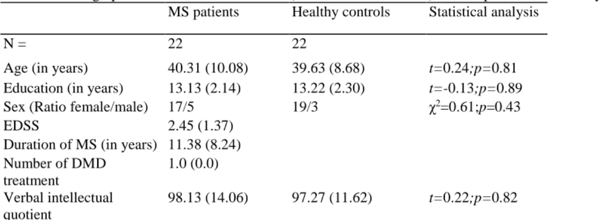
Table 1: Demographical and clinical data Mean (and standard deviation) for MS patients and healthy controls MS patients Healthy controls Statistical analysis
N = 22 22
Age (in years) 40.31 (10.08) 39.63 (8.68) t=0.24;p=0.81
Education (in years) 13.13 (2.14) 13.22 (2.30) t=-0.13;p=0.89
Sex (Ratio female/male) 17/5 19/3 χ2=0.61;p=0.43
EDSS 2.45 (1.37)
Duration of MS (in years) 11.38 (8.24) Number of DMD treatment 1.0 (0.0) Verbal intellectual quotient 98.13 (14.06) 97.27 (11.62) t=0.22;p=0.82
EDSS Expanded Disability Status Scale, DMD Disease-Modifying Drug
Neuropsychological assessment
MS patients underwent a comprehensive neuropsychological baseline examination in the first session (see Table 2 for a complete description) in order to obtain descriptive data on their general cognitive status.
A second session was devoted to the AM/EFT assessment by means of the Autobiographical Interview (AI; Levine et al. 2002). We used an adapted version, enabling the assessment of the EFT component and the use of cue-words (Addis et al. 2009). MS patients and healthy controls were instructed to retrieve/imagine personal unique events, temporally and con-textually specific, occurring over minutes to hours (but not longer than 1 day) and to generate freely as much details as possible about the event. Regarding the AM condition, following Levine et al., three past events per life period were collected [i.e., four or five life periods, depending on the subject's age; 0-11 years, 12-20 years, 21 to (current age -1) or 21-35 years, 36 to (current age -1) and the previous year]. For the EFT component, subjects had to generate five future events that could plausibly occur within the next year. Participants were informed that the cue-words were intended to be used flexibly and no time limit was set to avoid the potential influence of the patients' slowed down cognitive processing speed on AM/EFT performance.
AI session were audio-recorded for later transcription and scored following Levine et al.'s standardized
procedure: after the identification of the central episodic event, details were classified as internal details (i.e., an episodic details related to the central event) or external (i.e., non-episodic information such as semantic details, metacognitive statements, repetitions or else episodic details unrelated to the central event). For each participant, the number of internal details was averaged across the 12 or 15 past events, and across the five future events. A complementary analysis was also conducted, taking into account only the previous year for the AM and EFT scores. The rationale for this additional analysis was to ensure that a similar pattern of results would be observed with a matched temporal interval in both conditions, and thus guarantee the further comparisons for the fMRI tasks (see below).
Following Levine et al.'s recommendations, the interrater reliability was verified for 10 % of the past and future events, which were scored by a second scorer blind to the group membership. Coefficients were assessed with intraclass correlation and showed for all measures a high interrater reliability (between 0.82 and 0.99).
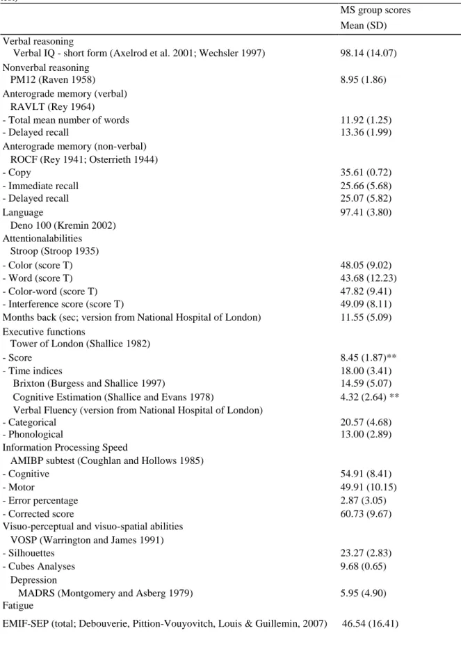
Table 2 Neuropsychological baseline examination scores: Mean (and SD) for the MS patient group in
comparison with the normative data of each test (mean and SD score, 5th percentile or cut-off depending on the test)
MS group scores Mean (SD) Verbal reasoning
Verbal IQ - short form (Axelrod et al. 2001; Wechsler 1997) 98.14 (14.07) Nonverbal reasoning
PM12 (Raven 1958) 8.95 (1.86)
Anterograde memory (verbal) RAVLT (Rey 1964)
- Total mean number of words 11.92 (1.25)
- Delayed recall 13.36 (1.99)
Anterograde memory (non-verbal) ROCF (Rey 1941; Osterrieth 1944)
- Copy 35.61 (0.72) - Immediate recall 25.66 (5.68) - Delayed recall 25.07 (5.82) Language 97.41 (3.80) Deno 100 (Kremin 2002) Attentionalabilities Stroop (Stroop 1935) - Color (score T) 48.05 (9.02) - Word (score T) 43.68 (12.23) - Color-word (score T) 47.82 (9.41)
- Interference score (score T) 49.09 (8.11)
Months back (sec; version from National Hospital of London) 11.55 (5.09)
Executive functions
Tower of London (Shallice 1982)
- Score 8.45 (1.87)**
- Time indices 18.00 (3.41)
Brixton (Burgess and Shallice 1997) 14.59 (5.07)
Cognitive Estimation (Shallice and Evans 1978) 4.32 (2.64) **
Verbal Fluency (version from National Hospital of London)
- Categorical 20.57 (4.68)
- Phonological 13.00 (2.89)
Information Processing Speed
AMIBP subtest (Coughlan and Hollows 1985)
- Cognitive 54.91 (8.41)
- Motor 49.91 (10.15)
- Error percentage 2.87 (3.05)
- Corrected score 60.73 (9.67)
Visuo-perceptual and visuo-spatial abilities VOSP (Warrington and James 1991)
- Silhouettes 23.27 (2.83)
- Cubes Analyses 9.68 (0.65)
Depression
MADRS (Montgomery and Asberg 1979) 5.95 (4.90)
Fatigue
Source of normative data for each test: Verbal IQ (Wechsler 1997); PM12: Progressive Matrices 12 (National Hospital, London); RAVLT: Rey Auditory Verbal Learning Test (Strauss et al. (2006). Compendium of Neuropsychological Tests, 3rd Ed., 2006); ROCF: Rey-Osterrieth Complex Figure (Fastenau et al. 1999); Déno 100 (Kremin 2002); Stroop test (Golden 1978); Months back (National Hospital, London); Tower of London (Collette and Van der Linden 1993); Brixton (Burgess and Shallice 1997); Cognitive Estimation Task (Shallice and Evans 1978); Verbal Fluency (National Hospital, London); AMIPB: Information Processing Speed test from the Adult Memory Information Processing Battery (Coughlan and Hollows 1985); VOSP: Visual Object and Space Perception (Warrington and James 1991); MADRS: Montgomery and Asberg Depression Rating Scale (Montgomery and Asberg 1979); EMIF-SEP: Echelle de Mesure de 1 Impact de la Fatigue (Debouverie et al., 2007); ** Scores under the normal range
Neuroimaging session
fMRI task
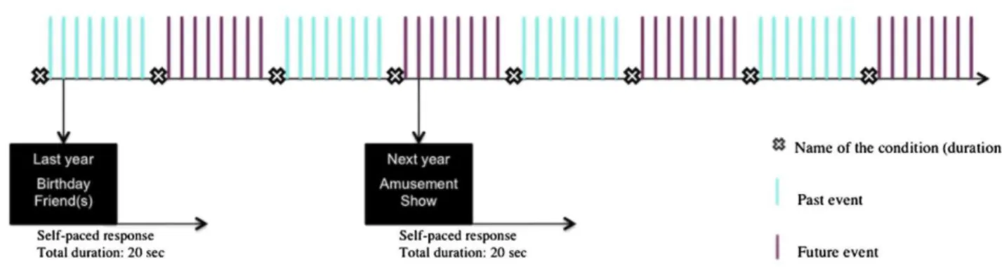
The fMRI session comprised two experimental tasks: the past events and the future events conditions. The tasks consisted in the evocation of unique personal past/future events, contextu-ally specific, occurring over minutes or hours, but not more than 1 day. The participants were reminded about the similarity with the event's
characteristics of the AI. More precisely, it was specified that events following the same criteria than those present during the behavioral assessment (conducted during the first session, systematically before the MRI session) had to be evoked during the fMRI tasks. This precision helped the participants to better understand what corresponded to a unique personal event. However, participants were instructed to not provide the same events as those previously mentioned during the AI. After the fMRI session, the absence of events' repetition was also verified based on the post-scan questionnaire (see below). Following these instructions and immediately prior to scanning, participants completed a computerized practice trial for each task in order to be familiar with the experimental design and timing of presentation of the stimuli. A particular attention was paid to the patients' practice trial to optimize the further completion of the tasks, especially since the fMRI examination imposes time constraints. To ensure that the participants' understood the distinction between the construction and elaboration phases, they were asked to verbalize their answers during the practice trial. A particular attention was paid to the congruence between the button press and the answer reflecting the construction phase. Further instructions and practice trials were given when necessary.
Based on Addis et al. (2007a), two phases were distinguished in the evocation of events: (i) the construction phase corresponding to the search and initial building up of the event, and (ii) the elaboration phase
corresponding to the retrieval/imagination of details associated with the event. For both tasks, 32 pairs of words were proposed to probe personal events (e.g., for the past condition: 'sand-sun'; for the future condition: 'meal-family'), covering the same life periods mentioned above for the AI. The rationale to present cues by pairs, instead of a single cue-word, was clinically based. Indeed, taking into account that the included MS patients showed impaired AM and EFT performance, the use of pairs of words was deemed better adjusted to obtain a sufficient number of events per condition, especially given that fMRI tasks require time-constraints. Within each pair, the two words were thematically related and their relevance to probe past and future events was verified in a pilot study with 12 healthy participants (unpublished data). The participants were instructed about the fact that the cue-words were meant to generate past or future events in a flexible way.
Each trial had a fixed duration of 20 s modulated by the subject's response: once an event was retrieved/imagined, participants pressed a button on a four-button response box to mark the end of the construction phase. Then, a central fixation-cross indicated the elaboration phase which lasted during the remaining time. Importantly, participants were instructed to press the button only if an event came to their mind. In the absence of an answer from the participant, the next trial was automatically presented after the fixed trial duration of 20 s.
The experimental design was organized in eight functional runs of 8 stimuli (four functional runs per condition), alternating between past events and future events conditions. In both tasks, each trial was followed by short periods of fixation that were of jittered duration (mean duration=1.5 s, range=1 to 2 s). At the beginning of each sequence, the name of the condition was displayed on the screen for 6 s. The presentation order of stimuli within each condition was randomized. A schematic illustration of the fMRI experimental design is presented in the Fig. 1. The programming and response collection was done with E-Prime 2 software (Psychology Software Tools, Inc.). Words were displayed on a screen in white text with a black background and viewed using a mirror incorporated in the head-coil.
Immediately following scanning, a post-scan questionnaire was completed in order to verify response accuracy and exclude invalid trials. For each past or future event, participants indicated the type of events (unique, repetitive, extensive, semantic or absent). The different types of events were defined as follows: (i) unique: specific or particular occurrence of events, within a specific time and space frame, no longer than 1 day; (ii) repetitive: composed of event memories, which are usual and repeated, and thus lack episodicity; (iii) extended: includes events whose duration is longer than 1 day (e.g., my week of holidays in Rome), without the mention of
a specific incident; (iv) semantic: encompasses general, semantic associations with the cue-words not self-relevant (i.e., there is often snow at Christmas); (v) absent: corresponds to the absence of response in the scanner (i.e., no button press to end the construction phase). Participants were also asked to provide the spatio-temporal context of events, its emotional valence (categorized in positive, neutral or negative event) and rated on two visual-analogous scales of 10 cm (0=0 cm, corresponding to low degree of difficulty or amount of details; 10=10 cm, corresponding to a high degree of difficulty or amount of details) the difficulty of access/imagination and the amount of details. For the spatio-temporal context, participants were asked to write down the most detailed account they were able to about the location of the event and when it occurred/will occur. Regarding the type of event, while participants initially determined the specificity of events, a further control of this aspect was made by the experimenter (A.E), based on the spatial-temporal context of events. More precisely, immediately after the completion of the post-scan questionnaire, in the absence of a specific spatio-temporal context (necessary to consider an event as unique) or in the case of doubt regarding the classification, participants were asked to provide additional details about the event to determine its precise nature.
fMRI data acquisition
Images were acquired using a 3 T MRI scanner (Siemens Verio). The image sequence was a T2*-weighted echo planar imaging sequence (TR=2500 ms, TE=30 ms, Matrix=64x 64 voxels, FOV=224 mm, FA=90). The anterior commissure and posterior commissure were identified in the midsagittal slice, and 45 contiguous slices (each 4 mm thick) were prescribed parallel to the anterior commissure and posterior commissure plane to cover the whole brain.
Fig. 1 Schematic representation of the experimental design
STATISTICAL ANALYSES
Behavioral analyses
Regarding the AI scores, the mean number of internal details for the past and future conditions were submitted to a repeated measures ANOVA with Group (MS vs. healthy controls) as between-subject factor and Temporal Direction (Past vs. Future) as within factor. Similar statistical analyses were applied for the data collected during the post-scan questionnaire, including the type of events (mean number of events), the emotional valence (percentage of positive, neutral or negative events), difficulty of access/imagination and amount of details (mean score on visual-analogous scales) and for the construction phase (mean reaction time in sec). Analyses were followed by Tukey HSD post hoc test when appropriate.
MRI image preprocessing and statistical analysis
Functional images were realigned to the first volume to correct for motion artifacts, spatially normalized to a standard EP1 template based on the Montreal Neurological Institute reference brain in Talairach space (Talairach and Tournoux 1988) and then spatially smoothed using an 8 mm full-width at half-maximum isotropic Gaussian kernel.
For both conditions, evoked hemodynamic response were time locked to the onset of the cue presentation (construction phase) and were modeled with a canonical hemodynamic response function. Hemodynamic activity related to the elaboration phase was modeled with a boxcar function of 10 s-duration that started
immediately after the end of the construction phase (indicated by button press). The 10 s-duration interval for the elaboration phase was fixed to allow a sufficient time interval to details search and the trials with a construction phase longer than 10 s were excluded.
as well as for future events (Addis et al. 2004a, b, 2011), both type of events were included as regressors of interest. This methodological choice has been made to ensure that a sufficient number of valid trials would be obtained for MS patients.
Statistical analyses were conducted using SPM8 (Statistical Parametric Mapping,
http://www.fil.ion.ucl.ac.uk/spm/). Following St. Jacques et al. (2011a, b), group differences in brain activity modulated by the task and the phase of evocation were explored by means of a 2 (Group: MS, healthy controls) × 2 (Task: past events, future events) × 2 (Phase: construction, elaboration) ANOVA. Additional analyses on the results of the ANOVA were conducted as appropriate to establish the directions of the effects. For the main effects, follow up t-tests were specified as established in SPM8 for ANOVA procedure (MS vs. HC, HC vs. MS, past vs. future, future vs. past, construction vs. elaboration and elaboration vs. construction). For the interactions, the signal from representative voxels showing the significant interactions was extracted and further submitted to statistic analysis outside SPM (using Statistica software) to determine the direction of the interaction.
For all the fMRI statistical analyses, a statistical threshold of p<0.05 (corrected for multiple comparisons, FWE) was used. However, since no previous study has been conducted in mental time travel in MS patients, in order to have a more informative overview of the results, in the case of no significant result, exploratory analyses were also conducted with a more lenient threshold of p<0.001 (uncorrected) and a minimum extent threshold of ten contiguously activated voxels (note however that the latter threshold has been extensively used in the context of neuroimaging studies exploring the neural substrates of mental time travel, e.g., Addis et al. 2007a or Mullally et al. 2012).
RESULTS Behavioral results
The patients' neuropsychological baseline scores were in the normal range for all cognitive functions, with two exceptions, planning and cognitive estimations abilities (Table 2).
AI performance
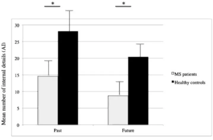
At the behavioral level, focusing first on the AI performance, we showed a significant main effect of group
F(1,42)=73.80, p<0.001,η2
P=0.63, with MS patients exhibiting poorer performance for number of internal details than healthy controls, irrespective of the temporal direction (Fig. 3). A main effect of temporal direction was also highlighted, F(1,42)=55.65, p<0.001„η2
P=0.60, showing a lower mean number of internal details for the future than for the past condition, irrespective of the group of subjects. A similar pattern of results was exhibited when taking into account only the previous year for the AM condition versus the EFT score, with lower performance for the MS patients than for the healthy controls, F(1,42)=38.98, p<0.001,η2
P=0.48,and lower performance for the EFT than for the AM condition, F(1, 42)=55.35, p<0.001 η2
P=0.57. No interaction effect reached the threshold of statistical significance (Fig. 2).
fMRI behavioral results
Reaction time
Turning to the fMRI behavioral results (Table 3), no significant difference was observed for the mean construction reaction time between the two groups, F(1,42)=1.46, p=0.23, η2
P=0.03, or between the two temporal directions, F(1, 42)=2.81, p=0.10, η2
P=0.06.
Type of past and future events
A first repeated measure ANOVA aimed to compare the mean number of events from each type (unique, repetitive, extended, semantic, absent) provided by patients and healthy controls. The results showed neither a main effect of group, F(1, 42)= 1.92, p=0.17, η2
P=0.04, nor of temporal direction, F(1, 42)= 1.61,
p=0.21,η2
P=0.03. However, a main effect of type of event was revealed, F(4, 168)=174.77, p<0.001, η2
P=0.80,with a higher number of unique events in comparison with the four other types (p<0.001 in all the cases), irrespective of groups. Post hoc comparisons showed also a higher number of repetitive events in comparison with extended (p<0.001), semantic (p<0.001) and absent (p=0.04) events. However, an equivalent number of extended, semantic and absent events was shown (statistical threshold between p=0.15 and p=0.99).
Emotional valence
A second repeated measure ANOVA focused on emotional valence, with the comparison of the percentage of positive, neutral and negative past and future events between the two groups. No significant main effect of group, F(1, 42)=0.00, p=0.82,η2
P=0.001, or of temporal direction, F(1, 42)=1.00, p=0.25, η2P=0.03, was observed. However, a significant main effect of emotional valence was shown, F(2, 84) = 105.95,
p<0.001,η2
P=0.71,with a higher rate of positive events in comparison with neutral (p<0.001) and negative events
(p<0.001), irrespective of the temporal direction. The mean rate of neutral and negative events was equivalent
(p= 0.73). A Temporal direction x Emotional valence interaction was obtained, F(2, 84)=14.38, p<0.001, η2
P=0.25, showing a lower rate of negative events for the future than for the past event condition (p<0.001), whereas positive and neutral events showed similar scores across the two temporal directions (respectively
p=0.17 and p=0.36).
Fig. 2 Mean number of internal details provided by MS patients and healthy controls during the AI for the AM
and EFT conditions (p<0.001; * significant difference)
Difficulty of access/imagination and amount of details
Finally, repeated measure ANOVAs were conducted to compare the mean score for the difficulty of access and the mean number of details (rated on two visual-analogous scales) across past and future conditions and between patients and healthy controls. Results showed no significant difference for the difficulty of access/imagination between the groups, F(l,42)=0.65, p=0.42, η2
P=0.01, or between the two temporal directions, F(1, 42)=2.46,
p=0.12, η2
P=0.05. However, a main effect of group has been obtained for the amount of details, F(1, 42)= 4.85,
p=0.03, η2
P=0.10, with a lower self-assessed amount of details for the MS patients than for the healthy controls. However, no significant difference between the two temporal directions was observed, F(1, 42)=0.18, p=0.66, η2
P=0.004.
Neuroimaging results
Brain regions differently engaged by MS patients and healthy controls
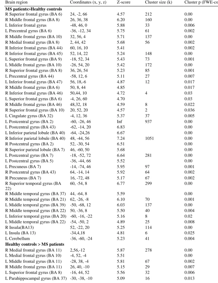
Statistical analyses revealed a significant main effect of group (Table 4), with extensive additional brain activations in MS patients in comparison with healthy controls during the execution of the past and future tasks, irrespective of the recall phase (Fig. 3a). These increased cerebral activations were mainly observed in the bilateral frontal regions (inferior, middle, superior and medial gyri, as well as the supplementary motor area) and extended to the bilateral parietal regions (inferior/superior gyri and the somatosensory cortex) and the bilateral temporal structures (inferior, middle and superior portions). More posterior cerebral activations were also exhibited in the bilateral precuneus, as well as activations in the anterior part of the cortical midline, namely the bilateral cin-gulate gyrus. Brain activations were also reported in the bilateral insular cortex and the left cerebellum.
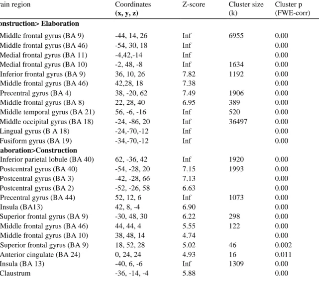
The reverse contrast revealed distinct brain regions that were more activated in healthy controls than in MS patients (Fig. 3b). These brain regions encompassed on the one hand the medial, superior and middle portions of the prefrontal cortex, bilaterally and on the other hand, the left parahippocampal gyrus (BA 37) and the right uncus (BA 20).
Differential brain activations for past and future events
Analyses focusing on the brain activation differences for the past and future tasks (Table 5) demonstrated a main effect of temporal direction. While no greater recruitment for the past relative to the future was observed, several additional cerebral activations were highlighted for the future task, including the frontal regions (medial and supplementary motor area) and the parietal regions (supramarginal gyrus and somatosensory areas), all right-lateralized.
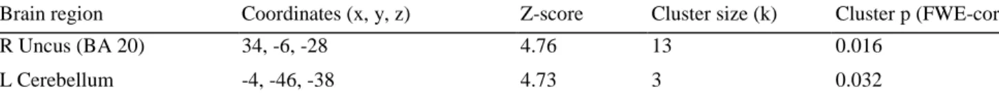
Differential brain activations for the construction and elaboration phases
A main effect of recall phase was obtained (Table 6). In particular, several brain regions showed an enhanced recruitment during the construction phase, encompassing predominantly the bilateral frontal areas (inferior, middle, medial and motor regions), but also the right middle temporal gyrus (BA 21), and left posterior brain regions (middle occipital, lingual and fusiform gyri, extending from BA 18 to BA 19). The reverse contrast also highlighted some brain regions showing increased activations during the elaboration phase, including
predominantly the right frontal regions (middle and superior gyri), the left parietal lobe (inferior parietal lobule and somatosensory cortex), the left anterior cingulate (BA 24) and the left transverse temporal gyrus (BA 41). In addition, bilateral activations of the insular cortex and the left claustrum were observed.
Distinct influence of the group on the past and future task
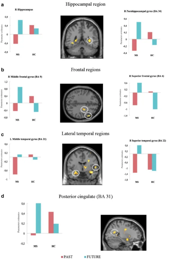
A significant Group x Task interaction was obtained (only evident at p<0.001 ; k=10 voxels), which revealed differential neural substrates between the two groups during past and future simulations (see Table 7 for coordinates). Overall, several brain regions showed an enhanced recruitment in MS patients compared with healthy controls during the future relative to the past task (Fig. 4). Statistical analysis revealed that this enhanced brain network for future simulation in MS patients involved the right middle (BA 9) and superior frontal gyri (BA 6), the left middle temporal gyrus (BA 21), the right superior (BA 22) and transverse temporal (BA 41) gyri, and also the right sub-gyral temporal region (BA 20). Significant activations were also observed in the right anterior (BA 24) and posterior (BA 31) portions of the cingulate cortex, as well as in the left amygdala, the bilateral insular cortex, the bilateral thalamus, the bilateral basal ganglia and the right cerebellum. Increased activations in MS patients were also displayed in the right hippocampus and parahippocampal gyrus during the future task. Moreover, the patients also showed greater activation in the left hippocampus for future than for past events, contrasting with the healthy controls who demonstrated the opposite result, i.e., greater activation in the left hippocampus for the past than for the future events.
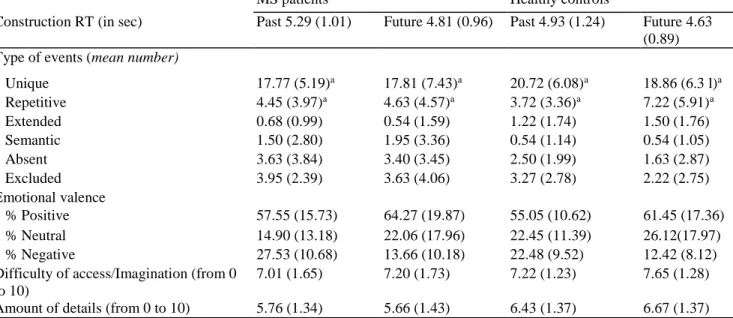
Table 3: Mean (and standard deviation) for the fMRI behavioral results for MS patients and healthy controls
MS patients Healthy controls
Construction RT (in sec) Past 5.29 (1.01) Future 4.81 (0.96) Past 4.93 (1.24) Future 4.63
(0.89) Type of events (mean number)
Unique 17.77 (5.19)a 17.81 (7.43)a 20.72 (6.08)a 18.86 (6.3 l)a Repetitive 4.45 (3.97)a 4.63 (4.57)a 3.72 (3.36)a 7.22 (5.91)a Extended 0.68 (0.99) 0.54 (1.59) 1.22 (1.74) 1.50 (1.76) Semantic 1.50 (2.80) 1.95 (3.36) 0.54 (1.14) 0.54 (1.05) Absent 3.63 (3.84) 3.40 (3.45) 2.50 (1.99) 1.63 (2.87) Excluded 3.95 (2.39) 3.63 (4.06) 3.27 (2.78) 2.22 (2.75) Emotional valence % Positive 57.55 (15.73) 64.27 (19.87) 55.05 (10.62) 61.45 (17.36) % Neutral 14.90 (13.18) 22.06 (17.96) 22.45 (11.39) 26.12(17.97) % Negative 27.53 (10.68) 13.66 (10.18) 22.48 (9.52) 12.42 (8.12)
Difficulty of access/Imagination (from 0 to 10)
7.01 (1.65) 7.20 (1.73) 7.22 (1.23) 7.65 (1.28)
Amount of details (from 0 to 10) 5.76 (1.34) 5.66 (1.43) 6.43 (1.37) 6.67 (1.37)
RT reaction time; a Mean total number of personal events included as regressors of interest in neuroimaging statistical analyses for MS patients: past= 22.22; future=22.45; and for healthy controls: past=24.44; future=26.08. b Mean number of trials with a construction phase longer than 10 s
Table 4 Brain regions differentially engaged in mental time travel in MS patients and healthy controls (Main effect of group; FWE p<0.05)
Brain region Coordinates (x, y, z) Z-score Cluster size (k) Cluster p (FWE-corr)
MS patients>Healthy controls
R Superior frontal gyrus (BA 6) 24, -2, 66 4.57 212 0.00
R Middle frontal gyrus (BA 8) 26, 36, 38 6.20 160 0.00
L Inferior frontal gyrus -48, 46, 0 5.88 33 0.006
L Precentral gyrus (BA 6) -36, -12, 34 5.75 61 0.002
R Middle frontal gyrus (BA 10) 32, 56, 4 5.71 137 0.00
R Medial frontal gyrus (BA 8) 2, 44, 42 5.68 56 0.002
R Inferior frontal gyrus (BA 44) 60, 16, 10 5.41 0.002
R Inferior frontal gyrus (BA 45) 52, 14, 22 5.24 148 0.00
L Superior frontal gyrus (BA 9) -18, 52, 34 5.43 73 0.001
L Middle frontal gyrus (BA 10) -26, 54, 20 5.42 172 0.00
R Superior frontal gyrus (BA 8) 36, 26, 54 5.23 85 0.001
L Precentral gyrus (BA 44) -58, 12, 6 5.11 27 0.007
L Inferior frontal gyrus (BA 47) 56, 18,-6 4.87 12 0.017
R Middle frontal gyrus (BA 6) 50, 8, 44 4.85 0.017
R Inferior frontal gyrus (BA 46) 50,44, 10 4.72 4 0.03
L Superior frontal gyrus (BA 6) -4, 26, 60 4.70 0.03
R Middle frontal gyrus (BA 46) 48,32, 18 4.59 8 0.022
R Superior frontal gyrus (BA 10) 20, 52, 20 4.57 2 0.036
L Cingulate gyrus (BA 32) -4, 12, 36 5.37 37 0.005
L Postcentral gyrus (BA 2) -60, -26, 46 Inf 937 0.00
L Postcentral gyrus (BA 43) -62, -14, 20 6.83 0.00
L Inferior parietal lobule (BA 40) -64, -24,26 6.67 0.00
R Inferior parietal lobule (BA 40) 48, -44, 56 7.24 1051 0.00
R Postcentral gyrus (BA 2) 52, -30, 54 6.51 0.00
R Superior parietal lobule (BA 7) 46, -60, 50 5.68 0.00
L Postcentral gyrus (BA 7) -18, -52, 72 6.64 281 0.00
L Postcentral gyrus (BA 5) -36, -44, 66 5.52 0.00
L Precuneus (BA 7) -14, -74, 46 5.95 97 0.001
R Postcentral gyrus (BA 43) 64, -14, 14 5.92 64 0.002
R Precuneus (BA 7) 16, -72, 48 5.17 67 0.002
R Superior temporal gyrus (BA 22)
60, -54, 8 6.77 299 0.00
R Middle temporal gyrus (BA 37) 44, -64, 8 5.59 0.00
R Middle temporal gyrus (BA 21) 62, -26, -8 6.10 70 0.001
L Middle temporal gyrus (BA 39) -50, -68, 12 6.03 137 0.00
R Middle temporal gyrus (BA 22) 50, -36, 8 5.50 40 0.004
L Inferior temporal gyrus (BA 20) -60, -16, -22 5.16 8 0.02
L Middle temporal gyrus (BA 22) -54, -50, 2 4.89 25 0.008
R Insula(BA13) 52, -22, 20 5.25 114 0.00
L Insula (BA 13) -34,4,18 4.81 6 0.025
L Cerebellum -36, -60, -24 5.23 41 0.004
Healthy controls > MS patients
R Medial frontal gyrus (BA 11) 2,56,-12 5.87 278 0.00
L Medial frontal gyrus (BA 10) -4, 52, -4 5.51 0.00
L Middle frontal gyrus (BA 11) -28, 38, -4 5.81 67 0.002
R Middle frontal gyrus (BA 11) 26, 40, -10 5.15 29 0.007
L Superior frontal gyrus (BA 8) -16, 44, 52 5.56 32 0.006
Brain region Coordinates (x, y, z) Z-score Cluster size (k) Cluster p (FWE-corr)
R Uncus (BA 20) 34, -6, -28 4.76 13 0.016
L Cerebellum -4, -46, -38 4.73 3 0.032
Distinct influence of the group on the construction and elaboration phases
Statistical analyses evidenced a significant Group x Task interaction (see Table 8 for coordinates; again only evident at p<0.001; k=10 voxels).
A first set of results showed that different brain regions were recruited by MS patients and healthy controls during the construction phase. During this phase, healthy subjects recruited to a greater extent than MS patients primarily left-lateralized regions, including the medial frontal gyrus (BA
10), the cuneus (BA 18/19) and the precuneus (BA 31). Conversely, MS patients demonstrated increased brain activations during the construction phase in the right superior and left middle temporal gyrus (BA 22), the right inferior parietal lobule (BA 40) and the right superior parietal lobule (BA 7; Fig. 5). Within the group of MS patients, we showed greater left-lateralized activations during the construction phase (vs. the elaboration phase) in the inferior frontal gyrus (BA 9), the precuneus (BA 7) and the insula (BA 13).
Finally, in the healthy control group, additional brain activations were observed during the elaboration phase (vs. the construction phase) which were mainly located in the left parietal lobe, namely the postcentral gyrus (BA 1/2/7) and the inferior parietal lobule (BA 40), but also in the right insular cortex.
No significant Task x Phase interaction was shown.
Fig. 3 Brain regions showing increased activity during mental time travel in MS patients (a) and healthy
controls (b); FWE, p<0.05; see Table 4 for coordinates)
DISCUSSION
The aim of the present study was to explore mental time travel performance in RR-MS patients, with a double approach in clinical neuropsychology and functional neuroimaging. The main findings showed patients' generally normal performance in baseline tests. Their performance on the AI was poorer than controls in both temporal conditions, with both groups performing better in the past relative to the future condition. FMRI behavioral results highlighted a higher number of unique events and higher rate of positive events in general, while a lower rate of negative events for the future condition relative to the past condition was observed, in both patients and healthy controls. Finally, patients' self-assessment showed lower amount of details compared with controls. Neuroimaging analyses highlighted (i) increased bilateral cortical engagement in the patient group (especially in the bilateral prefrontal regions), while the controls showed increased bilateral cortical and subcortical engagement (e.g., in the bilateral frontal regions and the left parahippocampal gyrus). (ii) The future condition recruited significantly more cortical regions than the past (e.g., in the right medial frontal region and the right parietal regions), (iii) The two groups showed different patterns of cerebral activation following the temporal direction, with patients recruiting significantly more extensive brain regions for the future condition (e.g., in the hippocampus and the lateral temporal regions bilaterally, as well as in the right frontal regions), (iv)
The construction and elaboration phases showed also different cortical activation loci (construction: e.g., mostly in the bilateral frontal regions and the left posterior brain regions; elaboration: e.g., in the bilateral fronto-parietal regions), (v) During the construction vs. elaboration phases, the patients recruited significantly more right but also left lateralized regions (e.g., in the lateral temporal and fronto-parietal regions, bilaterally). The latter were different from those significantly more activated by the controls.
Clinically, the present group of MS patients showed impaired performance in both AM and EFT, expressed by a lower number of episodic details in comparison with healthy controls. Moreover, better performance were observed for the AM than for the EFT condition in both groups, replicating the results from previous studies (D'Argembeau et al. 2008; Addis et al. 2009; Ernst et al. 2014a, b). Importantly considering either the memories belonging only to the last year or memories across lifespan did not influence the results; in both cases, EFT performance remained lower than AM performance.
Regarding the phenomenological properties of past and future events evoked during the fMRI session, a higher rate of positive events has been reported, relative to neutral and negative events. This tendency to provide a greater number of emotionally positive personal events was particularly important for future events. These results are in agreement with the robust positivity bias and the 'rosy future effect' described in the literature dealing with AM and EFT (Szpunar et al. 2012; Rasmussen and Bernsten 2013; Walker et al. 2003). Equivalent performance were obtained for the post-scan questionnaire variables between the two groups (with the exception of the amount of details, showing a lower score for MS patients). Similar to Hach et al.'s (2014) results with depressed patients, despite the presence of mental time travel impairment at the clinical level, our MS patients were still able to perform adequately the fMRI tasks. This observation warrants the interpretation of the patients' cerebral activation changes as resulting from pathology, and not from, for instance, poor task performance.
Table 6 Brain regions differentially engaged in the construction and the elaboration phases of past and future
events (Main effect of recall phase; FWE p<0.05)
Brain region Coordinates
(x, y, z)
Z-score Cluster size (k)
Cluster p (FWE-corr) Construction> Elaboration
L Middle frontal gyrus (BA 9) -44, 14, 26 Inf 6955 0.00
L Middle frontal gyrus (BA 46) -54, 30, 18 Inf 0.00
L Medial frontal gyrus (BA 11) -4,42,-14 Inf 0.00
L Medial frontal gyrus (BA 10) -2, 48, -8 Inf 1634 0.00
R Inferior frontal gyrus (BA 9) 36, 10, 26 7.82 1192 0.00
R Middle frontal gyrus (BA 46) 42,28, 18 7.38 0.00
R Precentral gyrus (BA 4) 38, -20, 62 7.49 1906 0.00
R Middle frontal gyrus (BA 8) 22, 28, 40 6.95 389 0.00
R Middle temporal gyrus (BA 21) 56, -6, -16 Inf 520 0.00
L Middle occipital gyrus (BA 18) -24, -86, 20 Inf 36497 0.00
L Lingual gyrus (B A 18) -24,-70,-12 Inf 0.00
L Fusiform gyrus (BA 19) -34,-70,-12 Inf 0.00
Elaboration>Construction
R Inferior parietal lobule (BA 40) 62, -36, 42 Inf 1920 0.00
L Postcentral gyrus (BA 40) -54, -28, 20 7.15 1993 0.00
L Postcentral gyrus (BA 3) -42, -28, 66 7.13 0.00
L Postcentral gyrus (BA 2) -52, -26, 58 6.63 0.00
R Precentral gyrus (BA 44) 52, 12, 6 Inf 1073 0.00
R Insula (BA13) 42, 8, -4 6.90 0.00
L Superior frontal gyrus (BA 9) -30, 48, 30 6.22 298 0.00
R Middle frontal gyrus (BA 46) 44, 44, 4 5.55 122 0.00
R Middle frontal gyrus (BA 10) 38, 48, 14 4.74 0.00
R Superior frontal gyrus (BA 9) 18, 52, 28 5.02 46 0.002
L Anterior cingulate (BA 24) 0, 24, 24 4.93 16 0.011
L Insula (BA 13) -40, 6, -6 Inf 1309 0.00
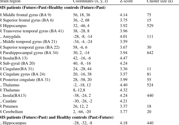
Table 7 Brain regions showing significant activations for the Group x task interaction (p <0.001 uncorrected;
k= 10 voxels)
Brain region Coordinates (x, y, z) Z-score Cluster size (k)
MS patients (Future>Past>Healthy controls (Future>Past)
R Middle frontal gyrus (BA 9) 56, 18, 36 4.14 75
R Superior frontal gyrus (BA 6) 36, -2, 68 3.75 15
R Hippocampus 32, -46, 4 3.92 529
R Transverse temporal gyrus (BA 41) 38, -28, 8 3.96
L Amygdala -28, -8, -14 4.01 111
L Middle temporal gyrus (BA 21) -34, -4, -24 3.59
R Superior temporal gyrus (BA 22) 58, -6, 6 3.67 30
R Parahippocampal gyrus (BA 34) 30, 2, -14 3.94 642
R Insula(BA 13) 42, -16, -6 4.47
R Sub-gyral (BA 20) 40, -8, -16 4.24
R Cingulate(BA 31) 24, -28, 44 3.36 11
R Cingulate gyrus (BA 24) 20, -16, 38 3.57 81
R Posterior cingulate (BA 31) 28, -58, 20 3.99 55
L Thalamus -2, -18, 12 4.40 524 R Thalamus 8,-12,8 4.32 L Insula(BA13) -38, -24, 2 4.24 440 L Caudate -30, -26, -2 4.21 R Putamen 26, 12, 2 3.37 18 R Cerebellum 2, -66, -20 3.77 20
MS patients (Future>Past) and Healthy controls (Past>Future)
L Hippocampus -28, -32, -8 4.18 440
Cluster corrected threshold of p<0.05 (FWE) reached with a cluster size of k=208 (AlphaSim utility in REST software; Song et al. 2011; http://www.restfmri.net)
Concerning the neuroimaging results, the present report constitutes, to our knowledge, the first neuroimaging study on mental time travel in MS patients. Consistent with previous neuroimaging examinations on future thinking in healthy subjects, we reported greater brain activations only for the future condition (in comparison with the past condition), located in the frontal and parietal regions (Addis et al. 2007a; Szupnar et al. 2007). The same is true for the comparison between the two recall phases, showing two sets of brain regions differentially engaged for the construction and the elaboration phases. Consistent with our hypothesis, the pattern of brain activations observed in MS patients showed many overlaps with the typical mental time travel core network (Schacter et al. 2012). As expected, increased brain activations in the bilateral frontal regions were evidenced during the evocation of personal past and future events, relative to healthy controls. This was also accompanied by enhanced bilateral parietal activations, notably in the inferior parietal lobule. Recently, St. Jacques et al. (2011b) introduced the notion of the "frontoparietal network", which is related to executive control processes, and therefore, especially involved in the initial construction of personal events. Thus, the enhanced activity in this frontoparietal network could reflect the actual higher cognitive demand required during the generation of events in our MS patients. The latter observation is in agreement with the dysexecutive hypothesis at the origin of mental time travel impairment in MS patients put forward by Ernst and colleagues (2014a, b). Also supporting this view, higher frontoparietal activations (left inferior frontal gyrus, right inferior and superior parietal lobules) were particularly evident in our MS patients during the construction phase and higher frontal activations (right middle frontal gyrus and right superior frontal gyrus) were also observed during the evocation of future events, which both highly required executive control processes.
Fig. 4 Significant brain activations for the Group x Task interaction in the right hippocampus and right
parahippocampal gyrus (BA 34), the right middle (BA 9) and superior (BA 6) frontal gyri, the left middle (BA 21) and right superior (BA 22) temporal gyri, and the posterior cingulate (BA 31); (p<0.001 uncorrected; k= 10 voxels)
Table 8 Brain regions showing significant activations for the Group x Recall phase interaction (p <0.001
uncorrected; k= 10 voxels)
Brain region Coordinates (x, y, z) Z-score Cluster size (k)
Healthy controls (construction>elaboration)>MS patients (construction>elaboration)
L Medial frontal gyrus (BA 10) 0, 54, -10 4.94 291
L Cuneus (BA18) -2, -88, 22 4.10 32
R Cuneus (BA19) 22, -88, 22 3.58 17
L Precuneus (BA 31) -10, -60, 22 3.45 26
MS patients (construction>elaboration)>Healthy controls (construction>elaboration)
R Superior temporal gyrus (BA 22) 60, -54, 8 4.08 43
L Middle temporal gyrus (BA 22) -56, -48, 2 3.99 57
R Inferior parietal lobule (BA 40) 48, -44, 56 3.40 34
R Superior parietal lobule (BA 7) 26, -68, 54 3.26 16
L Inferior frontal gyrus (BA 9) -44, -2, 24 4.82 312
L Precuneus (BA 7) -16, -72, 44 3.49 45
L Insula (BA13) -34, 4, 18 3.99 312
Healthy controls (elaboration>construction)>MS patients (elaboration>construction)
L Postcentral gyrus (BA 2) -62, -30, 42 4.21 171
L Inferior parietal lobule (BA 40) -64, -24, 26 3.77
L Postcentral gyrus (BA 1) -64, -16, 22 3.70
L Postcentral gyrus (BA 7) -16, -52, 72 3.49 22
R Insula (BA 13) 52, -22, 24 3.35 14
Cluster corrected threshold of p<0.05 (FWE) reached with a cluster size of k=208 (AlphaSim utility in REST software; Song et al. 2011; http://www.restfmri.net)
Fig. 5 Significant activations for the Group x Recall phase interaction in the left middle (BA 22) and right
superior (BA 22) temporal gyri, the right inferior (BA 40) and superior (BA 7) parietal lobule (p<0.001 uncorrected; k= 10 voxels)
Regions from the bilateral temporal lobes (in particular the BA 20/21/22) were also more widely recruited by MS patients. These regions typically pertain to the AM/EFT core network and sustain personal semantic knowledge. This cognitive process has a primary role during event generation, where general personal knowledge serves as a basis for the research of more specific episodes, but is also involved during the further research of details, in relation to semantic representations of the recollected details (Svoboda et al. 2006). Interestingly, the greater recruitment of these temporal regions in the patient group was mostly observed during the construction phase and the EFT task that particularly require personal semantics (Svoboda et al. 2006; Viard et al. 2012). Additionally, these temporal activations were accompanied by greater fronto-parietal activations, likely reflecting the fact that the construction phase and the future condition are particularly demanding in executive control processes.
In parallel, no greater recruitment of the hippocampal region was demonstrated in MS patients when past and future events were analyzed together. Interestingly though, the bilateral hippocampus and the right
parahippocampal gyrus showed a higher activity for the future vs. past events in MS patients. This higher hippocampal activity for the future condition followed the same pattern than the increased fronto-parietal and lateral temporal region activations. This finding indicating an increased hippocampal activity for future relatively to past events is in accord with the observation in the healthy subject that constructing future events engages the hippocampus more than remembering past events (Addis et al. 2011). And also with the evidence showing that the hippocampus although important to both past and future simulations seems to be doubly important to future simulation because it enables (besides drawing out relevant past details), the recombination and integration of those details into coherent future events (Addis et al. 2009, see the constructive episodic simulation hypothesis, Schacter and Addis 2007). Taking together, it appears that this enhanced neural activity was triggered by increased cognitive demand.
Regarding healthy controls, the opposite pattern (in comparison with MS patients) was highlighted for the left posterior hippocampus, showing an increased activity for the past vs. the future condition. While this latter observation is in agreement with previous reports (Abraham et al. 2008; Botzung et al. 2008; Weiler et al. 2010), it is not in accordance with Addis et al. (2007a) who showed greater right anterior hippocampal activation for the future than for the past. The hippocampus plays different roles in past and future events. In particular, the hippocampus serves to index, retrieve and bind together the different aspects of a memory trace (Addis et al. 2007a, b). Regarding its role in future events, the hippocampus is associated to the retrieval and integration of disparate details into a coherent future event, but also to the encoding of the simulation product (Addis and Schacter 2012). In addition, different roles have been attributed to the hippocampus according to its laterality and along its antero-posterior axis (see Viard et al. 2012 for a review). With respect to the right hippocampus, it has been suggested that this structure is especially involved in the integration of novel events (Addis et al. 2007a, b), in emotional processes (Denkova et al. 2006), in the spatial context and sense of reliving associated with memories (Viard et al. 2011). The left hippocampus is particularly responsive to the episodic quality of past/future events and supports the generation of complex coherent scene (Viard et al. 2011). The anterior portion of the hippocampus is mainly involved in the flexible recombination of details while the posterior hippocampus is especially related to spatial memory but is also responsive to the amount of details generated for past and future events (Viard et al. 2012). Taken together, it appears that the left posterior hippocampus is particularly involved when strictly episodic events triggered by specific (i. e., personal) cues are evoked (Viard et al. 2012). Distinct time course of the hippocampal engagement has also been highlighted across the evocation phases for past and future conditions in healthy controls (Weiler et al. 2010). In particular, Weiler et al. (2010) found a greater hippocampal activity during the early construction of past events while this stronger hippocampal activity was only observed during the late elaboration phase of future events. Importantly, as previously
highlighted by Addis et al. (2007a, b), in the case of subtle differences in brain activations between the construction and the elaboration phases, collapsing the analyses across these two phases could influence the patterns of activity reported. In this context, the greater hippocampal engagement for the past condition found in the present study could have been influenced by the combined analysis of the construction and elaboration phases.
The posterior brain regions, and particularly those of the cortical midline and its adjacent structures (i.e., the posterior cingulate gyrus and the precuneus), are generally associated with mental visual imagery process (Greenberg and Rubin 2003; Szupnar et al. 2007; Summerfield et al. 2009) and contribute to the generation of details retrieved/imagined and to the subjective sense of remembering/imagining. The anterior portion of the cortical midline, including the anterior cingulate gyrus and the medial frontal gyrus, are involved in
self-referential processes (Northoff et al. 2006). In the case of MS patients, increased brain activations were observed in the bilateral precuneus and anterior cingulate gyrus during mental time travel. Moreover, both anterior and posterior cin-gulate gyri were particularly engaged during future simulation in the patient group, likely reflecting the current setting up of visual imagery and self-referential processes. Despite this greater brain recruitment, the patients did not reach the same level of episodic details (i.e., amount of details) than healthy controls.
Increased brain activations were also observed in MS patients in the basal ganglia and the cerebellum, especially during the simulation of future events. Brain activations in the cerebellum have been frequently reported in the literature during the retrieval of memories and the imagination of personal future events (see for instance
Svoboda et al. 2006; Addis et al. (2007a, b); Szupnar et al. 2007). While its precise role in the context of AM and EFT is not fully elucidated, it has been suggested that the cerebellum plays a role in the imagination of one's body movements (Hesslow 2002). In addition, the cerebellum and the frontal cortex are connected via the cerebello-thalamocortical pathway (Middleton and Strick 2001) and its involvement in various cognitive functions is well established, especially in executive functions. The same is true for the basal ganglia, which share strong interactions with the frontal cortex, and for which an involvement in various cognitive tasks has been described (Hesslow 2002). In this context, the greater brain activations observed in the cerebellum and the basal ganglia could be related to the main functional changes reported in our MS patients in the frontal region, especially since they pertain to the frontoparietal network, for which the involvement in AM has been recently described by St. Jacques et al. (2011a, b).
Conversely, only a few additional cerebral activations have been reported in the healthy controls, located in key nodes of the AM/EFT cerebral network. In particular, they showed a greater neural activity in the medial frontal regions, but also in the middle and superior frontal gyri, the parahippocampal gyrus and the adjacent uncus during the simulation of past and future events. Moreover, the medial frontal gyrus and posterior brain regions (i.e., the precuneus and the cuneus) showed an early engagement during the construction phase in healthy controls, while these same activations seemed delayed to the elaboration phase in MS patients. Increased activations in healthy controls were also observed in the somatosensory regions during the elaboration phase (vs. the construction phase), not evident in the patient group. In the AM literature, the somatosensory cortex is closely linked to the retrieval of somatosensory representations related to the event, the emotional intensity, and is also especially engaged during the elaboration of events (Daselaar et al. 2008; Botzung et al. 2010).
In summary, distinct and common functional changes were observed in MS patients during the simulation of personal past and future events. In fact, as it was expected, brain activation changes were particularly obvious during the most executive demanding conditions, namely the EFT condition in the construction phase. Our results provided therefore supportive arguments to the executive related-deficit hypothesis suggested by Ernst et al. (2014a, b). Our findings are also consistent with previous studies showing brain activation changes in clinical conditions in the case of EFT impairment in patients with depression (Hach et al. 2014) or in semantic dementia (Viard et al. 2014). In this context, future studies conducted in different clinical conditions could help to improve our understanding of the underlying mechanisms sustaining mental time travel impairment.
This last point is of particular importance considering the central role of AM and EFT in everyday life
functioning, for instance in identity, social situations, coping skills or goal attainment, but also more generally in well-being (Szupnar 2010; Rasmussen and Habermas 2011). Overall, the present study shows that MS patients presenting with selective mental time travel deficit, show, in parallel with findings in the healthy subject, common and distinct neuro-cognitive mechanisms for past and future conditions.
Acknowledgments
We are grateful to the "Fondation pour la Recherchesur la Sclérose en Plaques" (ARSEP; Ile de France; grant to L.M) for research funding and to the Ministry of National Education and Research (A.E.'s PhD grant). AE is now a postdoctoral researcher in the LEAD (CNRS UMR5022) at the University of Burgundy, supported by a research funding from the Region Bourgogne (France) accorded to Dr. Chris Moulin and Dr. Céline Souchay (LEAD, CNRS UMR5022, University of Burgundy). We thank Dr. B. Levine for personal communication of the AI to LM. We also thank C. Vinet-Gasse, B. Journault and A. Botzung for helping with testing and V.
Voltzenlogel for interrater reliability scoring, N. Heider, S. Graves, F. Ernwein, A. Clerc-Renault, E. Montaut and the NCC MA students for the transcriptions of the AI audio-recordings. We are grateful to the "Imagerie in vivo" Platform (ICube laboratory), where the MRI sessions were done, the CIC of the Strasbourg University Hospitals, where patients and controls were examined, C. Marrer for fMRI technical assistance and O. Després for initial participation in E Prime programming.
A. Ernst, V. Noblet, E. Denkova, F. Blanc, J. De Seze, D. Gounot, and L. Manning declare no competing financial interests.
REFERENCES
Abraham, A., Schubotz, R. I., & von Cramon, D. Y. (2008). Thinking about the future versus the past in personal and non-personal contexts.
Brain Research, 1233, 106-119.
Addis, D. R, & Schacter, D. L. (2012). The hippocampus and imagining the future: where do we stand? Frontiers in Human Neuroscience, 5, 1-15.
Addis, D. R., Moscovitch, M., Crawley, A. P., & McAndrews, M. P. (2004a). Recollective qualities modulate hippocampal activation during autobiographical memory retrieval. Hippocampus, 14, 752-762.
Addis, D. R, McIntosh, A. R, Moscovitch, M., Crawley, A. P., & McAndrews, M. P. (2004b). Characterizing spatial and temporal features of autobiographical memory retrieval networks: a partial least squares approach. Neurolmage, 23, 1460-1471.
Addis, D. R, Wong, A. 1, & Schacter, D. L. (2007a). Remembering the past and imagining the future: common and distinct neural substrates during event construction and elaboration. Neuropsychologia, 45, 1363-1377.
Addis, D. R., Moscovitch, M., & McAndrews, M. P. (2007b). Consequences of hippocampal damage across the autobiographical memory network in left temporal lobe epilepsy. Brain, 130, 2327-2342.
Addis, D. R, Wong, A. 1, & Schacter, D. L. (2008). Age-related changes in the episodic simulation of future events. Psychological Science,
19,33-41.
Addis, D. R., Sacchetti, D. C, Ally, B. A., Budson, A. E., & Schacter, D. L. (2009). Episodic simulation of future events is impaired in mild Alzheimer's disease. Neuropsychologia, 47, 2660-2671.
Addis, D. R, Cheng, T., Roberts, R. P., & Schacter, D. L. (2011). Hippocampal contributions to the episodic simulation of specific and general future events. Hippocampus, 21, 1045-1052.
Axelrod, B. N., Ryan, J. 1, & Ward, L. C. (2001). Evaluation ofseven-subtest short forms of the Wechsler Adult Intelligence Scale-III in a referred sample. Archives of Clinical Neuropsychology, 16, 1-8.
Bendfeldt, K., Kuster, P., Traud, S., Egger, H., Winklhofer, S., Mueller-Lenke, N., et al. (2009). Association of regional gray matter volume loss and progression of white matter lesions in multiple sclerosis - a longitudinal voxel-based morphometry study. Neurolmage, 45, 60-67. Benedict, R H. B., Bakshi, R., Simon, J. H., Priore, R., Miller, C, & Munschauer, F. (2002). Frontal cortex atrophy predicts cognitive impairment in multiple sclerosis. Journal of Neuropsychiatry and Clinical Neurosciences, 14, 44-51.
Berntsen, D., & Bohn, A. (2010). Remembering and forecasting: the relation between autobiographical memory and episodic future thinking.
Memory and Cognition, 38, 265-278.
Botzung, A., Denkova, E., & Manning, L. (2008). Experiencing past and future personal events: functional neuroimaging evidence on the neural bases of mental time travel. Brain and Cognition, 66, 202-212.
Botzung, A., Rubin, D. C, Miles, A., Cabeza, R., & LaBar, K. S. (2010). Mental hoop diaries: emotional memories of a college basketball game in rival fans. The Journal of Neuroscience, 30, 2130-2137.
Burgess, P., & Shallice, T. (1997). The Hayling and Brixton Tests. Test manual. Bury St Edmunds, UK: Thames Valley Test Company. Chiaravalloti, N. D., & DeLuca, J. (2008). Cognitive impairment in multiple sclerosis. Lancet Neurology, 7, 1139-1151.
Collette, F., & Van der Linden, M. (1993). Une adaptation du test de la Tour de Londres. Bruxelles: Service de Neuropsychologie, Université de Liège.
Coughlan, A., & Hollows, S. (1985). The Adult Memory and Information Processing Battery. Saint James Hospital, Leeds. D'Argembeau, A., & van der Linden, M. (2004). Phenomenal characteristics associated with projecting oneself back into the past and forward into the future: influence of valence and temporal distance. Consciousness and Cognition, 13, 844-858.
D'Argembeau, A., Raffard, S., & Van Der Linden, M. (2008). Remembering the past and imagining the future in schizophrenia. Journal of
Abnormal Psychology, 117, 247-251.
Daselaar, S. M., Rice, H. J., Greenberg, D. L., Cabeza, R, LaBar, K. S., & Rubin, D. C. (2008). The spatiotemporal dynamics of autobiographical memory: neural correlates of recall, emotional intensity, and reliving. Cerebral Cortex, 18, 217-229.
De Vito, S., Gamboz, N., Brandimonte, M. A., Barone, P., Amboni, M., & Delia Salla, S. (2012). Future thinking in Parkinson's disease: an executive function? Neuropsychologia, 50, 1494-1501.
Debouverie, M., Pittion-Vouyovitch, S., Louis, S., & Guillemin, F. (2007). Validity of a French version of the fatigue impact scale in multiple sclerosis. Multiple Sclerosis, 13,1026-1032.
Denkova, E., Botzung, A., Scheiber, C, & Manning, L. (2006). Implicit emotion during recollection of past events: a nonverbal fMRI study.
Brain Research, 1078, 143-150.
Ernst, A, Blanc, F., de Seze, J., Sellal, F., Chauvin, B., & Manning, L. (2014a). Impaired mental simulation of specificpast and future personalevents in non-depressed multiple sclerosis patients. Journal of the Neurological Sciences, 345, 68-74.
Ernst, A., Noblet, V, Denkova, E., Blanc, F., de Seze, J., Gounot, D., & Manning, L. (2014b). Functionalcerebral changes in multiple sclerosis patients during an autobiographicalmemory test. Memory, 22, 1-17.
Fastenau, P. S., Denburg, N. L., & Hufford, B. J. (1999). Adult norms for the Rey-osterrieth complex figure test and for supplemental recognition and matching trials from the extended complex figure test. The Clinical Neuropsychologist, 13, 30-47.
Golden, C. J. (1978). Diagnosis and rehabilitation in clinical neuropsychology. Springfiels: Charles C. Thomas. Greenberg, D. L., & Rubin, D. C. (2003). The neuropsychology of autobiographical memory. Cortex, 39, 687-728.
Hach, S., Tippett, L. J., & Addis, D. R. (2014). Neural changes associated with the generation of specific past and future events in depression. Neuropsychologia, 65, 41-55.
Hassabis, D., Kumaran, D., Vann, S. D., & Maguire, E. A. (2007). Patients with hippocampal amnesia cannot imagine new experiences.
Proceedings of the National Academy of Sciences, 104, 1726-1731.