ECOLE DE TECHNOLOGIE SUPÉRIEURE UNIVERSITÉ DU QUÉBEC
MANUSCRIPT-BASED THESIS PRESENTED TO ÉCOLE DE TECHNOLOGIE SUPÉRIEURE
IN PARTIAL FULFILLMENT OF THE REQUIREMENTS FOR THE DEGREE OF DOCTOR OF PHILOSOPHY
Ph. D.
BY
Martin BRUMMUND
STUDY OF THE OCCLUSION EFFECT INDUCED BY AN EARPLUG: NUMERICAL MODELLING AND EXPERIMENTAL VALIDATION
MONTREAL, DECEMBER 19, 2014 © Copyright 2014 reserved by Martin Brummund
© Copyright reserved
It is forbidden to reproduce, save or share the content of this document either in whole or in parts. The reader who wishes to print or save this document on any media must first get the permission of the author.
BOARD OF EXAMINERS (THESIS PH.D.) THIS THESIS HAS BEEN EVALUATED BY THE FOLLOWING BOARD OF EXAMINERS
Mr. Frédéric Laville, Thesis Supervisor
Department of Mechanical Engineering at École de technologie supérieure
Mr. Yvan Petit, Thesis Co-supervisor
Department of Mechanical Engineering at École de technologie supérieure
Mr. Franck Sgard, Thesis Co-supervisor
Scientific Division at Institut de recherche Robert-Sauvé en santé et en sécurité du travail
Mrs. Nicola Hagemeister, President of the Board of Examiners
Department of Automated Manufacturing Engineering at École de technologie supérieure
Mr. Jérémie Voix, Member of the jury
Department of Mechanical Engineering at École de technologie supérieure
Mr. Raymond Panneton, External Evaluator
Department of Mechanical Engineering at Université de Sherbrooke
THIS THESIS WAS PRENSENTED AND DEFENDED
IN THE PRESENCE OF A BOARD OF EXAMINERS AND PUBLIC NOVEMBER 27, 2014
ACKNOWLEDGMENT
I would like to express my deepest gratitude to my thesis supervisors Professors Franck Sgard, Yvan Petit and Frédéric Laville who have guided and supported me on countless occasions throughout this research project. Your expertise and dedication make you exceptional mentors and I feel privileged to be given the opportunity to research with and learn from you. Thank you for everything you have contributed to realize this study.
I would like to gratefully acknowledge the Institut de recherche Robert-Sauvé en santé et en sécurité du travail (IRSST) for funding this work. Additionally, I would like to gratefully acknowledge the funding received in form of prizes and scholarships from: École de technologie supérieure (ÉTS), Équipe de recherché en sécurité du travail – Réseau de recherche en santé et en sécurité du travail du Québec (ÉREST-RRSSTQ), Canadian Acoustical Association (CAA) and Acoustical Society of America (ASA).
I would like to thank the members of the Scientific Advisory Committee: Elliott H. Berger (3M), William J. Murphy (NIOSH), Karl Buck (ISL), and Nicolas Trompette (INRS) for sharing a tremendous amount of expert knowledge and experience. Your continued guidance throughout this research project has contributed significantly to the outcome of this study. I feel privileged to have been given the opportunity to present our research work to some of the most renowned researchers in hearing protection.
I would like to gratefully acknowledge Stefan Stenfelt (Linköping University) and Sabine Reinfeldt (Chalmers University of Technology) for kindly sharing occlusion effect data for the present study. Additionally, I would like to thank Hugues Nélisse (IRSST), Cécile Le Cocq (ÉTS) and Jérôme Boutin (IRSST) for providing experimental occlusion effect measurements for the present work and for their help and support during the experimental testing of the artificial external ear test fixture.
I would like to thank Professor Jérémie Voix of the Sonomax-ÉTS industrial research chair in in-ear technologies for his expertise and support as well as for providing earplug samples for the present study.
I would like to thank Annie Levasseur and Jaëlle Tremblay (both LIO - HSCM) for their help and support with the medical image processing and material testing.
I would like to acknowledge Mario Corbin and Olivier Bouthot (both ÉTS) for the technical support and help during the implementation of the artificial external ear prototype and the subsequent experimental tests.
I would like the express my gratitude to the board of examiners for their active participation during my doctoral exam and for evaluating the present doctoral thesis.
I would like to thank my colleague and great friend Guilhem Viallet for the many great moments we shared together at the office, at conferences and after work. Thanks to you I am starting to master the French language, my friend! I would also like to thank Sylvain Boyer for being a great colleague and friend.
Finally, I would like to thank my fiancée Vanessa Tamim for her love, help and support throughout the past years.
Gewidmet meinen Eltern Eure tatkräftige Unterstützung trug maßgeblich zum Entstehen dieser Arbeit bei.
ÉTUDE DE L’EFFET D’OCCLUSION INDUIT PAR UN PROTECTEUR AUDITIF DE TYPE BOUCHON D’OREILLE : MODÉLISATION NUMÉRIQUE ET
VALIDATION EXPÉRIMENTALE Martin BRUMMUND
RESUME
Malgré l’existence de limites d’exposition au bruit en milieu du travail, la perte d'audition professionnelle constitue un problème important au Québec et à l’échelle mondiale. Plusieurs approches existent pour protéger les travailleurs exposés à des niveaux sonores nocifs. La solution à court terme la plus souvent rencontrée en pratique demeure l’utilisation de protecteurs individuels contre le bruit (PIB), tels que les bouchons d'oreilles et les protecteurs de type coquille.
Les PIBs représentent un moyen de protection efficace et ils se distinguent d’autres approches par leur coût économique faible et leur facilité d’utilisation. Néanmoins, à cause de problèmes d’inconfort il arrive souvent que la durée de port recommandée pour limiter l’exposition au bruit n’est pas respecter par les travailleurs et qu’ils risquent par conséquent de développer une perte d’audition professionnelle.
L’inconfort associé au port des PIBs peut être associé à deux grandes catégories. L’inconfort « physique » regroupe, pour les PIBs de type bouchon d’oreille, par exemple une sensation de pression sur les parois du conduit et l’échauffement de l’oreille. L’inconfort « auditif » regroupe des phénomènes liés à la modification de la perception de sons et de la parole ainsi que des difficultés de communication. Une source d’inconfort « auditif » qui joue un rôle important est l’effet d’occlusion.
L'effet d'occlusion apparaît lors de l’insertion d’un bouchon d’oreille dans le canal auditif et décrit un phénomène d’amplification sonore en basses fréquences par rapport au niveau habituel en oreille ouverte. Cette amplification sonore est perceptible par le porteur et mesurable par le niveau de pression dans le canal auditif (mesure objective) et la modification du seuil d’audition (mesure subjective). De plus, l'effet d'occlusion entraîne une sensation désagréable qui résulte du fait que la propre voix du porteur est déformée (elle est perçue comme plus caverneuse) et que les bruits physiologiques (tels que la respiration et la circulation sanguine) sont également amplifiés par le port des bouchons.
La réduction de l'effet d'occlusion contribuerait à augmenter le confort auditif des PIBs et donc aiderait à prévenir la surdité professionnelle. Afin de contribuer à résoudre les problématiques présentement associées au port des PIBs, l’institut de recherche Robert-Sauvé en santé et en sécurité du travail (IRSST) et l'École de technologie supérieure (ÉTS) ont mis en place un projet conjoint de recherche scientifique visant à mieux comprendre le fonctionnement des PIBs dans un but d’une meilleure conception acoustique.
La présente étude fait partie de cette collaboration et a pour but d’étudier l'effet d'occlusion induit par un bouchon d'oreille inséré dans le canal auditif grâce au développement de nouveaux modèles numériques prédictifs et de méthodes expérimentales.
Deux modèles couplés elasto-acoustiques linéaires à géométrie complexe et à géométrie simplifiée (axisymétrique) sont présentés pour simuler l’effet d’occlusion induit par un bouchon d’oreille dans le cadre d’une excitation par voix osseuse.
Le premier modèle 3D à géométrie complexe est utilisé pour simuler l’effet d’occlusion d’un bouchon d’oreille à plusieurs profondeurs d’insertions. Des bilans de puissances sont utilisés pour étudier la circulation d’énergie dans l’oreille occluse à faible, moyenne et profonde insertion pour mieux comprendre comment contribuent la surface intérieure du bouchon ainsi que les parois non-occlues à l’effet d’occlusion. Les effets d’occlusions numériques obtenus à partir du modèle sont validés à l’aide de mesures expérimentales disponible dans la littérature. Le modèle numérique 3D est utilisé pour étudier les modèles qualitatifs de l’effet d’occlusion à l’aide de bilans de puissances.
Le deuxième modèle à géométrie axisymétrique est utilisé pour étudier les fonctions de transferts (oreille ouverte et occluse) ainsi que l’effet d’occlusion dans trois conditions d’excitation différentes. Tout d’abord une excitation purement par voie osseuse est considérée. Ensuite un bruit aérien est ajouté de manière incohérente à la stimulation par voie osseuse pour étudier l’effet d’une excitation mixte. Cette excitation mixte est ensuite étudiée dans le cas (i) d’une occlusion parfaitement étanche et (ii) sous présence de petites fuites acoustiques dans les modèles géométriques du bouchon. Chaque type d’excitation est considéré pour quatre configurations de conditions aux limites et d’excitation. Toutes les simulations numériques sont comparées à des mesures expérimentales.
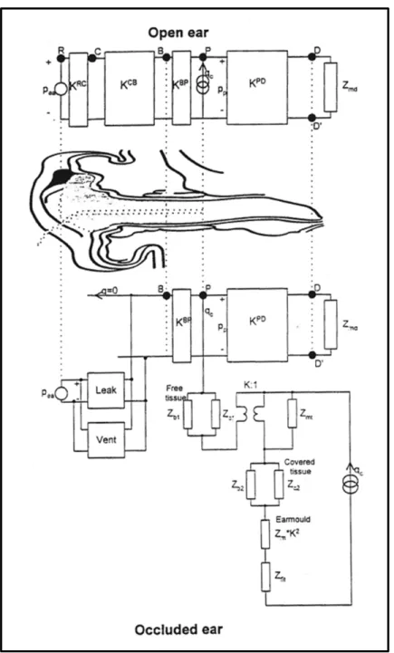
Le modèle axisymétrique est utilisé pour simuler l'effet d'occlusion pour étudier le rôle que joue le type du bouchon par rapport à l'effet de l'occlusion. Ce travail est réalisé en trois temps. Dans un premier temps le modèle numérique est validé à l’aide de mesures expérimentales faites sur deux groupes indépendant de sujets humains, chaque groupe étant équipé d’un bouchon différent. Dans un deuxième temps le modèle axisymétrique est validé à l’aide de deux modèles électriques équivalents de l’effet d’occlusion issus de la littérature. Dans un troisième temps des calculs de bilans de puissances sont effectués pour mieux comprendre comment l’énergie circule dans le système dépendamment du type de bouchon utilisé. Ces calculs sont effectués dans le canal occlus ainsi que dans les bouchons en contact avec les parois du canal à une moyenne et une très faible profondeurs d’insertions.
Le prototype d'un nouveau dispositif expérimental d’une oreille externe artificielle est proposé pour mesurer l’effet d’occlusion de manière objective et standardisé. Des résultats expérimentaux issus de ce prototype sont présentés pour démontrer la fonctionnalité du dispositif dans le but de mesurer l'effet d'occlusion d'un bouchon d'oreille. Premièrement la mise œuvre d’une oreille externe artificielle axisymétrique est présentée. Deuxièmement, le dispositif expérimental est utilisé pour étudier la contribution des différents chemins de transmission sonore. Troisièmement l’effet d’occlusion d’un bouchon en mousse à moyenne
profondeur d’insertion est mesuré à l’aide du dispositif expérimental. La mesure de l’effet d’occlusion est répétée à plusieurs niveaux d’excitation mécanique pour étudier la linéarité du système.
Mots-clés : Santé et sécurité du travail, protection auditive, effet d’occlusion, méthode d’éléments finis, étude numérique, mesure expérimentale, dispositif expérimental
STUDY OF THE OCCLUSION EFFECT INDUCED BY AN EARPLUG: NUMERICAL MODELLING AND EXPERIMENTAL VALIDATION
Martin BRUMMUND ABSTRACT
Despite existing limits for occupational noise exposure, professional hearing loss remains a high priority problem both in Québec and worldwide. Several approaches exist to protect workers from harmful noise levels. The most frequently employed short term solution includes the distribution of hearing protection devices (HPD) such as earplugs and ear muffs. While HPDs offer an inexpensive (e.g. direct cost) and efficient means of protection workers often only tend to wear HPDs for limited amounts of time and, thus, remain at risk of developing professional hearing loss.
Discomfort while using HPDs contributes to HPD underutilization and non-use. Two more general categories of discomfort can be distinguished. The category physical discomfort includes, for instance, problems such as heating of the ear and irritation of the ear canal that that occur upon earplug insertion. The category auditory discomfort refers to alterations in the auditory perception of sounds and one’s own voice as well as hindered workplace communications. One important auditory discomfort that promotes HPD non-use is the occlusion effect.
The occlusion effect occurs upon earplug insertion and describes sound amplification phenomena in the occluded ear canal at the low frequencies. The sound amplification is both perceivable and measurable (e.g., open and occluded sound pressure levels, hearing threshold shift). Additionally, the occlusion effect causes the HPD wearer to perceive his/her own voice as being distorted (e.g. hollow sounding) and physiological noises (e.g. respiration, blood circulation) are amplified also subsequent to earplug insertion.
Reducing the occlusion effect has the potential to increase the auditory comfort of HPDs and could help preventing occupational hearing loss in the future. In order to improve this and other shortcomings observed with currently existing HPDs a large research collaboration between the Robert-Sauvé research institute in occupational health and safety (IRSST) and the École de technologie supérieure (ÉTS) has been launched.
The present study represents a part of this collaboration and aims at studying the occlusion effect of the system earplug – ear canal through the development of novel numerical models and experimental methods.
A 3D (complex geometry) and an axisymmetric (simplified geometry) linear elasto-acoustic finite element model are presented in this study to simulate the objective bone conduction earplug occlusion effect.
The 3D model of complex geometry is used to predict the occlusion effect induced by a silicone earplug at several insertion depths. Power balance computations are used to explain how the ear canal walls and the medial earplug surface contribute to observed occlusion effect magnitudes at varying occlusion depths. The numerical occlusion effect predictions are validated with experimental reference data that were retrieved from the literature. The 3D model is used to investigate two well established qualitative occlusion effect models using power balance computations.
The axisymmetric occlusion effect model is used to predict open and occluded transfer function levels as well as the occlusion effect across three different excitation scenarios. First, only structure borne excitation is considered. Next, airborne noise is added incoherently to the structure borne excitation to study the effect of a mixed excitation. The mixed excitation is considered (i) for a leak free (perfect seal) earplug insertion and (ii) under the presence of small earplug leaks. Each stimulation scenario is examined across four different boundary and load conditions. All predicted transfer function levels and occlusion effects are compared to experimental data.
An adapted version of the axi-symmetric occlusion effect model is employed to investigate the contribution of the earplug type to the occlusion effect magnitude. First, the numerical model is validated with the help of experimental occlusion effect data which were measured in two independent human reference groups which each use a different earplug type (silicone earplug and foam earplug). Second, the numerical model is further validated through comparison with two gold standard lumped element occlusion effect models which were drawn from the literature. Third, power balance computations are employed to investigate the power flow inside the occluded ear canal cavity as well as the earplug body (coupled to the ear canal walls) of the numerical external ear model. The power balances are computed both for the foam and the silicone earplug models at medium and very shallow earplug insertion depths.
A prototype of a novel artificial external ear test fixture for objective and standardized measurement of the occlusion effect is developed. Details on the implementation of the artificial external ear and the assembly of the occlusion effect test fixture are presented. The experimental test fixture is used to investigate the contribution of the structure and airborne sound transmission pathways. Experimental data is provided to demonstrate that the test fixture is functional and that it can be used to measure the occlusion effect of a foam earplug. The occlusion effect measurement is repeated for several mechanical stimulation levels to study the system’s linearity.
Keywords: Occupational health and safety, hearing protection, occlusion effect, finite element method, numerical study, experimental measurements, artificial test fixture
TABLE OF CONTENTS
INTRODUCTION ...1
0.1 Research problem...1
0.1.1 Hazardous occupational noise exposure and means of protection...1
0.1.2 Causes for HPD underutilization...2
0.1.2.1 General causes...2
0.1.2.2 Definition of the occlusion effect...3
0.1.2.3 The role of the occlusion effect in the context of HPD underutilization...3
0.2 Research objectives...4
0.2.1 General research objective...4
0.2.2 Specific research objectives...5
0.3 Thesis structure and research methodology...9
0.3.1 Chapter 1: Literature review...9
0.3.2 Chapter 2: Development of a 3D human external ear model of complex geometry...9
0.3.2.1 Chapter contents...9
0.3.2.2 Overview of the methodology used to implement the 3D external ear model...10
0.3.3 Chapter 3: Implementation of a simplified axisymmetric external ear model of average geometry...12
0.3.3.1 Chapter contents...12
0.3.3.2 Overview of the methodology used to implement the axisymmetric external ear model...12
0.3.4 Chapter 4: Numerical investigation of the earplug type dependent occlusion effect using an axisymmetric external ear model...14
0.3.4.1 Chapter contents... 15
0.3.4.2 Overview of the methodology used to investigate the earplug type dependent occlusion effect...15
0.3.5 Chapter 5: Implementation of an artificial external ear test fixture to measure the occlusion effect...17
0.3.5.1 Chapter contents...17
0.3.5.2 Overview of the methodology used to implement artificial occlusion effect test fixture...18
0.3.6 Chapter 6: Synthesis and conclusion...19
CHAPTER 1 LITERATURE REVIEW ...21
1.1 Brief overview of the anatomy of the human ear ...21
1.2 Bone conduction sound transmission pathways ...24
1.3 The outer-ear transmission path and occlusion effect ...26
1.3.1.1 The high pass filter effect model (low frequencies) ... 27
1.3.1.2 The ear canal resonance shift model (mid frequencies) ... 28
1.3.2 Experimental analysis of the occlusion effect ... 28
1.3.2.1 Subjective and objective occlusion effect ... 28
1.3.2.2 The contribution of the external ear components to the sound pressure in the occluded ear ... 29
1.3.2.3 The contribution of the occlusion volume to the occlusion effect magnitude ... 31
1.3.2.4 The contribution of the stimulation position to the occlusion effect magnitude ... 33
1.3.2.5 The contribution of the earplug to the occlusion effect ... 33
1.3.3 Occlusion effect modelling ... 34
1.3.3.1 The Schroeter and Poesselt (1986) model ... 34
1.3.3.2 The Stenfelt and Reinfeldt (2007) model ... 36
1.3.3.3 Hansen’s (1998) model ... 38
1.4 Concluding remarks……….40
CHAPTER 2 ARTICLE 1: THREE-DIMENSIONAL FINITE ELEMENT MODELING OF THE HUMAN EXTERNAL EAR: SIMULATION STUDY OF THE BONE CONDUCTION OCCLUSION EFFECT ...41
2.1 Abstract ...42
2.2 Introduction ...42
2.3 Methodology ...45
2.3.1 Geometrical reconstruction of the external ear ... 45
2.3.2 Material properties of external ear tissues ... 47
2.3.3 Geometrical and material properties of earplug model ... 49
2.3.4 Boundary conditions and excitation... 51
2.3.5 Finite element modeling ... 52
2.3.6 Computation of acoustical quantities ... 54
2.3.6.1 Transfer function levels and occlusion effects ... 54
2.4 Computation of exchanged powers ...54
2.5 Results and discussion ...55
2.5.1 Finite element modelling of the occlusion effect ... 55
2.5.2 Comparison of modelling results and experimental data ... 59
2.5.3 The high-pass filter effect removal and the ear canal resonance frequency shift ... 63
2.6 Conclusions ...69
2.7 Acknowledgements ...71
CHAPTER 3 ARTICLE 2: PREDICTION OF THE BONE CONDUCTION OCCLUSION EFFECT USING A TWO-DIMENSIONAL AXI-SYMMETRIC FINITE ELEMENT MODEL ...73
3.1 Abstract ...74
3.3 Methodology ...76
3.3.1 Geometrical layout of the axi-symmetric external ear model ... 76
3.3.2 Material properties of the external ear tissues and the earplug ... 79
3.3.3 Boundary and load conditions ... 81
3.3.4 Finite element modeling ... 83
3.3.5 Calculation of estimated transfer function levels and the numerical occlusion effects... 84
3.3.6 Experimental measurements ... 85
3.3.6.1 Subjects ... 85
3.3.6.2 Apparatus ... 85
3.3.6.3 Protocol ... 86
3.3.6.4 Experimental transfer function level approximation and occlusion effect calculation ... 87
3.4 Results ...88
3.4.1 Transfer function level and occlusion effect prediction with bone conduction stimulation ... 88
3.4.2 The combined effect of airborne noise and structure borne excitation on transfer function levels and occlusion effects ... 91
3.4.3 The role of earplug leaks in combination with airborne and structure borne excitation ... 94
3.5 Discussion ...99
3.5.1 Transfer function level and occlusion effect prediction with bone conduction stimulation ... 99
3.5.2 The combined effect of airborne noise and structure borne excitation on transfer function levels and occlusion effects ... 100
3.5.3 The role of earplug leaks in combination with airborne and structure borne excitation ... 101
3.6 Conclusions ...102
3.7 Acknowledgements ...104
CHAPTER 4 ARTICLE 3: AN AXI-SYMMETRIC MODEL TO STUDY THE EARPLUG CONTRIBUTION TO THE BONE CONDUCTION OCCLUSION EFFECT ...105
4.1 Abstract ...106
4.2 Introduction ...106
4.3 Methodology ...109
4.3.1 Geometrical layout of the axi-symmetric external ear model ... 109
4.3.2 Material properties of the external ear tissues and the earplug models ... 112
4.3.3 Boundary and load conditions ... 114
4.3.4 Finite element modeling ... 116
4.3.5 Computation of the numerical occlusion effects and exchanged powers ... 117
4.3.6 Experimental measurements ... 118
4.4.1 Occlusion effect predictions for foam and silicone earplugs ... 119
4.4.2 Comparison of modeling results with existing lumped OE models ... 123
4.4.3 Comparison of the exchanged and dissipated powers in the occluded numerical models ... 127
4.5 Conclusions ...133
4.6 Acknowledgements ...136
CHAPTER 5 IMPLEMENTATION OF A SIMPLIFIED, ARTIFICIAL EXTERNAL EAR TEST FIXTURE FOR MEASUREMENT OF THE EARPLUG INDUCED AUDITORY OCCLUSION EFFECT ...137
5.1 Introduction ...137
5.2 Methodology ...139
5.2.1 Implementation of the artificial axi-symmetric external ear ... 139
5.2.2 Assembly of the entire test apparatus ... 142
5.2.3 Working principle of the occlusion effect test fixture ... 144
5.2.4 The role airborne noise corruption ... 148
5.3 Preliminary results and discussion ...149
5.3.1 Sound field reproducibility ... 149
5.3.2 Analysis of transmission pathways ... 151
5.3.3 Measured occlusion effect ... 152
5.3.4 Linearity analysis ... 153
5.4 Conclusions and future work ...154
5.5 Acknowledgements ...156
CHAPTER 6 SYNTHESIS AND CONCLUSIONS ...157
6.1 Synopsis of the research problematic as well as the general and specific objectives of the present study ...157
6.2 Article 1: Contributions, limitations and future recommendations ...158
6.3 Article 2: Contributions, limitations and future recommendations ...161
6.4 Article 3: Contributions, limitations and future recommendations ...163
6.5 Chapter 5: Contributions, limitations and future recommendations ...166
6.6 General conclusion...168
LIST OF TABLES
Page
Table 2.1: Material properties of tissue domains included in the finite element model. For the air filled ear canal cavity ρair = 1.20 kg/m3 and
cair = 343.20 m/s were used ...49
Table 2.2: Material properties of earplugs tested with the finite element model ...51 Table 3.1: Overview of the dimensions used in the
axi-symmetric external ear model ...79 Table 3.2: Material properties employed for modelling of the external ear
tissues. The density of the air and the speed of sound of the air inside the ear canal are ρair = 1.20 kg/m3 and cair = 343.20 m/s ...80
Table 3.3: Material properties of the earplug model used to occlude the
external ear model ...81 Table 4.1: Geometrical parameter range studied during the sensitivity analysis
concerning the external ear geometry ...110 Table 4.2: Overview of the geometrical dimensions used in the axi-symmetric
external ear model subsequent to the implementation the geometrical sensitivity analysis ...112 Table 4.3: Material properties used for modeling of the external ear tissues ...113 Table 4.4: Material properties of earplugs used in the present study to
occlude the external ear model ...114 Table 5.1: Dimensions of the synthetic ear model ...141
LIST OF FIGURES
Page
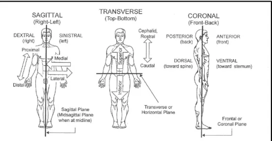
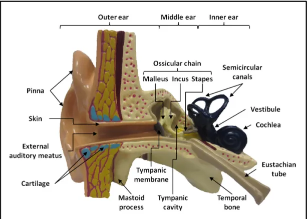
Figure 1.1: Overview of the anatomical orientations used throughout this chapter. The original image can be found in (Gelfand, 2010) ...21 Figure 1.2: Cross sectional schematic view of the human outer,
middle and inner ear ...22 Figure 1.3: Schematic illustration of the BC conduction pathways. 1) sound
radiation into the ear canal, 2) middle ear ossicle inertia, 3) inertial
movement of cochlea fluids, 4) elastic compression of cochlea walls ...25 Figure 1.4: Schematic illustration of the relationship between the occlusion
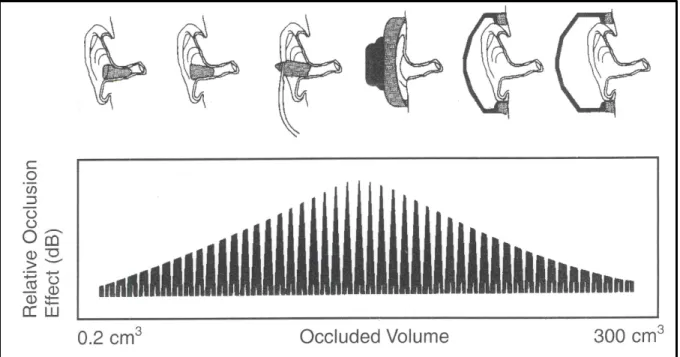
volume and the occlusion effect magnitude for earplug and
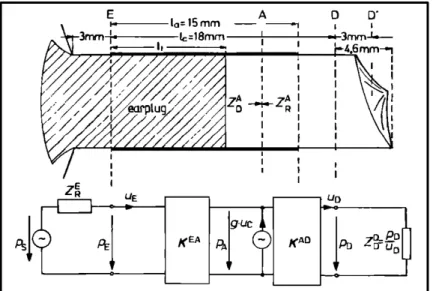
earmuff HPDs (Taken from Berger et al. (2003)) ...32 Figure 1.5: Layout of Schroeter and Poesselt’s (1986) ear canal model. The
original image can be found in (Schroeter and Poesselt, 1986) ...35 Figure 1.6: Layout of Stenfelt and Reinfeldt’s (2007) occlusion effect model.
Adapted version of original images (Stenfelt and Reinfeldt, 2007) ...37 Figure 1.7: Layout of Hansen’s (1998) occlusion effect model. The original
image can be found in (Hansen, 1997) ...39 Figure 2.1: Final external ear geometry as obtained after 3D reconstruction using
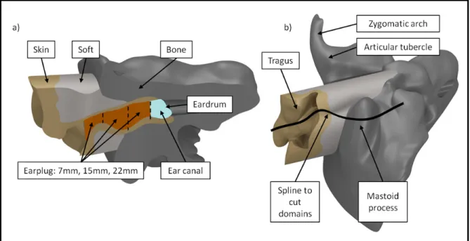
images of a human cadaver head (The Visible Human Project®). Left: Posterior-lateral view of temporal bone, cartilage tissue, and skin tissue at the ear canal entrance. Right: Medial view of the temporal bone geometry. Anatomical landmarks are indicated with arrows ...47 Figure 2.2: a) Anterior sectional view of occluded external ear model including all
modeled tissue domains. The different occlusion depths are indicated using dashed lines. b) Inferior view of the complete model. Due to the bends in the ear canal the domains were cut along a spline curve
instead of a plane. This also explains the narrow entrance region in a) ....50 Figure 2.3: a): occlusion effect simulations obtained with 3D finite element model
for silicone earplug at shallow, medium, and deep occlusions. All data are 1/3rd octave band filtered. b): corresponding transfer function levels at the center of the tympanic membrane in the open and occluded ear models for a silicone earplug. All data are 1/3rd octave band filtered ...55
Figure 2.4: Power balance plot for the 22 mm occlusion condition
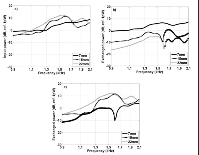
(silicone earplug). The time averaged power that is dissipated at the eardrum (solid line) as well as the time averaged powers that are exchanged between the ear canal wall – ear canal (dashed line) and earplug – ear canal (dotted line) are depicted. Markers on the dashed curve indicate frequencies at which the ear canal walls receive power from the ear canal cavity (e.g. negative power flowing from the ear canal to walls). Narrow band spectra are shown ...56 Figure 2.5: a): sum of the time averaged input power to the ear canal. b): time
averaged power exchanged between the ear canal walls and the ear canal cavity. c): time averaged power exchanged between the medial earplug surface and the ear canal cavity. The markers on the graphs indicate frequencies at which the ear canal wall or earplug receives power from the ear canal cavity (e.g. negative power flowing from
the ear canal to walls or earplug). Narrow band spectra are shown ...58 Figure 2.6: Comparison of experimental data and simulations (thick solid line with
markers) for a silicone earplug and a foam earplug (thick
dashed line) inserted at an occlusion depth of 15mm. The errorbar- plot (mean ± S.D) and individual occlusion effect plots (thin solid lines) define a reference zone that was measured experimentally by
Stenfelt and Reinfeldt (2007). All data are 1/3rd octave band filtered ...60 Figure 2.7: Time averaged power radiated into the environment normalized by the
time averaged input power exchanged at the ear canal wall - ear canal cavity interface for the open ear model.
Narrow band spectra are shown ...63 Figure 2.8: Time averaged power radiated into the environment (solid line) of the
open ear. Difference in time averaged power dissipated at the eardrums of the occluded and open ear models (dashed line). Above 1.4 kHz (not depicted) the power dissipated at the open eardrum exceeds that of the occluded eardrum. Narrow band
spectra are shown ...65 Figure 2.9: Time averaged input power from the ear canal walls for the open and
occluded ear (infinite impedance at the ear canal entrance) models for frequencies below 1.5 kHz. Narrow band spectra are shown ...66 Figure 2.10: a): average mean square velocity of the ear canal walls obtained for
the open and occluded (infinite impedance at ear canal entrance) ear models for frequencies below 1.5 kHz. b): real part of the surface averaged acoustical specific input impedance of the ear canal walls for the open and occluded (infinite impedance at ear canal
entrance) ear models for frequencies below 1.5 kHz. Narrow
band spectra are shown ...68 Figure 3.1: a) Anterior sectional view of a 3D reconstructed external ear
geometry (The Visible Human Project® images were used). b) Simplified axi-symmetric geometry of the external ear including ear canal, earplug, skin tissue, cartilage tissue and bony tissue. The axis of symmetry is indicated through a dashed-dotted line. The model’s dimension identifiers (see Table 3.1) are
included using text boxes ...77 Figure 3.2: Overview of the four boundary and load conditions tested in the
present study. Arrows indicate the locations at which the force was introduced normally on the surface. Inclined dashes indicate fixed boundaries. The brackets at the entrance and the end of the ear canal indicate impedance boundary conditions. The unmarked boundaries adjacent to the ear canal entrance (next to Zdisc)
indicate free boundaries ...82 Figure 3.3: Top view of the experimental measurement setup used to
determine the open and occluded TFLs and the OE subsequent to bone condition stimulation at the right ipsilateral mastoid process. Right hand side, top view (schematic) of the instrumented test subject inside the semi-anechoic room. Left hand side, data
acquisition chain (schematic) ...86 Figure 3.4: a) numerical open ear TFL predictions obtained for the four boundary
condition configurations. b) numerical occluded ear TFLs obtained for each tested boundary condition configuration. In addition to the numerical results the experimental (mean ± S.D.) open and occluded TFLs are included for comparison (dashed lines) ...88 Figure 3.5: Numerical OE predictions (solid lines) obtained from the TFLs
when only structure borne excitation is being considered. The OEs are provided for the experimental insertion depths 10.4mm, 11.7mm, and 13mm for all boundary condition configurations. The experimental OE (mean ± S.D.) is included for comparison (dashed error bar graph) ...90 Figure 3.6: Comparison of the experimental sound pressure level readings
obtained at the ipsilateral tragus (open ear) and inside the open
ear canal (mean ± S.D.) ...91 Figure 3.7: a) numerical open ear TFL predictions. b) numerical occluded
ear TFLs. All boundary condition configurations were considered (solid lines) for the open and occluded model under both structure borne and airborne excitation from the bone transducer. In addition
to the numerical results the experimental (mean ± S.D.) open and
occluded TFLs are included for comparison (dashed error bar graphs) ....92 Figure 3.8: Numerical OE predictions (solid lines) obtained for both
structure borne and airborne excitation from the bone transducer. The OEs are provided for the experimental insertion depths 11.7mm ± 1.3mm (mean ± S.D.) for all boundary condition configurations. The experimental OE (mean ± S.D.) is included for comparison
(dashed error bar graph) ...93 Figure 3.9: Experimental insertion loss data (mean ± S.D.) obtained for the
custom molded earplugs (right ear only) under pink noise excitation from a loudspeaker ...95 Figure 3.10: Numerical TFLs (occluded ear model only, 11.7mm) for earplug
leak diameters ranging from 0.2mm to 0.5mm for configurations a), b), c) and d). Numerical TFLs obtained for both structure and airborne excitation from the bone transducer are depicted. For comparison the TFLs obtained for the leak free condition
(solid lines with markers) are included ...96 Figure 3.11: Numerical OE predictions (insertion 11.7mm) for earplug
leak diameters ranging from 0.2mm to 0.5mm together with structure and airborne excitation from the bone transducer for configurations a), b), c) and d). For comparison the experimental
OEs (mean ± S.D.) are included (dashed error bar plot) ...98 Figure 4.1: Geometrical layout of the axi-symmetric FE model including the ear
canal, earplug, cartilaginous tissue, skin tissue and bony tissue. The axis of symmetry is indicated through a dash-dotted line.
All model identifiers are provided in Table 4.1 ...109 Figure 4.2: Boundary and load conditions employed in the present study for
the open (a) and occluded (b) external ear model. Arrows indicate boundaries at which loading was introduced in normal direction. Brackets denote impedance boundary conditions used to model the eardrum and the ear canal entrance (open ear only). Unmarked
boundaries indicate free boundary conditions ...115 Figure 4.3: Comparison of numerical OE predictions and experimental OE
measurements. a) foam earplug occlusion device inserted 11.1 mm into the ear canal with respect to the ear canal entrance in both numerical and experimental conditions. b) silicone earplug occlusion device at 11.7 mm insertion with respect to the
Figure 4.4: a) insertion loss data (mean ± S.D.) measured for foam earplug test subjects for a mean earplug insertion depth of 11.1mm. b) sound pressure level data (mean ± S.D.) obtained for the open ear and occluded ear measurements of the foam earplug test subjects.The solid black line corresponds to the sound pressure level at the open in-ear microphone. The dashed grey line indicates thesound pressure level at the ipsilateral tragus reference
microphone (open ear). The dotted grey line indicates the sound pressure level at the ipsilateral tragus reference microphone (occluded ear). The dotted black line represents the noise floor
of the in-ear microphone (open ear) ...122 Figure 4.5: a) comparison of OE predictions obtained with the FE model to OE
predictions obtained by Stenfelt and Reinfeldt (2007) at shallow (7 mm) and deep (22 mm) earplug insertion. b) comparison of OE prediction obtained with the FE model to prediction obtained by
Schroeter and Poesselt (1986) at one occlusion depth (9.2 mm) ...125 Figure 4.6: Occluded ear canal cavity power balance computation for foam
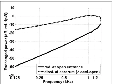
(11.1 mm insertion) and silicone (11.7 mm insertion) earplugs for frequencies up to 1 kHz. The following example demonstrates reading the figure. At 0.25 kHz about -10 dB of power gets dissipated (right hand ordinate) at the eardrum (black diamond marker) when the foam earplug numerical model is used. Of these -10 dB, 77% stem from the medial earplug surface (dark grey bar, left hand ordinate) and 23% stem from the unoccluded
ear canal walls (light grey bar, left hand ordinate) ...128 Figure 4.7: Occluded ear canal cavity power balance computation for foam
(11.1 mm insertion) and silicone (11.7 mm insertion) earplugs for the 2 kHz third octave band. Diamond markers refer to right hand ordinate and vertical bars refer to left hand ordinate. In this third octave band the power flow changes its sign and two zones can be distinguished. An example on how to read this figure can be found
in the caption of Figure 4.6 ...131 Figure 4.8: Power balance computation inside foam (11.1 mm insertion) and
silicone (11.7 mm insertion) earplugs for frequencies up to 2 kHz. Diamond markers refer to right hand ordinate and vertical bars refer to left hand ordinate. An example on how to read this figure can
be found in the caption of Figure 4.6 ...132 Figure 5.1: Schematic of the designed (built and assembled in-house) artificial
external ear model, its domains and its dimensions. a) ear canal, b) soft tissue domain, c) bony tissue, d) skin tissue at ear
canal entrance and on ear canal walls. For further reference see
also Table 5.1 ...140 Figure 5.2: Schematic of the assembled test apparatus. a) square steel plate,
b) slip-on flange, c) artificial external ear, d) IEC 60711 coupler, e) cage for transmission of mechanical excitation,
f) mini-shaker K2007E01 ...143 Figure 5.3: a) Back view: Polyurethane cylinder (beige) is press-fitted into
flange (black). Anterior disc of excitation cage is press-fitted around bony tissue b) Front view: Polyurethane cylinder after press-
fitting into flange. Remaining volume of central bore hole to be filled with two different types of silicone. c) Front view: After injection of the first silicone the silicone is cured overnight (grey). The insert (white) delimits the soft tissue cylinder and is removed after curing process. d) Front view: Final result after molding of soft tissue cylinder. e) Front view: A second insert (white) is placed concentrically in the soft/bony tissue subassembly. Its diameter is chosen so that the second silicone can fill the remaining volume (corresponds to ear canal walls and the skin tissue around the ear canal entrance). f) Front view: following the molding and curing
of the skin tissue domain, the second ABS insert is removed. ...144 Figure 5.4: Function principle of test fixture a) components important for
structure borne sound transmission. b) schematic of the
structure borne transmission ...145 Figure 5.5: Schematic representation of the structure borne sound
transmission in the synthetic external ear and sound radiation into
the open ear canal ...146 Figure 5.6: Experimental setup used to reproduce the airborne noise sound field
emitted by the transmission cage ...149 Figure 5.7: Sound pressure level readings obtained at the ¼-inch reference
microphone subsequent to excitation with the shaker setup (solid
line) and the loudspeaker setup (dashed line). Third octave
band filter applied ...150 Figure 5.8: Sound pressure level readings obtained at the IEC 60711-coupler
subsequent to excitation with the shaker setup (solid line) and the
loudspeaker setup (dashed line). Third octave band filter applied ...151 Figure 5.9: Third octave band Occlusion effect measurement of a foam earplug
Figure 5.10: Occlusion effect measurements of a foam earplug (constant insertion depth about 20mm) for pink noise excitation at three difference levels (90dB, 100dB, 110dB).
LIST OF ABREVIATIONS ABS Acrylonitrile butadiene styrene
AC Air conduction
ANSI American National Standards Institute ASA Acoustical Society of America
ASTM American Society for Testing and Materials ATF Artificial test fixture
BC Bone conduction
ÉTS École de technologie supérieure FE Finite element
FEM Finite element method HL Hearing loss
HPD Hearing protection device
IEC International Electrotechnical Commission
IRSST Institut de recherche Robert-Sauvé en santé et en sécurité du travail ISO International Organization for Standardization
L Length
NRR Noise reduction rating OE Occlusion effect
PIB Protecteur individuel contre le bruit
PDTM Power dissipated at the tympanic membrane PSD Power spectral density
REAT Real-ear attenuation at threshold RMS Root mean square
S.D. Standard deviation SPL Sound pressure level T Thickness
TFL Transfer function level VHP Visible human project
INTRODUCTION
0.1 Research problem
0.1.1 Hazardous occupational noise exposure and means of protection
Exposure to high noise levels can cause permanent damage to the auditory system. In the context of occupational health and safety prolonged noise exposure is often inevitable as the work task (e.g. construction industry) might require workers to spend entire shifts in noisy environments. To protect these employees the Canadian federal and provincial legislations have defined maximum noise exposure levels that must be met. For instance, in Québec the maximum noise exposure during an 8 hour work shift is limited to 90 dB(A) (Québec, 2014).
Despite the existing limits for occupational noise exposure professional hearing loss remains a high priority problem. In Québec, impairment of the sense of hearing and the ear was the most frequently encountered professional disorder in 2011, 2012 and 2013 (Lamarche et al., 2011, 2013, 2014). In 2013 the number of accepted cases even increased from 2600 (accepted cases in 2012) to 3303 accepted cases (Lamarche et al., 2014). Worldwide, an estimated 120 million workers are regularly exposed to noise levels that can permanently damage the auditory system (Organisation mondiale de la santé (OMS), 2001).
Several approaches exist to protect workers from harmful noise exposure. They include (i) treating the noise emitting source and surrounding workspace in an attempt to achieve more moderate noise levels, (ii) structurally reorganizing the work tasks to limit the cumulated noise exposure and (iii) distributing hearing protection devices (HPD) such as earplugs and earmuffs to attenuate the ambient noise levels in such way that existing noise exposure limits are met. Due to financial, spatial and temporal restrictions observed in the first two methods HPDs remain, to date, the most frequently utilized short term solution to protect the workers, because they are easy-to-use, fast in implementation and at the same time cost efficient (e.g. direct cost).
The acoustic performance of a HPD is influenced by the achieved noise attenuation and amount of time the protector is being worn (Sgard et al., 2008). If worn correctly, HPDs provide an efficient means of protection (Berger et al., 2003). When HPDs are removed even during short periods of time their overall performance is reduced. The decrease in HPD net-performance due to temporary protector removal is hereby greater for higher noise reduction rated (NRR) HPDs. For instance, removing an HPD of nominal NRR 25 dB for only 15 minutes during an 8 hour shift reduces the net NRR of that protector to 20dB. On the other hand if the nominal NRR equals 15dB, the net NRR reduces to about 14 dB if the protector is not being worn for 15 min throughout the entire work shift (Berger et al., 2003).
0.1.2 Causes for HPD underutilization
0.1.2.1 General causes
Numerous empirical studies have been conducted to examine HPD use across different industries and countries. These studies unambiguously express the need to increase consistent HPD use especially among non-users and occasional HPD users. Melamed et al. (1994) and Tak et al. (2009), for example, provide overviews of some of the research work that has been carried out in order to better explain why HPDs are underutilized in the workplace environment. Contributing factors include:
- Unavailability of HPDs in noisy environments
- Lack of knowledge on the ability of an HPD to reduce harmful noise - Lack of confidence to adequately use HPDs
- Physical discomfort while using HPDs (e.g. irritation of the ear canal, headband force) - Interference of the HPD with other work equipment (e.g. headgear)
- Interference of the HPD with the work task
- Fear of a decrease in the worker’s prestige among co-workers - Fear of blocking out useful auditory cues such as warning signals
- A perceived sense of isolation while wearing a HPD (Hughson et al., 2002) - Impression that the HPD negatively affects the communication with co-workers
Many of the aforementioned causes that contribute to non-use of HPDs can be approached through educational and motivational steps as part of a broader hearing conservation program (Berger et al., 2003) and adequate HPD selection.
Another important factor that contributes to HPD non-use is the occlusion effect. Often the occlusion effect is not specifically mentioned as cause for HPD non-use by test subjects. This could, hypothetically, originate from a certain degree of unawareness among the examined test subjects. As the present work is dedicated to studying the occlusion effect induced by an earplug the link between the occlusion effect and HPD underutilization is going to be discussed in detail in section 0.1.2.3. Prior to this discussion, a definition of the occlusion effect is presented as support for the reader.
0.1.2.2 Definition of the occlusion effect
The occlusion effect describes perceivable and measurable (e.g. sound pressure level inside the ear canal, hearing threshold shift) sound amplification phenomena that occur upon earplug insertion into the ear canal. Thus, the occlusion effect causes the HPD wearer to perceive his/her own voice as being distorted, hollow sounding and, most noticeably, amplified at the lower frequencies. Additionally, physiological noises (e.g. respiration, blood circulation) are amplified also subsequent to earplug insertion (Berger and Kerivan, 1983).
0.1.2.3 The role of the occlusion effect in the context of HPD underutilization
As was previously mentioned, the occlusion effect is often omitted as cause for HPD non-use by test subjects. Nevertheless, the occlusion effect contributes considerably for instance to the impression that HPDs hinder workplace communication as was discussed in Berger et al. (2003). Berger et al. (2003) provide a summarizing overview of the research work that has been carried out with regards to the effects of HPDs on auditory perception. It can be seen that HPDs can negatively affect speech intelligibility in conditions where the HPD wearer is listening to his own voice and in situations where co-workers, that are each wearing HPDs,
communicate (e.g. Howell and Martin, 1975). While the talker experiences the occlusion effect, the ambient noise is attenuated through his HPD at the same time. Thus, the talker gets the impression that his/her voice is louder with respect to the ambient noise than actually is the case and tends to lower his/her vocal effort. The latter decrease in signal level and signal to noise ratio hinders the intelligibility on the listener side and thus causes an overall reduction in speech intelligibility. Consequently, the HPD wearers are prone to removing their HPDs and, thus, remain at risk of developing professional hearing loss.
0.2 Research objectives
The preceding paragraphs provide a contextual overview on how the occlusion effect contributes to HPD underutilization and occupational hearing loss. In the following paragraphs the general and specific research objectives of the present study are presented.
0.2.1 General research objective
To improve the shortcomings observed in currently existing HPDs the Institut de recherche Robert-Sauvé en santé et en sécurité du travail (IRSST) and the École de technologie supérieure (ÉTS) have launched a research collaboration that aims at the development of tools and methods to better assess and design hearing protectors (IRSST research project 0099-7630).
The present study represents a sub domain of the outlined research collaboration. It aims at better understanding the occlusion effect induced by an earplug through the development of novel numerical models and experimental methods. The developed numerical models and experimental methods have the potential to provide valuable insight into the manner in which the earplug contributes to the occlusion effect and to identify earplug constituents (e.g. material properties) crucial to reducing the occlusion effect magnitude. In a future study, this work could serve as a foundation to specifically guide the design and implementation of a passive low occlusion effect earplug. As was outlined in section 0.1 the occlusion effect is an important factor that contributes to HPD non-use. It is, thus reasonable to assume that
reducing the occlusion effect could contribute to increasing the auditory comfort of HPDs. In return, this could help to reduce HPD non-use among workers that are exposed to harmful noise levels and, thus, assist in preventing occupational hearing loss in the future. Note that the present study only considers earplug type HPDs. Ear muff type HPDs are not considered in this work, because incorporating ear muffs would increase model complexity as the pinna and more of the head would have to be considered. In the future such model could definitely be implemented presently, however, it goes beyond the scope of the present study.
0.2.2 Specific research objectives
For the present study four specific objectives are defined to achieve the general objective outlined in the preceding paragraph. Under objective 1 a numerical model of complex geometry is implemented to model the human external ear as realistically as possible (note that several limitations apply which are outlined throughout the study). Under objective 2 a second numerical model is implemented. The degree of geometrical complexity of this model is reduced to increase the ease of use. Under objective 3 the numerical model implemented under objective 2 is used to further study the earplug induced occlusion effect. Lastly, objective 4 aims at developing a novel experimental method to measure the earplug occlusion effect. Note that detailed explanations of the relevance are provided with each specific objective. The four research objectives are:
O1: Implement a 3D human external ear finite element model of complex geometry using anatomical images to simulate and study the earplug induced occlusion effect in the case of a mechanical stimulation applied on the ipsilateral mastoid process. Validate the numerical occlusion effect data obtained with the model against experimental measurements from the literature.
Relevance of a 3D human external ear model: To date only a few studies have aimed at modelling the occlusion effect (see section 1.3.3 for details). All existing models rely on lumped elements (electrical equivalent circuits) to model the occlusion effect. While obtained results generally tend to be in good agreement with experimental measurements, lumped
element models seem less suitable than finite element models, especially, to improve the earplug design. For instance, finite element models make it possible to analyze three-dimensional sound propagation (lumped elements limited to one-three-dimensional sound propagation) in substructures of the external ear (e.g. ear canal walls) and the earplug. Furthermore, the link between the model parameters and the physical properties of, for instance, the earplug are clearer in a finite element model than in a lumped model and it is easier to represent the tissue domains that surround the ear canal. Additionally, finite element models allow for calculating the power flows between subsystems of the external ear in 3D. Exchanged and dissipated powers can also be calculated from 3D displacement fields in the tissues and the earplug and from pressure fields in the ear canal. These indicators can be very helpful to further understand the occlusion effect mechanisms and particularly how the earplug contributes to the occlusion effect and which earplug parameters (e.g. material properties) play key roles.
O2: Implement an axi-symmetric simplified external ear model on the basis of average external ear dimensions taken from the literature to simulate and study the occlusion effect in the case of a mechanical stimulation applied on the model’s circumference. Validate the numerical data with experimental measurements.
Relevance of an axisymmetric human external ear model: While 3D finite element models allow anatomically correct reconstruction of the geometrical complexity of the ear canal and its surrounding tissues (e.g. temporal bone, cartilage tissue, skin tissue), which facilitates adequate model excitation, they are also accompanied by several disadvantages. For instance, the 3D reconstruction of the external ear is very tedious especially when skin and cartilage tissues ought to be distinguished. The number of anatomical datasets available for 3D reconstruction of the external ear is very limited. 3D models can, due to their geometrical complexity, be very difficult to manipulate (e.g. meshing) and can require excessive computational resources. In addition to that, the geometrical complexity makes 3D models also cumbersome for the implementation of sensitivity analyses (e.g. earplug material properties, external ear geometry). Lastly, it is very challenging to implement an artificial test fixture on the basis of an anatomically correct 3D external ear geometry. For example, it is
very difficult to cast the skin covered ear canal walls. During the casting process an insert would be required. Upon curing, the insert would have to be removed from the ear canal which is challenging given the ear canal’s s-shape. Besides, 3D reconstructed external ear geometries are based on individual data. For an artificial test fixture it would be desirable to have an average ear canal model. Axi-symmetric, finite element models could contribute to alleviate the abovementioned difficulties, because they rely on simpler geometries, are easier to manipulate and require less computational resources. Existing averaged geometrical data of the human external ear (ear canal length, wall skin thickness etc.) can be employed for the implementation of such an axi-symmetric model which renders the tedious 3D ear reconstruction unnecessary. Additionally, an axi-symmetric external ear model can also serve as a blueprint for the implementation of an artificial external ear test fixture, because they can be manufactured more easily (e.g. see preliminary work by Brummund et al. (2013) and chapter 5). In this context the question arises whether a simplified axi-symmetric model is sufficient to model the occlusion effect or whether the geometrical complexity of the external ear needs to be considered. Stinson and Lawton (1989) demonstrated that the sound pressure level inside the human ear canal can be approximated using a cylinder for frequencies up to 6 kHz. Unfortunately, Stinson and Lawton (1989) limited their study to the open ear canal (no surrounding tissues) and airborne excitation. During the present work the feasibility to develop a 3D equivalent, simplified axi-symmetric occlusion effect model could be demonstrated. Obtained results were presented to the scientific community at 162nd Meeting of the Acoustical Society of America (Brummund et al., 2011). The presentation was granted the “Best Paper Award for Young Presenters in Noise”.
O3: Numerically predict the occlusion effects of two earplug types using the simplified model developed under O2, validate the obtained occlusion effects with experimental data and interpret observed occlusion effect differences with the help of power balance calculations.
Necessity to further investigate earplug type dependent occlusion effect: To date only very few experimental studies have aimed at examining the occlusion effect as a function of earplug type (please refer to section 1.3.2.5 for a review of the existing literature). Obtained
experimental results suggest that the earplug type can influence the occlusion effect at medium insertion depth, whereas it appears to be of secondary importance at very shallow insertion. These experimental results are highly relevant, because they imply that it could be possible to reduce the occlusion effect through an improved earplug design (e.g. material properties, geometry). Yet, it is not well understood how different earplug types contribute to the observed occlusion effect differences. Numerical occlusion effect modeling could
contribute to bridge this gap through an analysis of the exchanged and dissipated time averaged acoustical powers both inside the ear canal cavity (e.g. unoccluded ear canal walls, medial earplug surface, eardrum) as well as inside the earplug domain (e.g. occluded ear canal walls, earplug body, medial earplug surface). Note that no occlusion effect model can be found in the literature that has aimed at studying the contribution of the earplug to the occlusion effect numerically.
O4: Design and implement an artificial occlusion effect test fixture of similar geometry as the simplified numerical occlusion effect model (see O2) to examine the general feasibility of such a device and to experimentally measure the earplug occlusion effect objectively.
Relevance to implement an artificial external ear test fixture: To date, no commercially available artificial test fixture allows measuring the occlusion effect of either earplugs or earmuffs. In part, this might be due to the circumstance that it is challenging to implement a structure borne excitation that would result in a vibration of the ear canal walls and sound radiation into the ear canal cavity (see section 1.3 for details on the occlusion effect mechanisms). Some information on the advantages of using a simplified external ear geometry for the implementation of an artificial occlusion effect test fixture was already provided with objective 2 (e.g. easier to manufacture as complex model, easier to implement an average external ear geometry). Once implemented such a test fixture could be employed to test and rate existing earplug designs with respect to the occlusion effect they cause. Additionally, a test fixture equivalent numerical model could serve to better understand the contribution of each part of the system ear canal/earplug to the occlusion effect (e.g. using power balances). In the future, this mixed experimental-numerical approach could contribute
to more efficiently guide the earplug design process in order to realize an earplug that exhibits both adequate noise attenuation and a reduced occlusion effect.
0.3 Thesis structure and research methodology
The previous paragraph lists the specific research objectives of the present work. The following section details the structure of the present thesis along with an overview of the methodology used to attain each specific objective.
0.3.1 Chapter 1: Literature review
Chapter 1 provides a comprehensive literature review on hearing by bone conduction and the occlusion effect. A brief overview of the anatomical structures of the human ear that are crucial to bone conduction hearing is provided. The different bone conduction sound transmission pathways are discussed. The bone conduction pathway that is most important for the occlusion effect is presented along with qualitative models that have been established to explain the occlusion effect. Additionally, experimental literature data is reviewed which explains the contributions of the chosen measurement technique (objective versus subjective), the occlusion volume, the bone conduction stimulation position, and the earplug occlusion device to the occlusion effect magnitude. Lastly, existing lumped element occlusion effect models are reviewed.
0.3.2 Chapter 2: Development of a 3D human external ear model of complex geometry
0.3.2.1 Chapter contents
In chapter 2 the first research article is presented. This article is entitled “Three-dimensional finite element modeling of the human external ear: simulation study of the bone conduction occlusion effect”. The purpose of this research article is to complete specific objective 1. A
3D coupled linear elasto-acoustic finite element model of the human ear canal and surrounding structure to simulate the occlusion effect is proposed.
0.3.2.2 Overview of the methodology used to implement the 3D external ear model
Geometrical external ear reconstruction: To reconstruct the 3D human external ear model, anatomical color images of a female cadaver head are retrieved from the Visible Human Project® database of the US National Library of Medicine. The images are segmented using a contour detection tool in order to separate bony, cartilaginous and skin tissues as well as the ear canal. Upon completion of the segmentation the outer surfaces of each anatomical structure is created. The resulting geometry is imported into a computer assisted design tool for post-processing of the 3D solid model.
Material properties of external ear tissues and earplugs: The 3D model is imported into a commercially available finite element simulation tool. The material properties of the bony, cartilage and skin tissues are applied to each domain. The isotropic, linear elastic material properties (density, Young’s modulus, Poisson’s ration, loss factor) used in the model are drawn from the literature, when possible. While the assumption of linear elasticity is reasonable for small tissue deformations, assuming isotropy represents a simplification of the considered human tissues which are anisotropic and viscoelastic. Due to a lack of more accurate material property data, however, such simplification seems presently inevitable. Additionally, the material properties of two earplug types (silicone and foam earplugs) are applied to the earplug models of the occluded external ear. The earplug models are assumed to adapt to the shape of the ear canal without deforming its walls and to occlude the ear canal leak free. Again, isotropic linear elastic material behavior is assumed and all material properties are taken from the literature. To model the ear canal itself the speed of sound and density of air at 20°C are applied to this domain.
Boundary conditions and excitation: Boundary conditions are applied to the circumference of the external ear domains to express the connections that exist between the model domains
and their environment. The skin tissue of ear canal entrance region and the boundaries of the temporal bone are modeled as free boundary conditions. The circumference of the skin and cartilage tissue that back the ear canal are fixed. Between solid domains continuity of stress vectors and displacements is assumed. Interfaces between a solid domain and ear canal cavity are expressed through fluid-structure coupling. The eardrum is expressed using a locally reacting specific acoustical impedance boundary condition. Another locally reacting impedance boundary condition which corresponds to the radiation impedance of a baffled flat circular piston is applied over the entrance of the open ear canal. The external ear system is excited mechanically via the ipsilateral mastoid process using a constant total force of 1N which applies normally to a flat surface area.
Model meshing: The 3D model is meshed using tetrahedral quadratic elements for all domains. The mesh uses a convergence criterion of four elements per wavelength at 3 kHz. The meshed model is solved for the sound pressure at the eardrum. The sound pressures of the open and occluded external ear models are used to calculate the earplug occlusion effect. Other acoustical indicators such as the acoustical normal particle velocity are retrieved to calculate exchanged and dissipated powers.
Data analysis: Obtained numerical results are used to investigate the influence of the temporal bone’s boundary conditions on the transfer function levels and occlusion effect magnitude. The open ear and occluded transfer function levels and occlusion effect are predicted at several earplug insertion depths. The predicted occlusion effects at medium insertion depth are compared to experimental occlusion effect data measured in human subjects (the data were kindly provided by Stenfelt and Reinfeldt (2007)). Note that comparing the 3D finite element (FE) predictions to experimental data is presently the most suitable way to validate the numerical model due to the circumstance that no model equivalent test fixture exists. Besides, implementing an artificial external ear test fixture of complex geometry is a challenging task by itself. Power balances are used to explain how the ear canal walls and the medial earplug contribute to observed occlusion effect magnitudes at varying occlusion depths between 1 kHz and 2 kHz. Furthermore, power balance
computations and transfer function level predictions are used to investigate Tonndorf's (1966) and Huizing's (1960) qualitative occlusion effect models. Detailed discussions of the obtained results along with conclusions can be found in CHAPTER 2 where the journal article is presented. In section 6.2 a synthesis of the main results is presented along with a critical discussion of the scientific and technological contributions as well as the limitations. Future recommendations are drawn based on current limitations.
0.3.3 Chapter 3: Implementation of a simplified axisymmetric external ear model of average geometry
0.3.3.1 Chapter contents
In chapter 3 the second research article entitled “Prediction of the bone conduction occlusion effect using a two-dimensional axi-symmetric finite element model” is presented. The purpose of this article is to specify an axi-symmetric linear elasto-acoustic finite element model of the human external ear for simulation of the bone conduction occlusion effect using geometrical and material properties found in the literature. This purpose is coherent with specific research objective 2 (see section 0.2.2).
0.3.3.2 Overview of the methodology used to implement the axisymmetric external ear model
Geometrical model layout: The implementation of the axisymmetric external ear geometry is carried out using commercially available finite element software. The ear canal and earplug (occluded model) are represented as straight cylinders of identical cross sectional areas. The ear canal walls are formed by a cylindrical skin layer. Half of the ear canal walls are surrounded by a cylindrical cartilage layer and the remaining half are surrounded by a bone layer. The anatomical landmarks of the pinna are disregarded mainly to ensure axisymmetry. To model the ear canal entrance region (zone where ear canal merges into the cavum conchae) the cartilage layer and the ear canal walls are continued outwards in such
way that they protrude with respect to the ear canal entrance. The dimensions of the external ear domains (e.g. ear canal length, ear canal radius, ear canal wall thickness, entrance protrusion, bone and cartilage thicknesses) are obtained on the basis of an extensive literature review.
Material properties of external ear tissues and earplug: The material properties of the bony, cartilage and skin tissues are applied to the finalized axisymmetric geometry. The same isotropic, linear elastic material properties density, Young’s modulus, Poisson’s ration, loss factor) that are used for the 3D model of complex geometry (see section 0.3.2.2) are applied to each of the tissue domains of the simplified axisymmetric model. One silicone earplug type is considered with this model. The earplug model is assumed to adapt to the shape of the ear canal without deforming its walls. Both a perfect seal and leaks of small diameters are investigated. When earplug leaks are included, airborne noise is presented in phase to study their combined effect. Note that the hypothesis of the airborne noise and the structure borne excitation being in phase is a simplification which is presently inevitable as it is very challenging to measure the phase difference. The isotropic linear elastic material properties of the silicone earplug are taken from the literature. The speed of sound and density of air at 20°C are applied to the ear canal cavity.
Boundary and load conditions: Four different boundary and load conditions are considered with the axisymmetric model. All configurations use fixed, free, impedance and boundary load conditions. The four configurations mainly differ in terms of how much of the bone and/or cartilage tissue is stimulated by the boundary load. The power spectral density of a bone transducer’s RMS-force (obtained trough calibration with an artificial mastoid) serves as boundary load. The latter is always introduced normally and uniformly. This loading condition represents an idealization of the real stress vectors which act on the human tissues and which likely vary in terms of amplitude and direction along the tissue boundaries. Additionally, the RMS-force measured at the artificial mastoid (which would also apply to the mastoid process of a human subject) likely changes in terms of magnitude once it reaches the external ear. Applying this RMS-force directly to the circumference of the axisymmetric