Delivery of Granulocyte-Macrophage Colony-Stimulating Factor in
Bioadhesive Hydrogel Stimulates Migration of Dendritic Cells in Models of
Human Papillomavirus-Associated (Pre)Neoplastic Epithelial Lesions
Pascale Hubert,1 Brigitte Evrard,2 Catherine Maillard,3 Elizabeth Franzen-Detrooz,1 Luc Delattre,2 Jean-Michel
Foidart,3 Agnès Noël,3 Jacques Boniver,1 and Philippe Delvenne1
Department of Pathology, CRCE,1 Department of Pharmaceutical Technology,2 and Laboratory of Tumor and Development Biology,3 University of Liege, Liege, Belgium
Abstract
Because of the central role of dendritic cells and/or Langerhans cells (DC/LC) in the induction of cellular immune responses, pharmacological agents that modulate the recruitment of these cells might have a clinical interest. The present study was designed to evaluate the capacity of several pharmaceutical formulations to topically deliver granulocyte-macrophage colony-stimulating factor (GM-CSF) on human papillomavirus (HPV)-associated genital (pre)neoplastic lesions. The formulations were evaluated for their bioactivity and for their potential to recruit DC in organotypic cultures of HPV-transformed keratinocytes. We found that a
bioadhesive polycarbophil gel (Noveon) at pH 5.5 is able to maintain the bioactivity of GM-CSF at 4 or 37°C for at least 7 days, whereas a decreased activity of GM-CSF was observed when the molecule is included in other polymer gels. GM-CSF incorporated in the polycarbophil gel was also a potent factor in enhancing the colonization of DC into organotypic cultures of HPV-transformed keratinocytes since the infiltration of DC in the in vitro-formed (pre)neoplastic epithelium was very low under basal conditions and dramatically increased in the presence of GM-CSF gel. We next demonstrated that GM-CSF incorporated in polycarbophil gel induces the recruitment of human DC in a human (pre)neoplastic epithelium grafted into NOD/SCID mice. The efficacy of GM-CSF in this formulation was equivalent to that observed with liquid GM-CSF. These results suggest that GM-CSF incorporated in polycarbophil gel could play an important role in the recruitment of DC/LC in mucosal surfaces and be useful as a new immunotherapeutic approach for genital HPV-associated (pre)neoplastic lesions.
Morbidity and mortality caused by human papillomavirus (HPV) infection is a major health problem in developing countries and in the industrialized world, where direct and indirect costs from disease are very high. HPVs are common sexually transmitted pathogens inducing a spectrum of diseases ranging from benign genital warts to invasive carcinoma. Some types of HPV have been shown to be directly involved in the malignant transformation process, especially in the uterine cervix. Up to 99% of cases of cervical cancer and its precursors (squamous intraepithelial lesions [SILs]) may be attributed to infection by oncogenic HPV (3). However, HPV alone is not sufficient for tumor progression (17). The role of intrinsic immunity in controlling HPV infection and the subsequent development of SILs is shown indirectly by the increased frequency of HPV-associated lesions in patients with depressed cell-mediated immunity (11, 34). Although viral antigens are expressed in the majority of (pre)neoplastic lesions, progression to invasive cancer may occur, suggesting the existence of some qualitative and/or quantitative perturbations in the antigenic presentation function. This hypothesis is reinforced by the observation that most genital warts and SILs are characterized by a decreased density and function of Langerhans cells (LC) compared to the normal paired squamous epithelium (16, 29, 30). Since the immune system clearly plays an important role in influencing the natural history of the disease, there is some evidence that immune response modifiers may have therapeutic value for these lesions. There is now accumulating evidence that epithelial cells may influence immune reactions in squamous mucosa through the production of cytokines and/or chemokines. Indeed, keratinocytes are capable of producing a large array of cytokines, such as granulocyte-macrophage colony-stimulating factor (GM-CSF), interleukin-1α (IL-1α), tumor necrosis factor alpha, chemokines (e.g., macrophage inflammatory protein 3α [MIP3α]/CCL20, monocyte chemoattractant protein 1[MCP-1], and RANTES), and β-defensins, all of which can importantly influence the migration, activation and/or differentiation potential of LC and/or dendritic cells (LC/DC) (5, 9, 22, 31, 44). Interestingly, SILs have been associated with an intermittent pattern of staining for MIP3α compared with normal exocervix (16). Moreover, an inverse correlation between the expression of the MCP-1 gene and the HPV oncogenes E6 and E7 in cervical carcinoma cell lines has been described (38). The production profile of these cytokines and/or chemokines is most likely influenced by the complex differentiation state of the keratinocytes and thus has the potential to be altered after tumorigenesis (41). One of the most potent cytokines/chemokines used to modify the
immunogenicity of tumors appears to be GM-CSF (10). GM-CSF, produced by keratinocytes, acts as a selective chemoattractive molecule for the migration of DC into the epithelium and mediates the maturation of LC and DC (23, 32). Exposure of DC/LC to GM-CSF in vitro prolongs the survival of these cells and increases their capacity to present antigens to lymphocytes (26). Moreover, a correlation has been observed in vivo between the levels of GM-CSF produced by some carcinomas and the distribution and/or differentiation of tumor-associated DC (6). It has previously been demonstrated, by using the organotypic raft culture system, that GM-CSF is a potent factor in enhancing the colonization of DC/LC in a (pre)neoplastic epithelium formed in vitro (20). Moreover, the infiltration of organotypic cultures by DC specifically induced the apoptosis of keratinocytes transformed by HPV, whereas DC were not affected (18). These data suggest that the recruitment of DC into a virus-infected and/or (pre) neoplastic epithelium might be beneficial, not only by stimulating antiviral and antitumor immune responses but also by inducing the death of virus-infected and -transformed cells. Because of the central role of DC/LC in the induction and regulation of cellular immune responses, pharmacological agents that modulate the recruitment and function of these cells might be of clinical interest.
The present study was designed to evaluate the capacity of several pharmaceutical formulations to topically deliver GM-CSF to HPV-associated lesions.
One major problem associated with genital drug delivery is that the physiological conditions imposed by the protective mechanisms of the cervix or the vagina often lead to a limited contact time of administered drugs with the mucosa and a short duration of therapeutic efficacy, making a frequent dosing regimen necessary (39, 42). Patients are known to tolerate gels better than other conventional dosage forms such as inserts or ointments. Some bioadhesive polymers have attracted considerable attention for their opportunity to prolong the contact of drug with a mucosal surface without inducing adverse local effects on the epithelium (37).
In this study, we investigated the properties of hydroxypropyl cellulose, polycarbophil, carbomer, and poloxamer polymers to deliver bioactive GM-CSF. Cellulose derivatives have been widely used as thickening agents in cervicovaginal formulations (15, 27, 28). Carbopols and polycarbophils are acrylic acid polymers, which have good bioadhesive properties and prolonged retention of formulation at the site of administration (25). Poloxamer, a block copolymer made of polyoxyethylene and polyoxypropylene, is also known for its excellent
compatibility. It forms a gelling liquid in situ that has been recognized as a convenient dosage form for topical application. The liquid applied to mucosal area turns into a gel with temperature increases (4).
After incorporation of GM-CSF into these hydrogels, the different formulations were evaluated for their bioactivity after storage at 4 or 37°C and for their potential to recruit DC in HPV-associated (pre)neoplastic epithelium formed in vitro (organotypic cultures of HPV-transformed keratinocytes) or maintained in vivo (in a NOD/SCID mice model).
MATERIALS AND METHODS Preparation of the hydrogels.
Poloxamer P407 (Lutrol) was supplied from BASF (Ludwigshafen, Germany). Carbomer (Carbopol 974P) and polycarbophil (Noveon AA1) were gifts from Noveon (Brussels, Belgium). Hydroxypropyl cellulose (Klucel GF) was obtained from Hercules (Düsseldorf, Germany). GM-CSF was received from Novartis (Brussels, Belgium). All other chemicals were of analytical grade. The carbopol gel (1.3% [wt/wt]) was prepared by dispersing the carbopol resin in purified water. The mixture was stirred until thickening occurred and then neutralized by dropwise addition of 40% (wt/wt) tromethamine until a transparent gel appeared. The quantity of tromethamine was adjusted to achieve a gel pH of 6.9. The polycarbophil gel (1.0% [wt/wt]) was prepared by dispersing Noveon AA1 in purified water. The mixture was stirred until thickening occurred and then neutralized by dropwise addition of 40% (wt/wt) tromethamine until a transparent gel appeared. The quantity of
tromethamine was adjusted to achieve a gel pH of 6.9 or 5.5. The poloxamer gel was prepared by the cold method. Poloxamer P407 (20% [wt/wt]) was slowly added under gentle mixing to 0.05 M phosphate buffer (pH 6.9) at a temperature of 4°C. The mixture was allowed to dissolve overnight at 4°C until a clear solution was obtained. Finally, 7% (wt/wt) hydroxypropyl cellulose was slowly added to 0.05 M phosphate buffer (pH 6.9) under gentle mixing until a transparent gel appeared.
GM-CSF bioassay with TF-1 cells.
A total of 900 pg of GM-CSF, diluted in RPMI 1640 medium with 2% fetal calf serum (FCS), was incorporated per gram of different gels by gentle mixing. The gels were kept for different periods of time at 37 or 4°C and
protected from light to avoid degradation. After 0, 1, 2, 3, and 7 days, the gels were diluted with 6 ml of RPMI 1640-2% FCS. The final concentration of GM-CSF was 150 ng/ml. This concentration was chosen to be in the exponential growth curve of TF-1 cells (data not shown). After filtration on 0.45-µm-pore-sized filters (Millex-HV; Millipore, Bedford, Mass.), solutions were stored at -20°C before the bioassay. GM-CSF in solution (150 ng/ml) was also kept during the different intervals of time at 37 and 4°C and filtered before storage.
The factor-dependent human cell line TF-1 (ATCC CRL-2003) was cultivated following previously published recommendations (24) with minor modifications. Briefly, the cells were incubated in RPMI 1640 medium containing 10% fetal bovine serum and GM-CSF (5 ng/ml) at 37°C and with 5% CO2. Cells were deprived of
GM-CSF for 24 h and were cultured in the presence of 2% FCS. The cells were plated in a round-bottomed 96-well microtiter plate at 50,000 cells per 96-well, in 100 µl of RPMI 1640-2% FCS and 100 µl of GM-CSF previously included in the different polymers for various intervals of time. The tests were performed in quintuplicate. The cultures were incubated for 24 h in the presence of 3H-labeled thymidine (2 µCi/well, 7
Ci/mmol; Moravek Biochemicals, Brea, Calif). Cells were harvested by an automated sample harvester
(Canberra Packard, Tilburg, The Netherlands) and counted in a Top Count liquid scintillation counter (Canberra Packard).
DC culture and labeling with lipophilic fluorescent cell tracer.
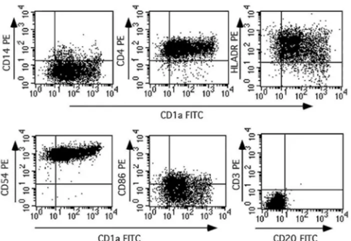
DC were generated from the adherent fraction of human peripheral blood mononuclear cells (PBMCs) with 800 U of GM-CSF (Novartis)/ml and 20 U of IL-4 (Bio-source Europe, Nivelles, Belgium)/ml, as previously described (19). DC generated for this study constituted a 90% pure cell population based on several criteria including morphology, forward- and side-scatter values observed by fluorescence-activated cell sorter analysis, and surface phenotype characteristics (CD1a+, HLA-DR+, major histocompatibility complex class I positive,
CD4+, CD54+, CD86dim, CD3-, CD20-, and CD14-).
The DC were labeled with a lipophilic fluorescent marker (CM-DiL; Molecular Probes, Leiden, The
Netherlands) according to a previously described procedure with minor modifications. Briefly, 4 x 10⁶ DC were
resuspended in 1 ml of phosphate-buffered saline (PBS) and heated to 37°C. The CM-DiL marker was diluted in 1 ml of PBS preheated to 37°C to obtain a final concentration of 16 µg/ml. The dye was mixed for several seconds until the dye was evenly distributed. This solution was immediately transferred to the cell suspension and rapidly mixed by pipetting. The cells were incubated for 2 min at 37°C and then for 2 min on ice. Finally, they were transferred in 40 ml of PBS at 4°C, centrifuged, and resuspended in the appropriate medium. Organotypic cultures.
Organotypic cultures of HPV-transformed keratinocytes (SiHa cell line) (13) were prepared as previously reported (20). After a 2-week stratification of keratinocytes, labeled DC were seeded on top of the in vitro-formed epithelium at a concentration of 2 x 10⁵ cells/50 µl of culture medium. The organotypic culture was then
placed on a polycarbophil gel containing either 800 U of GM-CSF/ml or no gel. The positive control was a solution of liquid GM-CSF at a concentration of 800 U/ml. After 48 h at 37°C, the collagen rafts were harvested. The cultures were then embedded in optimal cutting temperature compound (Tissue Tek; Sakura, Zouterwoude, The Netherlands) at -70°C and sectioned with a cryostat. The organotypic cultures were counterstained with the fluorescent dye 4',6-diamidine-2-phenylindole dihydro-chloride (DAPI; Roche Diagnostics, Brussels, Belgium). Infiltration of fluorescent DC was visualized with a Leica (Heidelberg, Germany) DMLB fluorescent microscope equipped with a 40X lens objective.
Assessment of CD1a+ cell infiltration in organotypic cultures.
The density of DC migration into the epithelial layer was assessed by the avidin-biotin-peroxidase technique (Vectastain ABC kit; Vector Laboratories, Burlingame, Calif.) with an anti-CD1a monoclonal antibody (clone NA1/34; Dako, Glostrup, Denmark). Nine-micrometer frozen sections were fixed in cold acetone for 3 min, and endogenous peroxidases were blocked with 0.1% H2O2 for 30 min. Sections were then incubated sequentially
with anti-CDla antibody (at a 1/40 dilution in PBS-2% bovine serum albumin-0.01 M NaN3) or with an
isotype-matched control antibody for 1 h, a biotinylated mouse anti-immunoglobulin antibody for 30 min, and streptavidin-horseradish peroxidase AB complex for another 30 min. Positive cells were visualized by a 3,3'-diarninobenzidine substrate. The sections were counterstained with hematoxylin. The DC infiltration in
organotypic cultures was evaluated by measuring the surface of immunostained cells with a computerized system of image analysis (CAS; Becton-Dickinson, Erembodegem, Belgium) following a previously described method (7). The entire surface of each culture section was analyzed, and five sections of each culture were stained with
anti-CD1a. The positive surface was expressed as the percentage of the total surface analyzed. Statistical analysis was performed with the Student t test (Instat Mac 2.01 software; Graph-Pad Software, San Diego, Calif.). Xenograft assay with NOD/SCID mice.
HPV-transformed keratinocytes (2 x 10⁵) from the CasKi cell line (ATCC CRL-1550) were plated on collagen
gel (2 mg of type I collagen isolated from rat tail tendons/ml) inserted in Teflon rings (Renner, Dannstadt, Germany) and maintained in culture for 1 day before transplantation onto mice. Twelve-week-old NOD/SCID mice were used for this study. They were housed under sterile conditions, and water and feed were provided at will. Before transplantation, the cells were washed and the medium was drained. The cell-coated collagen gels were then covered with a silicone transplantation chamber (Renner) and implanted entirely onto the dorsal muscle fascia of mice as previously described (2,14). The mice were killed 24 days later. The transplants were excised, embedded in optimal cutting temperature compound (Tissue Tek; Sakura), and frozen on dry ice for cryostat sectioning. In DC recruitment experiments, 2 x 10⁶ fluorescence-labeled DC were intravenously injected
at day 21. PBS or polycarbophil gel alone or with chemoattractants (GM-CSF liquid, 4 x 10⁵ U/ml; or GM-CSF
incorporated in polycarbophil gel, 4 x 10⁵ U/g of gel) was injected twice (each injection, 200 µl) into the
transplantation chamber 24 and 48 h before the mice were sacrificed. Transplant sections were counterstained with the fluorescent dye DAPI (Roche Diagnostics). Infiltration of fluorescent DC was visualized with a Leica DMLB fluorescent microscope equipped with a 40x objective. Fluorescent DC in the epithelium were counted and, after anti-keratin immunolabeling, the epithelial surface was evaluated with the CAS computerized system of image analysis (Becton-Dickinson). Infiltration results were expressed as the numbers of DC per square millimeter of epithelium. Statistical analysis was performed with the Student t test (Instat Mac 2.01 software; Graph-Pad).
Immunofluorescent labeling.
Graft cryosections (each, 5 µm in thickness) were fixed in acetone at -20°C and in 80% methanol at 4°C. They were then incubated for 60 min at room temperature with the primary antibodies anti-mouse type IV collagen (SIF105, rabbit anti-mouse; a gift of A. Noël) and anti-human keratin (KL1, mouse anti-human; Immunotech, Marseille, France). The appropriate secondary antibodies were then applied for 30 min: swine anti-rabbit conjugated to Texas red (Dako) and sheep anti-mouse conjugated to fluorescein-isothiocyanate (Sigma-Aldrich). For double immunofluorescence-la-beling studies, sections were first incubated with the two primary antibodies and then with fluorescein isothiocyanate- and Texas red-conjugated secondary antibodies. After being washed in PBS, coverslips were mounted, and labeling was analyzed under a microscope equipped with epifluorescence optics.
RESULTS
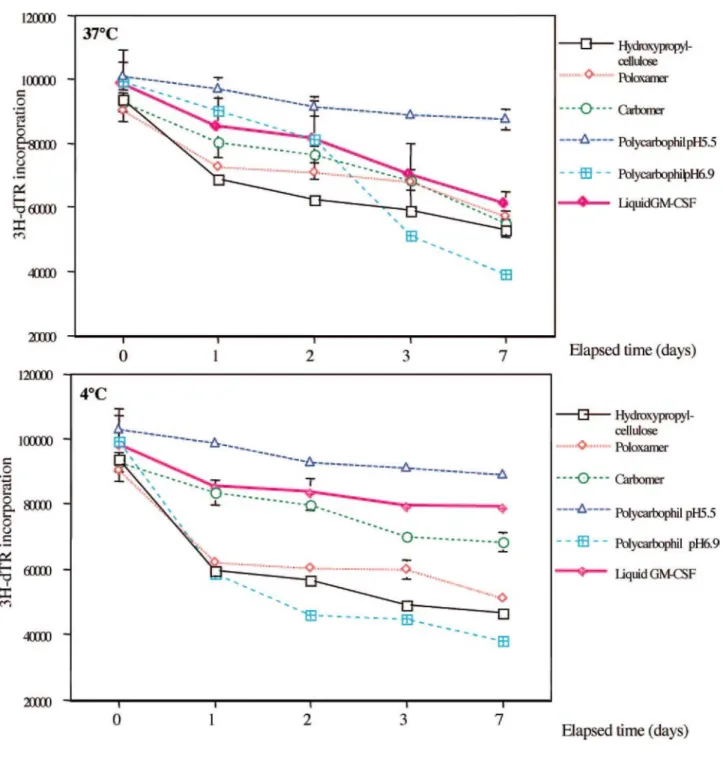
Analysis of GM-CSF bioactivity following incorporation into different polymers at 4 and 37°C by a bioassay with TF-1 cells.
The biological activity of GM-CSF was measured at different time intervals after incorporation into the hydrogels by a bioassay with the GM-CSF-dependent TF-1 cell line. The injectable solution (Leucomax; Novartis) was chosen as a positive control. The four polymers were tested at a pH value of 6.9, which was reported to be optimal for GM-CSF stability and which corresponds to the pH value of the injectable solution (data from Novartis). As polycarbophil was found to be a particularly convenient polymer for cervicovaginal formulations; as it is usually recommended that cervicovaginal formulations be developed at acidic pH values, the polycarbophil hydrogel was also tested at pH 5.5.
As shown in Fig. 1, there was a decrease in the bioactivity of the injectable solution of GM-CSF, more important at 37°C than at 4°C, which is in agreement with previous data (35). We also observed an important reduction in GM-CSF activity after incorporation in the hydroxypropyl cellulose (Klucel) or in the poloxamer hydrogel (Lutrol) at both temperatures. In contrast, polycarbophil gel (Noveon) at pH 5.5 induced protection against GM-CSF degradation. However, this protective effect was not observed at pH 6.9. Carbomer (Carbopol) conserved an activity of GM-CSF similar to that observed with the injectable solution at both temperatures and during at least a week. However, a pH reduction to 5.5 induced a rapid decrease in the activity of GM-CSF incorporated into carbomer gel at 4 and 37°C (data not shown).
These results suggest that the polycarbophil gel (Noveon) at pH 5.5 could be convenient for topical application of GM-CSF on a squamous cell genital mucosa.
Influence of GM-CSF incorporated in polycarbophil gel on the recruitment of DC in in vitro-formed (pre)neoplastic epithelium.
We investigated whether GM-CSF included in a polymer modulates the ability of DC to infiltrate an in vitro-formed (pre)neoplastic epithelium, reminiscent of high-grade (pre) neoplastic lesions observed in vivo.
Monocytic precursors from the plastic adherent fraction of human PBMCs cultivated in the presence of 800 U of GM-CSF/ml and 20 U of IL-4/ml for 7 days showed a morphology of DC with abundant dendritic membrane protrusions (data not shown). By fluorescence-activated cell sorter analysis (Fig. 2), these cells also displayed phenotypical features typical of DC with the presence of CDla, CD4, CD86, and CD54 (intercellular adhesion molecule 1) antigens; a moderate expression of major histocompatibility complex class II (HLA-DR) molecules; and the absence of macrophage (CD14), B-cell (CD20), and T-cell (CD3) markers. DC were layered on top of organotypic cultures of HPV-transformed keratinocytes in the presence of GM-CSF incorporated or not into the polycarbophil gel. The effect of GM-CSF on DC infiltration was assessed by examining cryosections of organotypic cultures frozen 48 h after the addition of GM-CSF. The ability of the GM-CSF gel to modulate the infiltration of DC was determined by the evaluation of fluorescent DC into the organotypic cultures. DC layered onto organotypic cultures of SiHa poorly infiltrated the epithelial cell layers in the absence of GM-CSF, whereas GM-CSF in solution or incorporated in the gel caused a significant increase in the density of DC observed within the epithelial sheet. Quantitative analysis of DC infiltration was performed with organotypic cultures derived from the HPV-transformed cell line SiHa (Fig. 3). The density of DC in the in vitro-formed epithelium was quantified by evaluating the surface labeled with the anti-CDla antibody throughout the full thickness of
organotypic cultures. Under basal conditions (in the absence of GM-CSF, either liquid or when gel incorporation had been carried out), the level of DC infiltration in the cultures was low (1.64 ± 0.78% of CD la-labeled surface). When the medium of organotypic cultures of HPV-positive cell lines was supplemented with GM-CSF, the infiltration of DC significantly improved (28.33 ± 8.11% of CD1a-labeled surface); the density of DC observed in these conditions was similar to that induced by the GM-CSF included in the gel (25.38 ± 5.81% of CDla-labeled surface).
GM-CSF included in polycarbophil gel stimulates the migration of human DC in xenografts of HPV-transformed keratinocytes.
To address the in vivo efficacy of GM-CSF included in polycarbophil gel, HPV-transformed transplanted keratinocytes cultured on a collagen gel were grafted onto the backs of NOD/SCID mice. Twenty-four days after transplantation, the mice were killed and the grafts were removed for immunohistological analysis. Histological examination of thin sections revealed that the growth pattern of the grafts reproduced a high-grade squamous (pre)neoplastic lesion and that the collagen gel was replaced by granulation tissue (data not shown).
Transplantation of HPV-positive keratinocytes induced an angiogenic response in the host tissue starting from vessels of the dorsal muscle and subsequently extending far up into the collagen gel. The vessels sprouted into the tumor epithelium, which started to invade the newly formed granulation tissue (data not shown).
To demonstrate the efficacy of GM-CSF incorporated into polycarbophil gel, two groups of five mice each were treated by injecting 200 µl of GM-CSF, in PBS or in the gel, in the transplantation chamber covering the (pre)neoplastic epithelium, daily for 2 days prior to killing the mice. A control group of mice was treated with PBS alone. At the first application of GM-CSF, 2 x 10⁶ human DC prelabeled with fluorescent tracer were
intravenously injected into the mice. The grafts were harvested 1 day after the last administration of GM-CSF and processed for microscopic examination. The DC infiltration in transplants was assessed by looking for stained DC in histological sections. The application of GM-CSF in the transplantation chamber resulted in a significant (P < 0.001) recruitment of DC in the tumor grafts of treated animals, while DC were rarely detected in the epithelia of control mice. Quantitative analysis of DC infiltration was performed by determining the number of DC per square millimeter of epithelial tissue (Fig. 4). The rate of DC infiltration in the grafts was very low (1.1 ± 1.4 DC/mm2) in the absence of CSF and drastically increased after treatment with liquid
GM-CSF (16.6 ± 14.6 DC/mm2). Similar results were observed when GM-CSF was incorporated into polycarbophil
FIG. 1: Proliferation response of TF-1 cell line after incubation with GM-CSF extracted at different time intervals from the hydrogels stored at 4 or 37°C. Growth was measured by 3H-labeled thymidine incorporation. Each point shows the mean of six replicates, and bars represent standard errors.
FIG. 2: Double staining and flow cytometry analysis of DC generated from cultures of adherent human PBMCs in the presence of GM-CSF and IL-4 during 7 days. Results are displayed as dot plots. Cells were labeled with CD1a-fluorescein isothiocyanate antibody versus indicated phycoerythrin-conjugated antibodies. Quadrant setting was performed according to the reactivities of isotype-matched controls. One representative experiment is shown out of more than 10 experiments performed.
FIG. 3: Quantitative evaluation of DC infiltration into organotypic cultures of HPV-transformed keratinocytes under the influence of GM-CSF incorporated or not into polycarbophil gel. The penetration of DC was followed by immunolabeling with CD1a. Results are expressed as the mean percentages of surface labeled with anti-CD1a compared to unlabeled surface of the epithelial sheet ± the standard deviation (five experiments). Asterisks indicate statistically significant differences (***,P < 0.001).
FIG. 4: Quantitative evaluation of DC infiltration into transplants of HPV-transformed keratinocytes. The results are expressed as numbers of DC per square millimeter of HPV-positive epithelium ± the standard deviation (five experiments were conducted for each condition). Asterisks indicate statistically significant differences (***, P < 0.001).
DISCUSSION
Different mucoadhesive drug delivery systems based on hydrogels have been recently developed (12, 33, 40). Their ability to interact with mucus glycoproteins and to remain localized to a specific site might help to deliver molecules important for antiviral or antitumor immune responses. It has previously been shown that GM-CSF increases the ability of monocyte-derived DC to infiltrate a (pre)neoplastic epithelium formed in vitro, suggesting that the effect of a treatment based on the local administration of GM-CSF might be a recruitment into the dysplastic epithelium of LC (20). LC are the main professional antigen-presenting cells in squamous mucosa. These cells are able to capture viral or tumor antigens and, after migration in the draining lymph nodes, to present these antigens to T cells. Subsequently, the sensitized T lymphocytes could reach the squamous epithelium and kill the cells bearing the antigens at the origin of the sensitization.
In this study, we analyzed the stability and bioactivity of GM-CSF in different formulations of hydrogels. As the quantitative and functional deficit of DC/LC is a constant immune alteration in genital HPV-associated lesions (43), we used epithelial sheets of HPV-transformed keratinocytes (7, 8, 20, 21, 36) maintained in vitro (on a collagen gel) or in vivo (in NOD/SCID mice).
We first demonstrated that polycarbophil gel at pH 5.5 conserves the bioactivity of GM-CSF at 4 or 37°C for at least 7 days, whereas a decrease in the biological activity of GM-CSF was observed mainly after incorporation into hydroxypropyl cellulose or poloxamer hydrogels at both temperatures. This could be due to interactions with the polymers that are present in the formulations at a high concentration to achieve the suitable viscosity. The residual bioactivity of GM-CSF incorporated into both gels at pH 6.9 was surprisingly different. These data could be attributed to the carboxyl group present in the polycarbophil gel and absent in the hydroxypropyl cellulose and poloxamer formulations. Indeed, similar results were reported with carboxylic acid containing polymers and carbomers, which were shown to inhibit the degradation of insulin and other peptide drugs (1). GM-CSF bioactivity evaluated after storage in the polycarbophil hydrogel formulated at a pH value of 5.5 was better than that observed for GM-CSF in solution. Moreover, the fact that the pH must be decreased to observe the protective effect of the polymer is completely compatible with the physiological cervicovaginal pH. Since the bioassay with TF-1 cells required the dilution of the different hydrogels, the bioactivity detected was that of solutions of GM-CSF. It was, therefore, important to determine whether the gel affected the activity of GM-CSF or not. Indeed, due to their viscosity, the hydrogels could interfere with the diffusion and the bioavailability of GM-CSF. We have demonstrated that GM-CSF included in the polycarbophil gel at pH 5.5 stimulated the infiltration of DC into an in vitro equivalent of HPV-associated (pre)neoplastic epithelium, suggesting that this hydrogel does not impair protein release in vitro.
Besides the stabilization of GM-CSF with a gel formulation and its efficacy in vitro, this report also describes the potential interest of a mouse xenograft model for the in vivo testing of new immunopharmacological treatments. The use of the dorsal muscle of the mouse as a transplantation site and a transplantation chamber were shown to be useful to evaluate the effects of topical formulations. Moreover, the model reported here has the advantage of using transformed keratinocytes derived from cell lines, available in unlimited quantities and capable of
generating reproducible data. By using this model, we have demonstrated that GM-CSF included in
polycarbophil gel is able to stimulate the intraepithelial migration of human DC in vivo. It remains to determine whether this DC recruitment with GM-CSF will be effective at inducing efficient immune responses against viral or tumor antigens.
Taken together, these results suggest that the polycarbophil gel (Noveon) is compatible with the diffusion of bioactive GM-CSF with the advantage of stabilizing the protein. Moreover, GM-CSF included in this gel formulation is also able to recruit DC into HPV-transformed (pre)neoplastic epithelial tissues in vitro and in vivo. This formulation, which is recommended for genital application, might restore some immune functions that have been shown to be altered in HPV-associated lesions.
ACKNOWLEDGMENTS
This work was supported by the Centre de Recherche Interuniversitaire en Vaccinologie with grants from the Walloon Region (DGTRE) and SmithKline Beecham Biologicals, the Belgian Fund for Medical Scientific Research, the Centre Anti-Cancereux près l'Université de Liège, and the Interuniversity Attraction Poles Programme-Belgian Science Policy (Brussels, Belgium). P. Delvenne is a Senior Research Associate of the Belgian National Fund for Scientific Research (FNRS).
REFERENCES
1. Bai, J., L. Chang, and J. Guo. 1996. Effects of polyacrylic polymers on the degradation of insulin and peptide drugs by chymotripsin and trypsin. J. Pharm. Pharmacol. 48:17-22.
2. Bajou, K., A. Noël, R. D. Gerard, V. Masson, N. Brunner, C. Holst-Hansen, M. Skobe, N. E. Fusenig, P. Carmeliet, D. Collen, and J. M. Foidart. 1998. Absence of host plasminogen activator inhibitor 1 prevents cancer invasion and vascularization. Nat. Med. 4:923-928.
3. Bosch, F. X., M. M. Manos, N. Munoz, M. Sherman, A. M. Jansen, J. Peto, et al. 1995. Prevalence of human papillomavirus in cervical cancer: a worldwide perspective. J. Natl. Cancer Inst. 87:796-802.
4. Chang, J., Y. Oh, H. Choi, and Y. Kim. 2002. Rheological evaluation of thermosensitive and mucoadhesive cervico-vaginal gels in physiological conditions. Int. J. Pharm. 241:155-163.
5. Charbonnier, A. S., N. Kohrgruber, E. Kriehuber, G. Stingl, A. Rot, and D. Maurer. 1999. Macrophage inflammatory protein 3α is involved in the constitutive trafficking of epidermal langerhans cells. J. Exp. Med. 190:1755-1768.
6. Colasante, A., G. Castrilli, F. Bianca, M. Brunetti, and P. Musiani. 1995. Role of cytokines in distribution and differentiation of dendritic cell/ langerhans'cell lineage in human primary carcinomas of the lung. Hum. Pathol. 26:866-872.
7. Delvenne, P., W. Al-Saleh, C. Gilles, A Thiry, and J. Boniver. 1995. Inhibition of growth of normal and human papillomavirus-transformed keratinocytes in monolayer and organotypic cultures by interferon-gamma and tumor necrosis factor-alpha. Am. J. Pathol. 146:589-598.
8. Delvenne, P., P. Hubert, N. Jacobs, S. L. Giannini, L. Havard, I. Renard, D. Saboulard, and J. Boniver. 2001. The organotypic culture of HPV-transformed keratinocytes: an effective in vitro model for the development of new immunotherapeutic approaches for mucosal (pre)neoplastic lesions. Vaccine 19:2557-2564.
9. Dieu, M. C, B. Vanbervliet, A Vicari, J. M. Bridon, E. Oldham, S. Ait-Yahia, F. Briere, A. Zlotnik, S. Lebecque, and C. Caux. 1998. Selective recruitment of immature and mature dendritic cells by distinct chemokines expressed in different anatomic sites. J. Exp. Med. 188:373-386.
10. Dranoff, G., E. Jaffee, A. Lazenby, P. Golumbek, H. Levitsky, K. Brose, V. Jackson, H. Hamada, D. Pardoll, and R. C. Mulligan. 1993. Vaccination with irradiated tumor cells engineered to secrete murine granulocyte-macrophage colony-stimulating factor stimulates potent, specific, and long-lasting antitumor immunity. Proc. Natl. Acad. Sci. USA 90:3539-3543.
11. Ellerbrock, T. V., M. A. Chiasson, T. J. Bush, X. W. Sun, D. Sawo, K. Brudney, and T. C. Wright. 2000. Incidence of cervical squamous intraepithelial lesions in HIV-infected women. JAMA 283:1031-1037.
12. Eouani, C, P. Piccerelle, P. Prinderre, E. Bourret, and J. Joachim. 2001. In-vitro comparative study of buccal mucoadhesive performance of different polymeric films. Eur. J. Pharm. Biopharm. 52:45-55.
13. Friedl, F., I. Kimura, T. Osato, and Y. Ito. 1970. Studies on a new human cell line (SiHa) derived from carcinoma of uterus. I. Its establishment and morphology. Proc. Soc. Exp. Biol. 135:543-545.
14. Fusenig, N. E. 1994. Epithelial-mesenchymal interactions regulate keratin-ocyte growth and differentiation in vitro, p. 71-94. In I. Leigh, B. Lane, and F. Watt (ed.), The keratinocyte handbook. Cambridge University Press, Cambridge, England.
15. Gauger, L. J. 1983. Extemporaneous preparation of a dinoprostone gel for cervical ripening. Am. J. Hosp. Pharm. 40:2195-2196.
16. Giannini, S. L., P. Hubert, J. Doyen, J. Boniver, and P. Delvenne. 2002. Influence of the mucosal epithelium microenvironment on Langerhans cells: implications for the development of squamous intraepithelial lesions of the cervix. Int. J. Cancer 10:654-659.
17. Herrington, C. S. 1995. Human papillomaviruses and cervical neoplasia. II. Interaction of HPV with other factors. J. Clin. Pathol. 48:1-6.
18. Hubert, P., S. L. Giannini, A. Vanderplasschen, R. Greimers, E. Franzen-Detrooz, N. Jacobs, J. Boniver, and P. Delvenne. 2001. Dendritic cells induce the death of papillomavirus-transformed keratinocytes. FASEB J. 15:2521-2523.
19. Hubert, P., R. Greimers, E. Franzen-Detrooz, J. Doyen, P. Delanaye, J. Boniver, and P. Delvenne. 1998. In vitro propagated dendritic cells from patients with human-papilloma virus-associated preneoplastic lesions of the uterine cervix: use of Flt3 ligand. Cancer Immunol. Immunother. 47:81-89.
20. Hubert, P., F. van den Brule, S. L. Giannini, E. Franzen-Detrooz, J. Boniver, and P. Delvenne. 1999. Colonization of in vitro-formed cervical human pap-illomavirus-associated (pre)neoplastic lesions with dendritic cells: role of granulocyte/macrophage colony-stimulating factor. Am. J. Pathol. 154:775-784.
21. Jacobs, N., M. P. Moutschen, E. Franzen-Detrooz, V. Boniver, J. Boniver, and P. Delvenne. 1998. Organotypic culture of HPV-transformed keratino-cytes: a model for testing lymphocyte infiltration of (pre)neoplastic lesions of the uterine cervix. Virchows Arch. 432:323-330.
22. Jonuleit, H., J. Knop, and A. H. Enk. 1996. Cytokines and their effects on maturation, differentiation and migration of dendritic cells. Arch. Dermatol. Res. 289:1-8.
23. Kaplan, G., G. Walsh, L. S. Guido, P. Meyn, R. A. Burkhardt, R. M. Abalos, J. Barker, P. A. Frindt, T. T. Fajardo, R Celona, and Z. A. Cohn. 1992. Novel responses of human skin to intradermal recombinant granulocyte/ macrophage-colony-stimulating factor: Langerhans cell recruitment, keratin-ocyte growth and enhanced wound healing. J. Exp. Med. 175:1717-1728.
24. Kitamura, T., T. Tange, T. Terasawa, S. Chiba, T. Kuwaki, K. Miyagawa, Y. F. Piao, K. Miyazono, A. Urabe, and F. Takaku. 1989. Establishment and characterization of a unique human cell line that proliferates dependently on GM-CSF, IL-3, or erythropoietin. J. Cell. Physiol. 140:323-334.
25. Knuth, K., M. Amiji, and J. Robinson. 1993. Hydrogel delivery system for cervico-vaginal and oral applications: formulations and biological consideration Adv. Drug Deliv. Rev. 11:137-167.
26. Koch, F., C. Heufler, E. Kampgen, D. Schneeweiss, G. Bock, and G. Schuler. 1990. Tumor necrosis factor α maintains the viability of murine epidermal Langerhans cells in culture, but in contrast to granulocyte/macrophage colony-stimulating factor, without inducing their functional maturation. J. Exp. Med. 171:159-165.
27. Lynas, K. W. 1980. Preparation of prostaglandin F2α cervico-vaginal gel. Aust. J. Hosp. Pharm. 10:88-92.
28. MacKenzie, I. Z., A. J. Davies, and M. P. Embrey. 1979. Fetal death in utero managed with cervico-vaginal prostaglandin E2 gel. BMJ 1:1764-1765.
29. McArdle, J. P., and H. K. Muller. 1986. Quantitative assessments of Langerhans' cells in human cervical intraepithelial neoplasia and wart virus infection. Am. J. Obstet. Gynecol. 154:509-515.
30. Morelli, A. E., G. Belardi, G. DiPaola, A. Paredes, and L. Fainboim. 1994. Cellular subsets and epithelial ICAM-1 and HLA-DR expression in human papillomavirus infection of the vulva. Acta Derm. Venereol. 74:45-50.
31. Nakamura, K., I. R Williams, and T. S. Kupper. 1995. Keratinocyte-derived monocyte chemoattractant protein 1 (MCP-1): analysis in a transgenic model demonstrates MCP-1 can recruit dendritic and Langerhans cells to skin. J. Investig. Dermatol. 105:635-643.
32. Pastore, S., E. Fanales-Belasio, C. Albanesi, L. M. Chinni, A. Giannetti, and G. Girolomoni. 1997. Granulocyte macrophage colony-stimulating factor is overproduced by keratinocytes in atopic dermatitis. J. Clin. Investig. 99: 3009-3017.
33. Peppas, N. A., and J. J. Sahlin. 1996. Hydrogels as mucoadhesive and bioadhesive materials. Biomaterials 17:1553-1561.
34. Petry, K. IL, D. Scheffel, U. Bode, T. Gabrysiak, H. Koche, E. Kupsch, M. Glaubitz, S. Niesert, H. Kuhnle, and I. Schedel. 1994. Cellular immunodeficiency enhances the progression of human papillomavirus-associated cervical lesions. Int. J. Cancer 57:836-840.
35. Pettit, D. K., J. R Lawter, W. J. Huang, S. C. Pankey, N. S. Nightlinger, D. H. Lynch, J. A. Schuh, P. J. Morrissey, and W. R Gombotz. 1997. Characterization of poly(glycolide-co-[scap]d,l-lactide)/poly([scap]d,l-lac-tide) microspheres for controlled release of GM-CSF. Pharm. Res. 14:1422-1430.
36. Renard, I., D. Mezzanzanica, S. Canevari, S. Ferrini, J. Boniver, P. Delvenne, and N. Jacobs. 2002. Anti-CD3/anti-epidermal growth factor recep-tor-bispecific antibody retargeting of lymphocytes against human neoplastic keratinocytes in an autologous organotypic culture model. Am. J. Pathol. 160:113-122.
37. Richardson, J., J. Whetstone, A. Fisher, P. Watts, N. Farraj, M. Hinchcliffe, L. Benedetti, and L. Ilium. 1996. Gamma-santigraphy as a novel method to study distribution and retention of a bioadhesive cervico-vaginal delivery in sheep. J. Control Release 42:133-142.
38. Riethdorf, S., L. Riethdorf, N. Richter, and T. Loning. 1998. Expression of the MCP-1 gene and the HPV 16 E6/E7 oncogenes in squamous cell carcinomas of the cervix uteri and metastases. Pathobiology 66:260-267.
39. Robinson, R., and W. Bologna. 1994. Cervico-vaginal and reproductive system treatments using a bioadhesive polymer. J. Control Release 28:88-94.
40. Shawesh, A., S. Kallioinene, L. Hellen, O. Antikainen, and J. Yliruusi. 2002. Pluronic F-127 gels as a vehicle for topical formulations of indomethacin and rheological behaviour of these formulations. Pharmazie 57:186-190.
41. Smedts, F., F. Ramaekers, R E. Leube, K. Keijser, M. Link, and P. Vooijs. 1993. Expression of keratins 1, 6, 15, 16, and 20 in normal cervical epithelium, squamous metaplasia, cervical intraepithelial neoplasia, and cervical carcinoma. Am. J. Pathol. 142:403-412.
42. Valenta, C, C. Kast, I. Harich, and C. Bernkop-Schnurch. 2001. Development and in vitro evaluation of a mucoadhesive cervico-vaginal delivery system for progesterone. J. Control Release 77:323-332.
43. Viac, J., Y. Chardonnet, M. C. Chignol, and D. Schmitt. 1993. Papilloma viruses, warts, carcinoma and Langerhans cells. In Vivo 7:207-212.
44. Yang, D., O. Chertov, S. N. Bykovskaia, O. Chen, M. J. Buffo, J. Shogan, M. Anderson, J. M. Schroder, J. M. Wang, O. M. Howard, and J. J. Oppenheim. 1999. Beta-defensins: linking innate and adaptive immunity through dendritic and T cell CCR6. Science 286:525-528.