The anti-inflammatory properties of intravenous immunoglobulin in
a murine model of allergic airway disease
Effects on the development of regulatory T-cells
par
Amir Hossein Massoud
Département de Microbiologie et Immunologie Faculté de Médecine
Thèse présentée à la Faculté des études supérieures et postdoctorales en vue de l’obtention du grade de Philosophiæ Doctor (Ph.D.)
en immunologie
Avril 2013
Faculté des études supérieures et postdoctorales
Cette thèse intitulée:
The anti-inflammatory properties of intravenous immunoglobulin (IVIg) in a murine model of allergic airway disease
Effects on the development of regulatory T-cells
Présenté par : Amir Hossein Massoud
a été évaluée par un jury composé des personnes suivantes :
Dr Idriss Djilali-Saiah, président-rapporteur Dr Walid M. Mourad, directeur de recherche
Dr Bruce D. Mazer, co-directeur Dr Ali Ahmad, membre du jury Dr James G. Martin, examinateur externe Dr Paolo Renzi, représentant du doyen de la faculté
Résumé
Les immunoglobulines intraveineuses (IVIg) constituent une préparation polyclonale d’IgG isolée et regroupée à partir du plasma sanguin de multiples donneurs. Initialement utilisé comme traitement de remplacement chez les patients souffrant d’immunodéficience primaire ou secondaire, les IVIg sont maintenant largement utilisées dans le traitement de plusieurs conditions auto-immunes, allergiques ou inflammatoires à une dose élevée, dite immuno-modulatrice. Différents mécanismes d’action ont été postulés au fil des années pour expliquer l’effet thérapeutique des IVIg dans les maladies auto-immunes et inflammatoires. Entre autre, un nombre grandissant de données issues de modèles expérimentaux chez l’animal et l’humain suggère que les IVIg induisent l’expansion et augmentent l’action suppressive des cellules T régulatrices (Tregs), par un mécanisme qui demeure encore inconnu. Également, les patients atteints de maladies auto-immunes ou inflammatoires présentent souvent un nombre abaissé de Tregs par rapport aux individus sains. Ainsi, une meilleure compréhension des mécanismes par lesquels les IVIg modulent les cellules T régulatrices est requise afin de permettre un usage plus rationnel de ce produit sanguin en tant qu’alternative thérapeutique dans le traitement des maladies auto-immunes et inflammatoires.
Par le biais d’un modèle expérimental d’allergie respiratoire induite par un allergène, nous avons démontré que les IVIg diminuaient significativement l’inflammation au niveau des voies aériennes ce, en association avec une différenciation des Tregs à partir des cellules T non régulatrices du tissu pulmonaire. Nous avons également démontré qu’au sein de notre modèle expérimental, l’effet anti-inflammatoire des IVIg était dépendant des cellules dendritiques CD11c+ (CDs) pulmonaires, puisque cet effet pouvait être complètement reproduit par le
transfert adoptif de CDs provenant de souris préalablement traitées par les IVIg. À cet effet, il est déjà établi que les IVIg peuvent moduler l’activation et les propriétés des CDs pour favoriser la tolérance immunitaire et que ces cellules seraient cruciales pour l’induction périphérique des Tregs. C’est pourquoi, nous avons cherché à mieux comprendre comment les IVIg exercent leur effet sur ces cellules. Pour la première fois, nous avons démontré que la fraction d’IgG riche en acide sialique (SA-IVIg) (constituant 2-5% de l’ensemble des IgG des donneurs) interagit avec un récepteur dendritique inhibiteur de type lectine C (DCIR) et active une cascade de signalement intracellulaire initiée par la phosphorylation du motif ITIM qui est responsable des changements observés en faveur de la tolérance immunitaire auprès des cellules dendritiques et des Tregs. L’activité anti-inflammatoire de la composante SA-IVIg a déjà été décrite dans des études antérieures, mais encore une fois le mécanisme par lequel ce traitement modifie la fonction des CDs n’a pas été établi. Nous avons finalement démontré que le récepteur DCIR facilite l’internalisation des molécules d’IgG liées au récepteur et que cette étape est cruciale pour permettre l’induction périphérique des Tregs.
En tant que produit sanguin, les IVIg constitue un traitement précieux qui existe en quantité limitée. La caractérisation des mécanismes d’action des IVIg permettra une meilleure utilisation de ce traitement dans un vaste éventail de pathologies auto-immunes et inflammatoires.
Mots-clés : immunoglobulines intraveineuses, asthme, inflammation des voies respiratoires,
récepteur lectine de type C, cellule dendritique, cellules T régulatrices, maladies auto-immunes et inflammatoires
Abstract
Intravenous immunoglobulin (IVIg) is a therapeutic preparation of normal human polyclonal IgG derived from pooled plasma from a large number of healthy donors. Initially used as replacement therapy for patients with primary and secondary immune deficiencies, IVIg is now also widely used for the treatment of a variety of autoimmune, allergic and systemic inflammatory disorders, at high immunomodulatory doses. The beneficial effect of IVIg in autoimmune and inflammatory diseases has been attributed to different mechanisms. Increasing evidence shows that IVIg induces expansion and enhances the suppressive function of regulatory T cells (Tregs) in different experimental animal models and human subjects, through an unknown mechanism. Human inflammatory and autoimmune diseases are known to be associated with Treg deficiency. Therefore, a more precise understanding of the mechanisms by which IVIg modulate Treg populations seems to be needed for more rational use of this compound as an alternative therapy in context of various inflammatory and autoimmune disorders.
Using a robust antigen-driven model of allergic airway disease, we have demonstrated that IVIg markedly attenuates airway inflammation and this effect is associated with the induction of Tregs from non-regulatory T cells in pulmonary tissues. We have also demonstrated that the anti-inflammatory actions of IVIg, in our model are dependent on a population of pulmonary CD11c+ dendritic cells (DCs), as the action of IVIg could be completely replicated by adoptive transfer of CD11c+ DCs from IVIg-treated mice. we have shown that tolerogenic DCs involve in the peripheral induction of Tregs. Given the requirement of DCs in the induction of Tregs, we explored the mechanism by which IVIg interacts and modulate these cells and for the first time demonstrated that the purified sialylated fraction of human IgG (SA-IVIg) (that consists 2-5% of
whole IgG) interacts with an inhibitory C-type lectin receptor on dendritic (DCIR) and this interaction triggers an ITIM intracellular signaling cascade. This subsequently results in rendering tolerogenic activities to DCs and peripheral induction of Tregs. The anti-inflammatory activity of SA-IVIg has been shown in previous studies, but the mechanism by which it modulates DCs functions is not well understood. We also demonstrated that DCIR facilitates the internalization of IgG molecules into DC and this internalization appears to be a crucial step for induction of Tregs.
IVIg is a costly therapeutic compound. Characterization of the mechanism of action of IVIg can lead to a better application of this plasma based therapy in a wide range of autoimmune and inflammatory diseases.
Keywords: intravenous immunoglobulin, asthma, airway inflammation, C-type lectin receptor,
Contributions of Authors
Article I
Gabriel N. Kaufman performed the research resulted in the generation of figures 1a,b,c, 3 and 4 and Table 1 and made the main contribution in writing and editing the manuscript. Amir Massoud carried out all the experiments resulted in the generation of figures 1d-g, 2, 5, 6 ,7, and participated in writing and editing the manuscript. The coauthors: Andree-Anne Banville-Langelier, Yufa Wang, Julie Guay and Jonathan A. Garellek, were given authorship in acknowledgement for their roles in teaching and performing experiment in flow cytometry, ELISA and RT-PCR in this study. Severine Audusseau performed the proliferation assays. Drs. Christine McCusker, Walid Mourad and Ciriaco Piccirillo were integral for their mentorship and advises on this project and aided in editing process of this manuscript. Dr. Bruce Mazer was the principal investigator and responsible for generating the main hypotheses and coordinating this project. All authors reviewed the manuscript and had access to primary data.
Article II
Dr. B. Mazer and Dr. C. Piccirillo and Amir Massoud were responsible for generating the main hypotheses and designing the experimental work and had a direct supervision on some experimental works employed to examine these hypotheses. Amir Massoud had the major contribution in writing and editing the manuscript. Julie Guay, and Karim Shalaby and Yasaman Nouhi were acknowledged as a coauthor for their role in teaching and helping with RT-PCR, flow cytometry and FlexiVent. Aidan Ablona and Daniel Chan made contributions in making solutions and culturing cells. Eva Bjur aided with breeding and providing the Tg mice used in
different experiments. Drs. Christine McCusker, Walid Mourad and Ciriaco Piccirillo were integral for their mentorship and advices on this project as well as in the editing process of this manuscript. Dr. Bruce Mazer was the principal investigator and responsible for generating the hypotheses and supervising the experimental works in this project. All authors reviewed the manuscript and had access to primary data.
Article III
Amir Massoud and Dr. Bruce Mazer were responsible for generating the main hypotheses, designing the experimental work, had direct supervision on all the experiments employed to examine these hypotheses and played the main role in writing and editing the manuscript. Madelaine Yona took an active role in helping with some experiments and preparing some solutions and contributed in editing the manuscript. Drs. Walid Mourad and Ciriaco Piccirillo were integral for their mentorship and advices on this project as well as in the editing process of this manuscript. Dr. Bruce Mazer was the principal investigator and supervised and coordinated this project. All authors reviewed the manuscript and had access to primary data.
Table of contents
Résumé ... iii
Abstract ... v
Contribution of Authors ... vii
Table of Contents ... ix
List of Figures ... xi
List of Tables ... xiv
List of Abbreviations ... xv
Acknowledgements ... xx
Chapter I Introduction, Review of the literature ... 1
1- A history of intravenous immunoglobulin and its clinical applications ... 2
2- Purification of IVIg from human plasma ... 8
3- Side effects ... 11
4- Anti-inflammatory Mechanisms of IVIg ... 12
4-1- The importance of “natural antibodies” for the immune-regulatory actions of IVIg ... 13
4-2- Modulatory effects IVIg on adaptive immunity ... 15
4-2-1- Modulatory effects of IVIg on helper T-cell subsets ... 16
4-2-2- Modulatory effects of IVIg on B cells ... 18
4-2-3- IVIg-mediated regulation of Antigen-Presenting Cells ... 20
4-2-3-1- Fcγ Receptor-dependent regulatory action of IVIg ... 23
5- The action of IVIg on the development and the activation of regulatory T cells ... 35
5-1- The mechanism by which IVIg affects the Treg compartment ... 38
6- Use of IVIg in Th-2 mediated atopic diseases ... 42
6-1- Proposed anti-inflammatory mechanisms of IVIg in allergic asthma ... 46
7- Concluding remarks and rationales ... 50
8- Hypothesis and objectives ... 50
Chapter II Studies on the anti-inflammatory effects of intravenous immunoglobulin, in a murine model of allergic airway disease ... 52
Article I Intravenous immune globulin attenuates airway hyper-responsiveness in a murine model of allergic asthma ... 53
Article II Intravenous immunoglobulin attenuates airway inflammation via the induction of Foxp3+ regulatory T cells in a murine model of allergic asthma ... 83
Article III C-Lectin Receptor Dendritic Cell (DCIR) mediates the tolerogenic effects of intravenous mmunoglobulin in pulmonary inflammation ... 116
Chapter III General discussion ... 150
List of Figures
Chapter I
Figure 1. Schematic representations of IgG structure and five immunoglobulin isotypes. ... 3
Figure 2. Representation of monomeric and dirmeric IgG ... 10
Figure 3. Effects of IVIg on differentiation and functions of dendritic cells ... 22
Figure 4. IVIg increases the clearance of pathogenic Abs by saturation of FcRn ... 25
Figure 5. Natural immunoglobulins non-specifically block the FcRs by their Fc fragments ... 27
Figure 6. IVIg induces the up-regulation of Fcexpressionandactivates the associated ITIM signaling pathway ... 31
Figure 7. Schematic structure and glycosylation of IgG... 33
Figure 8. Proposed role of Tregitopes (Antibody-derived Treg epitopes) in the immune-regulatory action of IgG. ... 39
Figure 9. A schematic depiction of the cellular components of the immune system that could be affected by IVIg. ... 49
Chapter II Article I Figure 1. IVIG diminishes lung inflammation in allergic airways disease. ... 74
Figure 2. IVIG inhibits Bronchial responsiveness to Methacholine. ... 76
Figure 3. IVIG attenuated OVA-driven splenocyte proliferation. ... 77
Figure 4. IVIG attenuates in vitro cytokine production. ... 78
Figure 5. Induction of mRNA for Notch ligands and Th2 transcription factor GATA3. ... 79
Figure 7. Assessment of T-regulatory cells in local and systemic tissues. ... 81
Article II Figure 1. IVIg induces de novo Tregs from Foxp3 negative T-cells. ... 105
Figure 2. IVIg induced Treg are greatly enriched in inflamed pulmonary tissues. ... 107
Figure 3. IVIg modulates homing receptors to encourage trafficking of Treg to pulmonary tissues. ... 108
Figure 4. IVIg induces antigen specific Treg... 109
Figure 5. Treg from IVIg treated mice are highly suppressive. ... 110
Figure 6. IVIg primes CD11c+ pulmonary dendritic cells to induce Treg. ... 111
Figure 7. Adoptive transfer of pulmonary DCs from IVIg treated-mice recapitulates the protective effect of IVIg therapy by inducing Treg. ... 112
Figure 8. Adoptive transfer of pulmonary DCs from IVIg treated-mice protects against increased AHR and pulmonary inflammation. ... 113
Article III Figure 1. The sialylated fraction of IgG abrogrates allergic airways disease ... 136
Figure 2. SA-IVIg interacts with CD11c+ DC by binding to DCIR ... 138
Figure 3. DCIR mediates binding and internalization of IgG into DC ... 140
Figure 4. DCIR is critically involved in the inhibition of AHR by IVIg/SA-IVIg. ... 142
Figure 5. The effect of SA-IVIg is not dependent on Fc receptors ... 144
Figure 6. Inhibition of IgG internalization abrogates the modulatory effects of IVIg on DC. .. 146 Figure 7. Ligation of DCIR by IVIg/SA-IVIg induces phosphorylation of SHIP-1 and SHP-2..
Chapter III
General discussion
Figure 1. Innate function of C-type lectin receptors (CLRs) in antigen uptake and signaling processes in antigen-presenting cells. ... 159 Figure 2. The known effects of IVIg on Treg compartment ... 165
List of Tables
Chapter I
Table I. Potential Anti-inflammatory and Immunomodulatory Activities of IgG. ... 41
Chapter II Article I
Table I. Cytokine measurements within purified splenic CD11c+ dendritic cells ... 82
Article II
Table I. Ki67 expression was monitored by flow cytometry on CD4+CD25+Foxp3+ Treg from the lungs under each experimental condition. ... 114 Table II. Cytokine production from induced-Tregs. ... 114 Table III. Phenotypic characteristics of CD11c+DC indicating increased expression of IL-10, Delta-like -4, and CD8 following IVIg treatment. ... 115
List of Abbreviations
(E): Elastance(R): Resistance
3[H]thy: tritiated Thymidine Ab: Antibody
ADCC: Antibody-dependent cytotoxicity Ag: Antigen
AHR: Airway hyperresponsiveness APC: Allophycocyanin
APCs: Antigen presenting cells B6: C57BL/6 mice
BHR: Bronchial hyperresponsiveness
BM-DC: Bone marrow-derived dendritic cells CD: Clusters of differentiation
CDR: Carbohydrate recognition domain CFSE: Carboxyfluorescein succinimidyl ester CHO: Chinese hamster ovary
CIDP: Chronic inflammatory demyelinating polyneuropathy CLR: C-type lectin receptor
CLR: C-type lectin receptor CNS: Central nervous system CON A: Concanavalin A
CTLA-4: Cytotoxic T-Lymphocyte Antigen 4 DAB 1: Diaminobenzidine
DC: Dendritic cell
DCIR: Dendritic Cell Immunoreceptor DEAE: Diethylaminoethanol
dLN: Draining lymph node dsDNA: Double stranded DNA
EAE: Experimental autoimmune encephalomyelitis ECL: Chemoluminescence
ELIZA: Enzyme-linked immunosorbent assay Fab: Fragment antigen-binding
FBS: Fetal bovine serum Fc: Fragment crystallizable FcR: Fc-gamma receptor
FEV1: Forced expiratory volume FITC: Fluorescein isothiocyanate Foxp3: Forkhead box protein 3 GBS: Guillain–Barré syndrome GFP: Green fluorescent protein
GITR: Glucocorticoid-induced TNFR-related protein
GM-CSF: Granulocyte macrophage colony-stimulating factor H&E: Hematoxylin and eosin
HLA: Human leukocyte antigen HSA: human serum albumin HSV: Herpes simplex virus I.N: Intranasal I.P: Intraperitoneal I.T. Intratracheal IFN: Interferon IgE: Immunoglobulin E IgG: Immunoglobulin G IHC: Immunohistochemistry IL: Interleukin
IPEX: Immunodysregulation polyendocrinopathy enteropathy X-linked syndrome ITAM: Immunoreceptor tyrosine-based activation motif
ITIM: Immunoreceptor tyrosine-based inhibition motif ITP: Immune thrombocytopenic purpura
iTreg: induced regulatory T cells IVIg: Intravenous immunoglobulin IVIg: Intravenous immunoglobulin KD: Kawasaki disease
KD: Knockdown
KD-DC: Knockdown dendritic cells KO: Knockout
LN: Lymph node MCh: Methacholine
MHC: Major histocompatibility complex MLR: Mixed leukocytes reactions MS: Multiple sclerosis
MZ: Marginal zone
NTN: Nephrotoxic nephritis
nTreg: Natural occurring regulatory T cells OVA: ovalbumin
PBS: Phosphate buffered saline PBS: phosphate-buffered saline pDC: Plasmacytoid dendritic cells PE: Phycoerythrin
PEEP: Post-expiratory end pressure PHA: Phytohaemagglutinin
PWM: Pokeweed mitogen
RT-PCR: Reverse transcription polymerase chain reaction RW: Ragweed
SA-IVIg: Sialic-acid enriched IgG siRNA: Small interfering RNA SLE: Systemic lupus erythematosus SOCS: Suppressor of cytokine signaling TCR: T cell receptor
Teff: Effector T TH: T helper
TNF: Tumor necrosis factor TNF: Tumor necrosis factors Treg: Regulatory T-lymphocytes WT: Wild type
Acknowledgments
I would like to express my deepest gratitude to my advisors, Dr. Walid Mourad and Dr. Bruce Mazer, for their excellent guidance, and providing me with an excellent atmosphere for doing research. To me, Dr. Mazer is more than a research director; his passion and care for every member of the team is far beyond expectation. I also would like to thank my supervisor, Dr. Walid Mourad who has consistently guided me in the right direction for the past 5 years of my PhD study. His help has been indispensable.
I would like to thank my committee members Dr. Idriss Djilali, Dr. James Martin and Dr. Ali Ahmad, who were unconditionally willing to help me and give me their best suggestions. I would never have been able to finish my dissertation without the guidance of my committee members. I would also like to thank Dr. Ciro Piccirillo for guiding me over the past several years and helping me to develop my background in Immunology.
From the heart, I would like to thank all my team members, colleagues at Meakins-Christie Laboratories, technicians, co-workers, and friends, who lift my spirits and generously support me in all my endeavors. I will keep in my mind each and every enjoyable moment I have spent with them.
Last but not least, I would like to thank my parents and my sister who were always supporting me and encouraging me with their best wishes. They always stood by me through the good times and bad.
Chapter I
Introduction
1- A history of Intravenous Immunoglobulin and its clinical applications
Intravenous immunoglobulin (IVIg) is a therapeutic preparation of normal human polyclonal IgG antibodies obtained from pooled plasma samples of more than 10000 healthy blood donors.
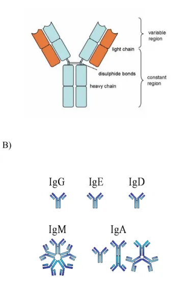
Antibodies are glycoproteins belonging to the immunoglobulin superfamily. They are produced by B-lymphocytes and are typically made of basic structural units, each with two large heavy chains and two smaller light chains. Immunoglobulin is composed of two fragments, known as Fab and Fc (Fig.1 A). The Fab region is the variable part of the molecule, capable of recognizing specific antigens, and the Fc region is the constant region of immunoglobulin, with binding capacity to Fc-receptors on various immune cells. There are different isotypes of antibody heavy chains, and immunoglobulin molecules are grouped into these different isotypes based on which heavy chain they possess. Five different antibody isotypes are identified in mammals; immunoglobulin M (IgM), immunoglobulin A (IgA), immunoglobulin D (IgD), immunoglobulin G (IgG) and immunoglobulin E (IgE) (Fig.1 B).
A)
B)
Figure 1. Schematic representations of IgG structure and five immunoglobulin isotypes. Adopted from: Janeway CA Jr, Travers P, Walport M, et al. Immunobiology: The Immune System in Health and Disease. 5th edition. New York: Garland Science; 2001.
A) Schematically representation of the structure of conventional IgG consisting of a light and heavy chain.
B) The five major isotypes of immunoglobulin structure. The basic functional unit of each antibody is a monomer immunoglobulin. Secreted antibodies can form dimeric structures as with IgA or pentameric structures with five immunoglobulin units such as IgM.
Commercially available IVIg consists of ≥96% IgG, representing the normal IgG subclass distribution of human serum (Burckhardt et al., 1989), which includes IgG1, IgG2, IgG3 and IgG4. Each subclass serves somewhat different functions in protecting the body against infectious agents. For example, the IgG1 and IgG3 subclasses are rich in antibodies against proteins such as the toxins produced by the diphtheria and tetanus bacteria, as well as antibodies against viral proteins. In contrast, antibodies against the polysaccharide (complex sugar) coating (capsule) of certain disease-producing bacteria (e.g. pneumococcus and Haemophilus influenzae) are predominantly of the IgG2 type in older children and adults. Some of the IgG subclasses can easily cross the placenta and enter the unborn infant’s bloodstream, while others do not. IgG1 and IgG3 subclasses interact readily with the complement system, while others interact poorly, if at all, with complement proteins. Thus, an inability to produce antibodies of a specific class or subclass may render the individual susceptible to certain kinds of infections but not others (Boyle et al., 2006).
Immunoglobulin products from human plasma were first used in 1952 as a method of antibody replacement therapy for the treatment of primary immune deficiencies such as agammaglobulinemia or hypogammaglobulinemia, deficiencies in immunoglobulin production characterized by recurrent bacterial infections (Knezevic-Maramica and Kruskall, 2003). By providing antibodies to patients who have weakened immune systems, IVIg can help reduce the risk of infection. These first immunoglobulin transfusions were administered intramuscularly which limited the dosage that could be given.
Since the introduction of immunoglobulin by the intravenous routes, more aggressive dosing strategies have become possible. IVIg is increasingly being used as a method of treatment for a
wide range of autoimmune and inflammatory diseases, due to its immune-regulatory actions, when administered in high doses (generally 1-2 grams/kg body weight, as opposed to 200–400 mg/kg body weight in immune deficiency conditions). The immune-modulatory activity of IVIg in autoimmune disorders was initially discovered in 1981, by Imbach et al. (Imbach et al., 1981) who observed a rise in platelet counts after administrating IVIg to patients with congenital agammaglobulinemia who were also thrombocytopenic. In the same year, Imbach reported the successful treatment of immune thrombocytopenic purpura, an autoimmune condition resulting in low platelet counts, by administration of IVIg. Since then the efficacy of IVIg, as a therapeutic compound with immune-regulatory activities, has been reported in various autoimmune disorders, chronic inflammatory diseases (Emmi and Chiarini, 2002; Hemming, 2001; Kaveri et al., 1997; Shoenfeld and Katz, 2005), and neurologic pathologies (Dalakas, 1997).
IVIg is mainly used as treatment in three major categories:
a) Primary and secondary immune deficiencies b) Autoimmune and inflammatory diseases c) Acute infections
The US Food and Drug Administration has approved the use of IVIg for the following 8 conditions:
1. Primary immunodeficiency states
2. Chronic inflammatory demyelinating polyneuropathy (CIDP) 3. Immune thrombocytopenic purpura (ITP)
5. Pediatric human immunodeficiency virus (HIV) infection 6. Kawasaki Disease (KD)
7. Guillain-Barre Syndrome (GBS)
8. Prevention of graft vs. host disease in an adult bone marrow transplant recipients
Given the broad action of IVIg, it can also be used to treat a variety of other conditions, in which controlled trials establishing the safety and efficacy are still needed. Some of these off-label uses are:
1. Neurological diseases: Myasthenia gravis, Lambert-Eaton myasthenic syndrome, multifocal motor neuropathy and multiple sclerosis.
2. Dermatological diseases: Autoimmune blistering dermatoses, dermatomyositis, pemphigus, pemphigoid.
3. Rheumatological diseases: Rheumatoid arthritis (adult and juvenile), systemic lupus erythematosus, systemic vasculitides, dermatomyositis, polymyositis, inclusion-body myositis and wegeners granulomatosis.
4. Hematological diseases: Aplastic anemia, pure red cell aplasia, Blackfan-Diamond anemia, autoimmune hemolytic anemia, hemolytic disease of the newborn, acquired factor VIII inhibitors, acquired von Willebrand disease, immune mediated neutropenia and neonatal alloimmune/ autoimmune thrombocytopenia.
5. Infectious diseases: Acquired infectious diseases that could be deleterious in low birth weight baby (ie, less than 1500 g), extensive burns and HIV infection.
7. Miscellaneous: Acute idiopathic dysautonomia, acute disseminated encephalomyelitis, hemophagocytic syndrome, multiple myeloma, recurrent pregnancy loss and POEMS syndrome (or Crow–Fukase syndrome, a rare medical syndrome defined as the combination of a plasma-cell proliferative disorder typically myeloma and polyneuropathy, and effects many organs).
The half-life of IgG molecule is approximately 21-29 days, except in bone marrow transplant patients and febrile and septic patients who may have a hypermetabolic state which decreases the half-life of IgG to 10-14 days (Ramos-Medina et al., 2012). However, the effectiveness of IVIg therapy can last between 2 weeks and 3 months, suggesting potential alteration of immunoglobulin metabolism following infusion of IVIg.
Studies exploring the mechanisms of action of IVIg demonstrated the efficacy of IVIg therapy in various animal models of autoimmune and inflammatory diseases, which support the anti-inflammatory effect of this therapeutic compound in human trials. IVIg treatment has protective effects in murine models of severe coronary arthritis (a murine model of KD) (Duong et al., 2003). IVIg has also protective effects in murine model of ITP by rapidly raising the platelet counts (Tremblay et al., 2012). In experimental autoimmune encephalomyelitis (EAE) (the murine model of MS) infusion of IVIg is associated with reduced disease severity and improvement of underlying CNS pathology (Humle Jorgensen and Sorensen, 2005). IVIg therapy also results in reduction of anti-dsDNA antibody titers and improvement of renal pathology in NZB/W mice, (a mouse model for SLE), (Shoenfeld et al., 2002). In an allergen-driven murine models of allergic asthma, rabbit IgG also displayed anti-inflammatory activity, by decreasing airway inflammation and improving lung pathology (Yamamoto et al., 2010). The
protective effects of IVIg in murine models of inflammatory arthritis have also been elucidated (Kaneko et al., 2006).
IVIg has become the major plasma product on the global blood product market. The worldwide consumption nearly tripled between 1992 and 2003, from 19.4 to 52.6 tons per year (Buchacher and Iberer, 2006). The per capita use of IVIg in Canada increased by approximately 83% between 1998 and 2004 (and another 18% between 2004 and 2006), thus making Canada one of the highest per capita users of IVIg in the world. The demand is still increasing due to the expanding number of diseases that may be successfully treated by IVIg, and because IVIg treatment is well-tolerated. It is believed that the increased use of IVIg is, at least in part, attributed to the off-label use.However, due to the high cost of this plasma based therapy, as well as the increasing demand for human plasma, IVIg therapy requires careful study to ensure that it is used judiciously. In the next section we will describe the steps that are taken in the purification of IVIg from plasma.
2- Purification of IVIg from human plasma
IVIg is a product of immunoglobulin fractions isolated from the pooled plasma of human blood donors. Cohn ethanol fractionation is the dominant method for the isolation of IVIg; despite its low yield, it is easy to perform and also inactivates most human viruses including HIV (Pyne et al., 2002). In this method, the different fractions of plasma proteins are sequentially precipitated using increasing concentrations of ethanol. In general, the IgG yield of ethanol fractionation is between 3.5 and 4.2 g/L plasma as 40 to 50% of IgG are lost in the non-IgG supernatants or are co-precipitated with the impurities (Buchacher and Iberer, 2006).
To increase the efficiency of IgG recovery, an ion exchange chromatography step is added following the conventional ethanol fractionation. Ion exchange chromatography increases the purity of isolated IgG to approximately 97%. The IgG containing solution is first loaded to a DEAE sepharose column at pH 5.2. Next, the solution is transferred to a macropourous anion-exchange resin column at pH 6.5. Methods of virus inactivation (including pasteurization and treatment at low pH) are also integrated in the process (Hoppe et al., 1973).
Affinity chromatography using Protein A or G is another very efficient tool for the purification of different subclasses of IgG. However, for purification of large amounts of polyclonal IgG, this method becomes very costly (Schwart et al., 2001).
IVIg preparations contain monomeric and dimeric IgG molecules, which are in a dynamic equilibrium depending on concentration, pH, temperature, donor pool size, time and stabilizers added in order to keep the portion of dimeric IgG below a certain level (Bayry et al., 2004). Monomers represent the monomeric IgG (molecular weight approximately 150 KDa) and dimers represent a complex of 2 IgG molecules which can be oriented in various ways, including typical idiotype/anti-idiotype binding. Sugars, such as sorbitol, maltose, and sucrose, or amino acids are added to some formulations to prevent aggregate formation. The pH also affects stability of IVIg compound, and usually is adjusted to between 4.0-4.5 (Gelfand, 2006).
A. Monomeric IgG B. Dimeric IgG
Figure 2. Representation of monomeric and dirmeric IgG
A. Monomeric IgG recognizes antigens on the cell surface of a cell by their variable regions. B. The variable region of the anti-idiotypic antibodies in the immunoglobulin preparation binds
Size exclusion chromatography is the final phase of the separation process and allows for the separation of different IgG fractions based on size. Monomeric and dimeric fractions are isolated by this method. In size exclusion chromatography the purification processes is followed by two more chromatography steps; the first using Q-Sepharose or CM-Sepharose columns and then on Sephacryl S-300 containing columns (Bayry et al., 2004). After these steps, sugars are added to the purified IgG to stabilize the preparation.
Commercially available IVIg can vary from one another depending on the procedures taken by manufacturers that might cause slight differences in the specific composition of the final product. These differences in composition can have clinical impact when used in therapy and hence need to be taken into account in evaluation of the clinical efficacy and safety of different IVIg products.
3- Side effects
IVIg therapy is generally a well-tolerated treatment with side effects occurring in less than 5% of patients (Levy and Pusey, 2000). Differences in manufacturing strategies may alter product efficacy and tolerability. The approaches used to minimize formation of IgG aggregates and stabilize preparations account for most of the variations in composition. Product features that are important to consider include sugar content, sodium concentration, volume load, osmolality, and amount of IgA (Carbone, 2007). The carbohydrate content of IVIg is particularly important in patients with diabetes mellitus; glucose-containing products may acutely affect glycemic control (Orbach et al., 2004). In addition, both sugar and sodium contents affect osmolality, and increased osmolality may be of concern in patients with a history of renal dysfunction, elevated blood pressure or heart failure (Ballow, 2007). Lyophilized products needing reconstitution also
tend to have higher osmolality than liquid formulations of IVIg. Patients who cannot tolerate increased osmolality may not tolerate a high volume load (Ballow, 2007).
Most adverse effects associated with IVIg administration are mild and transient and occur typically right after infusions. This may include chills, fever, headaches, fatigue, rigors, tremors, nausea, myalgia and malaise (Stangel and Pul, 2006). More serious adverse events occur less frequently and include anaphylaxis, aseptic meningitis, acute renal failure, and venous thromboembolism (Gelfand, 2006). IVIg preparations contain a small amount of IgA, and patients with IgA deficiency rarely may be at risk of anaphylaxis because of their production of IgE anti-IgA antibodies, particularly with prior exposure to blood products containing IgA. Anaphylaxis is rare, however, and most patients with IgA deficiency tolerate products with low IgA content (Gelfand, 2006). All patients receiving IVIg should have their vital signs checked at least every 15 minutes during the first hour of infusion and periodically thereafter (Murphy et al., 2005). Surveillance for tolerability and adverse events should be done judiciously with each infusion session. Pretreatment with acetaminophen, antihistamines, such as diphenhydramine, or corticosteroids may help prevent or minimize adverse events. Markers of renal function should be monitored before initiating IVIg therapy and periodically thereafter. (Sederholm, 2010).
4- Anti-inflammatory Mechanisms of IVIg
High dose IVIg is used to treat a wide spectrum of autoimmune and inflammatory diseases. Several mechanisms of action have been advanced, in different contexts, to explain the immune-regulatory actions of IVIg. The proposed mechanisms involve most of the components of the immune system and include; blockage of mononuclear phagocytic system (by saturating Fc γ-receptors), autoantibody neutralization by anti-idiotype antibodies, accelerated pathogenic
autoantibody clearance by saturation of the neonatal Fc receptor, modulation of cytokine networks, neutralization of complement components or other inflammatory mediators, and attenuation of antigen presenting cell (APCs), T or B-lymphocyte activation. In this section the most important accepted immune-regulatory explanations for the action of IVIg are summarized.
4-1- The importance of “natural antibodies” for the immune-regulatory actions of IVIg
IVIg preparations are composed of pooled antibodies from over 10,000 human blood donors, and thus, contain natural antibodies (antibodies that are produced without any previous infection, vaccination, foreign antigen exposure or passive immunization) originating from these donors. These natural antibodies may account for some of the beneficial therapeutic effects of IVIg.
In the absence of circulating IgG, such as in X-Linked Agammaglobulinemia (XLA) which is associated with a deficiency of mature B-cells, autoimmune pathologies are more prevalent (Etzioni, 2003), and interestingly, administration of IVIg may ameliorate this condition.http://bloodjournal.hematologylibrary.org/content/111/2/715.long - ref-33 Similarly, B-cell deficient mice, when induced with Experimental Auoimmune Encephalitis (EAE), fail to recover, unlike their WT counterparts (Dittel et al., 2000). These observations indicate a dysregulation of the immune system in the absence of natural antibodies. Thus, the protective effects of natural antibodies could reflect an important physiologic phenomenon.
The mechanism of action of natural antibodies is proposed to be dependent mainly on the F(ab')2 fragment of IgG (Vani et al., 2008). IVIg contains different subsets of natural antibodies, including neutralizing antibodies that are able to interact with soluble and cell surface antigenic determinants. Neutralizing IgG in IVIg preparations can scavenge inflammatory molecules, such
as complement components and pro-inflammatory cytokines. Infusion of IVIg can result in rapid changes in the serum levels of pro- and anti-inflammatory cytokines (Gonzalez et al., 2004). IVIg has been shown to selectively trigger the production of interleukin-1 receptor antagonist, the natural antagonist of IL-1 (Aukrust et al., 1999) and also contains neutralizing antibodies to IL-1α. IVIg also has been shown to bind to a large number of foreign, cellular, and soluble antigens (reviewed by Crow and Lazarus and others) including the T-cell receptor beta chain, CD5, CD4, granulocyte-macrophage colony-stimulating factor, HLA class I molecule, glycolipids, superantigens, DNA, phospholipids, Fas and Fas-Ligand, T-cell receptors, IgE Fc receptors, galactose disaccharides, thyroglobulin, ferritin, red blood cells, CD40 and pathogenic organisms and their antigens (Hurez et al., 1994; Kaveri et al., 1996; Lazarus, 2010; Svenson et al., 1998; Vassilev et al., 1993; Viard et al., 1998).
IgG molecules are known to bind to a variety of targets, and therefore, are capable of forming dimers and aggregates in the form of small immune complexes through binding to molecule(s) in the serum (Siragam et al., 2006). These dimers have been hypothesized to contribute to the beneficial effects of IVIg in a manner dependent on activating Fcγ receptors (Park-Min et al., 2007) which will be addressed later.
Each group of neutralizing antibodies has its unique biological function, which may contribute to the modulatory effects of IVIg. For example, the presence of autoantibodies against Fas in IVIg was responsible of inducing caspase-dependent cell death in Fas-sensitive leukocytes (Reipert et al., 2008). Other important subsets of natural antibodies within IVIg preparation are anti-glycan antibodies that can of bind to a broad range of carbohydrate structures. The majority of carbohydrate-specific IgG antibodies are from IgG2 subclasses (von Gunten et al., 2009). Some
glycans can elicit an immune response which results in the production of antibodies, particularly when glycans are associated with carrier proteins.
IVIg also contains significant amount of anti-idiotype antibodies (Fig. 2). The body normally produces small amounts of autoantibodies to protein sequences. In turn, anti-idiotype antibodies are produced to facilitate the inhibition and clearance of autoantibodies, and prevent initiation of immunologic responses to self-antigens. In normal serum, very small amounts of both auto/idiotype and anti-idiotype antibodies can be detected. IVIg may contain numerous versions of these natural antibodies, capable of reacting with many thousands of epitopes. Anti-idiotype antibodies present in the pool of IgG can manipulate the immune system by inhibiting the binding of autoantibodies to their cognate antigens, cellular receptors, cytokines and many other immune mediators (Vani et al., 2008), and therefore maintain immune homeostasis.
4-2- Modulatory effect of IVIg on adaptive immunity
The main components of the adaptive immune system are B-lymphocytes (which produce immunoglobulins) and T-lymphocytes (responsible for cell-mediated immunity). Adaptive immunity also relies on the capacity of Antigen Presenting Cells (APC) that present antigens to T-cells, bridging innate immunity to the adaptive arm. Adaptive immune responses are initiated after T-cell recognition of an epitope within the context of major histocompatibility complex (MHC) molecules expressed on the surface of APC. This antigen-mediated T-cell activation triggers the differentiation of naive T cells into effector T cells, the expansion of the effector T cell repertoire and the subsequent activation of other immune cells. CD4+ T cells are key effectors of the adaptive immune system, driving maturation and development of other cells, thus giving them the title of helper T cells (Th cells). Upon activation, naïve CD4+ T cells
differentiate into different subsets of effector T cells with distinct functions. This differentiation is also governed by the cytokine milieu, and is regulated by activation or production of lineage-specific transcription factors within T cells (Reiner, 2009). The major subsets of helper T cells include Th1, Th2, regulatory T cells (Treg) and Th17 cells (Reiner, 2009).
4-2-1- Modulatory effects of IVIg on helper T-cell subsets
IVIg may directly interact with different subsets of T cells, attenuating production of pro-inflammatory cytokines, such as IL-2, IFN-γ and TNF-α, while enhancing the level of inhibitory cytokine production, such as IL-10 (Pashov et al., 1997; Pashov et al., 1998).
In addition, IVIg modulates activation and proliferation of T cells. Amran et al. (Amran et al., 1994) showed that addition of IVIg to anti-CD3 and tetanus-activated T cells inhibited the proliferation of these cells in a dose-dependent manner. Addition of exogenous IL-2 or IL-4 to these activated T cell cultures reversed the inhibitory effect of IVIg. This suggested that IVIg might interfere with cytokines, leading to attenuation of T cell proliferation. MacMillan et al. (MacMillan et al., 2009) showed that F(ab')2 fragments of IVIg were sufficient to inhibit T-cell proliferation, following stimulation with super antigens; the presence of APC was not required for this inhibition. This suggested that IVIg reduced polyclonal T-cell activation by directly affecting T cells. IVIg can also attenuate APC activation and thus indirectly arrest T cell responses.
IVIg contains antibodies that recognize T-cell recognition and activation molecules, including CD4, CD5, and the T cell receptor (TCR) (Hurez et al., 1994; Lake et al., 1995). Anti-CD4 antibodies isolated from IVIg by affinity-chromatography were able to bind human CD4+ T cells
and inhibit their activation (Hurez et al., 1994). These anti-CD4 antibodies inhibited proliferative responses in mixed leukocytes reactions and in proliferation following in vitro HIV-infection of human CD4+ T cells (Hurez et al., 1994). Therefore, the presence of anti-CD4 antibodies in IVIg may be relevant to its immune-regulatory actions. IVIg has also been found to contain anti-CD5 antibodies, and therapeutic concentrations of IVIg inhibited the binding of CD5 monoclonal antibodies to CD5-expressing B and T cells in culture (Vassilev et al., 1993). The anti-CD5 antibodies present in IVIg may be important in modulating autoimmune responses, for example, by inhibiting cytotoxic T cells or autoantibody-producing B cells. Using a biosensor analysis system, it was demonstrated that a subset of antibodies present in therapeutic IVIg can bind to nonpolymorphic, highly conserved residues on HLA-B7 class I molecules (Kaveri et al., 1996). This sequence is likely to be involved in the interaction of class I molecules with the cytotoxic T- cell receptors and this interaction may be essential in the maturation of CD8+ T cells (Kaveri et al., 1996). IVIg also contains trace amounts of soluble CD4, CD8, and HLA antigens, which may act as decoy ligands, interfering with antigen-mediated T cell activation and contributing to immunosuppression (Blasczyk et al., 1993; Lam et al., 1993). In addition, a number of other studies have demonstrated the inhibitory effect of IVIg in expression of activation markers by T cells, such as CD25 or CD69 (Heidt et al., 2009; Klaesson et al., 1993; Kondo et al., 1991; MacMillan et al., 2009; Stohl, 1985; Tawfik et al., 2012; Tha-In et al., 2006; Toyoda et al., 1994).
IVIg may contribute to the suppression of T cell differentiation to pro-inflammatory subsets. Maddur et al. (Maddur et al., 2011) demonstrated that IVIg inhibited differentiation of naïve CD4+ T cells, stimulated to differentiate to Th17 cells, and suppressed the lineage-specific transcription factor ROR-gamma production within stimulated cells (Maddur et al., 2011). This
was mediated by the F(ab')2 fragment of IVIg. Interestingly, in this study pure CD4+ T cells were used in the absence of other accessory immune cells (i.e. APC), suggesting that the inhibition of Th17 differentiation was achieved by specific interaction of F(ab')2 fragments to T-cells. This suggests a protective effect of IVIg in Th17 cell-mediated autoimmune conditions.
4-2-2- Modulatory effects of IVIg on B cells
The immune-regulatory activities of IVIg might involve inhibition of humoral immunity and immunoglobulin production by B cells, through various proposed mechanisms.
B cells, which represent 5-15% of the circulating lymphoid pool, are responsible for antibody production, which acts against extracellular pathogens. B cells are activated in response to a variety of stimuli and ultimately differentiate to memory B cells and plasma cells. Plasma cells are usually restricted to the secondary lymphoid organs, comprising less than 0.1% of the lymphocytes in circulation. Autoreactive B cells (B-cells producing auto-antibodies) may be stimulated by either autoantigens or through non-specific polyclonal activation. Soluble immunoglobulins, produced by plasma cells against autoantigens, are responsible for the majority of clinical features in antibody mediated autoimmune diseases (Jacob and Rajabally, 2009).
The interaction of anti-idiotypic antibodies, present in the IVIg pool, to auto-idiopathic or B cell receptors (BCR) contributes to modulation of humoral immunity in autoimmunity (Durandy et al., 2009). Dussault et al. (Dussault et al., 2008) have further demonstrated that interaction of IgG (in dimeric forms) with B cells receptors (BCR) results in cross-linking and phosphorylation of ERK1/2 (which is downstream of the BCR) and subsequently represses B cell activation and
immunoglobulin production. Stohl et al. (Stohl, 1985) demonstrated that monomeric IgGs within IVIg can inhibit polyclonal immunoglobulin production by normal peripheral blood mononuclear cells (PBMCs) stimulated with pokeweed-mitogen (PWM), which might contribute to the prevention of pathogenic autoantibody production by B cells.
Other immune-regulatory effects of IVIg that can affect humoral immunity include inhibition of B cell differentiation (Kondo et al., 1994; Stohl and Elliot, 1996), inhibition of interleukin-6 and TNF-α production (two crucial mediators in activation of humoral immunity) (Sundblad et al., 1994), induction of B cell apoptosis via binding with FAS-L (Toyoda et al., 2003) and down-regulation of specific autoreactive B cells (Sigman et al., 1998; Vassilev et al., 1999; Zhuang et al., 2002).
A recent study by Proulx et al. (Proulx et al., 2009) demonstrated that culturing purified human B cells in the presence of therapeutic concentrations of IVIg resulted in internalization of different subsets of IgG into the cells, that subsequently interact with a series of intracellular proteins involved in antigen processing and presentation, such as MHC I and II molecules. This internalization has been shown to be independent of BCR and/or Fcγ receptors (Proulx et al., 2009). This may lead to alteration in antigen processing and presentation, leading to attenuation of the subsequent T cell activation by the affected cells.
Despite the evidence of inhibitory effects of IVIg on B and T lymphocytes via direct interaction with surface molecules or inhibition of pro-inflammatory mediators secreted by those cells, newer data may require a reevaluation of these concepts. Padet et al. (Padet et al., 2011) have demonstrated that IVIg preparations contain subsets of IgG that are specific for phytohaemagglutinin (PHA), Concanavalin A (Con A) and PWM (pokweed mitogen), which are
frequently used as polyclonal stimulators in in vitro studies, and whose effects are neutralized by addition of IVIg to culture. Furthermore, they have shown that removal of PH or Con A-reactive IgG from IVIg results in a loss of inhibitory activity of IVIg on T cells in vitro. This finding challenges the previously accepted notion that IVIg exerts its anti-inflammatory effects by acting directly on T cells and suggests that effects of IVIg observed in T and B-cell inhibition in vitro is rather a consequence of the interaction of IVIg with polyclonal stimulators.
4-2-3- IVIg-mediated regulation of Antigen-Presenting Cells
IVIg may exert its immune-regulatory effects on antigen-presenting cells (APCs), particularly dendritic cells (DC). An essential step in the immunopathology of autoimmune disease involves APCs presenting antigen to autoantigen-specific CD4+ T helper cells. Thus, it is of interest to examine IVIg-mediated modulatory effects on APCs.
IVIg treatment of DC in vitro inhibits the differentiation and maturation of DC and reduces the capacity of mature DC to secrete pro-inflammatory cytokines upon stimulation, while enhancing the production of IL-10 (Bayry et al., 2003a) (an inhibitory cytokine which is crucial for maintenance of peripheral tolerance).
While the direct effect of IVIg on T cells has been demonstrated to be dependent on the F(ab')2 fragment of IgG (as there are no Fc-receptors on T-cells), the effect of IVIg on APCs is mainly attributed to the Fc fragments, although some studies have implicated the F(ab′)2 fragment (Fig. 3-2) (Bayry et al., 2003a).
Natural antibodies in the serum of healthy individuals (and therefore in the IVIg pool) contain self-reactive antibodies that target soluble molecules or cell surfaces antigens. Bayry et al. (Bayry et al., 2004) identified natural antibodies reactive with the CD40 molecule as an important participant in differentiation of DC from monocytes (Fig. 3-1). Treating DC with anti-CD40 monoclonal antibodies results in enhancing their activation and maturation whereas, Bayry et al. have shown that maturation of DC in the presence of CD40-reactive natural antibodies derived from IVIg decreased, accompanied by increased production of 10 and decreased IL-12 (Fig. 3-2). They also demonstrated that activation of cyclic adenosine monophosphate (cAMP)-response element-binding protein (CREB-1), involved in the initiation of transcriptional activation of the IL-10 gene, increased in IVIg-treated DC.
Figure 3. Effects of IVIg on differentiation and functions of dendritic cells. Adopted from (Kwekkeboom, 2012)
1) Anti-CD40 Abs stimulate the differentiation of monocytes to DC. 2) Anti-CD40 Abs stimulate production of anti-inflammatory IL-10 by DC. 3) Other Abs inhibit differentiation of monocytes to DC by binding to (an) unknown receptor(s). Abs may bind either via their F(ab’)2 part (by specific recognition of the receptor as an antigen) or via their Fc-part to such receptor(s). 4) Abs inhibit upregulation of MHC class II molecules and co-stimulatory molecules on DC, and suppress production of pro-inflammatory IL-12 by binding to (an) unknown receptor(s), resulting in decreased T cell stimulatory capacity. 5) Abs upregulate expression of inhibitory Fcγ receptor IIb on DC, resulting in inhibition of DC-function upon binding of immune complexes. 6) Abs inhibit binding of immune complexes to activating FcγRIII, thereby suppressing presentation of auto-antigen associated with immune complexes to T cells.
How IVIg-primed DC exert their regulatory activities and effect other components of the immune system in not known. It has been hypothesized that IVIg-exposed DCs are impaired in the priming and activation of T cells, however, Crow et al. (Crow and Lazarus, 2008) demonstrated the protective effects of both IVIg and IVIg-primed DC in SCID mice with ITP (which lack functional B and T cells). This led to the conclusion that modulation of DC by IVIg might suppress autoimmunity independent of the adaptive immune system.
The immunologically relevant molecule(s) on APCs that are targeted by IVIg have not been completely identified. Determining the surface molecules and signaling events that participate in the modulation of DC by IVIg will further contribute to elucidation of the mechanism underlying the complex immunoregulatory effects of IVIg.
4-2-3-1- Fcγ receptor-dependent regulatory actions of IVIg
The family of Fcγ receptors (FcγRs ) recognize the Fc portion of IgG antibodies (Nimmerjahn and Ravetch, 2008). FcγRs differ in function, cellular distribution, and affinity for the Fc portion of Ig. In humans, five activating FcγRs have been identified: the high-affinity receptor FcγRI, which can bind monomeric IgG, and four low-affinity receptors (FcγRIIA, FcγRIIC, FcγRIIIA and FcγRIIIB), which only bind multimeric IgG in the form of immune-complexes. Cross-linking of activating FcγRs by immune complexes results in the triggering of intracellular Immunoreceptor Tyrosine-based Activating Motif (ITAM) signaling pathways that are present either in the cytoplasmic domain of the receptor (FcγRIIA and FcγRIIC), or in the associated FcR γ-chain (FcγRI and FcγRIIIA), resulting in cell activation (Nimmerjahn and Ravetch, 2008). FcγRIIB is a low affinity and the only inhibitory Fcγ-receptor that contains an immunoreceptor tyrosine-based inhibitory motif (ITIM) in its cytoplasmic domain. The cytoplasmic tail is also
associated with different tyrosine kinase (TK) proteins (Smith and Clatworthy, 2010) that modulate cell signaling cascades. Cross-linking of FcγRIIB and B cell receptors by immune-complexes results in phosphorylation of ITIM-associated protein kinases that negatively regulates other activating signaling in B cells and aborts B cell activation. FcγRs differ in their cellular distribution; myeloid cells express FcγRI, FcγRIIA and FcγRIIIA, whereas granulocytes express FcγRI, FcγRIIA and FcγRIIIB. In these cells, immune complex-mediated activation of these receptors is negatively regulated by FcγRIIB (Nimmerjahn and Ravetch, 2008). FcγRs bind different IgG subtypes with differing affinity. For example, FcγRIIB binds with highest affinity to IgG3, followed by IgG1 and IgG4, whereas activating FcγRs have higher affinity to IgG1 (Smith and Clatworthy, 2010).
Evidence has been presented suggesting that the immune-modulatory effects of IVIg in many autoimmune diseases are mediated by the Fc –FcR interaction rather than via the F(ab')2 fragment (Nimmerjahn and Ravetch, 2008). The Fc fragment of IgG can saturate the neonatal receptor FcRn, a natural Fc receptor that binds to immunoglobulin and increases their half-life by protecting them from catabolism. It is proposed that IVIg saturation of FcRn might result in the prevention of pathogenic autoantibodies binding to receptors, therefore accelerating their natural degradation in serum (Fig. 4). In agreement with this, the effect of IVIg has been reversed in FcRn deficient mice. IVIg treatment of mice with ITP leads to clearance of anti-platelet antibodies, whereas in mice deficient in FcRn, IVIg failed to decrease pathogenic Abs (Fig. 4).
Figure 4. IVIg increases the clearance of pathogenic Abs by saturation of FcRn. Adopted from (Chong and Chong, 2010).
The neonatal Fc receptor (FcRn) is an Fc receptor which is similar in structure to MHC class I. FcRn has been well characterized in the transfer of passive humoral immunity from a mother to her fetus. In addition, throughout life, FcRn protects IgG from degradation, thereby explaining the long half-life of this class of antibody in the serum. IVIg can saturate binding sites on the neonatal FcR. This can lead to increased degradation of auto-Abs and reduction of auto-Ab titers.
Li et al. (Li et al., 2005) demonstrated that IVIg-treated wild-type, but not neonatal FcRn-KO mice, were protected from developing bullous skin disease when the animals were infused with antibodies from patients with pemphigus vulgaris. However, Crow et al. (Crow et al., 2011) have recently shown contrary data in a murine model of ITP, in which IVIg-treated FcRn deficient mice displayed no difference in increasing platelet numbers as compared to IVIg-treated C57Bl/6 wild type mice. They did not, however, assess the frequency of anti-platelet antibodies.
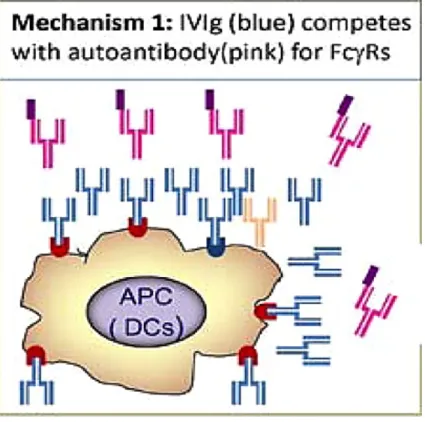
Another theory involves binding of IgG to activating FcγR or induction of FcγRIIB on APC. FcγRs are important in antibody-mediated effector functions of APCs, thus blocking of individual or all activating FcRs results in abrogation of antibody activity in triggering of inflammatory responses, as shown in a variety of autoimmune models such as ITP, nephrotoxic nephritis (NTN) and arthritis (Anthony et al., 2008b; Clynes and Ravetch, 1995) (Fig. 5).
FcγR-dependent actions of IVIg appear to involve APC (i.e. DC, macrophages and B cells). Activating and inhibitory FcγRs set a threshold for immune effector cell activation (Desai et al., 2007). While the activating FcγRs contribute to immune complex-mediated autoimmunity (Diaz de Stahl et al., 2002), FcγRIIB is the only inhibitory FcγR that contributes to the immune tolerance and regulates APCs activation (Fig. 6). A disturbed balance of activating and inhibitory FcγR has been implicated in the pathogenesis of many autoimmune diseases. In agreement with this concept, induction of FcγRIIB overexpression on B cells raises the threshold for B cell activation, which results in suppression of autoimmune disease, such as Lupus, despite the heterogeneity of factors influencing disease susceptibility (Svenson et al., 1998). IVIg has been shown to induce enhanced expression of FcγRIIB on macrophages and B cells, thereby increasing the threshold of immune complex-mediated cell activation.
Figure 5. Natural immunoglobulins non-specifically block the FcRs by their Fc fragments. Adopted from (Chong and Chong, 2010).
Fc fragments of immunoglobulin bind to activating FcγRs on APC, and reduce the clearance of auto-antigens coated with autoantibodies, abrogating the associated inflammatory responses initiated upon the ligation of FcγRs.
Siragam et al. (Siragam et al., 2005) and Bazin et al. (Bazin et al., 2006) proposed in separate studies that artificial immune complexes can confer protection to mice with ITP, comparable to IVIg, dependent on activating FcγRIII. They also showed that IgG molecules do not interact with inhibitory FcγRIIB (Siragam et al., 2006). IVIg-primed leukocytes from wild type mice could confer protection to mice with ITP, whereas IVIg-primed leukocytes from FcRγ chain–deficient mice could not. In another study, the effectiveness of immune complexes in ameliorating ITP was assessed, by generating IgG specific to a soluble antigen (OVA). Immune complexes of OVA/anti-OVA Ab recapitulated the protective effects of IVIg in ITP as well as in the K/BxN serum-induced arthritis model, through a mechanism dependent on FcγRIIB (Siragam et al., 2005). Moreover, in separate studies it was demonstrated that inhibition of FcγRIIB receptors, either by genetic deletion or with blocking antibodies, inhibits the protective effect of IVIg in ITP (Crow et al., 2003; Jee et al., 2011).
IVIg contains monomeric and dimeric IgG populations which are in a dynamic equilibrium depending on concentration, pH, temperature, donor pool size and stabilizers added to IVIg (Wymann et al., 2008). The frequency of dimeric IgG molecules within IVIg is usually at a very low level. Moreover, IVIg preparations are acid treated and stabilized at low pH routinely in order to dissociate the dimeric IgG (section 2). In addition high-performance liquid chromatography-purified IVIg monomers do not have significant protective effects in the murine model of ITP (Siragam et al., 2005). Monomeric antibodies in IVIg are not able to compete with pathogenic immune-complexes for binding to activating FcγRs because of their low affinity for these receptors (Nimmerjahn and Ravetch, 2007b). Thus it is unclear if the anti-inflammatory effect of IVIg is attributed to multimeric IgG molecules.
In addition, conflicting results have been found regarding the requirement of inhibitory FcγRIIB for the immune-regulatory action of IVIg. FcγRIIB was not reported to be the physical target of IgG in IVIg (Samuelsson et al., 2001; Siragam et al., 2006; Zhuang et al., 2002). IVIg can abrogate B-cell responses without direct interaction with FcγRIIB (Zhuang et al., 2002). Inhibition of human tonsillar B cell proliferation and immunoglobulin production, in response to IL-4 and CD40 stimulation, was not inhibited by IVIg when cells were co-incubated with both IVIg and antibodies against FcγRIIB, suggesting that FcγRIIB is not targeted by IVIg. In addition, adoptive transfer of IVIg-primed FcγRIIB deficient DC to mice with ITP led to the improvement of disease severity in recipients (Siragam et al., 2006). Expression of FcγRIIB was required by the recipient mice to achieve anti-inflammatory activity. This suggests that although FcγRIIB is not itself the physical target of IVIg, it plays a role in the downstream phase of IVIg’s function.
Aubin et al. (Aubin et al., 2010) have demonstrated that IVIg can inhibit antigen presentation to the same degree in bone marrow-derived DC from both wild-type and FcγRIIB-deficient mice. The overexpression of FcγRIIB on macrophages is not sufficient to achieve anti-inflammatory activity comparable to IVIg in murine models of arthritis and systemic lupus erythematosus (Baerenwaldt and Nimmerjahn, 2008; Diaz de Stahl et al., 2002). A recent study also indicated that there were strain-specific responses to IVIg. Leontyev et al. (Leontyev et al., 2012) demonstrated that both FcγRIIB-deficient and wild-type BALB/c recover from ITP with similar kinetics after IVIg treatment; while, this was not the case for C57BL/6 FcγRIIB-deficient mice. Thus, genetic factors might be important for the responsiveness to IVIg therapy.
Anthony et al. (Anthony et al., 2011a) recently proposed, using the K/BxN model of serum induced arthritis, that IVIg interacts with a C-type lectin immunorecetor (CLR). SIGN-R1 is expressed on the surface of marginal zone macrophages, and interaction with IVIg leads to the production of IL-33 by those cells and subsequently inducing production of IL-4 by basophils. IL-4 promotes increased expression of inhibitory Fc receptors (FcγRIIB) on effector APC (Fig. 6). This will be further elaborated in section 4-2-3-2.
Taken together, the inhibitory actions of IVIg on APC may involve interaction of IgG molecules with activating FcγRs, or unconventional FcγRs, resulting in induction of inhibitory signaling (Fig.6). Up-regulation of FcγRIIB on effector APCs may also contribute to the action of IVIg.
Figure 6. IVIg induces the up-regulation of FcγΙΙΒ expression and the associated ITIM
signaling pathway.
IVIg may induce surface expression of FcγRIIB on splenic macrophages and other APCs. Modulation of inhibitory (FcγRIIB) signaling is a potent mechanism for attenuating autoantibody-triggered inflammatory diseases. Upregulation of FcγRIIB is believed to be the result of interaction of IgG with activating FcγRs or certain c-type lectin receptors.
4-2-3-2- The immune-regulatory activity of sialylated-IgG
Recently work examining the glycosylation structure of IgG has led to fractionation of IVIg based on glycosylation sites. Glycosylation of IgG can determine the affinity and binding characteristics of IgG for FcγRs (Anthony et al., 2012). For example, IVIg can be separated into extensively sialylated and nonsialylated IgG fractions. The sialylated fraction (SA-IVIg), comprising only 5% or less of the IgG pool, appears to contribute importantly to the anti-inflammatory activity. Oligosaccharide residues present on the Fc of normal polyclonal IgG can attach at Asn 297, and sialic acid can be linked to the oligosaccharide chain either via a α2,3 or α2,6 linkage. Recent studies showed that α2,3 sialylation provides a better protection to the protein than α2,6 sialylation (Fig. 7).
In murine models of inflammatory arthritis, administration of high-dose IVIg protects mice from developing joint inflammation. Pretreated of IVIg with neuraminidase, removing the terminal sialic acid residues, abrogates the expected anti-inflammatory activity. Conversely, sialic acid– enriched IVIg preparation showed equivalent protection at one-tenth the dose of unfractionated IVIg (Anthony et al., 2008a).
Figure 7. Schematic structure and glycosylation of IgG. Adopted from (Scanlan et al., 2008)
The two conserved N-linked carbohydrate chains of IgG are attached to Asn-297 of the Cγ2 domains of each IgG heavy chain. A sialylated biantennary glycan is shown, with nonreducing sialic acid residues modeled according to likely linkage constraints. A range of other glycoforms also exists on different subsets of IgG.
Anthony et al. (Anthony et al., 2011a; Anthony and Ravetch, 2010; Anthony et al., 2008b) demonstrated that SIGN-R1, a C-type lectin receptor on murine splenic marginal zone macrophages, recognizes and can mediate anti-inflammatory effects of SA-IVIg in a murine model of serum-induced arthritis. They also demonstrated that sialylated-Fc fragments can bind to DC-SIGN, the human orthologue of SIGN-R1, with the similar affinity, that results in triggering of an inhibitory response in the ligated cells. It is important to note that DC-SIGN and SIGNR1 differ significantly in their cellular and tissue distribution and DC-SIGN is expressed particularly on human dendritic cells rather splenic or intestine macrophages.
Kaneko et al. (Kaneko et al., 2006) demonstrated that between 1 to 5% of IgG molecules within IVIg contain sialic acid moieties at the Asn297. They proposed that Fc-sialylated IgGs bind to a unique receptor on macrophages or other APCs and this interaction leads to the up-regulation of inhibitory FcγRIIB (Fig. 6). In addition to SIGNR1, SA-IVIg interacts with other C-type lectin receptors, such as CD22 (Siglec-2), which modulates B cell receptor signaling and promotes apoptosis in mature human B lymphocytes (Seite et al., 2010).
In a recent study sialic acid-enriched IgG was biochemically and functionally characterized. It was shown that the sialylation of IgG is not restricted to the Fc portion; high levels of sialic acid residues were mainly found on the Fab fragments. ex vivo LPS or PHA-stimulated human monocytes treated with enriched sialylated-Fab exhibited reduced CD54 expression and reduced secretion of MCP-1 (CCL2) (Kasermann et al., 2012). These data suggest that the anti-inflammatory activity of IVIg may be dependent on sialylation sites on both Fab and Fc.
The potential importance of sialylation IgG was underscored by evidence that the balance of sialylated vs. de-sialylated IgG influences autoimmune conditions. For example, in patients with