Adolescent idiopathic scoliosis associated
POC5 mutation impairs cell cycle, cilia length
and centrosome protein interactions
Amani Hassan1,2, Stefan Parent2, He´lène Mathieu2, Charlotte Zaouter2,
Sirinart Molidperee2, Edward T. Bagu3, Soraya Barchi2, Isabelle Villemure4, Shunmoogum A. Patten5, Florina MoldovanID1,2*
1 Faculty of Dentistry, Universite´ de Montre´al, Montre´al, Que´bec, Canada, 2 CHU Sainte-Justine Research Center, Montre´al, Que´bec, Canada, 3 Department of Basic Biomedical Sciences, Sanford Medical School, University of South Dakota, Vermillion, SD, United States of America, 4 E´ cole Polytechnique de Montre´al, Montre´al, Que´bec, Canada, 5 INRS–Institut Armand-Frappier, Universite´ du Que´bec, Laval, Montre´al, Que´ bec, Canada
*florina.moldovan@umontreal.ca
Abstract
Adolescent Idiopathic Scoliosis (AIS) is a spinal deformity that affects approximately 3 per-cent of human adolesper-cents. Although the etiology and molecular basis of AIS is unclear, several genes such as POC5 have been identified as possible causes of the condition. In order to understand the role of POC5 in the pathogenesis of AIS, we investigated the sub-cellular localization of POC5 in cilia of cells over-expressing either the wild type (wt) or an AIS-related POC5 variant POC5A429V. Mutation of POC5 was found to alter its subcellular localization and to induce ciliary retraction. Furthermore, we observed an impaired cell-cycle progression with the accumulation of cells in the S-phase in cells expressing
POC5A429V. Using immunoprecipitation coupled to mass spectrometry, we identified spe-cific protein interaction partners of POC5, most of which were components of cilia and cyto-skeleton. Several of these interactions were altered upon mutation of POC5. Altogether, our results demonstrate major cellular alterations, disturbances in centrosome protein interac-tions and cilia retraction in cells expressing an AIS-related POC5 mutation. Our study sug-gests that defects in centrosomes and cilia may underlie AIS pathogenesis.
Introduction
At present, the etiology and biological mechanisms that are involved in the pathogenesis of adolescent idiopathic scoliosis (AIS) remain unclear. Several etiologies and pathways such as neuroendocrine, neurological, muscular, biochemical and structural, hormonal, mechanical and genetic have been suggested to contribute to AIS [1–4]. We previously reported that muta-tions in the centrosomal protein-encoding genePOC5 are associated with familial idiopathic
scoliosis in French Canadian families [5]. The involvement ofPOC5 in AIS was further
con-firmed in a case-control study, where thePOC5 variant (rs6892146) was found to be associated
in individuals with AIS [6].
a1111111111 a1111111111 a1111111111 a1111111111 a1111111111 OPEN ACCESS
Citation: Hassan A, Parent S, Mathieu H, Zaouter
C, Molidperee S, Bagu ET, et al. (2019) Adolescent idiopathic scoliosis associated POC5 mutation impairs cell cycle, cilia length and centrosome protein interactions. PLoS ONE 14(3): e0213269.
https://doi.org/10.1371/journal.pone.0213269
Editor: Hemant Khanna, University of
Massachusetts Medical School, UNITED STATES
Received: July 23, 2018 Accepted: February 19, 2019 Published: March 7, 2019
Copyright:© 2019 Hassan et al. This is an open access article distributed under the terms of the
Creative Commons Attribution License, which permits unrestricted use, distribution, and reproduction in any medium, provided the original author and source are credited.
Data Availability Statement: All relevant data are
within the paper and its Supporting Information files.
Funding: This work was supported by: 1. Institut
de France (FR), Yves Cotrel Foundation to FM; 2. fonds de Recherche du Que´bec - Sante´ (FRQS) to FM; 3. Re´seau de Recherche en Sante´
Buccodentaire et Osseuse (RSBO) to FM; 4. Gouvernement du Canada | Canadian Institutes of Health Research (CIHR) to FM; 5. Scoliosis Research Society (SRS) to FM.
In humans, thePOC5 gene is on chromosome 5q13 and encodes an ubiquitously expressed
protein, abundant in the centrioles where it interacts with centrin and inversin [7]. POC5 is essential for assembling the distal half of the centriole and the elongation of the centrioles [7]. It is also involved in cell functions such as cell polarity, division, motility, and forms part of the cell cytoskeleton that is important for cell dynamics [7–9]. The localization of POC5 within photoreceptors is crucial for ciliary connection and retinal function [10].
Cilia are organelles that extend from the cellular surface of most eukaryotic cells [11]. There are two types of cilia, motile and nonmotile cilium, the latter is also known as primary cilium. Motile cilia are composed of a 9+2 axonemal structure with nine outer microtubule doublets surrounding two centrally located singlet microtubules, and additional accessory structures [10]. Primary cilium are found in almost all eukaryotic cells and are characterized by their 9+0 axoneme organization. They sense and transduce environmental signal and are critical for embryonic and postnatal development, as well as for tissue homeostasis in adulthood [12]. Due to their broad tissue distribution, defects in primary cilia will result in to a broad range of ciliopathies characterized by phenotypic variability and clinical features ranging from renal, retinal, hepatic, musculoskeletal and central nervous system defects [13–16]. Cilia abnormali-ties were recently associated with scoliosis and defects in the central nervous system [17]. For instance, in zebrafish, mutation of the protein-tyrosine kinase-7 was shown to affect the forma-tion and funcforma-tion of motile cilia in the central nervous system [17] suggested that the ciliary abnormalities caused a disturbance in the flow of cerebrospinal fluid (CSF) leading into spinal curvature. Given the roles of centrosomal proteins in ciliogenesis [18], it is very likely that mutations in POC5 would impact cilia function. However, this hypothesis remains to be explored.
In this study, we investigated the impact of mutations inPOC5 on primary cilia and the
subsequent implications in the pathogenesis of AIS. We show that an AIS-related mutation in POC5 induce ciliary retraction and impair cell-cycle. We further demonstrate that mutated POC5 loses its ability to interact with proteins that are important for cilia function as well as cytoskeleton organizations.
Materials and methods
Ethical considerations
All human tissue samples were collected in accordance with the policies regarding the ethical use of human tissues for research. The protocol used in this study was approved by the Centre hospitalier universitaire Sainte-Justine Ethics Committee (# 3704).
Cellular localization of POC5
All cells used in this study were cultured in DMEM media (Wisent cat: 319-015-CL) in an eight-well-chamber glass slide (Fisher scientific cat: 354108). HeLa cells were transfected with either Myc tagged wt-POC5 (Origene cat: RC211731) or POC5A429V, (generated by site directed mutagenesis [5]). Following, a 24 hours period post transfection with lipofectamine (Invitrogen, cat: 11668–019), the cells were processed for immunocytochemistry analysis. Briefly, the cells were fixed on ice using 70% ethanol and 0.2% triton, permeabilized with 0.1% triton in PBS (PBT), and then incubated for one hour at room temperature (RT), with rabbit polyclonal POC5 antibody (abcam cat: ab188330, 1/250) and mouse monoclonal acetylated-α-tubulin antibody (Sigma Aldrich cat: T7451 1/2000) diluted in 2% BSA/PBT. After incubation with the primary antibodies, the cells were washed three times with PBT, incubated for one hour at room temperature with secondary antibodies (1/500) Alexa fluor 488 anti-rabbit (life technologies cat: A11008) and Alexa fluor 555 anti-mouse (Life technologies cat: A21422).
Competing interests: The authors have declared
Samples were then mounted using prolong gold anti-fade reagent with diamidino-2-phenylin-dole dye for fixed cells (DAPI) (Life technologies cat: P36931). Confocal fluorescent images were captured using a Zeiss microscope, Z-stack digital imaging was performed in order to reveal more details of the position of POC5 with respect to cilia. The Z-stack technique com-bines multiple images taken at different focal distances to give a subsequent image with a greater depth of field. Acetylated-α- tubulin (a ciliary marker), POC5 protein and DAPI (the nuclear counterstain) were visualised as red, green, and blue, respectively.
To analyze expression and cellular localization of POC5 at different phases of the cell cycle, HeLa cells were cultured in DMEM Medium (Wisent cat: 319-015-CL) with 10% FBS (Wisent cat: 080–110) and 1% penicillin streptomycin Glutamine (Wisent cat: 50-202-EL) (PSG). When the HeLa cells were confluent, they were serum starved for 24 hours in order to syn-chronize their cell cycle. Serum starvation was performed by culturing confluent cells in DMEM medium without FBS, supplied with 1% PSG to block in G1 phase. To block cells in S phase, cells were then supplied with DMEM medium with FBS (10%) and 1% PSG for 24 hours. Cells were then examined by double immunofluorescence as described above. Immu-nofluorescence studies were also performed on normal human osteoblasts and cells carrying
POC5 variant mutation c.C1286T (p.A429V). Tissue samples were collected for mutation
anal-ysis of the osteoblasts from patients with scoliosis during surgery. Genomic DNA was ex-tracted from cells using pure link genomic DNA mini kit (cat: k 1820–01). Polymerase chain reaction was performed for exon 10 using primers: Forward: 5’CTTTTCATAAGGTGGGACC T3’ Reverse: 5’TCCGATGCCCTTACCAG3’. Bands corresponding to the correct molecular weight ofPOC5 were excised from gel and purified using GenElute Gel extraction kit (Cat:
NA1111-1KT). The purified DNA amplicons were then sequenced (University McGill and Ge´nome Que´bec Innovation Centre, Montre´al QC).
Preparation of nuclear and cytoplasmic extracts
Cells were washed with 4 ml of PBS (1x) and then harvested in PBS by scraping. The lysate was collected in eppendorf tube and spun for 5 minutes in a microcentrifuge at low speed (4000 rpm). Pellet was washed twice with 500μl of buffer A (Hepes 10mM, KCl 10mM, DTT 0.5mM)(w/o NP-40) and then cell pellet was resuspended in 20μl of buffer A (w/ NP-40 (1%)) and incubated at 4˚C for 10 minutes with rocking. The cell pellet was spun down in microcen-trifuge for 2 minutes at max speed. The supernatant is the cytoplasmic extract (C extract) by adding 55μl of buffer C and stored at –80˚C. The nuclear extract was resuspended in 55 μl of buffer C (Hepes 20 mM, glycerol 20%, KCl 500 mM, EDTA 0.2 mM, PMSF 0.5 mM, DTT 0.5 mM and MgCl2 1.5mM); and incubated at 4˚C for 15 minutes with rocking. The nuclei were then spun in microcentrifuge for 5–10 minutes at max speed. At this stage, the supernatant is the nuclear extract (N extract).
Immunoprecipitation coupled to mass spectrometry (IP–MS),
co-Immunoprecipitation and Western blot
To identify specific protein-protein interaction partners of POC5, immunoprecipitation cou-pled to mass spectrometry, co-immunoprecipitation, and Western Blot analysis was performed on HEK293 cells transfected with wt orPOC5A429Vexpressing vectors. HEK293 cells were cul-tured in DMEM medium (Wisent cat: 319-015-CL) with 10% FBS and 1% PSG. When conflu-ent, cells were transfected using lipofectamine 2000 (Invitrogen cat: 11668027) with control (mock-myc empty vector), Myc tagged wt orPOC5A429Vexpressing vectors. For transfection, cells were maintained in antibiotic free medium overnight. On the second day, cells were transfected based on the lipofectamine protocol provided by the manufacturer (Invitrogen).
For protein extraction, cells were lysed in IP lysis buffer (Pierce Thermoscientific, cat: 87787) supplied with 1 x protease inhibitor cocktail (Roche cat: 04693116001). Lysate was then centri-fuged for 15 minutes at 8000 rpm. Supernatant was collected for protein quantification by Bradford (Bradford Biorad cat: 500–0006). For immunoprecipitation, 3000μg of proteins were immunoprecipitated with 2.5μg/ml of anti-myc antibody (Origene, cat: TA15021) overnight (ON) at 4˚C. Magnetic beads (Biorad cat: 161–4023) were washed with PBS and blocked using 2% BSA/PBS (BSA from Sigma-Aldrich cat: A7906) ON at 4˚C. On the second day, beads were washed 3 x with PBS and then incubated with protein lysate and antibody complex at 4˚C ON. On the third day, the flow through was collected and the beads were heated for 10 minutes in laemmeli 2 x (biorad cat: 161–0737) at 55˚C. Protein samples and dual plus molecular weight ladders (Bio-rad cat: 161–0374) were separated by SDS-PAGE approximately 90 minutes at 100 Volts in running buffer (25 mM Tris base, 192 mM glycine, 1% SDS, pH 8.3). The gel was stained with Coommasie blue (Biorad cat G-250 #1610406) for 3 hours. After staining the gel with Coomassie blue, the bands corresponding to the molecular size of POC5 (63 kDa) were excised and analyzed by mass spectroscopy. The gels were subjected to trypsin digestion then an aliquot of the tryptic digest (prepared in 5% acetonitrile/ 0.1% trifluoroacetic acid in water) was analyzed by LC-MS on an LTQ-Orbitrap mass spectrometer system (ThermoElectron) coupled to a Dionex 3000 nano-LC system (Camberley). Mass spectroscopy analysis was evalu-ated using scaffold software [19]. Mass Spectroscopy was performed at the Mass spectroscopy plateform at the Institute of Research in Immunology and Cancer (IRIC).
In parallel, an aliquot of protein samples was transferred to nitrocellulose membranes (Bio-Rad cat: 9004700) and ran 90 min at 90 V and 250 mA. The total proteins on membranes were detected with Ponceau S staining (Sigma cat: P3504). Membranes were blocked with 20% non-fat milk (Santa-Cruz cat: Sc2324) in PBST (10 mM phosphate, 137 mM NaCl, 2.7 mM KCl, containing 0.05% Tween-20, pH 7.4) for 1 hour and then incubated with anti-POC5 antibody in PBST with 5% BSA in PBT at 4˚C ON. The secondary antibody was (anti-rabbit IgG sec-ondary antibody (Thermoscientific cat: 31460) diluted at 1: 10000 for one hr at RT. In order to validate the mass spectroscopy data, and confirm the interaction of several ciliary proteins with POC5, Western blot was performed by stripping the same membrane with a Restore stripping buffer (Thermofisher cat: 21059) and probing it with different antibodies. The fol-lowing antibodies were used Annexin2 (374394), Galectin 3 and 7 (32790 and sc-137085), CKAP5 374394), Desmocollin1 398590), CEP290 (ab84870), Septin9 (sc-293291), Acetylated-α-tubulin (Sigma Aldrich cat# T7451 1/2000), RAB11 (ab3612), EHD4 (sc-376373), Annexin5 (sc-32321), and CystatinA (sc-376759). All antibodies acquired from Santa Cruz (Sc) were mouse monoclonal and used at a dilution of 1/500. Rabbit polyclonal antibodies CEP290 and Rab11 were used at a dilution of 1/250. The secondary antibody (rabbit IgG secondary antibody (Thermoscientific cat: 31460) was diluted 1:10000, and anti-mouse IgG secondary antibody (Thermoscientific cat: 31430) was diluted 1:10000 for 1 hour at RT. Membranes were exposed to ECL prime Western blotting detection reagent (GE health-care cat: RPN2232) for 5 minutes at RT room.
Immunoprecipitation phosphatase assay
HEK293 cells were transfected with wtPOC5 myc tagged orPOC5A429Vmyc tagged vectors (as described above) and then were synchronized at G1 or S phases. To block cells at G1 phase, cells were serum starved for 24 hours. To block cells at S phase, cells were first starved for 24 hours and then complete medium with serum (10% FBS) was added for 24 hours. For immu-noprecipitation-phosphatase assays, total lysates were incubated with 2.5μg anti-myc antibody as described above. Beads were blocked with 1% BSA and then washed three times and
resuspended in 100μl PBS. Then, protein antibody complex were incubated with beads. Alka-line phosphatase (NEB cat: M0290S) was added to the mixture and incubated at 37˚C for 1 hour. Western blot was performed using POC5 antibody (Abcam cat: 188330) as described above.
Quantification and statistical analysis
The length of cilia was quantified using ImageJ by measuring the acetylated-α-tubulin signals [20]. For ciliation experiments with NOB and AIS cells, approximatly 300 cilia were counted per experiment. For cell cycle analysis, the number of cells in S phase and G1 phase were counted. The percentage of cells in each phase over the number of total cells was determined. Approximately 100 cells were counted per phase. Mean values of individual experiments were plotted in bar graphs with±SD between the individual sets. P-values were calculated by one-way analysis of variance (ANOVA). P<0.05 considered statistically significant.
Results
Altered subcellular localization of POC5 upon mutation
We first examined the subcellular distribution of wild-type humanPOC5 (wtPOC5) and an
AIS-related mutantPOC5A429V(mutPOC5) in HeLa cells. HeLa cells are p53-defective cell
lines and hence they do not arrest at G1 phase after the disruption of centrosomal proteins, unlike non-transformed cells that are arrested at G1 through p53 dependent pathway upon depletion of centrosomal proteins. The centriolar cycle of HeLa cells is well described [21,22]. For HeLa cell to progress through its life cycle, centriolar replication and pericentriolar changes occurs in synchrony with DNA synthesis and mitosis. Since POC5 localization and function has been well characterized in HeLa cells [7], we sought to first investigate the cellular effects of POC5 mutations in the HeLA cells.
In wtPOC5 cells, POC5 was found to localize to the acetylated-α-tubulin ring (Fig 1A). On the other hand, POC5 was mislocalized inPOC5A429Vcells (Fig 1B).
Aberrant POC5 localization in osteoblasts derived from AIS patients
bearing the
POC5
A429Vmutation
To confirm our observation in HeLa cells, we next investigated the effects ofPOC5 mutation
on the subcellular localization by comparing normal (non-scoliotic) to osteoblasts derived from AIS patients carrying thePOC5A429Vvariant (Fig 2). In normal control osteoblasts, POC5 expression was colocalized with acetylated-α-tubulin stainings, suggesting that centrio-lar protein POC5 is located at the cilium (Fig 2A–2D). In AIS osteoblasts, POC5 was rather located within the nucleus and not colocalized with acetylated-α-tubulin stainings (Fig 2E– 2H). Furthermore, a retraction of cilium was observed inPOC5A429Vosteoblasts (Fig 2H) com-pared to controls. More precisely, quantification of the ciliary length showed thatPOC5A429V
cells predominantly had shorter cilium of more than 3μm than that of control cells (Fig 2I). Altogether, these results suggest that mutations in POC5 affect its subcellular localization with respect to the cilium.
Defects in cell-cycle progression in cells overexpressing the mut
POC5
Because POC5 is localized to the distal portion of centrioles and is recruited to procentrioles for full centriolar maturation and normal cell-cycle progression, we next determined howPOC5A429Vprotein is localized during cell cycle progression. HeLa cells transfected with either wt orPOC5A429Vwere synchronized in the growth phase (G1) cell cycle phase and then stained
Fig 1. Differential localization of POC5 with acetylated-α-tubulin in wtPOC5 and POC5A429Vexpressing cells.
Confocal imaging of HeLa cells transiently transfected with wtPOC5 (a) or POC5A429V(b). POC5 and acetylated-α-tubulin were determined using specific antibodies and positive signal revealed by green and red coloration
respectively. Localization of POC5 with respect to acetylated-α-tubulin is shown: in cells transfected with wtPOC5 (a) where positive immunostaining was enriched at the perinuclear acetylated-α-tubulin ring, while in cells expressing
POC5A429V(b), POC5 was visualized inside the nucleus (disconnected from the perinuclear acetylated-α-tubulin ring). The Z-stack imaging (recording images at different focal planes) allows the visualization of the three-dimensional structure containing both POC5 and acetylated-α-tubulin (green and red) for the wtPOC5, while only POC5 (green) was visualized for thePOC5A429V. Images were taken using the Zeiss microscopy Mag x40. Scale bar 1.6μm. POC5 in green, acetylated-α-tubulin in red and DNA was stained with DAPI (blue).
https://doi.org/10.1371/journal.pone.0213269.g001
Fig 2. Human osteoblasts carryingPOC5A429Vshow short cilia. Representative immunodetection of POC5 and acetylated-α-tubulin by immunofluorescence in osteoblasts from normal (non scoliotic) (a, b, c, d) and cells with thePOC5A429V(e, f, g, h). Merged images (d and h) show differential colocalization of POC5 with respect to cilia. Zoomed image shows that POC5 is located at the cilium level marked by acetylated-α-tubulin. Human osteoblasts carrying the variant
POC5A429V(e, f, g, h), show decreased staining intensity and absence or retraction of cilium (h).POC5A429Vprotein was seen to be localized within the nucleus (zoomed image). POC5 (green coloration), acetylated-α-tubulin (red coloration) and the nucleus were stained with DAPI (blue). Images were taken using the Zeiss microscopy. Mag x 40. Scale bar 1.6μm. (I) The graphs represent the percentage of cilia for each length category in NOB and AIS cells (POC5A429V). Each bar represents the mean of three independent experiments (±SD). P< 0.05 considered statistically significant. (J) Sequence alignment with Sanger sequencing confirm AIS cells to have thePOC5A429Vmutation.
for POC5. In cells transfected with wtPOC5, POC5 protein was observed outside the nucleus
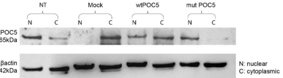
during the G1 phase (Fig 3A) and in the nucleus during the Synthesis (S)-phase (Fig 4B). How-ever, inPOC5A429Vexpressing cells, POC5 was found to be rather localized in the nucleus through the G1 and S phases (Fig 3C and 3D). Interestingly, a significant increase (4%) in the proportion of S-phase cells was detected inPOC5A429Vcells compared to wtPOC5 cells (Fig 3E). To confirm this observation, we performed cellular fractionation of cytoplasmic and nuclear extracts of cells overexpressing wt orPOC5A429V. The wtPOC5 was concentrated in both nuclear and cytoplasmic fractions with higher expression levels in the cytoplasm. Con-trary to wtPOC5, thePOC5A429Vwas exclusively found in the nucleus (Fig 4).
In order to check if the differential recruitment of wtPOC5 andPOC5A429Vvariant to the cilium is phosphorylation-dependent, we studied the pattern of phosphorylation of wt and
POC5A429Vduring the cell cycle progression. We found that thePOC5A429Vwas hyper phos-phorylated independently of the phase of the cell cycle. In all cell cycle phases, thePOC5A429V
protein had lower migration level than wtPOC5 protein and treatment with alkaline phos-phatase shifted backPOC5A429Vto an apparent molecular weight similar to wtPOC5 levels (S2 Fig).
POC5 interacting protein partners are mostly ciliary and cytoskeletal
proteins
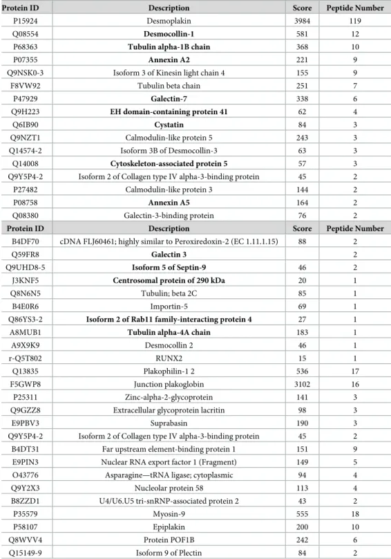
To gain a better understanding of the centrosome and cilium protein interaction landscape in AIS, mass spectrometry studies were conducted in cells transfected with either the wtPOC5 or POC5A429Vcells following myc-tagged POC5 pull-down assays. Immunoprecipitation of POC5 showed similar expression levels of POC5 in both wt andPOC5A429Vtransfected sam-ples (Fig 5A). We identified 85 proteins that interact predominantly with wtPOC5 but not
POC5A429V(Table 1) and only 5 proteins interacting withPOC5A429V(Table 2). Amongst these
Fig 3.POC5A429Vexpressing cells are arrested in S phase. Confocal imaging of HeLa cells overexpressing wtPOC5 or POC5A429Vspecific staining for POC5 was performed using POC5 antibody. During G1 phase, obtained by serum starvation (a), wtPOC5 is located within the cytoplasm, while in the S phase (b) (serum replacement after deprivation), POC5 is located within the nucleus. WtPOC5 expressing cells have normal progression through G1 and S phases, (a, b). In the cells overexpressingPOC5A429V, POC5 is located within the nucleus in both G1 and S phases (c, d). Cells are blocked in S phase and unable to progress through the cell cycle. E) The graphs represent the percentage of cells in G1 of S phase. Almost all wtPOC5 expressing cells are in G1 phase (45%), but
POC5A429Vcells are accumulating in S phase. Each bar represents the mean of three independent experiments (±SD). P<0.05 considered statistically significant. Images were taken using the Zeiss microscopy. POC5 (visualized in red), DNA was stained with DAPI (blue). Mag x 40. Scale bar 1.6μm.
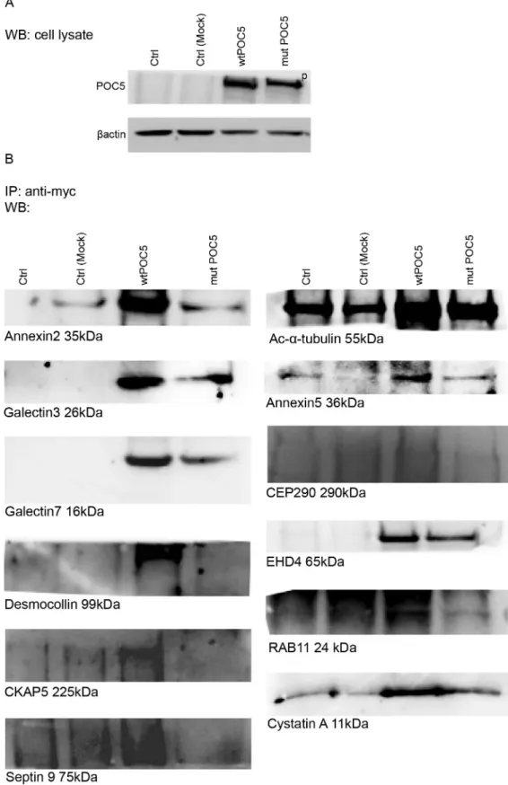
Fig 4. Validation of mass spectrometric results. Proteins identified by mass spectroscopy interacting with the
wtPOC5 were analysed by CO-IP. Coimmunoprecipitation of myc-POC5 using anti-Myc antibodies in Hek293 cells. Proteins in the immune complexes were revealed by Western blotting with different antibodies as described in materials and methods. Control (cells transfected with Mock pCMV-entry vector) was used along cells transfected with
proteins, we identified 12 ciliary and cytoskeletal proteins that have altered interaction with POC5 when mutated. For instance, Annexin2, Desmocollin1 and Centrosomal protein 290kDa (CEP290) were found to interact with wtPOC5, but not withPOC5A429V(Fig 5B). Weaker interactions between POC5 and Galectin 3, Galectin 7, Acetylated-α-tubulin, EH domain-containing protein 4 (EHD4), Annexin5 and Cystatin A were observed in cells expressingPOC5A429Vcompared to wtPOC5 (Fig 5BandS1B Fig). Protein disulfide-isomerase A4, iduronate 2-sulfatase, golgi resident protein GCP60, aminopeptidase B, cDNA FLJ53442— highly similar to poly (ADP-ribose) polymerase 1 were found a new interactors of POC5 upon mutation (Table 2). These findings suggest that an altered centrosomal and cilia protein inter-action may be involved in AIS.
myc tagged wt orPOC5A429Vexpressing cells. A) Western blot on total cell lysate using POC5 antibody. Wt and
POC5A429Vtranfected cells have same expression levels of POC5. Mock transfected sample has very low expression of POC5. POC5 is observed at the expected size 63kDa in the wtPOC5 and at higher level inPOC5A429V(indicated as p: phosphorylated). B) CO-IP shows the binding of Annexin2, Annexin 5, Desmocollin1 and CEP290, exclusively with wtPOC5. Very weak interaction was observed between POC5A429with Acetylated-α-tubulin, Galectin3 and 7, EHD4,
CystatinA compared to wtPOC5.
https://doi.org/10.1371/journal.pone.0213269.g004
Fig 5. Differential colocalization of wtPOC5 andPOC5A429Vwith respect to centrin. A) Immunoflourescence staining was performed in HeLa cells
non-transfected (NT) overexpressing pCMV-entry empty vector (mock), wtPOC5 orPOC5A429V. Staining of POC5 (red), centrin 2 (green) shows colocalization of wtPOC5 and centrin 2 (merge orange colour), and absence of colocalization withPOC5A429Vand centrin 2. B) Statistical analysis of centrin 2 expression, intensity and number of centrin foci per cell in mock, wtPOC5 and POC5A429Vexpressing HeLa cells. Percentage of centrin 2 number and intensity are reduced inPOC5A429Vexpressing cells. Unlike wtPOC5 cells, mostPOC5A429Vcells have one foci of centrin but majority of wtPOC5 expressing cells have two foci of centrin 2. Centrin count and intensity was performed using ZEN software. Images (n = 6) were used for quantification. Error bars are the mean± SD. p<0.05 considered statistically significant. POC5 (red), centrin (green) and DAPI (blue). NT: not transfected. NS: not significant.
Table 1. Mass spectroscopy results of proteins interacting exclusively with wtPOC5. Scaffold software was used for
the analysis of the identified proteins interacting with wtPOC5. Protein identification in wtPOC5 expressing cells, detected 85 candidates interacting with wtPOC5. Clustering proteins by biological function indicated: ciliary proteins (17 proteins); cell adhesion (7 proteins); cytoskeleton-associated protein (4 proteins); RNA processing (9 proteins); extracellular matrix (3 proteins); response to estrogen (1 protein); cell cycle and cytokinesis (1 protein). Most of the identified proteins interacting with POC5 are cytoskeletal proteins (As shown in the table) and those marked in bold were considered for further analysis. Other protein groups belong to cell cycle and cytokinesis, extracellular proteins, RNA processing, cell adhesion, and response to estrogen.
Protein ID Description Score Peptide Number
P15924 Desmoplakin 3984 119
Q08554 Desmocollin-1 581 12
P68363 Tubulin alpha-1B chain 368 10
P07355 Annexin A2 221 9
Q9NSK0-3 Isoform 3 of Kinesin light chain 4 155 9
F8VW92 Tubulin beta chain 251 7
P47929 Galectin-7 338 6 Q9H223 EH domain-containing protein 41 62 4 Q6IB90 Cystatin 84 3 Q9NZT1 Calmodulin-like protein 5 243 3 Q14574-2 Isoform 3B of Desmocollin-3 63 3 Q14008 Cytoskeleton-associated protein 5 57 3 Q9Y5P4-2 Isoform 2 of Collagen type IV alpha-3-binding protein 45 2
P27482 Calmodulin-like protein 3 144 2
P08758 Annexin A5 164 2
Q08380 Galectin-3-binding protein 76 2
Protein ID Description Score Peptide Number
B4DF70 cDNA FLJ60461; highly similar to Peroxiredoxin-2 (EC 1.11.1.15) 88 2
Q59FR8 Galectin 3 2
Q9UHD8-5 Isoform 5 of Septin-9 46 2
J3KNF5 Centrosomal protein of 290 kDa 20 1
Q8N6N5 Tubulin; beta 2C 85 1
B4E0R6 Importin-5 69 1
Q86YS3-2 Isoform 2 of Rab11 family-interacting protein 4 27 1
A8MUB1 Tubulin alpha-4A chain 183 1
A9X9K9 Desmocollin 2 46 1
r-Q5T802 RUNX2 15 1
Q13835 Plakophilin-1 2 536 17
F5GWP8 Junction plakoglobin 3102 16
P25311 Zinc-alpha-2-glycoprotein 141 3
Q9GZZ8 Extracellular glycoprotein lacritin 98 3
E9PBV3 Suprabasin 190 3
Q9Y5P4-2 Isoform 2 of Collagen type IV alpha-3-binding protein 45 2 B4DT31 Far upstream element-binding protein 1 151 9 E9PIN3 Nuclear RNA export factor 1 (Fragment) 149 5 O43776 Asparagine—tRNA ligase; cytoplasmic 94 4
Q9Y2X3 Nucleolar protein 58 113 4
B8ZZD1 U4/U6.U5 tri-snRNP-associated protein 2 43 2
P35579 Myosin-9 555 18
P58107 Epiplakin 200 10
Q8WVV4 Protein POF1B 242 6
Q15149-9 Isoform 9 of Plectin 84 2
WtPOC5 but not
POC5
A429Vcolocalizes with centrin
Localization of hPOC5 and centrin during the cell cycle has previously been described in liter-ature [7]. To precisely localize POC5 and to study the consequence of mutation on POC5 recruitment to cilia, we performed double immunostaining for POC5 and centrin in cells over-expressing wtPOC5 orPOC5A429V. In immunofluorescence microscopy, anti-hPOC5 antibody staining labeled two foci for wtPOC5 and one foci forPOC5A429V. These foci represent centri-oles, as confirmed by colocalization with human centrin (Fig 6A). Quantification of centrin percentage per cell revealed that there is significantly higher centrin staining intensity in wtPOC5 expressing cells thanPOC5A429Vcells and that thePOC5A429Vexpressing cells had mainly one or more than two centrin foci compared to wtPOC5 cells which had predomi-nantly 2 foci (Fig 6B).
Discussion
In this study, we demonstrated that the mutation in POC5A429V, previously described in patients with AIS [5], resulted in an altered subcellular localization of POC5 both in trans-fected and clinically-relevant patients cells. We also showed that several cilia, microtubules, cytoskeleton, and centrosomal proteins interact with POC5 and many of which are lost with an AIS-related POC5 mutation. We further found an impaired cell cycle progression and cili-ary retraction in cells expressing POC5A429Vcompared to controls (Fig 7).
In French families with AIS, various functional variants (c.G1336A (p.A446T), c.G1363C (p. A455P), and c.C1286T (p.A429V)) are contributing to the occurrence of AIS [5]. Recently, a common variant ofPOC5 was associated with the susceptibility to AIS (single-nucleotide
polymorphism (SNP) rs6892146)) in Chinese population [6]. This common SNP has a
Table 2. Mass spectroscopy results of proteins interacting exclusively with POC5A429V. Five proteins were found
to be interacting exclusively with POC5A429. Protein disulfide-isomerase A4, Iduronate 2-sulfatase, Golgi resident pro-tein GCP60, Aminopeptidase B and cDNA FLJ53442; highly similar to Poly (ADP-ribose) polymerase.
Protein ID Description Score Peptide Number
P13667 Protein disulfide-isomerase A4 226 9
P22304 Iduronate 2-sulfatase 138 5
Q9H3P7 Golgi resident protein GCP60 193 3
Q7RU04 Aminopeptidase B 95 2
B4E0E1 cDNA FLJ53442; highly similar to Poly (ADP-ribose) polymerase 1
61 2
https://doi.org/10.1371/journal.pone.0213269.t002
Fig 6. Differential subcellular localization of wtPOC5 andPOC5A429V. Nuclear and cytoplasmic cell extracts were
obtained using protocol as described in materials and methods. Equal nuclear and cytoplasmic protein samples loaded were determined usingβ-actin as loading control and subjected to immunoblotting using anti-POC5 antibody. Controls were cells non transfected or transfected with pCMV-entry vector. The wtPOC5 is mainly expressed in the cytoplasm and thePOC5A429Vis mostly nuclear. The results are from two independent experiments (n = 2).
significantly different distribution of minor allele frequency in patients with the GG and with CC genotype. Interestingly, in patients with the GG genotype, POC5 mRNA expression was found to be significantly increased when compared to the controls [6]; however, little is known about the function of POC5 and its role in AIS pathologies. ThePOC5 variant, POC5A429Vis a rare variant that was found in AIS patients [5].
In addition to the association of POC5 with AIS [5], recently, new mutations inPOC5 gene
were associated with Retinitis Pigmentosa (RP) [10]. Retinitis Pigmentosa is a photoreceptor degenerative disease, characterized by the degeneration of rod and cone photoreceptors and is classified as ciliopathy disease. Weisz et al [10] reported POC5 to be localized at the connection cilia in the photoreceptors, and when mutated, the length of photoreceptor outer segments is reduced. In ourin vitro work, in cells overexpressing POC5A429V, POC5 was not co-localized with the acetylated-α-tubulin (Fig 1B), dispatched from centrin (Fig 5) and solemnly localized in the nucleus (Fig 6). In contrast, the wtPOC5 was found to be interacting with several ciliary proteins (Table 1). We point out to a role of POC5 as a ciliary protein that is located at the base of the cilium as was confirmed by centrin staining (Fig 5).
Our immunofluorescence results showed also a differential localization of wt and
POC5A429Vwith respect to the centrin (Fig 5). Our mass spectroscopy and co-IP data (Fig 4) showed several ciliary proteins to be interacting exclusively with wtPOC5 but notPOC5A429V.
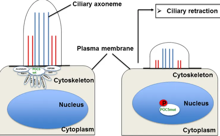
Fig 7. Proposed model for the mechanisms of ciliary retraction inPOC5A429Vexpressing cells. Under normal conditions, POC5 is an essential protein for
normal cell cycle progression, and this process is a tightly regulated mechanism. In cycling cells, with wtPOC5, POC5 protein is found interacting with several ciliary proteins that assemble before entering to G1 phase. Weak or no interaction ofPOC5A429Vwith ciliary proteins (revealed in this study by
immunofluorescence, mass spectroscopy, and CO-IP) results in incorrect assembly and cilium retraction.
Among these two proteins component of cilia were found; RAB11 and CEP290. Protein RAB11 is part of the multiple GTPases group that include RAB6, RAB11, and RAB8A which are involved in the trafficking to the cilium. Interestingly, several disorders associated with genetic mutations encoding defective proteins of cilia formation or function showed scoliosis as a secondary manifestation. Some studies have shown that the potential handover mecha-nisms may exist between RAB11 and RAB8 at the base of the cilium [23]. Rab8 and Rab11 were found to be associated with the Bardet-Beidl syndrome (BBS) pathway [24]. CEP290 is also known to be an important component of the primary cilium, localizing to the Y-links of the ciliary transition zone and having a role in the regulation of transport in and out of the cili-ary compartment [25]. Furthermore, CEP290 mutations lead to a range of ciliopathy syn-dromes with variable clinical manifestations in humans: BBS, Joubert syndrome and Meckel-Gruber syndrome [26]. Given that both proteins (POC5 and CEP290) are located at the base of the cilium (as also confirmed in this study by centrin immunostaining), and that CEP290 interacts solely with wtPOC5, this supports the role of POC5 as a ciliary protein operating at the base of the cilium.
Interestingly, two proteins previously associated with scoliosis, Annexin A2 and Calmodu-lin were found to interact with wtPOC5 were previously associated with scoliosis. Annexin A2, is highly expressed in osteoblasts, and differentiated growth plate chondrocytes and plays an essential role in bone mineralization which appears to be critical in AIS patients. Calmodulin-like protein 3 (immunoprecipitated with wtPOC5 but notPOC5A429V) is another interesting putative protein partner of POC5 in AIS. Calmodulin-like protein is a calcium sensor protein that is closely related to the ubiquitous calmodulin, which is considered potential key molecule in the etiology of scoliosis because of its effects on muscle contractility, that was reported defective in AIS pateients [27] (Table 1). Only five proteins were found to be associated with mutPOC5 protein 5. Interestingly, among those proteins is iduronate 2-sulfatase. Iduronate 2-sulfatase is associated with Hunter syndrome clinical disorder (mucopolysaccharidosis type II, MPS-II) in which patients may display, scoliosis [28,29]. Also, the cellular stress gene pro-tein disulfide isomerase (PDIA4) was found to be associated withPOC5A429V. PDIA4 is up-regulated in mouse models of brain neurodegenerative diseases involving protein misfolding. Although not much is known about the physiological role of PDIA4, studies indicate that this gene is upregulated following endoplasmic reticulum ER stress [30,31]. Another interesting observation was the hyper-phosphorylation of thePOC5A429V(S2 Fig). It is well known that posttranslational modifications are one of the ways to regulate protein activity, subcellular localization, and stability [32]. Further studies are needed to determine where exactly the phos-phate group is added and the accurate consequences on the physico-chemical properties, sta-bility, kinetics, and dynamics [32].
One of the most intriguing results of our study was the immunofluorescence on human cells carrying thePOC5 variant (c.C1286T; p.A429V mutation) showing the subcellular
mislo-calization of POC5. We observed ciliary retraction in scoliotic osteoblasts (Fig 2H) with the
POC5A429Vmutation (Fig 2H) when compared to the normal osteoblasts (Fig 2D). The pri-mary cilium is an antenna-like projection of the cell that plays a critical role in the perception and integration of environmental signals like mechanotransduction. Cilia are also essential for left-right (L-R) symmetry during embryonic development [33,34] and for cerebrospinal fluid (CSF) flow [35]. Motile cilia dysfunction may cause spinal deformity similar to the IS [17] and restoration of motile cilia activity stopped spinal curve progression, as evidenced recently in a zebrafish model [17]. This observation supports our results that show a defect in cilia organiza-tion as well as perturbed localizaorganiza-tion of POC5 with respect to the cilia in cells expressing a
POC5 mutation (Fig 2H). Another interesting observation is the defect in the cell cycle in
and a different subcellular localization of wt andPOC5A429Vat different stages of the cell cycle was observed. Not only the cellular localization of POC5 but also the number of centrin foci were used to confirm the accumulation ofPOC5A429Vin S phase (>2 foci of centrin) [7,36]. ThePOC5A429Vwas permanently located within the nucleus in G1 and S phases (Fig 3C and 3D). By cellular fractionation, we confirmed the different subcellular localization of wt and
POC5A429(Fig 6). Previous work [7] also showed that a depletion of POC5 in HeLa cells affects the progression through S phase. Human h-POC5-depleted cells had a significant increase in the proportion of S-phase cells; thus, hPOC5 depletion induced an accumulation of cells in S phase where procentriole assembly was probably initiated; however, these procentrioles failed to elongate. The cilia and neurosensory component of this pathology merits further investiga-tion through various funcinvestiga-tional tests in patients with AIS in order to evaluate the possible functional defect connected with the altered structures identified in this work. Further study, focused on the primary cilia-mediated function, will provide more insight into the molecular mechanisms and etiology of AIS.
Conclusion
This work identified specific protein interaction partners of POC5 and revealed that
POC5A429Valters the interaction with several ciliary and cytoskeletal proteins. Improved inter-action ofPOC5A429Vwith ciliary proteins resulted in the loss of POC5 co-localization with acetylated-α-tubulin, impaired cell cycle progression and cilium retraction These findings open new avenues for the understanding the role of POC5 in AIS at the molecular and cellular levels and suggest that centrosomes and cilia may underly AIS pathogenesis
Supporting information
S1 Fig. Acetylated-α-tubulin is highly enriched in precipitated wtPOC5 lysate but not POC5A429V. Hek293 cells were transfected with empty pCMV entry vector, wtPOC5 or
POC5A429Vmyc tagged vectors. Immunoprecipitation of POC5 was performed using myc anti-body (origene). A) Western blot of acetylated-α-tubulin after immunoprecipitation of POC5, shows high expression of acetylated-α-tubulin in the wtPOC5 expressing sample and lower levels inPOC5A429V. Very low levels are observed in mock transfected sample. B) Coommasie blue staining shows similar levels of POC5 expression in wt andPOC5A429Vtransfected sam-ples. POC5 is observed at the expected size 63kDa. Also the heavy and light chains of antibody are observed.
(TIF)
S2 Fig. Phosphorylation state of wtPOC5 andPOC5A429Vat G1 and S phases of cell cycle. The immunoprecipitated samples were either non treated or treated with alkaline phosphatase and then western blot was performed using POC5 antibody (abcam). The presence or absence of phosphorylation with wtPOC5 andPOC5A429Vis shown at G1 and S phase. POC5 wt is not phosphorylated, but thePOC5A429Vis phosphorylated, and treatment with phosphatase dephosphorylatesPOC5A429Vthat returns back to the same levels of wtPOC5. Phosphorylation of mutPOC5 is seen at both G1 and S phases.
(TIF)
Acknowledgments
Proteomics analyses were performed by the Proteomics Platform of the Institute of Research in immunology and Cancer (IRIC) in Montreal, Canada.
Author Contributions
Funding acquisition: Florina Moldovan. Investigation: Florina Moldovan.
Methodology: Amani Hassan, Charlotte Zaouter, Sirinart Molidperee, Shunmoogum A. Pat-ten, Florina Moldovan.
Project administration: Florina Moldovan. Resources: Florina Moldovan.
Supervision: Isabelle Villemure, Florina Moldovan. Validation: Amani Hassan.
Writing – original draft: Amani Hassan, He´lène Mathieu, Charlotte Zaouter, Sirinart Molid-peree, Edward T. Bagu, Soraya Barchi, Isabelle Villemure, Shunmoogum A. Patten, Florina Moldovan.
Writing – review & editing: Stefan Parent, He´lène Mathieu, Charlotte Zaouter, Edward T. Bagu, Soraya Barchi, Isabelle Villemure, Shunmoogum A. Patten, Florina Moldovan.
References
1. Parent S, Newton PO, Wenger DR. Adolescent idiopathic scoliosis: etiology, anatomy, natural history, and bracing. Instr Course Lect. 2005; 54:529–36. PMID:15948477.
2. Fadzan M, Bettany-Saltikov J. Etiological Theories of Adolescent Idiopathic Scoliosis: Past and Pres-ent. Open Orthop J. 2017; 11:1466–89.https://doi.org/10.2174/1874325001711011466PMID:
29399224; PubMed Central PMCID: PMCPMC5759107.
3. Burwell RG, Clark EM, Dangerfield PH, Moulton A. Adolescent idiopathic scoliosis (AIS): a multifactorial cascade concept for pathogenesis and embryonic origin. Scoliosis Spinal Disord. 2016; 11:8.https:// doi.org/10.1186/s13013-016-0063-1PMID:27252984; PubMed Central PMCID: PMCPMC4888516. 4. Adamson AD, Friedrichsen S, Semprini S, Harper CV, Mullins JJ, White MR, et al. Human prolactin
gene promoter regulation by estrogen: convergence with tumor necrosis factor-alpha signaling. Endo-crinology. 2008; 149(2):687–94.https://doi.org/10.1210/en.2007-1066PMID:18006630; PubMed Cen-tral PMCID: PMCPMC2342177.
5. Patten SA, Margaritte-Jeannin P, Bernard JC, Alix E, Labalme A, Besson A, et al. Functional variants of POC5 identified in patients with idiopathic scoliosis. J Clin Invest. 2015; 125(3):1124–8.https://doi.org/ 10.1172/JCI77262PMID:25642776; PubMed Central PMCID: PMCPMC4362221.
6. Xu L, Sheng F, Xia C, Li Y, Feng Z, Qiu Y, et al. Common variant of POC5 is associated with the sus-ceptibility of adolescent idiopathic scoliosis. Spine (Phila Pa 1976). 2017.https://doi.org/10.1097/BRS. 0000000000002490PMID:29189569.
7. Azimzadeh J, Hergert P, Delouvee A, Euteneuer U, Formstecher E, Khodjakov A, et al. hPOC5 is a cen-trin-binding protein required for assembly of full-length centrioles. J Cell Biol. 2009; 185(1):101–14.
https://doi.org/10.1083/jcb.200808082PMID:19349582; PubMed Central PMCID: PMCPMC2700515. 8. Dantas TJ, Daly OM, Conroy PC, Tomas M, Wang Y, Lalor P, et al. Calcium-binding capacity of centrin2
is required for linear POC5 assembly but not for nucleotide excision repair. PLoS One. 2013; 8(7): e68487.https://doi.org/10.1371/journal.pone.0068487PMID:23844208; PubMed Central PMCID: PMCPMC3699651.
9. Chang CW, Hsu WB, Tsai JJ, Tang CJ, Tang TK. CEP295 interacts with microtubules and is required for centriole elongation. J Cell Sci. 2016; 129(13):2501–13.https://doi.org/10.1242/jcs.186338PMID:
27185865; PubMed Central PMCID: PMCPMC4958302.
10. Weisz Hubshman M, Broekman S, van Wijk E, Cremers F, Abu-Diab A, Samer K, et al. Whole-exome sequencing reveals POC5 as a novel gene associated with autosomal recessive retinitis pigmentosa. Hum Mol Genet. 2017.https://doi.org/10.1093/hmg/ddx428PMID:29272404.
11. Pan J, Seeger-Nukpezah T, Golemis EA. The role of the cilium in normal and abnormal cell cycles: emphasis on renal cystic pathologies. Cell Mol Life Sci. 2013; 70(11):1849–74.https://doi.org/10.1007/ s00018-012-1052-zPMID:22782110; PubMed Central PMCID: PMCPMC3657316.
12. Satir P. CILIA: before and after. Cilia. 2017; 6:1.https://doi.org/10.1186/s13630-017-0046-8PMID:
28293419; PubMed Central PMCID: PMCPMC5343305.
13. Pazour GJ, Witman GB. The vertebrate primary cilium is a sensory organelle. Curr Opin Cell Biol. 2003; 15(1):105–10. PMID:12517711.
14. Mitchison HM, Valente EM. Motile and non-motile cilia in human pathology: from function to pheno-types. J Pathol. 2017; 241(2):294–309.https://doi.org/10.1002/path.4843PMID:27859258. 15. Braun DA, Hildebrandt F. Ciliopathies. Cold Spring Harb Perspect Biol. 2017; 9(3).https://doi.org/10.
1101/cshperspect.a028191PMID:27793968; PubMed Central PMCID: PMCPMC5334254.
16. Powles-Glover N. Cilia and ciliopathies: classic examples linking phenotype and genotype-an overview. Reprod Toxicol. 2014; 48:98–105.https://doi.org/10.1016/j.reprotox.2014.05.005PMID:24859270. 17. Grimes DT, Boswell CW, Morante NF, Henkelman RM, Burdine RD, Ciruna B. Zebrafish models of
idio-pathic scoliosis link cerebrospinal fluid flow defects to spine curvature. Science. 2016; 352(6291):1341– 4.https://doi.org/10.1126/science.aaf6419PMID:27284198.
18. Werner S, Pimenta-Marques A, Bettencourt-Dias M. Maintaining centrosomes and cilia. J Cell Sci. 2017; 130(22):3789–800.https://doi.org/10.1242/jcs.203505PMID:29142065.
19. Searle BC. Scaffold: a bioinformatic tool for validating MS/MS-based proteomic studies. Proteomics. 2010; 10(6):1265–9.https://doi.org/10.1002/pmic.200900437PMID:20077414.
20. Gadadhar S, Dadi H, Bodakuntla S, Schnitzler A, Bieche I, Rusconi F, et al. Tubulin glycylation controls primary cilia length. J Cell Biol. 2017; 216(9):2701–13.https://doi.org/10.1083/jcb.201612050PMID:
28687664; PubMed Central PMCID: PMCPMC5584158.
21. Robbins E, Jentzsch G, Micali A. The centriole cycle in synchronized HeLa cells. J Cell Biol. 1968; 36 (2):329–39. PMID:5638885; PubMed Central PMCID: PMCPMC2107360.
22. La Terra S, English CN, Hergert P, McEwen BF, Sluder G, Khodjakov A. The de novo centriole assem-bly pathway in HeLa cells: cell cycle progression and centriole assemassem-bly/maturation. J Cell Biol. 2005; 168(5):713–22.https://doi.org/10.1083/jcb.200411126PMID:15738265; PubMed Central PMCID: PMCPMC2171814.
23. Knodler A, Feng S, Zhang J, Zhang X, Das A, Peranen J, et al. Coordination of Rab8 and Rab11 in pri-mary ciliogenesis. Proc Natl Acad Sci U S A. 2010; 107(14):6346–51.https://doi.org/10.1073/pnas. 1002401107PMID:20308558; PubMed Central PMCID: PMCPMC2851980.
24. Westlake CJ, Baye LM, Nachury MV, Wright KJ, Ervin KE, Phu L, et al. Primary cilia membrane assem-bly is initiated by Rab11 and transport protein particle II (TRAPPII) complex-dependent trafficking of Rabin8 to the centrosome. Proc Natl Acad Sci U S A. 2011; 108(7):2759–64.https://doi.org/10.1073/ pnas.1018823108PMID:21273506; PubMed Central PMCID: PMCPMC3041065.
25. Drivas TG, Bennett J. CEP290 and the primary cilium. Adv Exp Med Biol. 2014; 801:519–25.https://doi. org/10.1007/978-1-4614-3209-8_66PMID:24664739.
26. Rachel RA, Li T, Swaroop A. Photoreceptor sensory cilia and ciliopathies: focus on CEP290, RPGR and their interacting proteins. Cilia. 2012; 1(1):22.https://doi.org/10.1186/2046-2530-1-22PMID:
23351659; PubMed Central PMCID: PMCPMC3563624.
27. Acaroglu E, Akel I, Alanay A, Yazici M, Marcucio R. Comparison of the melatonin and calmodulin in paravertebral muscle and platelets of patients with or without adolescent idiopathic scoliosis. Spine (Phila Pa 1976). 2009; 34(18):E659–63.https://doi.org/10.1097/BRS.0b013e3181a3c7a2PMID:
19680092.
28. Roberts SB, Tsirikos AI. Thoracolumbar kyphoscoliosis with unilateral subluxation of the spine and postoperative lumbar spondylolisthesis in Hunter syndrome. J Neurosurg Spine. 2016; 24(3):402–6.
https://doi.org/10.3171/2015.6.SPINE15268PMID:26588497.
29. Wilson PJ, Morris CP, Anson DS, Occhiodoro T, Bielicki J, Clements PR, et al. Hunter syndrome: isola-tion of an iduronate-2-sulfatase cDNA clone and analysis of patient DNA. Proc Natl Acad Sci U S A. 1990; 87(21):8531–5. PMID:2122463; PubMed Central PMCID: PMCPMC54990.
30. Galligan JJ, Petersen DR. The human protein disulfide isomerase gene family. Hum Genomics. 2012; 6:6.https://doi.org/10.1186/1479-7364-6-6PMID:23245351; PubMed Central PMCID:
PMCPMC3500226.
31. Kaplan A, Gaschler MM, Dunn DE, Colligan R, Brown LM, Palmer AG 3rd, et al. Small molecule-induced oxidation of protein disulfide isomerase is neuroprotective. Proc Natl Acad Sci U S A. 2015; 112(17):E2245–52.https://doi.org/10.1073/pnas.1500439112PMID:25848045; PubMed Central PMCID: PMCPMC4418888.
32. Nishi H, Hashimoto K, Panchenko AR. Phosphorylation in protein-protein binding: effect on stability and function. Structure. 2011; 19(12):1807–15.https://doi.org/10.1016/j.str.2011.09.021PMID:22153503; PubMed Central PMCID: PMCPMC3240861.
33. Yoshiba S, Hamada H. Roles of cilia, fluid flow, and Ca2+ signaling in breaking of left-right symmetry. Trends Genet. 2014; 30(1):10–7.https://doi.org/10.1016/j.tig.2013.09.001PMID:24091059.
34. Shinohara K, Hamada H. Cilia in Left-Right Symmetry Breaking. Cold Spring Harb Perspect Biol. 2017; 9(10).https://doi.org/10.1101/cshperspect.a028282PMID:28213464.
35. Huang BK, Choma MA. Microscale imaging of cilia-driven fluid flow. Cell Mol Life Sci. 2015; 72 (6):1095–113.https://doi.org/10.1007/s00018-014-1784-zPMID:25417211; PubMed Central PMCID: PMCPMC4605231.
36. Uetake Y, Loncarek J, Nordberg JJ, English CN, La Terra S, Khodjakov A, et al. Cell cycle progression and de novo centriole assembly after centrosomal removal in untransformed human cells. J Cell Biol. 2007; 176(2):173–82.https://doi.org/10.1083/jcb.200607073PMID:17227892; PubMed Central PMCID: PMCPMC2063937.