O
pen
A
rchive
T
OULOUSE
A
rchive
O
uverte (
OATAO
)
OATAO is an open access repository that collects the work of Toulouse researchers and
makes it freely available over the web where possible.
This is an author-deposited version published in :
http://oatao.univ-toulouse.fr/
Eprints ID : 16126
To link to this article : DOI:10.1039/c6en00116e
URL :
http://dx.doi.org/10.1039/c6en00116e
To cite this version :
Bour, Agathe and Mouchet, Florence and
Cadarsi, Stéphanie and Silvestre, Jérôme and Chauvet, Eric and
Bonzom, Jean-Marc and Pagnout, Christophe and Clivot, Huges and
Gauthier, Laury and Pinelli, Eric Impact of CeO2 nanoparticles on
the functions of freshwater ecosystems : a microcosm study. (2016)
Environmental Science Nano, 3. pp. 830-838. ISSN 2051-8153
Any correspondence concerning this service should be sent to the repository
administrator:
[email protected]
PAPER
Impact of CeO
2
nanoparticles on the functions of
freshwater ecosystems: a microcosm study
†
Agathe Bour,*
adFlorence Mouchet,
adStéphanie Cadarsi,
adJérôme Silvestre,
adEric Chauvet,
aJean-Marc Bonzom,
bdChristophe Pagnout,
cdHugues Clivot,
cdLaury Gauthier
adand Eric Pinelli
adWe investigated the impact of CeO2nanoparticles (NPs) with different sizes, shapes and coatings on the
function of a freshwater experimental ecosystem. We hypothesized that the different types of NPs would have different effects on the organisms involved in leaf litter decomposition and could differentially affect this process. Experiments were conducted in microcosm under environmentally relevant conditions with low CeO2NP concentrations (1 mg L−1). Leaf litter decomposition, leaf-associated fungal biomass, bacterial
community diversity and toxicity onChironomus riparius larvae were studied. A decrease in teratogenicity (mouthpart deformities) in chironomid larvae was observed with citrate-coated spherical NPs, suggesting a hormesis effect. In contrast, exposure to non-coated, spherical NPs led to increased teratogenicity in chi-ronomids, changes in bacterial community diversity and decreased leaf litter decomposition. Large, non-coated plates induced changes in bacterial assemblages, whereas no effect on fungal biomass was ob-served. These results are discussed and several hypotheses are presented to explain the results. Leaf litter decomposition is a marker that is frequently used to assess freshwater ecosystems' health. Extending its use to nano-ecotoxicology enables the study of NP impact on the function of ecosystems. This study shows that leaf litter decomposition and mouthpart deformities in chironomid larvae are sensitive, congru-ent markers of the environmcongru-ental impact of CeO2NPs under these experimental conditions.
1. Introduction
Cerium dioxide nanoparticles (CeO2NPs) are largely used as
fuel additives or wood coatings due to their catalytic proper-ties.1 Produced in large amounts in Europe,2 their growing production and use are expected to result in environmental release,3,4 making it crucial to determine their potential ecotoxicity. Once released, CeO2NPs are expected to be found
in aquatic environments,3 which represent the final sink for most contaminants. Furthermore, they tend to quickly aggre-gate and sediment in the water column,5,6and benthic organ-isms are predominantly exposed to these NPs. Disturbances in benthic systems could ramify and amplify throughout eco-logical networks and impact wider systems7 because many benthic organisms, such as microorganisms or macro-inver-tebrates, are the basis of food webs or play a major role in lit-ter decomposition and organic matlit-ter release.
Most studies are currently performed on single species and are poorly representative of the environmental condi-tions. Therefore, it is very important to use and develop inte-grative tools and methods to consider the complexity of eco-systems, as many biotic and abiotic parameters can modulate NP bioavailability and toxicity.8 Microcosms and mesocosms aEcoLab, CNRS, INPT, UPS, Université de Toulouse, Toulouse, France
bLaboratoire de Radioécologie et d'Ecotoxicologie, IRSN, DEI/SECRE, Cadarache,
France
cCNRS, LIEC UMR 7360, Université de Lorraine, Campus Bridoux, Metz, France dInternational Consortium for the Environmental Implications of Nanotechnology
(iCEINT), France
† Electronic supplementary information (ESI) available. See DOI: 10.1039/ c6en00116e
Nano impact
Ecotoxicity studies conducted under environmentally relevant conditions are crucial to understanding the long-term fate of nanoparticles and their impact on ecosystems. In this study, the impacts of CeO2NPs on an experimental aquatic ecosystem are assessed in microcosm. The results provide evidence that
CeO2NPs lead to teratogenicity in chironomid larvae and to a significant decrease in leaf litter decomposition. These effects might result in important
im-pacts on aquatic ecosystems by decreasing the available organic matter used by numerous primary consumers that are the basis of many food webs. The re-sults also show that effect endpoints, such as litter decomposition and teratogenicity in invertebrates, can be used as relevant and powerful markers of the long-term impact of NPs.
are used to approximate environmental conditions or to mimic natural phenomena, such as tidal cycles.9,10 They al-low the establishment of ecological processes, such as species interactions (e.g., predation and competition), biomass pro-duction and organic matter decomposition. Studying the im-pact of NPs on ecological processes would greatly expand our understanding of the potential ecosystem impact of NPs. Among the ecological processes, leaf litter decomposition has been shown to be a valuable tool for the evaluation of ecosys-tem function and freshwater ecosysecosys-tems' health assess-ment.11,12Leaf litter decomposition is indeed a vital process in freshwater ecosystems13and involves microorganisms and macro-invertebrates.7,14,15
In this study, we investigate the impact of CeO2NP
pollu-tion on ecosystem funcpollu-tion by focusing on the process of leaf litter decomposition. We hypothesized that CeO2 NPs with
different characteristics would differentially affect the organ-isms involved in leaf litter decomposition and therefore have a different impact on this process. For this purpose, we used three types of manufactured CeO2NPs to chronically
contam-inate aquatic indoor microcosms. Following long-term expo-sure, we assessed the NPs' impact on leaf litter decomposi-tion and the associated decomposer organisms: microbial communities and larvae of the benthic invertebrate Chironomus riparius. The relative contributions of microor-ganisms and macro-invertebrates to litter decomposition were also evaluated. The originality of this approach lies in the use of litter decomposition as a marker of NP toxicity. In-deed, this marker is frequently used in the field of ecology as an indicator of the proper function of ecosystems, but it has been rarely used to assess the environmental impact of NPs.16
2. Materials and methods
2.1 NanoparticlesThree types of CeO2 NPs, which are referred to as NP1, NP2 and NP3, were tested. NP1s are commercially available, small (2–5 nm) spherical NPs that are coated with a tri-ammonium citrate layer. NP2s are commercially available, small (2–5 nm), non-coated spherical NPs. NP3s are industrially pro-duced, larger (20–60 nm), non-coated plates.
Fresh NP suspensions were prepared before each contami-nation. Stock suspensions were homogenized in an ultra-sonic bath (Bioblock, type 570 HF, frequency 35 kHz) for 10 minutes and sampled to prepare fresh suspensions (93.4 mg L−1) in ultrapure water.
Stock suspensions were characterized by transmission electron microscopy (TEM, Jeol Jem 2100, 200 kV, HR; see the ESI,† Fig. S1) to determine the primary size and shape of the NPs. The NPs were also characterized in microcosms throughout the experiment. The Ce concentrations were mea-sured in water, in sediment and in organisms by ICP-MS (PerkinElmer, NexIon 300X; detection limit: 0.01μg L−1). Wa-ter samples were taken before the first NP addition (T0), be-fore chironomid larvae introduction and one week later (T1
and T2, respectively), and at the end of the experiment (T4) and analyzed for total and dissolved Ce. Surface sediment (10 mm) was sampled at T4 to determine the total Ce concentra-tions after acidic digestion. Water, sediment and organism samples were prepared as previously described.17Ce concen-trations were measured in triplicates (pools of 20 chironomid larvae).
2.2 Organisms and leaf litter
Microcosms contain microbial communities, diatoms and chironomid larvae. A microbial consortium was isolated from water filters of the freshwater Museum-Aquarium of Nancy (France). Microorganisms were centrifuged and suspended in 50% glycerol (2.7× 10¹⁰bacteria per mL), and then stored at −80 °C. The consortium was allowed to develop in mineral water (Volvic®) for 72 h prior to inoculation in the micro-cosms. Diatoms (Nitzschia palea and Navicula pelliculosa) were cultured at EcoLab laboratory in CHU 10 medium with Fe EDTA as iron source (6.4 < pH < 6.6). Chironomid larvae (Chironomus riparius) were obtained and grown at EcoLab laboratory according to standardized procedures.18 The microcosms also contain laboratory-grown larvae of the am-phibian species Pleurodeles waltl., which were used for pur-poses beyond the scope of this paper; hence, they are not de-scribed here.
Alder leaves (Alnus glutinosa Gaertn.) were collected from trees at abscission in an uncontaminated site and oven dried for 72 h at 30°C. Before the experiment, batches of identical weight were placed in deionized water to prevent breakage during handling and sterilized to limit the introduction of non-intended microbial species into the microcosms. The se-lected weight corresponds to a leaf surface covering approxi-mately one-third of the microcosm surface.
2.3 Microcosm experiment set-up
An initial experiment was conducted using large indoor microcosms (glass tanks; 75× 20 × 60 cm, L × W × H), as pre-viously described.19Briefly, the microcosms were filled with reconstituted sediment (6.5 L; 89% silica sand, 10% kaolin, and 1% calcium carbonate) and Volvic® water (56 L). The microcosms were equipped with a water recirculating system. Natural light was provided under 16 : 8 light–dark cycles and a constant temperature was maintained (21 ± 1°C) for organ-isms' needs and to prevent bias from temperature changes in litter decomposition. Temperature, conductivity, pH, redox potential, and dissolved oxygen were monitored continuously. The rates of NO3−, NO2−and NH4+production were controlled
every three days. The experimental conditions were analyzed in triplicate, with random assignation of the microcosms. Al-der leaves, diatoms and microbial consortium were intro-duced in the systems first. A biofilm was allowed to develop for two weeks before NP contamination. Contamination consisted of the chronic addition of NPs: fresh NP suspen-sions (50 ml of 93.4 mg L−1) were added 12 times over four weeks to obtain the final concentration of 1 mg L−1. Deion-ized water was added to the control microcosms in place of
the NP suspension. After one week of contamination, 700 chi-ronomid larvae aged 72 h were added to each microcosm. Amphibian larvae were added one week later and the experi-ment ended 12 days later. Alder leaves were then collected with microcosm water. The surface sediment was sampled for NP quantification and then sieved to collect remaining chironomid larvae, which were stored in 70% ethanol.
A second experiment was performed using the same expo-sure systems without NPs to study the relative contributions of microbial species and invertebrates to litter decomposi-tion. Two conditions were tested:“condition 1” comprised all of the species cited above (microbial communities, chirono-mid larvae and amphibian larvae), and “condition 2” com-prised the same species, with the exception of chironomid larvae. The kinetics of organism introduction/biofilm devel-opment were the same as described above.
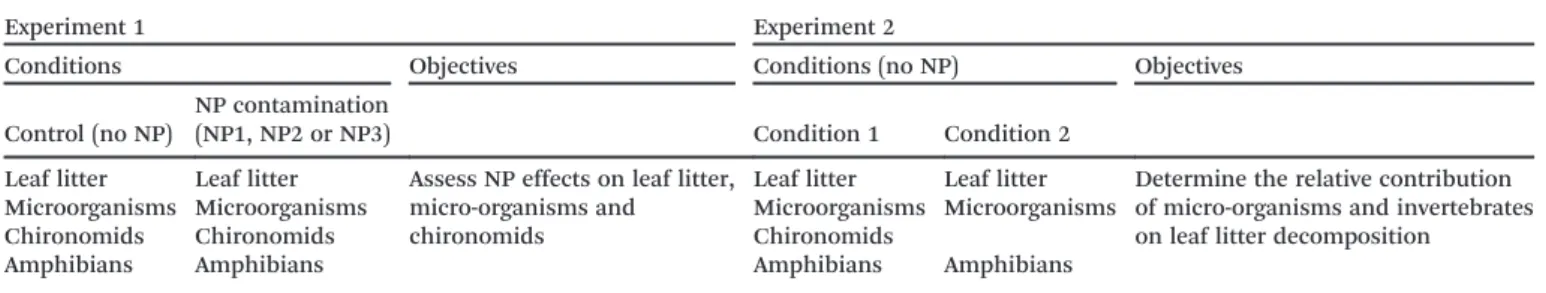
Table 1 summarizes the experimental conditions and objectives.
2.4 Leaf decomposition assessment
Leaf litter decomposition was assessed as described in Cornut et al.20After collection, leaves were gently rinsed with water from their respective microcosm to remove the sedi-ment. Sets of five discs (12 mm diameter) were cut from the leaves of each microcosm, avoiding the central vein, and promptly frozen at −20 °C until they were processed for er-gosterol extraction. The remaining leaf litter was dried at 105 °C to a constant mass and weighed to the nearest 0.01 g. The leaf material was then ground and portions of leaf material of approximately 500 mg were ashed (4 h at 550 °C) and weighed to determine the organic matter content. The remaining leaf mass was expressed as the ratio of the ash-free dry mass (AFDM) between the final and the initial leaf litter. Four unexposed batches of leaf litter were used to termine the initial AFDM according to the procedures de-scribed above.
2.5 Fungal biomass and bacterial diversity assessment Leaf-associated mycelial biomass was assessed through its er-gosterol contents, as previously described.20,21 Leaf material was lyophilized and weighed to the nearest 0.1 mg, and then lipids were extracted with alkaline methanol and heated at 80 °C for 30 min. The extracts were purified using solid-phase extraction cartridges (Oasis HLB, 60 mg, 3 cc, Waters,
Mil-ford, MA, USA) and ergosterol was quantified by high-performance liquid chromatography. The ergosterol concen-tration was corrected for the extraction efficiency (87–100%), which was measured for each sample series of controls to which known amounts of ergosterol were added. The ergos-terol concentration was converted into fungal biomass using a conversion factor of 5.5 mg ergosterol per g of fungal dry mass.22
After DNA extraction, the bacterial community structure in the water column was assessed by PCR-DGGE, as described in Clivot et al.23Pelagic microorganisms were sampled every week by water filtration (total volume of 100 ml, filtered at 0.45μm). Detailed information about the DNA extraction and DGGE analysis procedures are provided in the ESI.† Briefly, DNA was extracted from the filters using an isolation kit and then PCR amplified. The fragments were analyzed on a poly-acrylamide gel. Software analysis was performed to normalize and compare DGGE profiles. Data were analyzed by NMDS and ANOSIM.
2.6 Toxicity onC. riparius
Larval growth was determined by measuring the body length (ImageJ® software). Cephalic capsules of each larva were also measured to determine the larval instars24and assess poten-tial delays in development. Teratogenicity was evaluated in the cephalic capsules as mouthpart deformities, as described by Dias et al.25Briefly, the cephalic capsules were placed in 15% potassium hydroxide (Sigma, France), heated for 12 mi-nutes at 95°C, and then incubated in 70% ethanol overnight to stop the reaction. The capsules were then mounted with Eukitt® mounting medium (03989, Fluka, France) and ob-served under a microscope (Olympus CX41). Mouthpart de-formities were assessed and rated according to the methods described by Warwick and Tisdale26 and Vermeulen et al.27 The treatment groups were compared based on the incidence (individual or total deformities) and severity of the deformi-ties. The latter is evaluated by ratings based on the impacted mouthpart surfaces. Briefly, each tooth is virtually divided into four areas and the number of areas covered by a defor-mity is used as the tooth rate.
2.7 Data analysis
Differences in the AFDM contents and fungal biomass be-tween groups were tested for significance using a one-way analysis of variance (ANOVA) test followed by Tukey's test.
Table 1 Experimental conditions and objectives
Experiment 1 Experiment 2
Conditions Objectives Conditions (no NP) Objectives
Control (no NP)
NP contamination
(NP1, NP2 or NP3) Condition 1 Condition 2 Leaf litter Leaf litter Assess NP effects on leaf litter,
micro-organisms and chironomids
Leaf litter Leaf litter Determine the relative contribution of micro-organisms and invertebrates on leaf litter decomposition
Microorganisms Microorganisms Microorganisms Microorganisms
Chironomids Chironomids Chironomids
The differences in the body sizes and mouthpart deformity ratings of the chironomid larvae between groups were tested for significance using a Kruskal–Wallis one-way analysis of variance on ranks, followed by Dunn's test. The differences in the incidence of mouthpart deformities were tested for sig-nificance using a chi-square test. All of the statistical analyses were performed with SigmaPlot 12.0 software. The analysis of the DGGE profiles is described in the ESI.†
3. Results
3.1 Analysis of the physico-chemical parameters
No significant differences in the physico-chemical parameters of the system were observed between conditions (see the ESI,† Fig. S2). Temperature was maintained at 21 ± 1 °C. Con-ductivity ranged between 207 and 235μS cm−1(mean values) at the beginning and at the end of the experiment, respec-tively. The mean pH was 8.5, with a slight decrease (7.8) dur-ing the second week of NP contamination. The oxygen rates ranged between 110% and 95% (mean values) and were no less than 70% (mean values) throughout the experiment and in every condition.
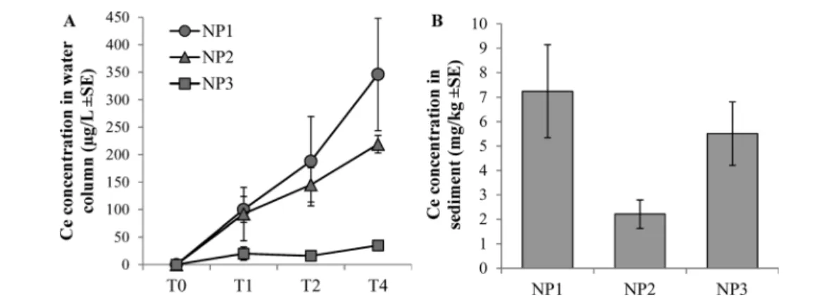
The Ce concentrations in water increased over time. NP3s were rapidly removed from the water column, whereas NP1s were the most stable in the column (Fig. 1A). No differences were observed in the Ce concentrations in sediment between conditions (Fig. 1B).
3.2 Impacts of the CeO2NPs on leaf litter decomposition and
organisms
After six weeks of incubation in the microcosms, the rates of alder leaf decomposition were rather high, with mean AFDM values of less than 15% at the end of the experiment (Fig. 2). A significant decrease ( p< 0.05) in litter decomposition was observed in the microcosms contaminated with NP2 com-pared to the control. The NP1 and NP3 conditions were not significantly different from the control condition.
The ergosterol contents showed that the fungal biomass on alder leaves was important in every condition at the end
of the experiment. No significant differences were observed between conditions (Fig. 3).
The DGGE analysis revealed slight changes in the bacterial communities that were exposed to NP2 or NP3. Although no differences were observed between groups at the beginning of the experiment, the bacterial community structures from NP2 and NP3 groups tended to differ from that of the control group, starting from the third week of contamination (Fig. 4). No changes in community diversity were observed between NP1 and the control group.
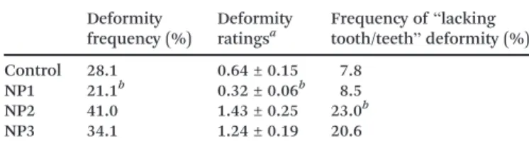
No significant differences were observed in the numbers of chironomid larvae observed at the end of the experiment, with a mean number of 79 ± 16 larvae out of the 700 that were initially introduced. No effects on growth and develop-ment were observed. No differences were observed in the mean larvae body sizes at the end of the experiment between groups (ESI,† Fig. S3). The measurements of the cephalic cap-sules showed that with few exceptions (2–3 larvae per condi-tion), all of the larvae had reached the fourth developmental instar in every condition, indicating that there was no devel-opmental delay. The teratogenicity results obtained from the study of mouthpart deformities are presented in Table 2. The teratogenicity observed in the presence of NP1 was signifi-cantly less ( p< 0.05) than that in the control condition; both
Fig. 1 (A) Ce concentration in the water column throughout the experiment (n = 3). (B) Ce concentration in sediment at the end of the experiment (n = 3). Data are corrected from background concentration determined in the control group.
Fig. 2 Remaining organic matter from leaf litter incubated in mesocosms exposed to CeO2NPs (mean values ± standard error).n =
the frequency and the seriousness of deformities were re-duced in the NP1 condition compared to the control. When considering all of the assessed deformities as a whole, the NP2 and NP3 conditions were not significantly different from the control, either in the frequency or in the seriousness of the deformities. However, a significantly larger ( p < 0.05) number of larvae lacking one or more teeth (Fig. 5) was ob-served in the NP2 condition compared to the other groups. No similar phenomenon has been reported for other individ-ually studied deformities. The NP concentrations in chirono-mid larvae ranged between 266 and 606 mg kg−1(Table 3).
3.3 Relative contributions of microorganisms and invertebrates to leaf litter decomposition
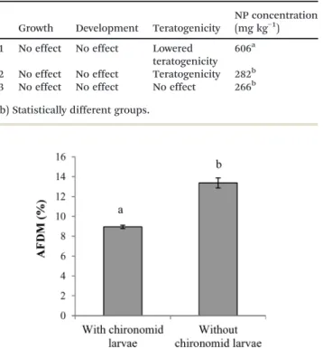
A patently increased decomposition was observed in the pres-ence of chironomid larvae; most of the alder leaves had been eaten, and only the hard parts of the leaves (i.e., veins) remained. In contrast, leaves that were incubated without chironomid larvae were still intact at the end of the experi-ment (ESI,† Fig. S4). The assessment of remaining organic matter from the leaves showed that the AFDM was signifi-cantly reduced ( p < 0.01) in the presence of chironomids (Fig. 6), indicating a greater decomposition rate.
4. Discussion
4.1 NP effects on microorganisms
The fungal biomass present on decomposing alder leaves was not impacted after four weeks of NP contamination with any
Fig. 3 Fungal biomass on alder leaves incubated in mesocosms exposed to CeO2NPs (mean values ± standard error).n = 3.
Fig. 4 NMDS plots of DGGE pelagic bacterial community profiles over time from control (grey icons) and NP (black icons) conditions (n = 3). T0: before NP contamination, T1: after one week of contamination, T2: after two weeks of contamination T3: after three weeks of contamination, T4: after four weeks of contamination. Circles indicate the most important differences between control and NP groups.
Table 2 Mouthpart deformities inC. riparius larvae exposed to NPs Deformity frequency (%) Deformity ratingsa Frequency of“lacking tooth/teeth” deformity (%) Control 28.1 0.64 ± 0.15 7.8 NP1 21.1b 0.32 ± 0.06b 8.5 NP2 41.0 1.43 ± 0.25 23.0b NP3 34.1 1.24 ± 0.19 20.6
aMean value ± standard error.bStatistically different from control
type of CeO2 NP. However, given the important fungal
bio-mass observed in all the conditions, it is possible that slight differences between conditions occurred but were not statisti-cally detected, given the low statistical power related to the relatively low replication. Bacterial communities were im-pacted by the CeO2 NPs. The analysis of the DGGE profiles
showed that communities exposed to NP2 or NP3 tended to be different from the control group at the end of the experi-ment. However, despite the strong tendencies, these differ-ences are at the limits of statistical significance due to the relatively low replication within conditions. The differences observed between conditions could be related to the NP con-centrations in the water column, with low concon-centrations resulting in a more important impact. Indeed, the Ce
concen-trations were reduced for NP2 and NP3 (Fig. 1A). However, a single NP concentration has been tested in this study, and dose–response studies would be necessary to confirm this hy-pothesis. Another hypothesis to explain these differences is that the impact of the NPs on the bacterial community is re-lated to the NP shape and coating.
Previous studies have shown that CeO2NPs are cytotoxic,
and contact between bacteria and the CeO2NPs may result in
lipid peroxidation, membrane damage and reactive oxygen species (ROS) production.28,29 It has also been shown that NPs with rough surfaces, corners and edges are more biologi-cally and chemibiologi-cally reactive.30,31 This result suggests that NP2 and, to a greater extent, NP3, which presents many cor-ners and edges, may have been cytotoxic and induced cellular damage and ROS production in bacteria. Therefore, the changes in the bacterial community may be due to the differ-ent sensitivities of differdiffer-ent species to CeO2 NP cytotoxicity.
For NP1, the citrate coating may have prevented direct con-tact between the bacteria and the CeO2 NPs and, therefore,
may have hindered ROS production and cellular membrane damage. The late response of the communities can be explained by different causes. The tested CeO2NPs could
im-pact bacterial communities only over a long period of time, and/or the NP concentrations were not high enough to im-pact bacteria in the first weeks of contamination. Indeed, the repeated addition of NPs in the microcosms results in an in-crease in NP concentration over the course of the experiment. 4.2 NP effects onC. riparius
In our conditions, the small number of chironomid larvae remaining at the end of the experiment is largely explained by predation from the amphibian larvae.19 Similar to previous studies,32,33no growth inhibition or developmental delays were observed in the chironomids regardless of the type of CeO2NP
tested. However, differences in teratogenicity were observed be-tween conditions in the microcosms, which were different from the observations after single-species exposure for 7 days.32The induction of mouthpart deformities has previously been shown in chironomid larvae that were exposed to uranium,25 organic compounds34 and heavy metals,35–38 but to the best of our knowledge, this is the first study to observed teratogenic effects on chironomids exposed to NPs. Teratogenicity was more im-portant in the presence of NP2 compared to the control group because the NP2 group had a significantly larger number of missing teeth. In contrast, a significant decrease in teratogenic-ity was observed with NP1 compared to the control group. The teratogenicity observed in the control group represents a nor-mal, naturally occurring background in chironomidae.35 More-over, laboratory breeding increases the natural background of teratogenicity.25,27The significantly decreased teratogenicity ob-served with NP1 suggests that this type of NP could have a pro-tective action on C. riparius larvae. As no differences in NP con-centrations were observed in the sediment where larvae are found, the differences in teratogenicity are mainly attributed to the NP characteristics. Ce quantification shows that the concen-trations of NP3 in the water column are 5- to 10-fold less than
Fig. 5 Microscopic observation of C. riparius larvae mouthparts (×400). The dashed line represents the horizontal symmetry axis and the red circle indicates the absence of a lateral tooth.
Table 3 Toxicity onC. riparius and NP body concentrations
Growth Development Teratogenicity
NP concentration (mg kg−1) NP1 No effect No effect Lowered
teratogenicity 606a
NP2 No effect No effect Teratogenicity 282b NP3 No effect No effect No effect 266b (a, b) Statistically different groups.
Fig. 6 Remaining organic matter from leaf litter in the presence or absence of chironomid larvae (mean values ± standard error).n = 3. Letters (a, b) indicate statistically different groups (p < 0.01).
those of NP1 and NP2, suggesting a greater amount of aggrega-tion and sedimentaaggrega-tion.5,39Thus, it can be hypothesized that NP3s, in the form of large aggregates, are less bioavailable for chironomid larvae once ingested. In contrast, NP2s are small, less likely to be aggregated, and may be more easily internal-ized, although further work is required to confirm their passage through the intestinal membrane. It can also be hypothesized that once ingested, NP2s clog the gut, as has been demon-strated for TiO2NPs,40carbon nanotubes41and colloidal clay,42
thus producing decreased nutrient absorption and physiologi-cal disturbances. In contrast, NP1s have a positive impact on teratogenicity. In a previous study, the authors observed a small number of chironomid larvae with deformities, possibly be-cause mortality due to uranium exposure eliminated the larvae with the lowest fitness, which are the most likely to present de-formities.25In our study, this hypothesis can be ruled out as we previously showed that NP1s are not acutely toxic to C. riparius.32 Therefore, the explanation of the differences ob-served between the CeO2NPs might lie in the mechanisms
in-volved in deformity induction. Deformities are defined as mor-phological features that depart from the normal chironomid larvae configuration.43Broken teeth, which are easily recogniz-able by their chipped or rough edges, are not included in defor-mities. These deformities are due to physiological disturbances during molting.44Because molting is regulated by hormones,34 it can be hypothesized that CeO2 NPs positively or negatively
impact hormonal processes, depending on the NP type. Other studies suggest that phthalate-induced upregulation of the HSP 40 and 90 genes might be correlated with the increased occur-rence of deformities in C. riparius larvae.45 It has also been shown that NPs induce changes in gene expression.46–48 To-gether, these results suggest that the effects of the CeO2NPs on
teratogenicity could be due to CeO2 NP-induced changes in
gene expression. Mouthpart deformities can also be considered as fluctuating asymmetry.49Some authors underscore the possi-bility that stress in organisms reduces energy reserves, and as the control over growth processes is energetically costly, the en-ergy allocated for developmental control decreases, likely lead-ing to an increase in developmental stability under stress.50 Therefore, in our study, the mouthpart deformities might be due to decreased energy resources related to stress induced by NP2 exposure. As previously mentioned, gut clogging by NPs could also limit nutrient absorption and decrease the energy re-sources. Concerning the results observed with NP1, it can be hypothesized that a hormesis phenomenon51 occurred, and overcompensation following NP1-induced stress resulted in in-creased developmental stability. Another hypothesis would be that citrate uptake subsequently increased the energy resources for the chironomids. The different hypotheses still need to be confirmed and have opened many perspectives for future research.
4.3 NP effects on leaf litter decomposition
Decreases in leaf litter decomposition have been observed in heavy metal-contaminated aquatic systems as a side effect of metal toxicity on microbial communities and
macro-invertebrates.13,52–54 Despite the relevance of this phenome-non, litter decomposition has only been studied once as a marker of NP impact on freshwater ecosystems.16 In our study, a significant decrease in alder leaf decomposition was observed in microcosms exposed to NP2. This decomposition may result from microbial and/or chironomid larval activity. Indeed, many studies have shown that microbial communi-ties are involved in litter decomposition and that contami-nant toxicity could result in decreased litter decomposition rates.13,16,52–54Several authors also reported that macro-inver-tebrates, particularly shredders, are also involved in leaf litter decomposition.7,14,15,54–56 Chironomid larvae are collector– grazers and preferentially feed on fine particulate organic matter or graze on biofilms at the surface of sediments. They can still decompose and directly feed on leaf litter, particu-larly in the absence of shredders.13,57,58To determine the rel-ative contributions of microorganisms and invertebrates to leaf litter decomposition, the second experiment was performed in the presence or absence of chironomid larvae. The patent decrease in litter decomposition observed in the absence of chironomids indicates that these organisms are mainly responsible for leaf litter decomposition in our experi-mental conditions. The results observed with the CeO2 NPs
are consistent with this finding. Decreased leaf litter decom-position was only observed with NP2, and a negative impact on chironomids was also observed with NP2. In contrast, the observed shift in the bacterial communities in the presence of NP2 could partially explain the effects on litter decomposi-tion, but litter decomposition was not altered with NP3, al-though a shift in the bacterial communities was observed with this type of NPs. Therefore, in contrast to what was ob-served with CuO NPs,16the impact of CeO2 NPs on litter
de-composition is not related to the biocidal effects of the NPs, but rather to their impact on macro-invertebrates. Teratoge-nicity and leaf litter decomposition were both impacted in the presence of NP2. Although no statistical correlation could be established between these two endpoints, likely due to the low statistical power, we cannot exclude the possibility that they are related. It can be hypothesized that mouthpart defor-mities hamper the chironomids' grazing activity and, there-fore, lead to a decrease in litter decomposition. Another hy-pothesis would be that a disturbance in feeding behavior was related to the ingestion of NP2. As previously mentioned, gut clogging by NPs could interfere with the digestive process, af-fect larval feeding behavior and result in decreased decompo-sition rates, as previously reported with heavy metal.59More work is still needed to explore and confirm these hypotheses.
Many studies have reported that leaf litter decomposition is a sensitive marker of natural stream contamination.11,16,35,52–54,60,61 Similarly, the induction of mouthpart deformities in chirono-mid larvae has proven to be a sensitive bioindicator of con-tamination in natural freshwater ecosystems.35,37,38,62In our study, despite the absence of a direct statistical link between mouthpart deformities in chironomids and leaf litter decom-position, the results observed with NP2 indicate that these two bioindicators are congruent concerning the effects of
CeO2 NPs. Similarly, MacDonald and Taylor35 reported that
leaf litter decomposition was congruent with mouthpart de-formities in chironomids in freshwater ecosystems exposed to municipal sewage effluent. Therefore, the present study shows that these markers can be used as valuable tools in microcosm experiments, particularly for the assessment of NP ecotoxicity. Moreover, they appeared to be more sensitive than other markers, such as growth inhibition or develop-mental delays.
5. Conclusion
This study aimed to investigate how CeO2NPs affect organisms
involved in leaf litter decomposition and impact this process. The most important impacts were observed with the small, uncoated spheres, which impacted the bacterial communities and teratogenicity on chironomid larvae and decreased litter decomposition. The small, citrate-coated spheres did not im-pact bacterial communities, but a significantly decreased tera-togenicity was observed on chironomids, suggesting a hormesis effect. The large, uncoated, cubic NPs only affected the bacte-rial communities. This study suggests that the differences in the observed effects are due to specific characteristics and be-haviors of the NPs. The observed effects on bacteria could be due to ROS production, resulting in membrane damage. The teratogenicity observed on chironomid larvae could be the re-sult of gene expression or hormonal disturbances or to de-creased energy resources due to general stress or limited nutri-ent absorption related to gut clogging by the NPs. We observed that toxicity in chironomids, which are the main decomposers in this system, has a significant impact on litter decomposi-tion, a vital process in freshwater ecosystems. Therefore, this study highlights the potential long-term, severe impact of CeO2
NPs on aquatic environments, resulting from sub-lethal toxicity to decomposer species. It also highlights that leaf litter decom-position and mouthpart deformities in chironomids are sensi-tive, congruent markers, as well as the advantages of using microcosms for NP ecotoxicity assessments. Thus, the simulta-neous use of microcosms and sensitive markers of toxicity should be more widely used in the field of nanoecotoxicology.
Acknowledgements
The authors gratefully acknowledge Dr. David Baqué for his as-sistance with ICP-MS analyses and Dr. Laurent Verneuil for his assistance with litter collection. Dr. Mélanie Auffan is also grate-fully acknowledged for her advice on microcosm experiments. The authors gratefully acknowledge French National Research Agency (ANR) for funding this project (ANR-10-NANO-0006/ MESONNET) and the CNRS for funding the GDRi ICEINT.
References
1 M. Auffan, A. Masion, J. Labille, M.-A. Diot, W. Liu, L. Olivi, O. Proux, F. Ziarelli, P. Chaurand, C. Geantet, J.-Y. Bottero and J. Rose, Environ. Pollut., 2014, 188, 1–7.
2 F. Piccinno, F. Gottschalk, S. Seeger and B. Nowack, J. Nanopart. Res., 2012, 14, 1–11.
3 F. Gottschalk and B. Nowack, J. Environ. Monit., 2011, 13, 1145–1155.
4 N. C. Mueller and B. Nowack, Environ. Sci. Technol., 2008, 42, 4447–4453.
5 J. T. K. Quik, I. Lynch, K. Van Hoecke, C. J. H. Miermans, K. A. C. De Schamphelaere, C. R. Janssen, K. A. Dawson, M. A. C. Stuart and D. Van De Meent, Chemosphere, 2010, 81, 711–715. 6 P. Zhang, X. He, Y. Ma, K. Lu, Y. Zhao and Z. Zhang,
Chemosphere, 2012, 89, 530–535.
7 J. Jabiol, B. G. McKie, A. Bruder, C. Bernadet, M. O. Gessner and E. Chauvet, J. Anim. Ecol., 2013, 82, 1042–1051.
8 A. Bour, F. Mouchet, J. Silvestre, L. Gauthier and E. Pinelli, J. Hazard. Mater., 2015, 283, 764–777.
9 J. L. Ferry, P. Craig, C. Hexel, P. Sisco, R. Frey, P. L. Pennington, M. H. Fulton, I. G. Scott, A. W. Decho, S. Kashiwada, C. J. Murphy and T. J. Shaw, Nat. Nanotechnol., 2009, 4, 441–444.
10 P.-E. Buffet, M. Richard, F. Caupos, A. Vergnoux, H. Perrein-Ettajani, A. Luna-Acosta, F. Akcha, J.-C. Amiard, C. Amiard-Triquet, M. Guibbolini, C. Risso-De Faverney, H. Thomas-Guyon, P. Reip, A. Dybowska, D. Berhanu, E. Valsami-Jones and C. Mouneyrac, Environ. Sci. Technol., 2013, 47, 1620–1628. 11 C. Pascoal, M. Pinho, F. Cássio and P. Gomes, Freshwater Biol.,
2003, 48, 2033–2044.
12 C. Pascoal, F. Cássio and P. Gomes, Int. Rev. Hydrobiol., 2001, 86, 407–416.
13 D. Campos, A. Alves, M. F. L. Lemos, A. Correia, A. M. V. M. Soares and J. L. T. Pestana, Ecotoxicology, 2014, 23, 830–839. 14 M. A. S. Graça, R. C. F. Ferreira and C. N. Coimbra, J. North
Am. Benthol. Soc., 2001, 20, 408–420.
15 B. R. Taylor and E. E. Chauvet, Hydrobiologia, 2014, 721, 239–250.
16 A. Pradhan, S. Seena, C. Pascoal and F. Cássio, Microb. Ecol., 2011, 62, 58–68.
17 M. Tella, M. Auffan, L. Brousset, J. Issartel, I. Kieffer, C. Pailles, E. Morel, C. Santaella, B. Angeletti, E. Artells, J. Rose, A. Thiéry and J.-Y. Bottero, Environ. Sci. Technol., 2014, 48, 9004–9013. 18 AFNOR, 2004.
19 A. Bour, F. Mouchet, S. Cadarsi, J. Silvestre, L. Verneuil, D. Baqué, E. Chauvet, J.-M. Bonzom, C. Pagnout, H. Clivot, I. Fourquaux, M. Tella, M. Auffan, L. Gauthier and E. Pinelli, Nanotoxicology, 2015, 1–11.
20 J. Cornut, H. Clivot, E. Chauvet, A. Elger, C. Pagnout and F. Guérold, Water Res., 2012, 46, 6430–6444.
21 M. O. Gessner, in Methods to Study Litter Decomposition, ed. M. A. S. Graça, F. Bärlocher and M. O. Gessner, Springer Netherlands, 2005, pp. 189–195.
22 M. O. Gessner and E. Chauvet, Appl. Environ. Microbiol., 1993, 59, 502–507.
23 H. Clivot, C. Pagnout, D. Aran, S. Devin, P. Bauda, P. Poupin and F. Guérold, Appl. Soil Ecol., 2012, 59, 116–123.
24 Environnement Canada, 1997.
25 V. Dias, C. Vasseur and J.-M. Bonzom, Chemosphere, 2008, 71, 574–581.
26 W. F. Warwick and N. A. Tisdale, Can. J. Fish. Aquat. Sci., 1988, 45, 1123–1144.
27 A. C. Vermeulen, P. C. Dall, C. Lindegaard, F. Ollevier and B. Goddeeris, Arch. Hydrobiol., 1998, 144(1), 103–125.
28 N. J. Rogers, N. M. Franklin, S. C. Apte, G. E. Batley, B. M. Angel, J. R. Lead and M. Baalousha, Environ. Chem., 2010, 7, 50. 29 I. Rodea-Palomares, S. Gonzalo, J. Santiago-Morales, F.
Leganés, E. García-Calvo, R. Rosal and F. Fernández-Piñas, Aquat. Toxicol., 2012, 122–123, 133–143.
30 S. George, S. Lin, Z. Ji, C. R. Thomas, L. Li, M. Mecklenburg, H. Meng, X. Wang, H. Zhang, T. Xia, J. N. Hohman, S. Lin, J. I. Zink, P. S. Weiss and A. E. Nel, ACS Nano, 2012, 6, 3745–3759. 31 D. A. Pelletier, A. K. Suresh, G. A. Holton, C. K. McKeown, W.
Wang, B. Gu, N. P. Mortensen, D. P. Allison, D. C. Joy, M. R. Allison, S. D. Brown, T. J. Phelps and M. J. Doktycz, Appl. Environ. Microbiol., 2010, 76, 7981–7989.
32 A. Bour, F. Mouchet, L. Verneuil, L. Evariste, J. Silvestre, E. Pinelli and L. Gauthier, Chemosphere, 2015, 120, 230–236. 33 S.-W. Lee, S.-M. Kim and J. Choi, Environ. Toxicol. Pharmacol.,
2009, 28, 86–91.
34 G. Meregalli, L. Pluymers and F. Ollevier, Environ. Pollut., 2001, 111, 241–246.
35 E. E. MacDonald and B. R. Taylor, Hydrobiologia, 2006, 563, 277–287.
36 M. Dickman and G. Rygiel, Environ. Int., 1996, 22, 693–703. 37 A. Di Veroli, F. Santoro, M. Pallottini, R. Selvaggi, F. Scardazza,
D. Cappelletti and E. Goretti, Chemosphere, 2014, 112, 9–17. 38 A. Di Veroli, E. Goretti, M. L. Paumen, M. H. S. Kraak and W.
Admiraal, Environ. Pollut., 2012, 166, 212–217.
39 A. A. Keller, H. Wang, D. Zhou, H. S. Lenihan, G. Cherr, B. J. Cardinale, R. Miller and Z. Ji, Environ. Sci. Technol., 2010, 44, 1962–1967.
40 B. Campos, C. Rivetti, P. Rosenkranz, J. M. Navas and C. Barata, Aquat. Toxicol., 2013, 130–131, 174–183.
41 F. Mouchet, P. Landois, E. Sarremejean, G. Bernard, P. Puech, E. Pinelli, E. Flahaut and L. Gauthier, Aquat. Toxicol., 2008, 87, 127–137.
42 S. E. Robinson, N. A. Capper and S. J. Klaine, Environ. Toxicol. Chem., 2010, 29, 168–175.
43 O. N. Odume, W. J. Muller, C. G. Palmer and F. O. Arimoro, Phys. Chem. Earth, 2012, 50–52, 140–148.
44 L. Bisthoven, K. Timmermans and F. Ollevier, Hydrobiologia, 1992, 239, 141–149.
45 K. Park and I.-S. Kwak, Chemosphere, 2008, 74, 89–95.
46 P. M. Gopalakrishnan Nair and I. M. Chung, Comp. Biochem. Physiol., Part B: Biochem. Mol. Biol., 2015, 190, 1–7.
47 P. M. G. Nair, S. Y. Park and J. Choi, Chemosphere, 2013, 92, 592–599.
48 P. M. G. Nair, S. Y. Park, S.-W. Lee and J. Choi, Aquat. Toxicol., 2011, 101, 31–37.
49 G. M. Clarke, Environ. Pollut., 1993, 82, 207–211.
50 D. Lajus, A. Yurtseva, G. Birch and D. J. Booth, Mar. Pollut. Bull., 2015, 101, 758–767.
51 E. J. Calabrese and L. A. Baldwin, Hum. Exp. Toxicol., 2002, 21, 91–97.
52 S. Duarte, C. Pascoal, A. Alves, A. Correia and F. Cássio, Freshwater Biol., 2008, 53, 91–101.
53 I. Fernandes, S. Duarte, F. Cássio and C. Pascoal, Sci. Total Environ., 2009, 407, 4283–4288.
54 H. Roussel, E. Chauvet and J.-M. Bonzom, Environ. Toxicol. Chem., 2008, 27, 637–644.
55 M. A. S. Graça, Int. Rev. Hydrobiol., 2001, 86, 383–393.
56 A. Pradhan, S. Seena, C. Pascoal and F. Cássio, Chemosphere, 2012, 89, 1142–1150.
57 M. Callisto, J. F. Gonçalves Jr and M. A. S. Graça, Rev. Bras. Zootec., 2007, 24, 442–448.
58 L. S. da Silveira, R. T. Martins, G. A. da Silveira, R. M. Grazul, D. P. Lobo and R. da Gama Alves, J. Insect Sci., 2013, 13, 20.
59 F. Heinis, K. R. Timmermans and W. R. Swain, Aquat. Toxicol., 1990, 16, 73–85.
60 A. Medeiros, S. Duarte, C. Pascoal, F. Cássio and M. Graça, Int. Rev. Hydrobiol., 2010, 95, 12–26.
61 C. Pascoal and F. Cássio, Appl. Environ. Microbiol., 2004, 70, 5266–5273.
62 S. Al-Shami, C. S. M. Rawi, S. A. M. Nor, A. H. Ahmad and A. Ali, Environ. Entomol., 2010, 39, 210–222.