Association of Left Ventricular Global Longitudinal Strain
With Asymptomatic Severe Aortic Stenosis
Natural Course and Prognostic Value
E. Mara Vollema, MD; Tadafumi Sugimoto, MD; Mylène Shen, MSc; Lionel Tastet, MSc, MS; Arnold C. T. Ng, MD, PhD; Rachid Abou, MD; Nina Ajmone Marsan, MD, PhD; Bart Mertens, PhD;Raluca Dulgheru, MD; Patrizio Lancellotti, MD, PhD; Marie-Annick Clavel, DVM, PhD; Philippe Pibarot, DVM, PhD; Philippe Genereux, MD; Martin B. Leon, MD; Victoria Delgado, MD, PhD; Jeroen J. Bax, MD, PhD
IMPORTANCEThe optimal timing to operate in patients with asymptomatic severe aortic stenosis (AS) remains controversial. Left ventricular global longitudinal strain (LV GLS) may help to identify patients who might benefit from undergoing earlier aortic valve replacement. OBJECTIVETo investigate the prevalence of impaired LV GLS, the natural course of LV GLS, and its prognostic implications in patients with asymptomatic severe AS with preserved left ventricular ejection fraction (LVEF).
DESIGN, SETTING, AND PARTICIPANTSThis registry-based study included the institutional registries of 3 large tertiary referral centers and 220 patients with asymptomatic severe AS and preserved LVEF (>50%) who were matched for age and sex with 220 controls without structural heart disease. The echocardiograms of patients and controls were performed between 1998 and 2017.
EXPOSURES Both clinical and echocardiographic data were assessed retrospectively. Severe AS was defined by an indexed aortic valve area less than 0.6 cm2
/m2
. Left ventricular global longitudinal strain was evaluated on transthoracic echocardiography using speckle tracking imaging.
MAIN OUTCOMES AND MEASURES The prevalence of impaired LV GLS, the natural course of LV GLS, and the association of impaired LV GLS with symptom onset and the need for aortic valve intervention.
RESULTS Two hundred twenty patients (mean [SD] age, 68 [13] years; 126 men [57%]) were included. Despite comparable LVEF, LV GLS was significantly impaired in patients with asymptomatic severe AS compared with age- and sex-matched controls without AS (mean [SD] LV GLS, −17.9% [2.5%] vs −19.6% [2.1%]; P < .001). After a median follow-up of 12 (interquartile range, 7-23) months, mean (SD) LV GLS significantly deteriorated (−18.0% [2.6%] to −16.3% [2.8%]; P < .001) while LVEF remained unchanged. Patients with impaired LV GLS at baseline (>−18.2%) showed a higher risk for developing symptoms (P = .02) and needing aortic valve intervention (P = .03) at follow-up compared with patients with more preserved LV GLS (ⱕ−18.2%).
CONCLUSIONS AND RELEVANCESubclinical myocardial dysfunction that is characterized by impaired LV GLS is often present in patients with asymptomatic severe AS with preserved LVEF. Left ventricular global longitudinal strain further deteriorates over time and impaired LV GLS at baseline is associated with an increased risk for progression to the symptomatic stage and the need for aortic valve intervention.
JAMA Cardiol. doi:10.1001/jamacardio.2018.2288 Published online August 15, 2018.
Invited Commentary Author Audio Interview Supplemental content
Author Affiliations: Author
affiliations are listed at the end of this article.
Corresponding Author: Jeroen J.
Bax, MD, PhD, Department of Cardiology, Leiden University Medical Center, Albinusdreef 2, 2300RC Leiden, the Netherlands ([email protected]).
JAMA Cardiology |
Original Investigation
I
n patients with asymptomatic severe aortic stenosis (AS), the current guidelines recommend a watchful waiting strat-egy until symptoms or left ventricular (LV) systolic dys-function (ie, LV ejection fraction [LVEF]<50%) develop.1,2The optimal timing for intervention in these patients remains controversial.3-6To determine whether the patients are truly asymptomatic, exercise testing is an important diagnostic tool.7 However, in patients who are unable to perform this test, ad-ditional measurements are needed to better define the tim-ing of the intervention. The assessment of LV systolic func-tion by means of global longitudinal strain (GLS) by speckle tracking echocardiography has demonstrated that a signifi-cant proportion of patients with severe AS have impaired LV GLS despite having normal LVEF.8-13Impaired LV GLS has been associated with worse outcomes in patients with sympto-matic severe AS.14However, to our knowledge, the preva-lence of impaired LV GLS among patients with asymptomatic severe AS and normal LVEF and the natural course and prog-nostic value of LV GLS in this subgroup of patients has not been extensively elucidated. Accordingly, this study aimed to in-vestigate the prevalence of impaired LV GLS, as well as de-scribing the natural course of serial changes in LV GLS and its prognostic implications, in asymptomatic patients with se-vere AS and preserved LVEF.
Methods
Study Population and Data Collection
From a multicenter international registry of patients with AS (Leiden University Medical Center, Heart Valve Clinic, and In-stitut Universitaire de Cardiologie et de Pneumologie de Qué-bec), 220 patients with asymptomatic severe AS and pre-served LV ejection fraction (LVEF > 50%) were selected and included in this retrospective study. Patients were selected based on available echocardiographic data at baseline (de-fined as the date of the first diagnosis of severe AS) with a fea-sible speckle tracking analysis. The definition of severe AS was based on an indexed aortic valve area (AVA) of less than 0.6 cm2
/m2
and/or a mean aortic valve gradient of 40 mm Hg or more and/or a peak aortic jet velocity of 4 m per second or more.2,15,16When available, the last transthoracic echocardio-gram performed at the outpatient clinic or before aortic inter-vention was analyzed to evaluate the changes in valve hemo-dynamics, LV structure, and systolic function (including LV GLS). Measurements of the echocardiographic data were per-formed at each institution by experienced observers. Aortic valve intervention was defined as a surgical or transcatheter aortic valve replacement (AVR) or balloon valvuloplasty. The exclusion criteria were AS-related symptoms at baseline (eg, angina, syncope, or dyspnea), nonsevere AS, LVEF of less than 50%, having undergone prior aortic or mitral valve interven-tion, acute endocarditis at baseline, or the inability to mea-sure LV GLS.
In addition, an age- and sex-matched control group of 220 individuals without structural heart disease was included and used as a reference for measuring LV GLS. The transthoracic echocardiograms of this group of individuals were
per-formed at the Leiden University Medical Center. The referral reasons to perform echocardiography in this group were atypi-cal chest pain, palpitations, or syncope without the presence of a murmur.
Baseline patient demographics and clinical follow-up data were gathered and analyzed retrospectively using the depart-mental patient information systems and hospital records. This retrospective analysis of clinically acquired data was ap-proved by the respective institutional review boards of each participating center, and consent was waived due to the ret-rospective nature of the study.
Transthoracic Echocardiography
Transthoracic echocardiography was performed in all patients at rest in the left decubitus position using commer-cially available ultrasonography systems. Conventional LV dimensions and function as well as AVA were measured fol-lowing current recommendations.17
Additionally, LV GLS was measured with a 2-dimensional speckle tracking analy-sis on apical 2-, 3-, and 4- chamber views using commer-cially available software (Leiden University Medical Center: EchoPac, version 113; General Electric; Vingmed Ultrasound; Heart Valve Clinic Liège and Institut Universitaire de Cardi-ologie et de PneumCardi-ologie de Québec: 2D Cardiac Perfor-mance Analysis; TomTec Imaging Systems).17The frame rate of the 2-dimensional echocardiographic data was 40 frames per second or higher. Left ventricular GLS measures the shortening of the myocardial fibers in the longitudinal direc-tion and is convendirec-tionally presented as a negative value. Therefore, a less negative LV GLS (ie, closer to 0) represents worse LV systolic function.
Clinical and Echocardiographic Follow-up and End Points
Patients were routinely followed up at the outpatient clinic ac-cording to guideline recommendations.16The onset of AS-related symptoms was recorded. The medical treatment and timing for AVR was left at the discretion of the treating phy-sician of each institution. The time to symptom development and AVR, as well as the date of all-cause mortality, were re-corded as clinical end points for assessing the prognosis.Key Points
QuestionWhat is the natural course of left ventricular global longitudinal strain (LV GLS) and prevalence and prognostic value of impaired LV GLS in patients with asymptomatic severe aortic stenosis (AS) and preserved left ventricular ejection fraction? FindingsIn this registry-based study of 220 patients, asymptomatic patients with severe AS showed significantly impaired LV GLS compared with 220 controls with further deterioration over time. Patients with impaired LV GLS at baseline showed a higher risk for developing symptoms and requiring aortic intervention.
MeaningSubclinical myocardial dysfunction that is characterized by impaired LV GLS is often present in patients with asymptomatic severe AS and is associated with symptom development and the need for intervention.
Statistical Analysis
Categorical variables were presented as numbers (percent-age) and continuous variables as means (SD) if normally dis-tributed or medians (interquartile range [IQR]) if otherwise. Histograms were used to evaluate if a Gaussian distribution was present. Comparisons between the total asymptomatic se-vere AS group and the control group were performed using the t test or Mann-Whitney U test for continuous variables and the χ2test or Fisher exact test for categorical variables as ap-propriate. The group of patients with asymptomatic severe AS was divided according to the symptom status at the last
echo-cardiogram performed: symptomatic vs asymptomatic. Changes within and between these 2 groups were assessed using linear mixed models, with correction for age, sex, and time to follow-up. To further examine the prognostic value of LV GLS, the study population was divided according to the me-dian baseline LV GLS value. Cumulative event rates were cal-culated with the Kaplan-Meier method. Two end points were defined: new onset of symptoms and AVR. Comparisons be-tween the 2 groups were performed using log-rank tests. To assess the association between baseline LV GLS and the end points, Cox proportional hazards modeling was used. Spline Table 1. Baseline Clinical and Echocardiographic Characteristics of Patients With Asymptomatic Severe Aortic
Stenosis and an Age- and Sex-Matched Cohort of Individuals Without Structural Heart Disease
Variables Mean (SD) P Value Patients With Asymptomatic Severe AS (n = 220) Age- and Sex-Matched Cohort (n = 220) Age, y 67.9 (13.0) 65.7 (13.3) .08 Male, No. (%) 126 (57) 126 (57) >.99
Body surface area, m2 1.87 (0.2) 1.93 (0.2) .002
Hypertension, No. (%) 128 (59) 103 (48) .02
Hypercholesterolemia, No. (%) 103 (47) 54 (25) <.001
Diabetes, No. (%) 34 (16) 24 (11) .16
History of smoking, No. (%) 73 (38) 17 (11) <.001
Coronary artery disease, No. (%) 47 (22) 0 <.001
Prior myocardial infarction, No. (%) 15 (7) 0 <.001
Medication use, No. (%)
β-Blocker 78 (36) 49 (23) .002
ACE inhibitor/ARB II 88 (40) 65 (30) .02
Calcium antagonist 48 (22) 23 (11) .001
Diuretics 52 (24) 44 (20) .34
Statins 112 (51) 56 (26) <.001
Aspirin and/or clopidogrel 93 (43) 48 (22) <.001
Vitamin K antagonist or NOAC 31 (14) 3 (1) <.001
Creatinine level, IQR, mg/dL 0.91 (0.79-1.09) 0.90 (0.78-1.05) .56 Estimated GFR, IQR, mL/min/1.73 m2 76 (61-89) 80 (65-89) .48 Baseline Echocardiography
Valve anatomy, No. (%)
<.001 Tricuspid 170 (77) 220 (100) Bicuspid 50 (23) 0 Aortic valvea Mean gradient, mm Hg 39.4 (12.6) NA NA Peak velocity, m/s 4.0 (0.6) NA NA Area, cm2 0.86 (0.1) NA NA Area index, cm2/m2 0.46 (0.1) NA NA Stroke volume, mLa 81.4 (17.1) NA NA
Stroke volume index, mL/m2a 43.8 (9.1) NA NA
Left ventricular end-diastolic diameter, mm
45.4 (5.8) 48.5 (6.4) <.001
Left ventricular end-systolic diameter, mm
28.2 (5.0) 30.5 (6.5) <.001
Intraventricular septal thickness, mm 12.9 (2.3) 10.3 (1.7) <.001
Posterior wall thickness, mm 11.6 (1.9) 10.0 (2.0) <.001
Left ventricular mass index, g/m2 112.0 (27.7) 92.5 (21.4) <.001 Left ventricular ejection fraction, % 61.5 (5.9) 62.1 (6.3) .27 Left ventricular global longitudinal
strain, %
−17.9 (2.5) −19.6 (2.1) <.001
Abbreviations: ACE,
angiotensin-converting enzyme; ARB II, angiotensin II receptor blocker; AS, aortic stenosis; GFR, glomerular filtration rate estimated using the Chronic Kidney Disease Epidemiology Collaboration formula; IQR, interquartile range; NA, not applicable; NOAC, novel oral anticoagulants.
SI conversion factor: To convert creatinine to micromoles per liter, multiply by 88.4.
aAortic valve gradients, aortic valve area, and stroke volume were only measured for patients with AS.
models were fitted with overlaying confidence intervals for each end point vs LV GLS on the log-hazards scale, adjusting for age, sex, coronary artery disease, atrial fibrillation, and LV mass index. SPSS, version 23.0 (IBM) was used for the sta-tistical analyses. A P value of <.05 was considered statisti-cally significant.
Results
Characteristics of Patients With Asymptomatic Severe AS
vs Controls
In total, 220 patients with asymptomatic severe AS and preserved LVEF (mean [SD] age, 68 [13] years; 126 men [57%]) were evaluated in this study (Table 1). Despite com-parable LVEF, LV GLS was significantly impaired in the patients with asymptomatic severe AS compared with con-trols, suggesting that asymptomatic patients with severe AS can harbor subtle myocardial dysfunction. When using the mean LV GLS value of the control group as a reference to define normal (≤−19.6%) or impaired LV longitudinal systolic function (>−19.6%), 153 patients (70%) with asymptomatic severe AS had impaired LV GLS. The mean (SD) value of LV GLS was not significantly different across the centers (Lei-den, −18.2% [2.3%]; Québec, −18.0% [1.8%]; Liège, −17.4% [3.1%]; analysis of variance P = .15). In addition, there were no differences across the centers in the proportion of patients with impaired LV GLS (Leiden, 65%; Québec, 81%; Liège, 73%; P = .20).
Changes in LV GLS Over Time in Patients With AS
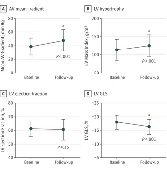
To evaluate the changes in LV GLS in patients with asymptom-atic severe AS, a subgroup of 150 patients (68.2%) with se-vere AS with an available second transthoracic echocardio-gram result (at the last clinical follow-up or before AVR) and a feasible speckle tracking analysis result was evaluated. The me-dian time interval between the 2 echocardiograms was 12 months (IQR, 7-23). The changes in valve hemodynamics and LV systolic function are displayed in eTable 1 in the Supple-ment. Over time, there were significant increases in mean trans-valvular gradients and LV mass index, whereas the AVA de-creased. While the mean (SD) LVEF remained unchanged (61.2% [5.7%] to 60.6% [7.6%]; P = .15), mean (SD) LV GLS showed significant impairment over time (−18.0% [2.6%] to −16.3% [2.8%]; P < .001) (Figures 1 and 2), demonstrating in-creasing subclinical LV dysfunction over time.
Of the 150 patients with echocardiographic follow-up and feasible speckle tracking analysis, 78 (52%) were sympto-matic at follow-up echocardiography and 72 patients (48%) re-mained asymptomatic. The median (IQR) time from baseline to follow-up echocardiography was similar between these 2 groups (symptomatic, 13 [8-28] months vs asymptomatic, 12 [6-20] months; P = .09). Compared with asymptomatic pa-tients, patients who developed symptoms at follow-up showed a higher prevalence of atrial fibrillation (22% vs 10%, respec-tively; P = .05) and had more frequent coronary artery dis-ease (27% vs 16%, respectively; P = .09). Table 2 outlines the changes in valve hemodynamics and LV systolic function over time in these patients divided by symptom status at follow-Figure 1. Patient With Asymptomatic Severe Aortic Stenosis With Preserved Ejection Fraction at Baseline
and Follow-up Baseline A Follow-up B −21 −24 −31 −27 −29
Peak Systolic Strain
Peak Systolic Strain
Sept LVEF, 63% LVMI, 100 g/m2 LVEF, 61% LVMI, 113 g/m2 GLS, −20.9% Ant Lat Inf Ant_Sept Ant_Sept Post Post −25 −31 −11 −19 −17 −9 −17 −19 −20 −17 −20 −16 −16 −15 −14 −17 −14 Sept GLS, −15.3% Ant Lat Inf −14 −11 −16 −15 −15 −18 −19 −16 −16 −14 −18 −16 −20.0 20.0 −20.0 20.0 5 5 0 10 15 10 15 AV Vmax AV Vmean AV maxPG AV meanPG AV VTI AV Env.Ti HR 4.70 m/s 3.55 m/s 88.41 mm Hg 56.16 mm Hg 114.5 cm 323 ms 65 BPM AV Vmax AV Vmean AV maxPG AV meanPG AV VTI AV Env.Ti HR 4.01 m/s 2.99 m/s 64.40 mm Hg 39.99 mm Hg 92.4 cm 309 ms 65 BPM v 0 15 5 10 v 15 10 [m/s] [m/s] −1 −2 −4 −6 −2 −3 −4 −5 −6 50 mm/s 50 mm/s −1 −1 −4 −4 5
Example of a patient with asymptomatic severe aortic stenosis and preserved left ventricular (LV) ejection fraction (LVEF) at baseline (A) and at follow-up (B). Over time, the aortic stenosis severity and LV hypertrophy progressed and LV systolic function as assessed with LV global longitudinal strain (GLS) deteriorated, whereas LVEF remained unchanged. LVMI indicates left ventricular mass index.
up. Within both groups, the progression of AS was observed over time with a concomitant increase in LV mass index and impairment in LV GLS without changes in LVEF. Between both groups, no significant differences were observed in valve he-modynamics and LV systolic function, although LV mass in-dex at follow-up was higher in patients with AS who devel-oped symptoms.
Prognostic Value of LV GLS in Symptom Development
and AVR
Of the 220 patients with asymptomatic severe AS, 118 (54%) developed symptoms during a median follow-up of 12 months (IQR, 5-24). After a median follow-up period of 13 months (IQR, 6-25), 162 patients (74%) received an aortic valve intervention (28 [17%] received transcatheter aortic valve implantation, 130 [80%] underwent surgical AVR, and 4 [3%] underwent balloon valvuloplasty). Most of these patients underwent aortic valve intervention because of symptom development (104 [64%]) or progression of AS severity (40 [25%]); only 18 patients (11%) received an AVR because of other reasons, such as an indication for coronary artery bypass grafting. During follow-up, 28 patients (13%) died; 8 patients (4%) died while scheduled for AVR or when receiving conservative treatment.
To evaluate the prognostic value of baseline LV GLS, the study population was divided into 2 groups according to the median value of baseline LV GLS (more preserved, ≤−18.2% vs more impaired, >−18.2%) (eTable 2 in the Supplement). Com-pared with patients with more preserved LV GLS, patients with more impaired LV GLS had a higher prevalence of coronary ar-tery disease (30% vs 15%, P = .01) and atrial fibrillation (26% vs 12%, P = .01). On transthoracic echocardiography, patients with more impaired LV GLS had a larger LV mass index and lower LVEF than patients with more preserved LV GLS, al-though mean LVEF was more than 60% in both groups (eTable 2 in theSupplement).
The cumulative event rates for developing symptoms were significantly higher in patients with a baseline LV GLS more than −18.2% compared with patients with an LV GLS −18.2% or less (59% vs 45% at 2-year follow-up, respectively, and 91% vs 79% at 5-year follow-up, respectively; log-rank P = .02) (Figure 3A). Similarly, for AVR, the cumulative event rates were significantly higher in patients with impaired baseline LV GLS (>−18.2%) compared with patients with more preserved base-line LV GLS (≤−18.2%) after 2 years (66% vs 57%, respec-Figure 2. Time Course of Valve Hemodynamics and Left Ventricular
Systolic Function in 150 Patients With Asymptomatic Severe Aortic Stenosis AV mean gradient A 80 40 60 20 0 Mean A V Gr adient , mm Hg Baseline Follow-up LV hypertrophy B 200 100 150 50 L V Mass Inde x , g /m 2 Baseline Follow-up LV ejection fraction C 80 60 70 50 40 L V Ejection F raction, % Baseline Follow-up LV GLS D −25 −15 −20 −10 −5 L V GLS, % Baseline Follow-up P <.001 P =.15 P <.001 a a P <.001 a
Time course of valve hemodynamics in a subgroup of 150 patients with asymptomatic severe aortic stenosis at baseline vs follow-up echocardiography by aortic valve (AV) mean gradient (A), left ventricular (LV) hypertrophy (B) and LV systolic function by LV ejection fraction (C), and LV global longitudinal strain (GLS) (D).
aP < .001.
Table 2. Echocardiographic Parameters in 150 Patients With Asymptomatic Severe Aortic Stenosis at Baseline vs Follow-up Divided by Symptom Status at Follow-up Echocardiography
Variable Symptomatic at Follow-up (n = 78) Asymptomatic at Follow-up (n = 72) P Value Intergroup Mean (SD) P Value Mean (SD)
P Value Baseline Follow-up
Baseline Follow-up Baseline Follow-up
Aortic valve Mean gradient, mm Hg 38.4 (11.6) 49.0 (15.8) <.001 39.4 (13.5) 46.6 (15.8) <.001 .71 .31 Peak velocity, m/s 3.9 (0.6) 4.4 (0.6) <.001 4.0 (0.6) 4.3 (0.6) <.001 .36 .25 Area, cm2 0.88 (0.1) 0.76 (0.1) <.001 0.85 (0.1) 0.79 (0.1) .05 .12 .30 Area index, cm2/m2 0.48 (0.1) 0.41 (0.1) <.001 0.46 (0.1) 0.42 (0.1) .02 .08 .41 Stroke volume, mL 81.1 (14.6) 80.6 (14.8) .17 81.7 (19.5) 82.1 (19.2) .71 .97 .91 Stroke volume index, mL/m2 44.6 (9.0) 43.7 (8.5) .34 43.6 (10.1) 43.5 (8.3) .94 .55 .84 LV
Mass index, g/m2 114.0 (27.1) 129.6 (29.2) <.001 113.7 (30.6) 121.0 (29.6) .01 .76 .06 Ejection fraction, % 61.4 (6.3) 61.6 (6.9) .25 61.0 (5.0) 59.5 (8.2) .28 .65 .06 Global longitudinal strain, % −17.7 (2.6) −16.3 (2.9) <.001 −18.2 (2.6) −16.4 (2.6) <.001 .21 .83 Abbreviations: AVR, aortic valve replacement; LV, left ventricular.
tively) and 5 years of follow-up (96% vs 82%, respectively; log-rank P = .03) (Figure 3B). The spline curves to assess the association between symptom development and aortic valve intervention across a range of LV GLS are shown in the eFig-ure in theSupplement. For both symptom development and aortic valve intervention, the linearity assumption was not violated (χ2
, 0.83; P = .67, and χ2
, 1.86; P = .41, respectively). For symptom development, a plateau can be seen (eFigure in
theSupplement). For aortic valve intervention, a clear
in-crease in hazard ratios can be observed for more impaired LV GLS (eFigure in theSupplement).
Discussion
This study demonstrated that in patients with asymptomatic severe AS and preserved LVEF, LV GLS assessed by speckle tracking imaging is impaired as compared with age- and sex-matched controls without structural heart disease. Over time, patients with asymptomatic severe AS showed a progression
of AS severity accompanied by increasing LV hypertrophy and further impairment of LV GLS, while LVEF remained rela-tively unchanged. Patients with impaired LV GLS at baseline showed a higher risk for developing symptoms and for need-ing aortic valve intervention at follow-up as compared with pa-tients with more preserved LV GLS. These findings suggest that LV GLS is a more sensitive marker for early myocardial dam-age than LVEF in this patient group and may help identify the patients who may benefit from earlier AVR.
LV GLS as a Marker for Subtle LV Dysfunction
in Asymptomatic Severe AS
Symptom development and LV systolic dysfunction are the main factors that determine the timing of AVR in patients with severe AS.1,2However, decreased physical activity in the ag-ing AS population may result in the underrecognition or late reporting of symptoms.18
Zilberszac et al19
demonstrated that 43% of elderly patients with asymptomatic severe AS who de-veloped symptoms presented with severe heart failure symp-toms (New York Heart Association class ≥III). The deteriora-tion of LV systolic funcdeteriora-tion defined by an LVEF of less than 50% can be regarded as a more objective parameter that indicates the need for AVR. However, this will only occur when the con-centric remodeled left ventricle fails to maintain normal wall stress because of significant afterload mismatch.20At this stage, LV remodeling is characterized by progressive myocardial fi-brosis, which is not reversible after an intervention.21,22
There-fore, more sensitive markers of LV systolic dysfunction are needed at an earlier stage to identify patients with severe AS who are at risk for irreversible myocardial damage. Recently, Stokke et al12
showed that by inducing concentric LV remod-eling with an increase in wall thickness and a reduction in di-ameter of the LV cavity, the LVEF can remain preserved, whereas LV GLS will be impaired. While the presence of im-paired LV GLS with preserved LVEF has been described in symptomatic severe AS,8,9,23
the prevalence of impaired LV GLS in asymptomatic severe AS has been less studied. Lafitte et al24 reported significantly impaired LV GLS in 65 patients with asymptomatic severe AS compared with 60 healthy partici-pants (−17.8% [3.5%] vs −21.1% [1.8%], respectively; P < .05), while no differences were observed in LVEF (64% [7%] vs 66% [5%], respectively).24This study extends these findings in a larger population. However, the mean value of LV GLS in this study was more preserved than that reported in previous studies (−18.0% vs −15% to −16.6%).25-29
This discrepancy could be explained by the inclusion of older patients in those studies. Furthermore, to our knowledge, this study is the first to report sequential measurements of LV GLS in the pe-riod between the initial AS diagnosis and intervention and to demonstrate a clear deterioration of LV GLS without a decline in LVEF.
Prognostic Value of LV GLS in Patients
With Asymptomatic Severe AS
Multiple echocardiographic predictors of mortality and other adverse cardiac events have been identified in asymptomatic severe AS with preserved LVEF (ie, peak aortic jet velocity >5.0
m/s,4,19,30,31aortic valve calcification,27,32small AVA,33
inap-Figure 3. Kaplan-Meier Estimates for Event Rates for Symptom Development and Intervention in Patients With Asymptomatic Aortic Stenosis Symptom development A 100 40 60 80 20 0 E v ent Rate, %
Time to Symptom Development, mo No. at risk 0 102 118 12 52 60 24 22 33 36 9 22 48 3 12 60 2 5 LV GLS >−18.2% LV GLS ≤−18.2% Intervention B 100 40 60 80 20 0 E v ent Rate, % Time to Intervention, mo No. at risk 0 102 118 12 58 66 24 25 36 36 14 22 48 4 14 60 2 7 LV GLS >18.2% LV GLS ≤18.2% LV GLS >−18.2% LV GLS >−18.2% LV GLS ≤−18.2% LV GLS ≤−18.2% Log-rank test; P = .03 Log-rank test; P = .02
Kaplan-Meier estimates for event rates for symptom development (A) and intervention (B) in patients with asymptomatic aortic stenosis. Cumulative event rates were compared with the study population that was divided according to left ventricular global longitudinal strain (LV GLS) at baseline more than −18.2% (orange line indicates more impaired) vs −18.2% or less (blue line indicates more preserved).
propriate LV hypertrophy,34and increased valvuloarterial impedance35). Data demonstrating the prognostic effect of LV GLS in severe AS and its incremental value over these deter-minants are accumulating. In a cohort of 395 patients with AS, including 302 patients with severe AS, Kusunose et al10 dem-onstrated that LV GLS was an independent predictor of all-cause mortality and had incremental prognostic value on top of known echocardiographic predictors and symptom status. However, only 21% of these patients with severe AS were asymptomatic, and mortality rates were high (25%). Lancel-lotti et al25showed in 163 exclusively asymptomatic patients with severe AS that LV GLS was independently associated with the occurrence of cardiac events (ie, symptom development, eventual AVR, and death). Other studies have investigated the prognostic effect of LV GLS in asymptomatic AS, but these of-ten had small patient samples, included moderate AS, or did not report symptom development as an end point.6,26-29
In con-trast, this study included a larger study population of 220 pa-tients with asymptomatic severe AS with low mortality rates at follow-up (28 patients [13%]) and a more preserved LV GLS at baseline, thus representing a lower-risk study population in an earlier disease stage of severe AS. In addition, this study demonstrated that the natural course of LV GLS is character-ized by further deterioration over time. These results provide further insights into the currently available literature by con-firming that LV GLS is a sensitive marker for subclinical myo-cardial dysfunction and might aid in identifying patients who are at risk for symptom development and the need for inter-vention. Therefore, the present evaluation corroborates that LV GLS holds promise in the preoperative assessment of pa-tients with asymptomatic severe AS without overt signs of LV dysfunction, although further prospective research is needed to determine the exact role of LV GLS in predicting AS pro-gression and severity.
Clinical Implications
In patients with symptomatic severe AS, it has been demon-strated that myocardial fibrosis can be present and persist af-ter AVR.21Diffuse myocardial fibrosis that was noninvasively assessed by native T1 mapping on cardiac magnetic reso-nance imaging was present in asymptomatic patients with se-vere AS and was associated with LV GLS that was measured by speckle tracking echocardiography.36This study shows that LV GLS is often impaired in asymptomatic severe AS and will further deteriorate if left untreated, while LVEF remains un-changed. This suggests that patients with impaired LV GLS at baseline have subclinical myocardial dysfunction that is
prob-ably secondary to diffuse fibrosis, which is not detected by the conventional echocardiographic parameters of LV systolic func-tion. Therefore, the evaluation of LV GLS and consideration of objective signs of AS-related cardiac damage in patients with asymptomatic severe AS with preserved LVEF (as recently sug-gested in a new AS staging classification37) may help to de-fine the optimal timing for AVR (before symptom develop-ment and irreversible myocardial damage occur).
Limitations
This study was limited by its retrospective design, which could have introduced a selection bias. Left ventricular GLS was measured using different platforms, which can lead to slight variations in the quantification of LV systolic dysfunc-tion when considering the current variability in LV GLS mea-surements across vendors. Although intervendor differences in LV GLS measurements have been reported to be statisti-cally significant, this bias was only moderate and the interob-server and intraobinterob-server reproducibility of LV GLS were com-parable with or superior to conventional echocardiographic parameters, such as LVEF.38,39Furthermore, the precision of LV GLS has been shown to be high even in observers with low experience levels.39
The differences in mean LV GLS values or in the prevalence of LV systolic dysfunction based on an LV GLS value of more impaired than −19.6% were not observed across the participating centers. Finally, as the par-ticipating centers are tertiary referral hospitals for AVR, refer-ral bias could be present, with subsequent increased rates of AVR. The decision of referral for AVR was left to the discre-tion of the treating cardiologist.
Conclusions
In asymptomatic severe AS, most patients have impaired LV GLS at the initial diagnosis despite preserved LVEF. Furthermore, during follow-up and before intervention, a further deteriora-tion of LV GLS occurred without a change in LVEF, whereas AS severity progressed and LV hypertrophy increased. Impaired LV GLS at baseline was associated with a higher risk of symp-tom development and need for aortic valve intervention. There-fore, assessing LV GLS holds promise in the risk assessment of asymptomatic severe AS, although further prospective stud-ies in larger patient populations are warranted to establish the exact role of LV GLS, integrated with other markers of AS se-verity and progression, in identifying patients who might ben-efit from earlier aortic valve intervention.
ARTICLE INFORMATION
Accepted for Publication: June 15, 2018. Published Online: August 15, 2018.
doi:10.1001/jamacardio.2018.2288
Author Affiliations: Heart Lung Center, Leiden
University Medical Center, Leiden, the Netherlands (Vollema, Abou, Marsan, Delgado, Bax); GIGA Cardiovascular Sciences, Department of Cardiology, Heart Valve Clinic, University of Liège Hospital, Centre Hospitalier Universitaire Sart Tilman, Liège, Belgium (Sugimoto, Dulgheru, Lancellotti); Institut
Universitaire de Cardiologie et de Pneumologie de Québec, Quebec Heart & Lung Institute, Laval University, Quebec City, Quebec, Canada (Shen, Tastet, Clavel, Pibarot); Department of Cardiology, Princess Alexandra Hospital, University of Queensland, Brisbane, Australia (Ng); Department of Medical Statistics, Leiden University Medical Center, Leiden, the Netherlands (Mertens); Gruppo Villa Maria Care and Research, Anthea Hospital, Bari, Italy (Lancellotti); Cardiovascular Research Foundation, New York, New York (Genereux, Leon); New York–Presbyterian Hospital, Columbia
University, Medical Center, New York (Genereux, Leon); Gagnon Cardiovascular Institute, Morristown Medical Center, Morristown, New Jersey (Genereux); Hôpital du Sacré-Coeur de Montréal, Université de Montréal, Montréal, Quebec, Canada (Genereux).
Author Contributions: Drs Vollema and Delgado
had full access to all of the data in the study and take responsibility for the integrity of the data and the accuracy of the data analysis.
Concept and design: Vollema, Ajmone, Delgado,
Bax.
Acquisition, analysis, or interpretation of data:
Vollema, Sugimoto, Shen, Tastet, Ng, Abou, Delgado, Bax.
Drafting of the manuscript: Vollema, Delgado, Bax. Critical revision of the manuscript for important intellectual content: Vollema, Sugimoto, Shen,
Tastet, Ng, Abou, Ajmone, Marsan, Dulgheru, Lancellotti, Clavel, Pibarot, Genereux, Leon, Delgado, Bax.
Statistical analysis: Vollema, Ng, Mertens, Delgado. Supervision: Delgado, Bax.
Conflict of Interest Disclosures: All authors have
completed and submitted the ICMJE Form for Disclosure of Potential Conflicts of Interest. The Department of Cardiology, Leiden University Medical Center received grants from Biotronik, Medtronic, Boston Scientific Corporation, and Edwards Lifesciences. Dr Delgado received speaker fees from Abbott Vascular. Dr Clavel received a research scholarship from Fonds de Recherche en Santé du Québec. Dr Pibarot holds the Canada research chair in Valvular Heart Diseases, his research program is funded by a foundation grant from Canadian Institutes of Health Research, he has core laboratory contracts with Edwards Lifesciences for which he receives no direct compensation, and he is a consultant for St Jude Medical. Dr Genereux received consultant fees and speaker fees from Edwards Lifesciences. Dr Leon is a member of the Placement of Aortic Transcatheter Valve Trial executive committee, for which he receives no direct compensation. No other disclosures are reported.
REFERENCES
1. Baumgartner H, Falk V, Bax JJ, et al; ESC
Scientific Document Group. 2017 ESC/EACTS guidelines for the management of valvular heart disease. Eur Heart J. 2017;38(36):2739-2791. doi:10 .1093/eurheartj/ehx391
2. Nishimura RA, Otto CM, Bonow RO, et al;
American College of Cardiology/American Heart Association Task Force on Practice Guidelines. 2014 AHA/ACC guideline for the management of patients with valvular heart disease: a report of the American College of Cardiology/American Heart Association Task Force on Practice Guidelines. J Am
Coll Cardiol. 2014;63(22):e57-e185. doi:10.1016/j .jacc.2014.02.536
3. Pellikka PA, Sarano ME, Nishimura RA, et al.
Outcome of 622 adults with asymptomatic, hemodynamically significant aortic stenosis during prolonged follow-up. Circulation. 2005;111(24): 3290-3295. doi:10.1161/CIRCULATIONAHA.104 .495903
4. Rosenhek R, Zilberszac R, Schemper M, et al.
Natural history of very severe aortic stenosis.
Circulation. 2010;121(1):151-156. doi:10.1161 /CIRCULATIONAHA.109.894170
5. Taniguchi T, Morimoto T, Shiomi H, et al;
CURRENT AS Registry Investigators. Initial surgical versus conservative strategies in patients with asymptomatic severe aortic stenosis. J Am Coll
Cardiol. 2015;66(25):2827-2838. doi:10.1016/j.jacc .2015.10.001
6. Généreux P, Stone GW, O’Gara PT, et al. Natural
history, diagnostic approaches, and therapeutic strategies for patients with asymptomatic severe
aortic stenosis. J Am Coll Cardiol. 2016;67(19):2263-2288. doi:10.1016/j.jacc.2016.02.057
7. Redfors B, Pibarot P, Gillam LD, et al. Stress
testing in asymptomatic aortic stenosis. Circulation. 2017;135(20):1956-1976. doi:10.1161
/CIRCULATIONAHA.116.025457
8. Ng AC, Delgado V, Bertini M, et al. Alterations in
multidirectional myocardial functions in patients with aortic stenosis and preserved ejection fraction: a two-dimensional speckle tracking analysis. Eur
Heart J. 2011;32(12):1542-1550. doi:10.1093 /eurheartj/ehr084
9. Kearney LG, Lu K, Ord M, et al. Global
longitudinal strain is a strong independent predictor of all-cause mortality in patients with aortic stenosis. Eur Heart J Cardiovasc Imaging. 2012;13 (10):827-833. doi:10.1093/ehjci/jes115
10. Kusunose K, Goodman A, Parikh R, et al.
Incremental prognostic value of left ventricular global longitudinal strain in patients with aortic stenosis and preserved ejection fraction. Circ
Cardiovasc Imaging. 2014;7(6):938-945. doi:10.1161 /CIRCIMAGING.114.002041
11. Klaeboe LG, Haland TF, Leren IS, et al.
Prognostic value of left ventricular deformation parameters in patients with severe aortic stenosis: a pilot study of the usefulness of strain
echocardiography. J Am Soc Echocardiogr. 2017;30 (8):727-735.e1. doi:10.1016/j.echo.2017.04.009
12. Stokke TM, Hasselberg NE, Smedsrud MK, et al.
Geometry as a confounder when assessing ventricular systolic function: comparison between ejection fraction and strain. J Am Coll Cardiol. 2017; 70(8):942-954. doi:10.1016/j.jacc.2017.06.046
13. Ng ACT, Prihadi EA, Antoni ML, et al. Left
ventricular global longitudinal strain is predictive of all-cause mortality independent of aortic stenosis severity and ejection fraction [published online July 28, 2017].Eur Heart J Cardiovasc Imaging.
14. Dahl JS, Videbæk L, Poulsen MK, Rudbæk TR,
Pellikka PA, Møller JE. Global strain in severe aortic valve stenosis: relation to clinical outcome after aortic valve replacement. Circ Cardiovasc Imaging. 2012;5(5):613-620. doi:10.1161/CIRCIMAGING.112 .973834
15. Baumgartner H, Hung J, Bermejo J, et al.
Recommendations on the echocardiographic assessment of aortic valve stenosis: a focused update from the European Association of Cardiovascular Imaging and the American Society of Echocardiography. Eur Heart J Cardiovasc Imaging. 2017;18(3):254-275. doi:10.1093/ehjci/jew335
16. Vahanian A, Alfieri O, Andreotti F, et al; Joint
Task Force on the Management of Valvular Heart Disease of the European Society of Cardiology (ESC); European Association for Cardio-Thoracic Surgery (EACTS). Guidelines on the management of valvular heart disease (version 2012). Eur Heart J. 2012;33(19):2451-2496. doi:10.1093/eurheartj /ehs109
17. Lang RM, Badano LP, Mor-Avi V, et al.
Recommendations for cardiac chamber quantification by echocardiography in adults: an update from the American Society of Echocardiography and the European Association of Cardiovascular Imaging. Eur Heart J Cardiovasc
Imaging. 2015;16(3):233-270. doi:10.1093/ehjci /jev014
18. Osnabrugge RL, Mylotte D, Head SJ, et al.
Aortic stenosis in the elderly: disease prevalence and number of candidates for transcatheter aortic valve replacement: a meta-analysis and modeling study. J Am Coll Cardiol. 2013;62(11):1002-1012. doi:10.1016/j.jacc.2013.05.015
19. Zilberszac R, Gabriel H, Schemper M, Laufer G,
Maurer G, Rosenhek R. Asymptomatic severe aortic stenosis in the elderly. JACC Cardiovasc Imaging. 2017;10(1):43-50. doi:10.1016/j.jcmg.2016.05.015
20. Carabello BA. Aortic regurgitation. A lesion
with similarities to both aortic stenosis and mitral regurgitation. Circulation. 1990;82(3):1051-1053. doi:10.1161/01.CIR.82.3.1051
21. Weidemann F, Herrmann S, Störk S, et al.
Impact of myocardial fibrosis in patients with symptomatic severe aortic stenosis. Circulation. 2009;120(7):577-584. doi:10.1161/CIRCULATIONAHA .108.847772
22. Treibel TA, López B, González A, et al.
Reappraising myocardial fibrosis in severe aortic stenosis: an invasive and non-invasive study in 133 patients. Eur Heart J. 2018;39(8):699-709. doi:10.1093/eurheartj/ehx353
23. Delgado V, Tops LF, van Bommel RJ, et al. Strain
analysis in patients with severe aortic stenosis and preserved left ventricular ejection fraction undergoing surgical valve replacement. Eur Heart J. 2009;30(24):3037-3047. doi:10.1093/eurheartj /ehp351
24. Lafitte S, Perlant M, Reant P, et al. Impact of
impaired myocardial deformations on exercise tolerance and prognosis in patients with asymptomatic aortic stenosis. Eur J Echocardiogr. 2009;10(3):414-419. doi:10.1093/ejechocard/jen299
25. Lancellotti P, Donal E, Magne J, et al. Risk
stratification in asymptomatic moderate to severe aortic stenosis: the importance of the valvular, arterial and ventricular interplay. Heart. 2010;96 (17):1364-1371. doi:10.1136/hrt.2009.190942
26. Zito C, Salvia J, Cusmà-Piccione M, et al.
Prognostic significance of valvuloarterial impedance and left ventricular longitudinal function in asymptomatic severe aortic stenosis involving three-cuspid valves. Am J Cardiol. 2011; 108(10):1463-1469. doi:10.1016/j.amjcard.2011.06 .070
27. Yingchoncharoen T, Gibby C, Rodriguez LL,
Grimm RA, Marwick TH. Association of myocardial deformation with outcome in asymptomatic aortic stenosis with normal ejection fraction. Circ
Cardiovasc Imaging. 2012;5(6):719-725. doi:10.1161 /CIRCIMAGING.112.977348
28. Nagata Y, Takeuchi M, Wu VC, et al. Prognostic
value of LV deformation parameters using 2D and 3D speckle-tracking echocardiography in asymptomatic patients with severe aortic stenosis and preserved LV ejection fraction. JACC Cardiovasc
Imaging. 2015;8(3):235-245. doi:10.1016/j.jcmg .2014.12.009
29. Carstensen HG, Larsen LH, Hassager C,
Kofoed KF, Jensen JS, Mogelvang R. Basal longitudinal strain predicts future aortic valve replacement in asymptomatic patients with aortic stenosis. Eur Heart J Cardiovasc Imaging. 2016;17 (3):283-292. doi:10.1093/ehjci/jev143
30. Kang DH, Park SJ, Rim JH, et al. Early surgery
very severe aortic stenosis. Circulation. 2010;121 (13):1502-1509. doi:10.1161/CIRCULATIONAHA.109 .909903
31. Bohbot Y, Rusinaru D, Delpierre Q,
Marechaux S, Tribouilloy C. Risk stratification of severe aortic stenosis with preserved left ventricular ejection fraction using peak aortic jet velocity: an outcome study. Circ Cardiovasc Imaging. 2017;10(10):e006760. doi:10.1161/CIRCIMAGING.117 .006760
32. Rosenhek R, Binder T, Porenta G, et al.
Predictors of outcome in severe, asymptomatic aortic stenosis. N Engl J Med. 2000;343(9):611-617. doi:10.1056/NEJM200008313430903
33. Maréchaux S, Ringle A, Rusinaru D, Debry N,
Bohbot Y, Tribouilloy C. Prognostic value of aortic valve area by doppler echocardiography in patients
with severe asymptomatic aortic stenosis. J Am
Heart Assoc. 2016;5(5):e003146. doi:10.1161/JAHA .115.003146
34. Cioffi G, Faggiano P, Vizzardi E, et al. Prognostic
effect of inappropriately high left ventricular mass in asymptomatic severe aortic stenosis. Heart. 2011; 97(4):301-307. doi:10.1136/hrt.2010.192997
35. Hachicha Z, Dumesnil JG, Pibarot P. Usefulness
of the valvuloarterial impedance to predict adverse outcome in asymptomatic aortic stenosis. J Am Coll
Cardiol. 2009;54(11):1003-1011. doi:10.1016/j.jacc .2009.04.079
36. Lee SP, Lee W, Lee JM, et al. Assessment of
diffuse myocardial fibrosis by using MR imaging in asymptomatic patients with aortic stenosis.
Radiology. 2015;274(2):359-369. doi:10.1148/radiol .14141120
37. Généreux P, Pibarot P, Redfors B, et al. Staging
classification of aortic stenosis based on the extent of cardiac damage. Eur Heart J. 2017;38(45):3351-3358. doi:10.1093/eurheartj/ehx381
38. Farsalinos KE, Daraban AM, Ünlü S, Thomas JD,
Badano LP, Voigt JU. Head-to-head comparison of global longitudinal strain measurements among nine different vendors: the EACVI/ASE inter-vendor comparison study. J Am Soc Echocardiogr. 2015;28(10):1171-1181, e2. doi:10.1016/j.echo.2015 .06.011
39. Negishi T, Negishi K, Thavendiranathan P, et al;
SUCCOUR Investigators. Effect of experience and training on the concordance and precision of strain measurements. JACC Cardiovasc Imaging. 2017;10 (5):518-522. doi:10.1016/j.jcmg.2016.06.012