Engralled genes in mammary development and tumorigenesis
par Nicole Martin
Programme de biologie moléculaire, Uhiversité de Montréal Faculté des études supérieures
Thèse présentée à la Faculté des études supérieures En vue de l’obtention dugrade de doctorat
En biologie moléculaire
May, 2006
Q
o
(î ) (‘\ r-N —ç (‘.N
Université
d1
de Montréal
Direclion des bibliothèques
AVIS
L’auteur a autorisé l’Université de Montréal à reproduire et diffuser, en totalité ou en partie, par quelque moyen que ce soit et sur quelque support que ce soit, et exclusivement à des fins non lucratives d’enseignement et de recherche, des copies de ce mémoire ou de cette thèse.
L’auteur et les coauteurs le cas échéant conservent la propriété du droit d’auteur et des droits moraux qui protègent ce document. Ni la thèse ou le mémoire, ni des extraits substantiels de ce document, ne doivent être imprimés ou autrement reproduits sans l’autorisation de l’auteur.
Afin de se conformer à la Loi canadienne sur la protection des renseignements personnels, quelques formulaires secondaires, coordonnées ou signatures intégrées au texte ont pu être enlevés de ce document. Bien que cela ait pu affecter la pagïnation, il n’y a aucun contenu manquant. NOTICE
The author of this thesis or dissertation has granted a nonexclusive ticense allowing Université de Montréal to reproduce and publish the document, in part or in whole, and in any format, solely for noncommercial educationaT and research purposes.
Ihe author and co-authors if applicable retain copyright ownership and moral rights in this document. Neither the whole thesis or dissertation, nor substantial extracts from it, may be printed or otheiwise reproduced without the author’s permission.
In compliance with the Canadian Privacy Act some supporting forms, contact information or signatures may have been removed from the document. While this may affect the document page count, it does flot represent any loss of content from the document.
Université de Montréal Faculté des études supérieures
Cette thèse intitulée:
Engraited genes in mammary development and tumorigenesis
présentée par Nicole Martin
a été évaluée par un jury composé des personnes suivantes:
Dr. Trang Hoang Présidente-rapporteuse Dr. Guy Sauvageau Directeur de recherche Dr. Sylvie Mader Membre du Jury Dr. Louis Gaboury Membre du Jury Dr. William Muller Examinateur externe Université de McGill Thèse acceptée 1e
RESUME EN FRANÇAIS ET MOTS CLES FRANÇAIS
Les gènes homéotiques EizgraiÏed-1 (En-1) et EngraiÏed-2 (En-2) sont d’importants régulateurs du développement du système nerveux central chez la souris. Leur implication dans le développement de la glande mammaire et leur contribution à la transformation de ce tissu demeurent inconnu. Le travail présenté dans cette thèse explore une approche de gène candidat afin de répondre aux questions suivantes: (i) Est-ce que Est-ces gènes sont impliqués dans le développement du tissu mammaire? (ii) Est-Est-ce que ces gènes participent à la transformation néoplasique du sein chez l’humain? Mes résultats démontrent que bien que l’expression d’EngraiÏed-2 n’est pas détectable dans le tissu mammaire murin au cours du développement fétal et chez la souris adulte, le gène paralogue En-1 lui est fortement exprimé dans l’épithélium mammaire de souris pré-pubaires. Mes travaux démontrent aussi que les souris chez qui le gène En-] est inactivé par recombinaison homologue présente une glande mammaire anormale caractérisée par une rareté d’arborisation des canaux mammaires. Des expériences de transplantations de glandes mammaires isolées de souris En-] mutantes confirment que ces observations sont probablement le résultat d’un défaut intrinsèque à l’arbre mammaire mutant. Ces études démontrent donc pour la première fois l’expression spatio-temporel d’En grailed-] dans la glande mammaire de souris et suggèrent un rôle important pour ce gène dans le développement mammaire. Mes travaux ont aussi démontrés que EN2 (mais pas EN]) est exprimé de façon ectopiquc dans environ 8% des cancer mammaires humains et chez près de la moitié des lignées cellulaires malignes. La sur-expression d’EN2 induit la transformation mammaire chez deux lignées non-transformées in vitro et la genèse d’adénocarcinomes in vivo. Des études d’interférence à l’ARN (RNAi) ont démontré qu’EN2 est requis pour la prolifération de cellules transformées humaines. De plus, des analyses par micro-puces effectuées chez 2 lignées cellulaires chez qui les niveaux d’EN2 furent expérimentalement modifiés ont permis d’identifions des gènes cibles potentiels de cette protéine. Ces résultats suggèrent qu’EN2 est impliqué dans la transformation du tissu mammaire humain et d’ENl contribue au développement de ce tissu.
Mots clés: Engraited, homéodomaine, glande mammaire, cancer du sein, oncogène, morphogénèse ductale, transplantation, puberté, RNAi, micro-puce
RESUME EN ANGLAIS ET MOTS CLES ANGLAIS
The homeobox genes Engrailed-] (En-1) and Engrailed-2 (En-2) occupy a prominent position in the developmental regulatory hierarchy and have been studied extensively in embryonic development, yet have received little attention with respect to mammary gland organogenesis and cancer. The studies presented in this thesis are the resuit of a candidate gene approach where the expression and potential role of these homeodomain containing proteins were investigated in the developing mammary gland and in breast tumors. While En-2 was neyer detected at any developmental timepoint in normal mouse or human breast tissue, we have defined the developmentally regulated expression of
En-] in the mammary epithelium of the prepubertal and early pubertal mouse manimary
gland. Moreover, using En-] mutant mice, we provide evidence that loss of En-1 function in the female rnammary gland resuits in severely impaired ductal growth. Pubertal En-] nuli mammary glands revealed a primitive ductal rudiment devoid of terminal end buds (TEBs), reminiscent of a prepubertal mammary gland, while a fully developed ductal system was seen inEn-1 heterozygous and wildtype sibÏings. En-1 nuil mammary epithelium transplanted into surgically cleared fat pads of syngeneic hosts displayed limited ductal outgrowth and a decrease in side branching. These studies demonstrate a unique spatio-temporal pattern ofEn-1 expression in mammary tissue and suggest a potential role for En-1 in the initial growth and morphogenesis of the epithelial ductal system during the onset of puberty. We also show that EN2 (but flot EN]) is ectopically expressed in a subgroup of human breast turnors and in a large proportion of breast cancer cdl unes and that its ectopic expression readily transforms mammary epithelial cells in vitro and promotes adenocarcinoma formation in vii’o. RNA interference studies show that EN2 expression is required for the maintenance of the transformed phenotype of human breast tumor ceils. Moreover, microarray analysis of both gain-of-function and loss-of-function of EN2 in two breast cancer ceil lines provided initial insight into putative EN2 responsive targets. These studies reveal thatEn genes have not only acquired a role in the postnatal development of the mouse mammary gland, but in addition, they have evolved to contribute to brcast tumorigenesis.
Mots clés: Engraited, homeobox, mammary gland, breast cancer, oncogene, ductal
TABLE DES MATIÈRES
RÉSUMÉ EN FRANÇAIS ET MOTS CLÉS FRANÇAIS iii
RÉSUMÉ EN ANGLAIS ET MOTS CLÉS ANGLAIS iv
TABLE DES MATIÈRES y
LISTE DES TABLEAUX ix
LISTE DES FIGURES x
LISTE DES SIGLES ET ABRÉVIATIONS xiii
LA DÉDICACE
REMERCIEMENTS xvii
CHAPITRE 1 1
INTRODUCTION
Investigating a Potential Role forEngrailedGenes in Mammary Gland Development
and Tumorigenesis 1
1.1 Homeobox genes in embryonic development 3
1.].] Homeobox genes are key developinental regulators 3 1.1.2 The hoinoeboxencodes a helix-turn-helix DNA-bindingdomain 4 1.1.3 Homeoproteiizsaretranscriptional regulators 5
1.2EngraiÏed homeobox gene family 6
1.2.] Engrailed genes represent one ofthe smatterfamilies of Ïzomeobox genes 6 1.2.2 Molecularstructure andproperties ofEngrailedproteins 7
1.2.3 En-1 and En-2 inmousedeveÏopmeizt 9
1.2.4 Conservation ofproteinfunction ainong the Engrailedfamily 12
1.2.5 Engraited is û target ofthe Wnt pathway 14
J .3 Mouse mammary gland development 15
1.3.1 Formation and differentiation ofthe embrvonic ,namnzarvgland 17 1.3.2 ReÏatively growth qitiescent prepttbertat inainînmygland deveÏopment 20 1.3.3 ProlUèration and moiplzogenesis in pubescent naimnaiy gland devetopment.20 1.3.4 Alveolar growth and secretorv dfferentiation duringpregnancv 22
1.3.5 Large quantities of milk production and secretion accompnay lactation 23 1.3.6 Apoptosis, regression and remodeling accompany involution after weaning ..24
1.3.7 Mammaiy gland transplantation 24
1 .4 Role of homeobox genes in the mammary gland .30 1.4.1 Homeobox genes have evoÏved ta have roles in mammary gland devetopment 30
1.5 Role of homeobox genes in cancer 31
1.5.] Homeobox genes are impÏicated in cancer 32
1.5.2 Misexpression ofhomeobox genes incarcinoma 32
1.5.3 Loss of hoineobox gene expression in carcinoma 34
1.5.4 Homeobox genes promote tumorigenesis 35
1.6 Breast cancer 36
1.6.] Breast cancerstem cetts 39
1.6.2 Breast cancer stem ceils astherapeutic targets 41
1.7 Objectives 41
Referen ces 44
CHAPITRE 2 51
ARTICLE 51
En-1 Deficiency Leads to Abnormal Ductal Development in the Mammary Gland ....51
Abstract 53
Introduction 54
Resuits 56
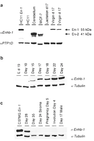
Temporal expression ofEn-] in the mouse maminaiy gland 56
En—] is expressedinceils lining the mammarv epithelial tree during pubert 5$
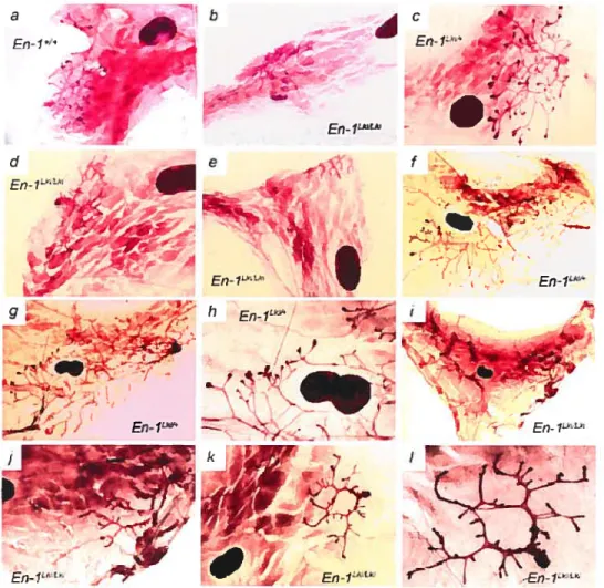
En-1nzanvnaiy glands exhibit impaired ductat growth and TEB formation 59
Reduced ductal moiphogenesis inEn—1 nuil mammaiyepithelial transplants 62
Discussion 64
Materi ais and Methods 67
Acknowledgments 71
References 72
CHAPITRE 3 74
ARTICLE 74
EN2 is a Candidate Oncogene in Human Breast Cancer 74
Abstract 76
Introduction 77
EngraiÏed genes arerareÏv activatedin Wnt—]—inducedmammarvtumors 7$ EN2 is ectopicalty expressed inhuman breast cancer sampÏes $1 Ectopic expression of En-2 readilv transfonns,nainmarvepitheliai ceti unes 83 Ectopic En-2 expressioninhibits differentiation of,nwnmaiyepithetial cetis and
renders them sensitive to 17-AAGtreatment $5
Transplanted En-2 -transduced HC]1 ceÏÏs generate adenocarcinomas that
,netastasize $7
siRNA-mediated suppression ofEN2 inhibits prottferation ofÏzuman breast cancer
ceils 91
Discussion 93
Matenaïs and Methods 95
Acknowledgments 101
References 103
CHAPITRE 4 107
Identification of Putative EN2 Transcriptional Targets by Microarray Analysis 107
Introduction 109
Resuits 110
EnzptoyingsiRNA to elticidatepotel?tiat downstream targtes ofEN2 110
Discussion 115
Materials and Methods 11$
Acknowledgments 121
References 126
CHAPITRES 128
Conclusions, Perspectives and Future Directions 12$
En-1 in mouse mammary gland development 130
EN2 in breast tumorigenesis 135
Perspectives 138
Conclusion 139
LiSTE DES TABLEAUX
Table 4.1 Subset of genes that were significantly (P <0.05) up-regulated 2 fold or down-regulated 0.5 fold in response to EN2 expression in either MDA-MB 435S or T-47D ce!! unes with the corresponding fold change observed in the
other ce!! !ine indicated 122
Table 4.2 Subset of regulated transcripts common to both ceil unes with significant induction or repression in response to EN2 expression with adjusted P values
less than 0.05 124
Table 4.3 Gene Ontology (GO) Mining Tool provided predictions that 1.01 genes would be associated with heat shock protein activity whule 4.25 genes with chaperone activity were actually among the first 477 probesets which were significant at the nominal 0.001 level ofthe univariate two-sample t-test 125
LISTE DES FIGURES
Fig. 1.1 The highiy conserved three-dimensional helix-tum-helix structure encoded by the homeodomain is the hailmark of ail horneodomain-containing proteins 5
Fig. 1.2 Moiecular structure ofthe Engraiied protein family 8
Fig. 1.3 Amino acid sequence comparison ofmouse En-l and En-2 10
Fig. 1.4 Location ofthe five pairs of visible ventral nipples and corresponding rnammary glands just under the skin in the female mouse 16
Fig. 1.5 Distinct stages within mammary gland deveiopment 17
Fig. 1.6 The inguinal (#4) mouse mamrnary gland depicted at different developmental
stages 19
Fig. 1.7 The proposed and elusive mammary epitheliai stem ceil (MSC) would give risc to distinct epithelial ccii lineages in the mammaiy gland 29
Fig. 2.1 En-1 protein is detected in the prepubertal and early pubertal female mouse
mammary gland 57
Fig. 2.2 En-] is expressed in duct-lining ceils of the mouse mammary gland 60
Fig. 2.3 Impaired ductal development in En-I nuil mutant mammary glands 61
Fig. 2.4 Abnormai ductal development of transplanted En-1 nuil mammary epithetium 64
Fig. 3.2 EN2 is not detectable in epithelial structures within normal human breast tissue $0
Fig. 3.3 EN2is ectopically expressed in human breast carcinornas 22
Fig. 3.4 HCÏ 1 and C57MG celis engineered to ectopically expressEn-2 by retroviral gene
transfer $4
Fig. 3.5 Ectopic expression ofEn-2 readily transforms HC1 I and C57MG ceil unes 86
Fig. 3.6 The proliferative advantage that accompanies ectopic En-2 expression in
mammary epithelial celis is lost after exposure to 17-AAG 8$
Fig. 3.7 Mammary glands reconstituted with eitherEn-2 orEn-2 +PBXIb transduced
HC11 cells develop adenocarcinomas $9
Fig. 3.8 EN2expression is required for proliferation ofMDA-MB-435S ceils 92
Fig. 4.1 RT-PCR analysis ofthe same totalRNAwhich was used in the microanay
analysis 111
Fig. 4.2 Heat map cluster diagram ofthe first 50 top-ranked differentially expressed filtered putativeEN2transcnptional targets whose expression correlates positively
with the presence ofEN2 112
Fig. 4.3 RT-PCR analysis of the five most differentially expressed transcriptional targets
identified by microarray analysis 113
LISTE DES SIGLES ET ABRÉVIATIONS
Abréviation Signification
1 7-AAG 1 7-(Allylamino)- 1 7-demethoxygeldanamycin; C31 H43N308 2ki EngraiÏed-2 knock-in
a.a. Amino acid
À
angstromA Adenine; purine base C5H5N5
ABC Avidin: Biotinylated enzyme Complex
Ade Adenine
Ag Antigen
AP Alveolar Precursor
ATP Adenosine triphosphate
bp base pair
fi-gal Beta-galactosidase fi-ME Beta-mercaptoethanol
BSA Bovine Semm Albumin
cDNA Complementary DNA
C Cytosine; pyrimidine base C4H5N30
CDS Coding sequence
ChIP Chromatin Immunoprecipitation CIHR Canadian Institute ofHealth Research
cDNA Complementary DNA
cRNA ComplementaryRNA
DIP Dexamethasone, Insulin and Prolactin DMEM Dulbeccos Modified EagÏe Medium DNA Deoxynbonucleic acid
dki DrosophiÏa engrailed knock-in
dpc days post coitum
DP Ductal Precursor
ECM Extracellular Matrix EGF Epidermal Growth Factor
EGFP Enhanced Green Fluorescent Protein EH Engrailed Homology region
en Drosoph lia engraiÏed gene En-] Murine EngraiÏed-] gene En-2 Murine EngraiÏed-2 gene EN] Human Engrailed-] gene EN2 Human Engrailed-2 gene EPC Epithelial Precursor Ceil ESA Epithelial-specific antigen EST Expressed Sequence Tag
FACS Fluorescence Activated Ccli Sorting
FCS Fetal CalfSenim
FITC Fluorescein isothiocyanate G Guanine; purine base C5H5N50 GFP Green fluorescent Protein GR Giucocorticoid receptor
HAM Ham’s F12 Medium
hd homeodomain deletion
KC)M-C Drosophiia Homeotic gene complex
HOX vertebrate HOXA-D clusters containing Homeobox genes HRP Horseradish Peroxidase
FISC Hemopoietic Stem Ccli
igG Immunoglobulin G
IRCM Institut de Recherches Cliniques de Montréal JVT In vitro transcription
K6 Cytokeratin 6
K18 Keratin 18
kDa kilodalton
L15 Leibovitz L15 tissue culture medium
lacZ E. cou ÏacZ gene; coding for the enzyme fl-galactosidase LCM Laser Capture Microdissection
Iki ÏacZ krrnck-in
LTC-IC Long-Term Culture Initiating Ceil LTR Long Terminal Repeat
MEF Mouse Embryonic Fibroblast MHC Major Histocompatibility Complex MMTV Mouse Mammary Tumor Virus rnRNA messenger RNA
MSC Mammary Stem Ccli MSCV Murine Stem Ccli Virus
MUC1 Mucin I, epithelial membrane antigen
N Normal Tissue
NeOT Neornycin resistance gene
N.D. Not Determined
NLS Nue lear Localization Signal
No R.T. No Reverse Transcriptase/No Reverse Transcription
NP4O Nonidet P-40 detergent
NT Not Transfected/Not Transduced PAGE Polyacrylamide Gel Electrophoresis PBS Phosphate-Buffered Sait
PCR Polymerase Chain Reaction PFA Paraformaidehyde
PGK Phosphoglycerate kinase PI Propidium lodine
PTP- 1 D Protein-tyrosine phosphatase 1 D Purot Puromycin resistance gene RMA Robust Multi-array Average RNA Ribonucleic acid
RNAi RNA Interference
rRNA nbosomal RNA
RPMI RPMI-1640 tissue culture medium PR Progesterone Receptor
RT-PCR Reverse Transcriptase-Polymerase Chain Reaction SDS Sodium Dodecyl Sulfate
siRNA Small interfering RNA $PF Specific Pathogen-Free
T Thymine; pyrimidine base C5H6N202
TCF T-CelI Factor
TdT Terminal deoxynucleotidyl transferase
TEB Terminal end bud
U Uracil; pyrimidine base C4H4N202 VSV Vesicular Stomatitis Virus
LA DÉDICACE
REMERCIEMENTS
I would like to thank my thesis director, Dr Guy Sauvageau, for bis continuai encouragement and guidance. He is an exemplary Clinician Scientist who unwaveringly dedicates so much of bis time and energy to lis work and the well being of the lab in general, inspiring ail those working under him to aspire to the same level of commitment. His enthusiasm for science is truly contagious and lis ability to aiways view experimental challenges and setbacks as invaluable learning experiences is instrumental in many of the projects in the lab persevering and eventually flourishing as they do.
I would also like to thank all the current and past members of the Sauvageau lab for their guidance, training, scientific discussions and equally importantly, their camaraderie. I have benefited greatly from the opportunity to work alongside such inspiring and uplifting colleaugues. I wouÏd also like to thank the Canadian Institute of Health (CIHR) and l’Institut de Recherches Cliniques de Montréal (IRCM) for their financial support during my studies.
Last, but certainly not least, I would have neyer accomplished such an ambitious endeavor and benefitted from such an invaluable educational experience without the momentous encouragement and support from my family, friends and fiancé.
CHAPITRE 1
INTRODUCTION
Investigating a Potential Role for EngraiÏed Genes in Mammary Gland Development and Tumorigenesis
The present Ph.D. thesis, consisting of five chapters, describes the expression, ftmction
and initial mechanistic insights of action ofEngraiÏed-] (En-1) and Engi-ailed-2 (En-2)
in normal mammary gland development and mammaiy tumorigenesis.
Chapter I is a titerature review which shah both encompass the most relevant studies from other groups to gain a further appreciation of this field and help identify the underlying motivation behind the research contained herein and is subdivided into six
sections. The first section briefly describes Homeobox genes in development, leading to
the second section, which specifically describes the EngraiÏed homeobox gene family. The third section outlines the different stages of mouse mammary gland development and imparts how the mouse is an invaluable model with which to investigate genes involved in mammary gland development. The fourth section highhights the role of excmplary homeobox genes in the developing mammary gland whule the flfth section summarizes the role of certain homeobox genes in cancer, with an emphasis in their potential involvement in breast cancer. The sixth section introduces the area of human hreast cancer in general. The Introduction was written by Nicole Martin under the supervision ofDr. Guy Sauvageau.
1.1 Homeobox genes in embryonic development
1.1.1 Homeobox genes are key developmental regulators
Homeobox genes comprise a large family of transcriptional regulatory proteins that are involved in a wide range of essentiai biological processes from embryonic development to terminal differentiation. They are defined by the presence of a 180-hp DNA sequence motif designated the homeobox. The homeobox was initialiy characterized as a sequence motif that was shared among Drosophita Homeotic genes (the HOM-C complex). The homeobox is now known to be evolutionarily conserved among many genes with over 1,000 homeobox genes having been identifled in severai species, ranging from hydra to humans as weii as fungi and plants .
A vast extent of what is presentiy known about homeobox genes and their corresponding functions is the culmination of decades of research that started witli dissecting Drosophita development and genetics. The HOM-C genes in Drosophila were originaliy identified as part of a hierarchy of genes that control embryonic development and play key roles in the determination and maintenance of cell fate and ccli identity. Mutations in these DrosophiÏa HOM-C or homeotic genes resuit in the conversion of one hody part or segment to the likeness or identity of another, and led to die coinïng of the terni ‘homeotic transformation’. Other classes of genes in this hierarchy, such as the gap, pair-ruied and segment polarity genes, which in turn play essential roles in the determination and maintenance of ceil fate and pattern formation in the developing Drosophila embryo, also contain members that are homeobox genes.
In mammals, homeobox genes reign over the specification of the overail body plan and are known to play crucial roles in a variety of developmental processes encompassing central nervous system and skeletal development, limb and digit specification, and organogenesis. The HOM-C genes are in many respects still considered to be the prototype homeobox genes and while they represent only a subset of ail known or predicted homeobox genes, their counterparts in mammais, the HOX genes, are arnong the most extensively studied family among vertebrate homeobox genes. Over evolutionary time, the number of homeobox genes lias increased and their functions
have been reengineered to meet the demands of increasingly diverse developmental processes. b date, it is estimated that the human genome contains at least 200 known or predicted homeobox genes, of which only 39 are members of the HOX family 2
1i mammals, mutations in homeobox genes can cause dramatic developmental defects including Ioss of specific structures as well as classical homeotic transformations. Some homeobox genes appear to have ce!! autonomous functions in differentiation and ce!! cycle contro! while others appear to have non-celi autonomous fonctions such as pattem formation and mediation ofreciprocal tissue interactions .
1.1.2 The homeodomain encodes a helix-turn-helix DNA-binding domain
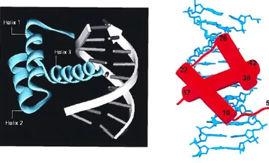
The high!y consewed 180-bp homeobox DNA sequence encodes the homeodomain, a
60 amino acid long DNA-binding domain that folds into three c-helices and a flexible
N-termina! arm. Helices 2 and 3 form a he!ix-turn-helix motif that is the hallmark ofa!! homeodomain-containing proteins. The third a helix, also known as the recognition helix, contacts specific bases in the major groove while the N-termina! extension contacts specific bases in the minor groove. The original 2.8
À
reso!ution structure ofthe engrailed homeodomain-DNA complex was the first crystal structure to reveal how thïs motif recognized DNA .A!though there is considerable variation in the primary sequence of the homeobox, the consensus amino acids, which are invariant among ail horneodomains, maintain the overal! fo!d and DNA-docking arrangement in the three dimensiona! structure of the homeodomain (fig. 1.1).
Evolutionaiy re!ationships and family c!assifications of homeobox genes are based on the leve! of simi!arity among their respective homeodomains and subsequent!y, by comparative analyses of amino acid sequences both amino-terminal and carboxy! terminal to the homeodomain, which vaiy considerably ftom protein to protein. These families vary in size from the relatively !arge ones, such as the HOX family, that comprises 39 members, to the small families, such as the Engrailed fami!y, which only has two members. The a.a. sequence diversity among homeodomain proteins is believed
to contribute to their distinct functional properties, by generating distinct DNA-binding
specificities, promoting unique protein-protein interactions, and other mechanisrns.
Fig. 1.1 The highly conserved tlwee-dimensionai helix-tum-helix structure encoded by
the homeodomain is the hallmark of ail homeodomain-containing proteins. The Ieft image depicts the DrosophiÏa engraiied homeodomain-DNA compiex where the 3 Œ
helices and the extended N-terminal arm of the engrailed homeodomain are shown in blue. The right image shows the complementary 2.2
Â
resolution structure of the Drosophita engrailed horneodomain bound to its optimal DNA site. DNA is depicted inblue and the protein backbone is depicted in red with Œ-helices represented by cylinders. Residue 5 is the first amino acid that could be reiiably modeied from the crystai structure while the other numbers indicate helix termini. Adapted from Fraenkel E., Rould M.A., Chambers K.A. and Pabo C.O. (199$) JMoÏ Bio!. 284:351-361.
1.1.3 Homeoproteins are transcriptional regulators
In accordance with their role as transcnptional regulators, homeobox genes encode
transcription factors, which are predominantly localized in the nucleus where they have been shown to function as activators, repressors or both. These homeoproteins are thought to instruct the ceil as to which genetic program they should further implement,
depending on which step these ceils have reached in the course of their development, or
in response to developmental cues. Although it is widely accepted that the binding
promiscuity of homeoproteins in vitro vs. their highly selective functions in vii’o, reflects their requirernent for cofactors, relatively few examples exist in which bona fide target genes have been identified and are regulated by specific homeobox genes in vivo. It is
now thought that the functional specificities of homeoproteins are dictated by several tiers of regulation, including post-transcriptional controls, nuclear-cytoplasmic transport and protein-protein interactions. For example, PBX andlor MEIS members bind DNA
cooperatively with HOX family members in vitro, which represents one mechanism that confers specificity5.6•
Although individual homeobox genes display unique expression patterns and specific biological functions, homeobox gene families can be distinguished by certain general features of their expression pattems and functional properties, as well as by their sequence similarities. For example, HOX gene expression is generally restricted to undifferentiated andlor proliferative celis during embryogenesis in patterns that reftect their biological functions and that specify positional information .
Other homeobox
genes have spatial and temporal expression patterns that are consistent with roles in
regulating epithelial-mesenchymal interactions that are required for tissue patterning during embryogenesis .
By contrast, the tissue-specific expression patterns of other
classes of homeobox genes in differentiated adult tissues are consistent with their functions as positive effectors of differentiation and homeostasis .
1.2 Engrailed homeobox gene family
1.2.1 Engraited genes represent one of the smaller families of homeobox genes
The Engrailed genes are widely regarded as devclopmental genes. They are involved in pattern formation, neurogenesis, and neuronal differentiation 10,1 ],12 In Drosophila, the
gene engrailed (en), which belongs to the segment polarity class of genes, was first
discovered as a spontaneously occurring mutation, which led to a homeotic transformation 13 Anterior transformations and ceil death occurs in the posterior
compartment of each segment in en mutants, and most mutants die as larvae with severely affected segmentation patterns 14 DrosophiÏa en is involved in regulating a number of key patteming processes, including segmentation of the epidermis and neurogenesis, and is an integral member of the highly complex cascade of developmentaÏ cues, which resuits in a fully developed fruit fty 13
The high degree of conservation among the Engraited gene family led to the rapid cloning of homologs from several species .
The ancestral Drosophila en gene bas undergone gene duplication to generate two Engrailed orthologs in vertebrates. However, genome duplication led to four EngraiÏed orthoiogs in Zebrafish, based on their homology to the mouse En-1 andEn-2 genes 16
1.2.2 Moiecular structure and properties of Engrailed proteins
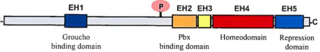
Although individual members of homeoprotein families often share littie sequence similarity outside of the homeobox, particular protein families such as Engrailed have additional conserved domains, which contribute to their distinct functional properties. Comparison of Engrailed homologs identified across several species revealed that ail En proteins have short stretches of conserved regions outside of the homeodomain 17 There are five distinct subregions within Engrailed proteins, designated EH 1—5 for Engrailed homology regions, where 1 through 5 refers to their N- to C-terminal positions 18 (Fig. 1.2). The EH4 domain is the largest and most conserved region and encompasses the homeodornain. The other domains are involved in proteïn-protein interactions. The EH1 and EH5 domains play a roie in active repression of transcription where the repressor function of Eh in vivois dependent upon its association with Groucho 19•
EH3 was defined as a region containing basic amino acids and whose primary sequence was the least conserved across species, although it is well conserved within cadi vertebrate class. In contrast, EH2 is 100% conserved across ail species, with tic exception of tic ftatworm where 17 of tic 19 amino acids are identical. Together, the EH2 and EH3 domains mediate interactions with PBX proteins .
EH2 and EH3 are similar to the hexapeptide motif and the adjacent linker region in HOX proteins,
respectively, which are also 5’ of the horneodomain and are responsible for mediating cooperative DNA binding interactions with PBX homeoproteins 20
This interaction has
a significant impact on the affinity of the Engrailed proteins to DNA, can redirect them to different targets, and can determine whether they act as activators or repressors of
transcription
There is n conserved phosphorylation site N-terminal to E1-12, which is post translationally modified by serine/threonine kinase 2 in insects and vertebrates 24
The phosphorylation of this site may also modulate the secretion of Engrailed protein in mammalian ceits 25 Despite being a transcription factor, which are predominantly localized in the nucleus, a small proportion of intracellular Engrailed protein (less then 5%) is actually found associated with membrane vesicles 26 becornes secreted, and is intemalized by cells 24,27 This occurs despite the protein lacking a classical secretion signal and depends on a short region in the horneodomain that is essential for nuclear export and extracellular release ofthe protein, in addition to the phosphorylation site28
EHI P EH2 EH3 EH4 EH5
N-I
I
L
Groucho Pbx Homeodomain Repression binding domain binding domain domain
fig. 1.2 Molecular structure ofthe Engrailed protein farnily. The five highly conserved
subregions designated EH1-EH5 for Engrailed homology regions l-5 are numerically based on their N- to C- terminal positions, and are designated as colored boxes within the full-length grey protein. The fourth and largest region, EH4, represents the horneodomain and is depicted as a red box. The letter P in the pink circle designates the conserved phosphorylation site N-terminal to EH2, which is putatively involved in modulating the secretion ofEngrailed proteins in mammalian celis.
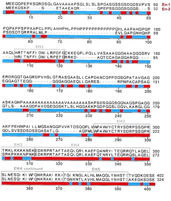
However, outside these regions of homoiogy, En proteins share littie identity across the phyla. At the protein level, the sequence differences between homologs and paralogs are significant. Whereas the homeobox domain is highly conserved, often reaching 90% amongst two different phyla, the sequence hornology drops below 30% for the remaining portion of the protein. This is evident where mouse En-1 shares greater amino
acid (a.a.) sequence identity with the human homolog ENY than with the mouse En-2
paralog. Overali, mouse En-Ï and human EN1 share 95% identity with each other while
mouse En-1 and En-2 proteins share approxirnately 55% a.a. identity with each other
and approx 35% a.a. identity with Drosophila en. Sirnilarly, human and mouse en-2 share 90% amino acid sequence identity with each other. The En-1 class is distinguished by the proline and alanine rich regions N-terminal to EH2 in ail En-1 proteins whule ail
En-2 proteins contain a unique serine ricli region. The 2 vertebrate En protein classes can be further distinguished by fine conserved amino acïds C-terminal to EH5 that are
specific for either the En-1 or En-2 class.
1.2.3 En-1 and En-2 in mouse development
In the mouse, En-] expression is first detected at the one-somite stage around 8.5 days
post coitum (dpc) in ceils of the anterior neural folds. En-2 expression, which occurs in a similar region, is first detected at the five-somite stage, but does not fully overlap with
En-1 expression until approximately $ somites have formed 29
En-2 continues to be
expressed, along with En-], within the neural plate during condensation of the first
somites and in overlapping domains in the mid-hindbrain region during embryonic development j.
At 9.5 dpc, En-1 expression is also detected in a rostral-to-caudal
pattern in two ventrolateral stripes along the hindbrain and spinal cord, in the dermomyatome of the somites, as well as in the ventral ectoderm of the limb buds, and additional En-] expression is later detected in the compact ceils of the somite-derived sclerotome 30 Indicative of their domains of expression during embryonic development,
En-1 and En-2 are required for midbrain and cerebellum development and En-1 also
10
MEEQCPEPKSQRD5GLGAV/sAAAP5GL5L5L5pGA5G5SG5]GDSVV5 50 En-1
- MEEKDSKP - - S ETAAEAQR QPEPSSGGGSGGGS s 32 En-2
•rt — .Z•?r !U
I I I I I
10 20 30 tQ 50
PCPAPPSPAAPCL PPLAHHPb_ PPbPPPPPPPPPOHLAAPÀhCPQP 100
PSDSDTGRRRALMLP EVLQAPGNHQHP 59
•
I I I I I
60 70 80 90 100
EH1
AAQLHRTTNFFI DNI LRPOFGCKKECP_PQLLVASAAAGGGAAAGGGSRV 150
-
-- -[HRITNFFI DNI LRPEFGRRKD AGTCCAGAGGARGG . . 95 b
—
ilo io o ‘40 io
ERDRGQTGAGRDPVHSLGTRASGAASLLCAPDANcGPCGSQpÂTAVG/G 200 EGGAGTTEGG - GGGAGGAEQLLGARES RPNACAPSAG 131 •flWn,. I I I I I 60 170 ‘80 190 200 b— nI i—,p 250 179 3i0 30 30 340 30
EH4 continued EH5
SLNESQ KI 1AFCNKRAK KKAITGI KNGLALbLMAQGLYNHSî7TVQDKDESE 402 SLNESQI KI ‘AFQNKRAK_KK/TGNKNTcAVhLMAQGLYNH5TtAKEGK5DSE 324
—la--I I I
360 370 380 390 400
Fig. 1.3 Amino acid sequence comparison ofmouse En-1 and En-2. The a.a. sequences are aligned and the five highly conserved regions EH 1-5 are indicated and enclosed in grey boxes. Orange regions undemeath the aligned sequences designate perfect matches while blue regions represent non-conserved a.a. Gaps introduced for alignment of the shorter 324 a.a. En-2 sequence with the 401 a.a. En-1 sequence are indicated by grey dashes. ASKAGNPAAAAAAAAAAAAAAVAAAAAAASKPSDSGGGSGGNAGSPGAQG GTLS• . AAAGDPAVDGEGGSKT.SLbGGAKKPGQPGGSLDGVLKARGLGQ -r m -r r 210 220 230 240 250 EH2
AK PEbNAI LLMGSANGGPVVKT DSCQPLVVIPAW/VCTRVSDRPSSGPR 300 GDLSVSSDSDSSQASATLG• - - - -. . AQPMLVPA1WCTRYSDRPSSGPRJ 222 260 2±0 260 260 360 EH4 TRKLKKKKNEKEDKRPRTAFTAEQLQRLKAEFQANRVI TEC6QTLAQEL 350 SRKPKKKNPNKEDkRPRTAFTAEQLQRLKÂEFQTNRYLTEC9RQSLAQEL, 272
--In the adult mouse, both En-1 and En-2 are coordinately expressed in groups of motor nuclei in the pons region and substantia nigra that are involved in motor control. In addition, En-2 is uniquely expressed in the granule and molecular layers of the cerebellar ceils while En-] is uniquely expressed in the postnatal limbs 31•
Their continuing expression in the nervous system into adulthood is thought to reflect an additional function in maintaining the integrity of the central nervous system The emergence of two En paralogs during evolution suggests that each gene likely serves a unique role by regulating the expression of distinct target genes in specific celi types. This notion is substantiated by the finding that unique phenotypes are associated with the disruption of individual En genes in mice.
Mice homozygous for En-] mutations die within a day of birth and have multiple abnormalities. The En-1 ‘itiM
(hd, homeobox deletion) mutants have a striking absence of the mid-hindbrain tissue that is apparent from 9.5 dpc onwards that results in the loss of the third and fourth cranial nerves and most of the cerebellum and colliculi 31
In addition, En-1 mutants have abnormally shaped forelimbs and exhibit skeletal defects in the 13th rib and sternum. Mutant forelimb paws are grossly deformed showing occasional ectopic ventral digits, truncations, and fusion of the digits, splaying outwards of the digits, supernumerary digits and a delay in ossification of the digits. The newborn mutants exhibited truncated sternums and a delay in ossification, as well as misalignment of the ribs and abnormal sternum ossification patterns. At birth, these mutants were readily distinguishable due to their forelimb abnormalities and by 12 hours after birth, it was obvious that these mutants were flot feeding as evidenced by the lack of rnilk in their stomachs 31• The mutants could move theirjaws and limbs so the Jack of feeding was attributed to a probable Jack of appropriate innervation from the CNS for feeding due to the deletion of brain tissue.
Phenotypic studies initially focused on newborns because En-] mutants die shortly after birth but subsequent analysis of viable mice homozygous for two other En-] mutant alleles, En-1 dki/Uki (Drosophila en knock-in) and En-1 2ki/2k1 (En-2 knock-in), and of rare En-1 ““
importance of En-1 in specifying postnatal ventral limb structures. Around 3-4 weeks of age, the above mutant limbs develop dorsally restricted hyperpigmentation and nail-like differentiation on the ventral epidermis, ectopic ventral hairs, and occasional ectopic ventral digits that emanate from the base of the proximal mutant paw pad 32•
The Eu-1 mutant brain defect provides evidence that patterning of the nervous system in mammais involves a phase of regionally controlled proliferation of cell precursors Mice lacking En-] function have a loss of midbrain and cerebellar structures that derive from the En ]-expressing brain region, suggesting that En-1 is required for the specification, survival, and differentiation of these neural precursors 31 In contrast, loss of En-1 function in the ventral ectoderm of the developing limb does flot lead to loss of En-] expressing ectodermal ceils, but instead results in an alteration of ventral ectoderm and mesoderm cell fate and limb patterning32•
Mice homozygous for En-2 mutations are viable but exhibit a distinct cerebellar phenotype where the mutant cerebellum exhibits a one-third reduction of the normal size and displays a specific alteration in the folding pattern lie En-2 mutant phenotype is milder than the En-1 mutant phenotype and is restricted to the brain. During postnatal development of the cerebellum, En-2 is required for the production of some of the cerebellar cdl precursors and for patterning and fusing of the fissures It is thought that the milder mid-hindbrain phenotype seen in En-2 mutants is due to the fact that En-1 and En-2 share partially redundant functions in the brain where En-] is expressed earlier thanEn-2 in the celis that will eventually express En-2.
1.2.4 Conservation of protein function among the Engrailed family
Despite the large sequence differences within the Engrailed family outside of the five conserved regions, the biochemical conservation of these genes is striking. Substituting mouse En-2 coding sequences in place of mouse En-1 coding sequences by gene targeting led to viable and fertile animais with a complete rescue of the brain defects, skeletal abnormalities and the embryonic limb patteming of the otherwise lethal En-1 null mutant 17 En-1 replaced with En-2 revealed that the two En proteins in the mouse have retained common biochemical functions throughout evolution since mouse En-2
can substitute for En-1, both in the neural tube where it is normally expressed as well as in regions such as the limbs, that normally express oniyE,?-] 17 The complete rescue of the newbomEn-] mutant phenotype suggested that the two paralogs were redundant and almost functionally equivalent. The main functional differences revealed by the nul! mutants for the two paralogs were thought to anse from the divergence in temporal and spatial expression rather than through divergence in biochemical function
Accordingly, the more severe deletion ofmid-hindbrain structures in En-1 nul! mutants, in comparison toEn-2 null mutants, was thought to be due to the lack of En ftinction in the anterior neural folds between the one- and eight-somite stages, before En-2 is expressed.
However, when surviving En-1 2ki/2k aduit mutants were analyzed, it became evident that although En-2 can rescue the embryonic limb defects ofEn-] hd/hd mutants, it cannot rescue the postnatal patterning defects of En-] hd%icl mutant limbs. En-] 2kt/2ki mice developed the hyperpigmentation and ventral nail-like structures ofEn-] 1’Mmutants 3-4 weeks after birth 36 This is a direct demonstration in mammals that the regulation of essential steps during embryogenesis has been conserved by the two paralogs, but the fact that En-2 cannot rescue the postnatal limb abnormalities shows that En-1 bas acquired some novel functions during evolution above those that are seemingly redundant with En-2.
The same replacement experiment was repeated by replacing the mouse En-] coding sequences withDrosophiÏa en coding sequences and clearly demonstrated the functional homology across phyla, when the ortholog from an invertebrate was able to substitute for a mammalian gene throughout the development of a highly ordered structure, the brain 36 The resulting mice were viable and fertile, further demonstrating the biochemical conservation over hundreds of millions of years of evolution. Mice expressingDrosophiÏa en in place ofEn-] results in a near complete rescue of the lethal En-] mutant brain defect and most skeletal abnormalities, reveating a common underlying molecular mechanism to their diverse developmental activities. in contrast, surviving adult En-1 dk,/dki mice demonstrated that expression ofDrosophiÏa en cannot
ftmctionally replace En-1 in the dorsal/ventrai patteming of the iimbs during cither embryonic or postnatal development 36•
Although neither En-2 nor en are capable of rescuing the postnatal iimb abnormalities that develop in rare En-1 1dM mutants that survive, these studies demonstrate that the
biochemical activity utilized in mouse to mediate brain development has been retained by Engraiied proteins across the phyla, and indicate that during evolution vertebrate En proteins have acquired two unique functions during embiyonic and postnatal iimb developrnent and that only En-1 can carry out the latter36
Ail En proteins share 5 conserved domains (EH1-EH5), and coding sequences in the
non-conserved regions of Drosophila en could have evolved a functionai domain
required for vertebrate limb development, which became further speciaiized in En-1
afier the secondEn gene (En-2) was formed by duplication 18
The ftmctional differences between the 3 En proteins may refiect the inabiiity of en and En-2 to interact with the full repertoire of En-1 accessory proteins, possibiy resuiting in aitered DNA binding affinities for selective targets.
1.2.5 Engrailed k a target of the Wnt pathway
Several upstream regulators that activate, repress or maintain engraiÏedexpression in the
developing Drosoph lia embryo have been identified, and similarly, several direct and
indirect targets of engrailed regulation have been identified in Drosoph lia but very few mammalian regulators or targetsofEngraiÏed-1 andlorEngraiied-2have been identified.
It has been shown, however, that the maintenance ofEn-1 expression during cmbiyonic developrnent by the Wnt signaling pathway has been conserved from files to mice Genetic analysis ofDrosophita development indicates that wingless (Wnt-1 homolog) signaling in the epidermis is required for the maintenance of engrailed expression in adjacent celis In Wnt-] mouse embryos, initial En-] andEn-2 expression is normal,
79
but subsequent expression of both are lost - . In addition, compound En-1 x En-2
mutants have a similar phenotype to that of Wnt-F’ mutants where the midbrain and
best shown when En-] expression, under the control of the Wnt-] enhancer, was sufficient to rescue the early rnidbrain and anterior hindbrain phenotypes in Wnt-T’ embryos s”. Wnt-] encodes a secreted growth factor that initiates a signaling cascade, which resuits in transcriptional activation mediated byfi-catenin[Tcf complexes 40•
1.3 Mouse Mammary Gland Development
The mammary gland forms as an appendage of the skin and lias its evolutionary origin in skin glands 41 All mammary glands reside just underneath the skin but the number and location vary among different classes of mammals. For example, only one pair of mammary glands develops in the thoracic region in humans whereas mice possess five ventral pairs of mammary glands (Fig. 1.4). The mammary gland consists of two main components. The epithelial component or parenchyma refers to the epithelial system composed of ducts and milk-producing alveolar ceils within the gland. This extensive ductal system is embedded in the surrounding stromal component, which refers to tlie fatty connective tissue of the mammary gland that supports the epithelial component. Adipocytes account for the majority of celis in the stromal compartment, but fibroblasts, celis of the hernatopoiefic system, blood vessels and neurons also reside in the fat pad41• The epitliclial celis form the branclied system of ducts that cliannel into a main primary duct, whicli opens up to the body surface through the nipple. A large proportion of epithelial ceils within the mammary gland are luminal secretory celis, which undergo functional differentiation during pregnancy to produce milk, which is then secreted into the inner lumen of the ducts. Basal myoepithelial cells surround the ductal system and these contractile ceils facilitate the detivery of milk from the milk secreting cells to the nipple during lactation.
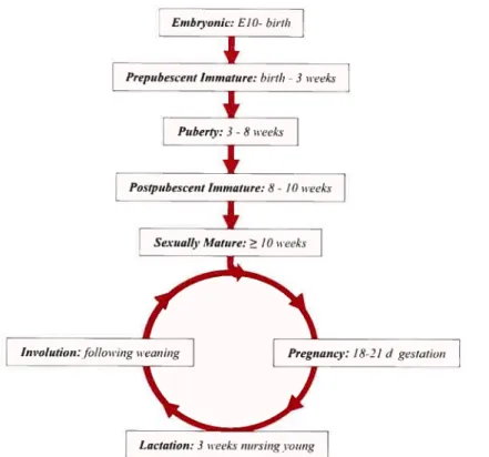
Unlike most mammalian organs, which develop primarily embryonically, the mammary gland is established during fetal development but the majority of expansion and development occurs postnatally 42• The structural and functional development of the gland involves the influence of hormones and growth factors like estrogen, progesterone and prolactin and development of the gland itself can be divided into roughly six distinct stages; embryonic, postnatal, puberty, pregnancy, lactation and involution (Fig. 1.5).
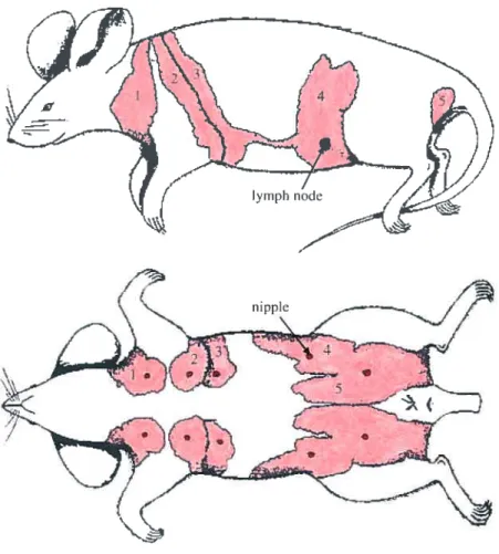
Fig. 1.4 Location of the five pairs of visible ventral nipples and corresponding rnammary glands just under the skin in the female mouse. The nipples are shown in red
and the mammary fat pad is shown in pink (the rnammary epithelial tree within the fatty
stroma is not depicted here). The adipose stroma of the gland provides a frame of support as well as a substrate within which the rnammary epithelial tree can grow and
function. The first 3 mammary glands are in the thoracic region while the 4t11
and 5th
mammary glands are in the abdominal region. The 4th
(inguinal) mammary gland is the most accessible and thus, is the gland of choice in most experirnental manipulations. The
4th
mammary gland also contains a central lymph node (shown in purpÏe), which serves
as a convenient reference point. Adapted from DeOme K.B. etal. (1959) J. Nati. Cancer
Embrionic: ElO-hirth
Prepubescent 1,unzuture: hirth-3weeks
Ir
__Puberfl’: 3-8 n’eeks‘I
Postpubescent hmnalnre: 8- 10 ii’eeks
Sexualle Mature:> 10teeks
Involution:/illou’ing neaning Pregnancv: 18—2 1 d gestation
Lactation: 3 eeks Inusing ozing
fig. 1.5 Distinct stages within mamrnary gLand development. The linear portion of the diagram represents the protiferative expansion and morphogenesis that takes place in the embryonic and virgin female. The circular portion ofthe diagram represents the cycle of proliferative expansion and morphogenesis, functional differentiation and eventual remodeling accornpanied with each pregnancy. Adapted from Lewis M.T. (2000) Breast CancerRes. 2:158-169.
1.3.1 Formation and differentiation of the embryonic mammary gland
The mammary epithelium is an ectodenrial derivative and therefore, the first distinction that must be made is the differentiation ofthe presumptive inammaiy epithelium from tissue that can also differentiate to formskin, hair follicles or other ectodermally derived structures. The future mammary anlage begins to differentiate from the ectodenn at day
epidermalÏy denved thickened epithelium that extend from the anterior to the posterior
Iimb bud, symmetrically displaced off the ventral midiine of the embryo. These streaks
represent the first morphological evidence of mammary pattem formation and differentiation before sexual differentiation of the gonads. The mammary anlage is first visible as small placodes which define the nipple region around El 1.5 and then appears
as five pairs of small epithelial buds on the ventral side of the embryo on E12 that
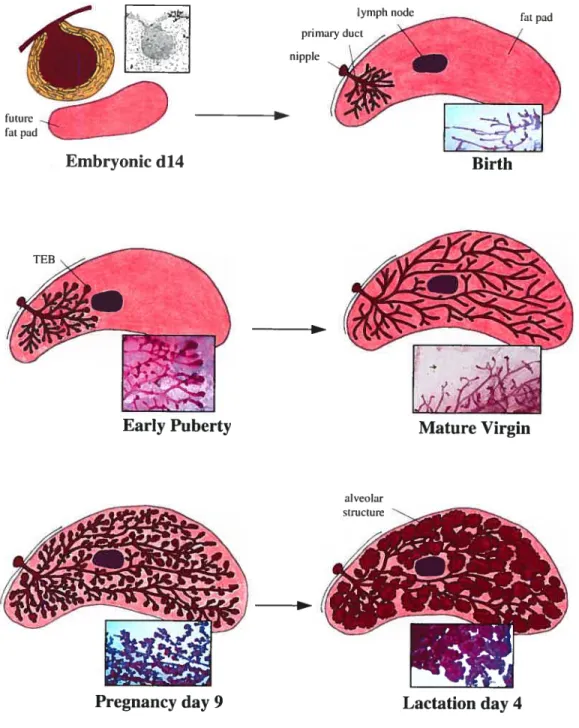
enlarge to form bulb-like structures by F14. During embryonic developrnent the mammary epithelium is associated with two types of mesenchyme; the mammary mesenchyme that is directly attached to the epidermal bud and the future mammary fat pad that is located below the epithelial bud in the deeper mesenchyme and consists of preadipocytes (fig. 1.6). The mammary mesenchyme consists of several layers of concentrically organized fibroblasts, which surround the epithelial bud and are more densely packed than the dermal cells while the mammary fat pad appears at E14 as undifferentiated mesenchyme and is required for future mammary epithelial morphogenesis.
The ccli identity of mammary epithelium is clearly estabiished as early as E 12.5 as shown by the capability of these epithelial buds at this stage to generate a ductal tree when transplanted into a cleared fat pad of a syngeneic host. Marnmary rudiments are formed in both sexes but the sexual phenotype of the rnammary gland is determined between E13 and F14. During this period the gland displays responsiveness to steroid hormones and is influenced by signais from the surrounding mesenchyme. In male embryos testosterone acts on the dense mesenchyme surrounding the epithelial mammary rudiment, which results in the subsequent detachment of the gland from the epidermis. In females the rnammary buds continue to grow and reciprocal interactions between the mammaiy bud and the mammary mesenchyme take place, which are crucial for elongation of the mammary bud. As the mammary bud elongates to form a primary sprout, it reaches and invades the second mesenchyme, the future mammary fat pad by
E 16. The primary sprotit then undergoes a srnall amount of branching morphogenesis,
Fig. 1.6 The inguinal (#4) mouse mammary gland depicted at different developmental stages. The adipose strorna of the mammary fat pad is shown in pink while the marnrnary epithelial system is depicted in red. The central lymph node, which serves as
a convenient reference point to evaluate ductal outgrowth in the #4 mammary gland. is
shown in purpie. The embryonic rnarnmary epithelial bud is sulTounded by rnammary
Iymph node primary duct nipple
Embryonic d14 Birth
si
Early Puberty Mature Virgin
alveolar structure
mesenchyme (yellow) while the future mammary fat pad lies below (pink). In the newborn mouse, the few rndimentary ducts emanate from a central primary duct that is connected to the nipple. At this stage, the mdimentary ductal system is proximal to the nipple and occupies littie space in the mammary fat pad. Rapid and invasive ductal elongation and branching commence with puberty and terminal end buds (TEBs) start to become visible. The ductal pattern of the mammary epithelial tree is created by the bifurcation and penetration of these TEBs through the underlying stromal fat pad. The growing ductal system reaches the central lymph node around 4 ½ weeks, and continues growïng until the ducts have reached the peripheral limits of the mammary fat pad around 6-8 weeks of age. Additional branching occurs in the postpubescent immature virgin and at 10-12 weeks the mammary gland reaches sexual maturity and ductal elongation ceases and TEBs regress to leave a branched system of ducts. Proliferation of mammary secretory epithelium during pregnancy leads to the formation of lobuloalveolar structures, which resemble grape-like clusters. At parturition the mammary fat pad is completely filled with secretory alveolar structures and milk proteins are secreted in large arnounts by the secretory epithelial celis.
1.3.2 Relatively growth quiescent prepubescent mammary gland development At birth, the mammary epithelial tree consists of 15-20 rudimentary branches emanating from a central primary duct, which is connected to the nipple (Fig 1.6). The rudimentary system of small ducts present in the newborn female occupies only a small portion of the mammary fat pad in the vicinity of the nipple. During the first three weeks of postnatal life, before the onset of gonadal hormone secretion, the marnmary ducts are in fact slowly elongating and branching into the underlying fatty stroma at a rate that is in pace with the overall growth of the entire animal.
1.3.3 Proliferation and morphogenesis in pubescent mammary gland development Functional development of the mammary gland proceeds in distinct stages that mostly coincide with the hormonal influences of puberty and pregnancy. Around 3-4 weeks of age, ovarian hormones stimulate accelerated and invasive ductal extension and
branching whereby the growth rate of the epithelial ductal system now exceeds the overail growth rate of the animal. Estrogen, atong with other hormones, plays a critical role in the expansion and morphogenesis of the growing ductal system.
The onset of puberty also coincides with the appearance of large club-shaped TEBs, highly proliferative and active structures found at the tips of the growing ductal branches. It is during the early pubertal developmental stage (3-7 weeks of age) when the TEBs are most prominent (Fig 1 .6). These TEBs are influenced by systemic steroid hormones and aid the ducts in linear growth as well as the regulation of branching pafferns. The ductal pattem of the mammary epithelial trec is created by the bifurcation and penetrating extension of these TEBs through the underlying stromal fat pad. The TEB is a specialized structure, comprising a solid mass of epithelial celis, which is composed oftwo distinct relatively undifferentiated ccli types. Actively proliferating cap celis make up the outerrnost layer of the end bud and interact with the sunounding stroma though a thin basal lamina as the subtending duct is formed while body ceils (about 6-10 layers thick), f111 the interior ofthe end bud. As the ducts elongate and the TEBs move forward, it is thought that the inner body ceils are the precursors of luminal epithelial celis and give risc to the inner luminal epitheliat ceil layer of the subtending duct and the outer cap cells are the precursors ofmyoepithelial ceils and give rise to the outer rnyoepithelial cell layer of the newly formed portion of the duct. Myoepithelial ceils are contractile ceils that form a sleeve around the primary ducts and become discontinuous around secondary and tertiaiy ducts and the TEBs themselves. Luminal epithelial ceils are generally used to refer to the non-myoepithelial component of the mammary epithelium system and these ceils line the lumen, the space within the centre ofthe mammary ducts.
Dichotomous branching of the growing ductal system occurs at the site of TEBs as they penetrate the underlying fat pad while monopodial branching of ducts occurs by budding from the body ofthe existing ducts. Ductal morphogenesis and iimer lumen formation is accomplished by a highly regulated process of both cdl proliferation and death in the 1FB. It has been demonstrated that apoptosis is an important mechanism in ductal
morphogenesis dunng puberty and occurs in the middle of the mass of body celis and adjacent developing luminal cells to generate the ductal lumen While a variety of ce!! types have been identified and their developmental capacities have begun to be explored, very little is known about how these cet! lineages and fates are established at this pubertal stage.
The growing ductal system reaches the central lymph node around 4 ,4 weeks, and continues growing until the ducts have reached the peripheral limits of the mamrnaiy fat pad around 6-8 weeks of age, at which time few TEBs rernain. Additional branching occurs in the postpubescent immature virgin and at 10-12 weeks the mammaiy gland reaches sexual maturity and ductal elongation ceases and TEBs regress to leave a branched system of differentiated ducts. The fully differentiated mature mammary ducts can now serve as channels for milk transport during lactation and consist of a discontinuous outer lining of myoepithelial cells lined by a single layer of luminal epithelial ceils. The gland becornes essentially quiescent in the mature virgin except for bnef periods during the ovulation cycle and the ducts will remain relatively quiescent as long as the mature female remains a virgin.
1.3.4 Alveo]ar growth and secretory differeutiation during pregnancy
Another phase of rapid proliferation takes place during pregnancy where 50% of the overali growth of the gland takes place from pregnancy day 12 until parturition. Hormonal changes during pregnancy initiate this cyctical phase of development which leads to a dramatic transition from a predominantly ductal morphology to a predominantly lobuloalveolar gland morphology. During pregnancy, the gland cornes under the influences of estrogen, progesterone and other placental hormones. New ductal outgrowth occurs from the lateral walls of the ducts and side buds increase in nurnber. In addition, lobuloalveolar progenitor ceils located in the ducts, mostly at the terminal ends of the ducts, proliferate and undergo alveolar development to form alveotar buds which differentiate to fom grape cluster-like structures containing lobuloalveolar units (Fig
1.6). Ibis vast expansion of the mammary epithelium fils in rnost of the fatty stroma
ceils on their outer surface surrounding columnar epithelial cells facing the lumen, the space in the centre of the alveoli where milk is initially secreted.
These morphological changes that the alveolar epithelium undergoes are accompanied by the development of secretory epithelial ceils within the alveoli that acquire the ability to produce milk proteins by midpregnancy. The capability to produce milk proteins represents the first stage in the transition to lactogenesis but the secretion of these milk proteins is inhibited during pregnancy. At parturition, secretory function is no longer inhibited and milk proteins are secreted in large amounts by the secretory epithelial cells, which represents the second and final stage in the transition to lactogenesis. Lobuloalveolar development and proliferation occur during pregnancy and resuit in the complete fihling of the fat pad by parturition while functional differentiation of the secretory epithelium coincides with parturition and lactation. Cell division occurs in both the alveolar and ductal celi populations throughout pregnancy and continues into the mid stage of lactation. Pregnancy terms vary slightly between 1$-21 days in different mouse strains.
1.3.5 Large quantities of milk production and secretion accompany lactation
Lactation involves the production and secretion of milk by the secretory epithelial celis. Mammary epithelial proliferation continues intoearly lactation where it is estimated that
20% of total mammary growth occurs during the first 14 days of lactation. By the time tlie mother is feeding lier pups, lier mamrnary glands are packed full of secretory epithelium witli little fat, the complete opposite of the situation in the virgin or non pregnant animal. The initiation of lactation is thouglit to be induced by the decrease in estrogen and progesterone and several hormones such as prolactin, insulin and glucocorticoids are involved in the maintenance of lactation. While the inner luminal celis of the alveoli produce the milk, the outer contractile myoepithelial cells form a basket-like network around the secretory alveoli and these cells are responsible for squeezing the rnilk out of the alveolï and down the ducts and out of the nipple in responsc to the hormone oxytocin.
1.3.6 Apoptosis, regression and remodeling accompany involution after weaning When lactation ceases after weaning, the mammary gland undergoes involution where the entire mammary alveolar compartment is remodeled. This remodeling elicits a dramatic change in the morphology of the mammary gland, restoring it to its pubescent state. The process of involution commences with the suspension of milk production, followed by the collapse of the mammary alveolar structures and removal of the secretory epithelial celis through programmed ceil death and phagocytosis. The alveolar structures collapse likely as a result of degradation of the extracellular matrix (ECM) and apoptosis of secretory epithelial cells and surrounding myoepithelial cells. The basement membrane and most of the epithelial cells are replaced with adipose tissue so that the mammary gland changes from the epithelial rich lactational state to the epithelial sparse non-parous state. After regressing to this state following weaning of the offspring the fat pad contains only well spaced ductal structures within the adipose matrix. With each subsequent pregnancy, a new cycle of lobulo-alveolar development occurs and can be repeated several times during the life of an animal.
1.3.7 Mammary gland transplantation
An essential feature of the mouse mammary gland is the regenerative capacity of its epithelium. Any portion of the epithelial tree can be transplanted into the mammary fat pad of a syngeneic female, whose endogenous epithelial tree has been removed, and reproduce a complete and functional mammary gland In a 3 week old female mouse, the epithelial ductal system occupies little space in the mammary fat pad and is confined to the most proximal portion of fat pad connected to, and in the vicinity of, the nipple (Fig. 1.6). This area containing the prepubertal epithelial ductal system can easily be surgically removed to generate a ‘cleared’ mammary fat pad into which cells or tissue fragments from another female can be transplanted where it can develop under the influences of a wildtype hormonal environment. In addition, endogenous epithelium can be left intact, providing identical mammary fat pad conditions to enable in situ comparisons between wildtype and transplanted epithelium.
The deletion ofa gene in the mouse genome can sometimes lead to the disruption of the normal development and functïon of more than one organ. The absence of some genes can influence ovarian function, which would obscure assigning direct or indirect effects on marnmary gland development. Moreover, some homozygous deletions are flot viable and postnatal mammary gland development cannot be studied directly. The above issues can be overcome by transpianting mammary epithelial celis from such mutants into a wildtype syngeneic host.
The ability to transplant portions of the mammaiyepithelium into the cleared fat pad of a syngeneic host allows one to examine both the morphogenic and tumorigenic capabilities of that particular marmnary epithelium. Ccli populations rcmoved from the mammary gland and mainmary epithelial ceils grown in vitro can also be transplanted. The 4 (inguinal) mammary gland is the most accessible and is the gland of choice in most experimental manipulations (Fig. 1.4).
The ability to delete genes from the mouse genome, in conjunction with tissue transplants to evaluate their physiologie role, has lcd to the identification of severai genes invoived in mammary gland development. Experimental manipulation of mammary tissue from wiÏdtype and knockout mice has shed light on distinct signaling networks activated by systernic hormones that induce or are involved in mammopoiesis whiie some of these signais act through reciprocal interactions between the mammary epithelium and the adipose stroma. The mammary transplantation method has been employed for decades to examine fundamental questions in mammary morphogenesis, senescence and tumorigenesis. Such experiments examine the role of genes in the context of a normal tissue environment with an endogenous hormonal milieu and can determine the potential role of oncogenes in preneoplastic and neoplastic transformations and the demonstration of oncogenic potential of unknown genes.
Daniel and colleagues used serial transpiantion studies to investigate the pattern of senescence in mammary cells and demonstrated that the proliferation potential of normal mammary celis declined with serial transplantation and was lost after 5-6 serial
transplants ‘.
The number of pnor ce!! divisions rather than chronological age of the transplant donor was the rnost important determinant for the onset of senescence or lack of division potential in the mammary gland. Moreover, they aiso demonstrated that
mouse mammary preneopiasias did flot senesce and were essentially immortal populations.
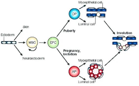
1.3.8 Adult mammary epithetial stem ceits
The observation that marnmary epithelial fragments or celis from adult mammary glands can generate a fuiiy differentiated mammary tree capable of lactating when transpianted into the cieared fat pad of a prepubertal mouse led to the speculation that the mammary epithelium must contain multipotential stem cells. Mammary epitheliai stem celis (MSCs) have been the focus of much recent interest because they are also likeiy the cells of origin of breast tumors. Some have suggested that the notion of breast stem ceils as a resident ccli population in the aduit breast is an oversimp!ified view and that perhaps a selective ce!! population has MSClike ability, possibly as a resuit ofits interactions with its rnicroenvironment or stem ccli niche. No definitive identification has been made of an adult MSC yet experimental evidence supports the notion that such an elusive stem celi exists and would give risc to the distinct mammary epithelial cdl lineages within the mammary gland and models of how these lineages might develop have been proposed (Fig. 1 .7). Currently, at least four distinct mammary ccli populations are thought to exist. One is the muitipotent MSC that is capable of self-renewal and recapitulating the entire mammary ductal system, and these MSCs would gives risc to committed epithelial precursor ce!ls (EPC5). The progeny of these EPCs give risc to two pre-committed epithelial progenitors that become restricted to a ductal or alveolar fate j.
The ductal precursor celis (DPs) form myoepithelial ceils and iuminal celis, the two ccli types that cxist in ductal structures. During pregnancy, the alveoli are thought to be generated from alveolar precursors (APs), which give riscto myoepithelial celis and luminal ceils.
The pre-committed epithelial progenitors and the stem cclls that are capable of giving risc to both ccli types, are thouglit to exist throughout the entire mammary epithelial tree but may be enriched in TEBs46• During the rapid expansion of the ductal system during
puberty where the body ceils and cap ceils proliferate and differentiate to generate new sections of the growing duct behind the active TEBs invading the underlying fat pad, stem ceil-like activity is thought to reside in the TEBs. Since some of the cap ceils can be seen to migrate into the body celi mass, the cap cells are the proposed stem ceils and symmetric ce!! divisions of the cap ceils within the TEBs as they move through the fat pad are thought to deposit MSCs in the growing ducts. In the mature virgin mammary gland, the location of putative MSCs is thought to be suprabasal, at the base of the luminal epithelial cell layer, adjacent to the myoepithelium and not contacting the lumen or the basement membrane This proposed stem cell niche would contain undifferentiated MSCs that do not express markers of either myoepithelial or luminal epithelial ceils. Divisions of this MSC would generate progenitor cells that, at least in its early stages, would be very difficuit to distinguish from its parental MSC until it acquires both myoepithelial and luminal lineage rnarkers before becoming committed to either the luminal or myoepithelial lineage In the proposed model, the ductal system contains multipotent stem cells and committed ductal and luminal precursor ceils nestled throughout the remodeled ductal system after involution (Fig. 1.7).
MSCs are conceivably the only celi population that would possess the replicative potential that would be needed to maintain the routine tissue renewal, the massive expansion in epithelial tissue that accompanies pregnancy and the cyclical process of subsequent pregnancies in the mamrnary gland. Sucli candidate stem cells would be the type that are quiescent until responding to physiological cues. In non-parous animals, a less expansive but similar process occurs as the estrus cycle progresses, as evidenced by the slight expansion and regression of the alveolar buds in response to the cyclic hormonal influences. Conversely, MSCs in the postnatal mouse mammary gland may also be involved in the replacement of luminal epithelial celis that are shed from the inner lining of the ducts into the lumen during routine celI turnover. Evidence to support this occurrence during lactation is provided by the fact that epithelial cells can be recovered from milk. Cells that are shed into the lumen of the alveolar and ductal systems must need to be continually replaced in order for the marnrnary epithelial tree to maintain its structure and integrity. Candidate stem celis that would participate in this
![fig. 2.2 En-] is expressed in duct-lining cells ofthe mouse marnmary gland.](https://thumb-eu.123doks.com/thumbv2/123doknet/2052221.5464/78.918.297.683.367.797/expressed-duct-lining-cells-ofthe-mouse-marnmary-gland.webp)